The Effects of Exposure Time on the Surface Microhardness of Three Dual-Cured Dental Resin Cements

Abstract
: This study evaluated the exposure time of light-curing of the polymers used for cementation on microhardness test in different storage times. The polymers (specifically called resin cements) were RelyX ARC, RelyX U100, and SET. Five specimens of each group were prepared and photo-polymerized with exposure times of 20 s and 180 s, using a LED polymerization unit with wavelength of 440 ˜ 480 nm and light output was consistently 1,500 mW/cm2. The Vickers hardness test was performed in a MMT-3 Microhardness Tester. Data were submitted to ANOVA and Tukey's test (α = 0.05). The values of RelyX ARC showed statistically significant difference to groups with light exposure when considering only chemical cure (p < 0.05). The groups with light exposure (20 s and 180 s) showed no significant difference between them (p > 0.05). The RelyX U100 cured only chemically showed statistically significant difference between 48 h and 7 days (p < 0.05). The SET resin cement showed no significant difference to groups without light exposure for all storage times (p > 0.05). The values of hardening of the dual-cured resin cements improved after setting by light and chemical activation demonstrating the importance of light curing.1. Introduction
Dental polymers, specifically resin cements, have been used considerably in recent years due to a larger application of dental adhesive procedures [1,2].
There are a variety of currently available cements on the market, though none is ideal for all clinical situations. Therefore, the choice of the luting agent must rely on its physical, biological and handling properties being allied to the characteristics of the prepared tooth and prostheses [3].
Factors, such as exposure time of light-curing and type of light-curing unit can influence the characteristic of the resin cement, changing the final quality of the restorations [4].
The dental resin cements can be classified based on the type of their cure such as chemical-, light-or dual-cure [5].
The chemical-cure polymerizes exclusively by chemical activation. The dual-cured resin cement is formulated to not depend only on light activation; the cure is started by photo-and chemical-initiators. These systems can be used locally where light transmission is limited. The light-cure resin cement is exclusively initiated by light, which offer the clinical advantages of extended working time, setting on demand, and improved color stability [6,7].
Hardness testing is commonly used as a simple and reliable method to indicate the degree of conversion [8].
The purpose of this study was to investigate the exposure time of light-curing of the dual-cured resin cements on microhardness test at different storage times.
2. Results and Discussion
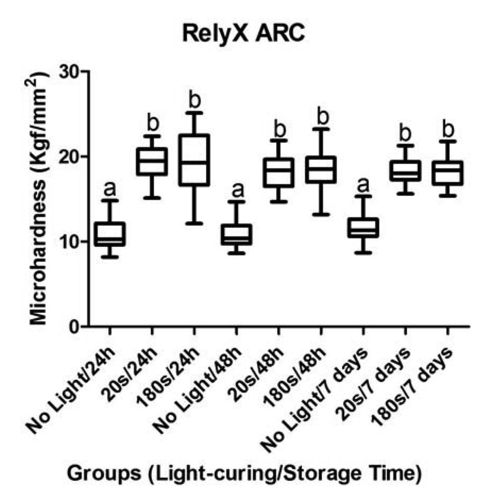
The data were analyzed separately for each dental material. The objective was to compare different exposure and storage times. Figure 1 shows the mean values (Kgf/mm2) for RelyX ARC. The values of RelyX ARC showed statistically significant difference to groups with light exposure when considering only chemical cure (p < 0.05). The groups with light exposure (20 s and 180 s) showed no significant difference between them (p > 0.05).
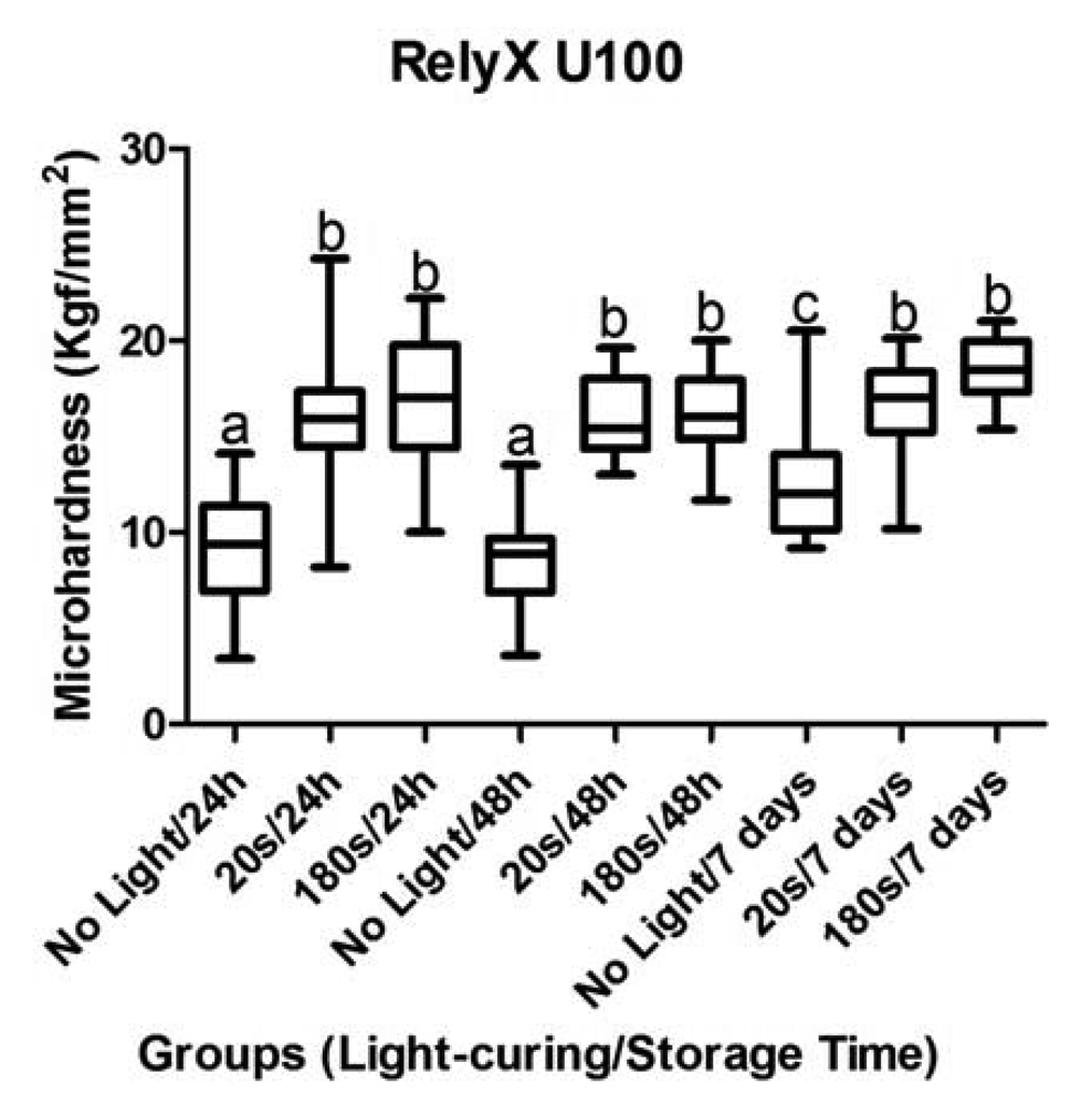
The RelyX U100 cured only chemically, showed statistical significance between 48 h and 7 days (p < 0.05). Others storage times showed no difference when compared with same light exposure (Figure 2).
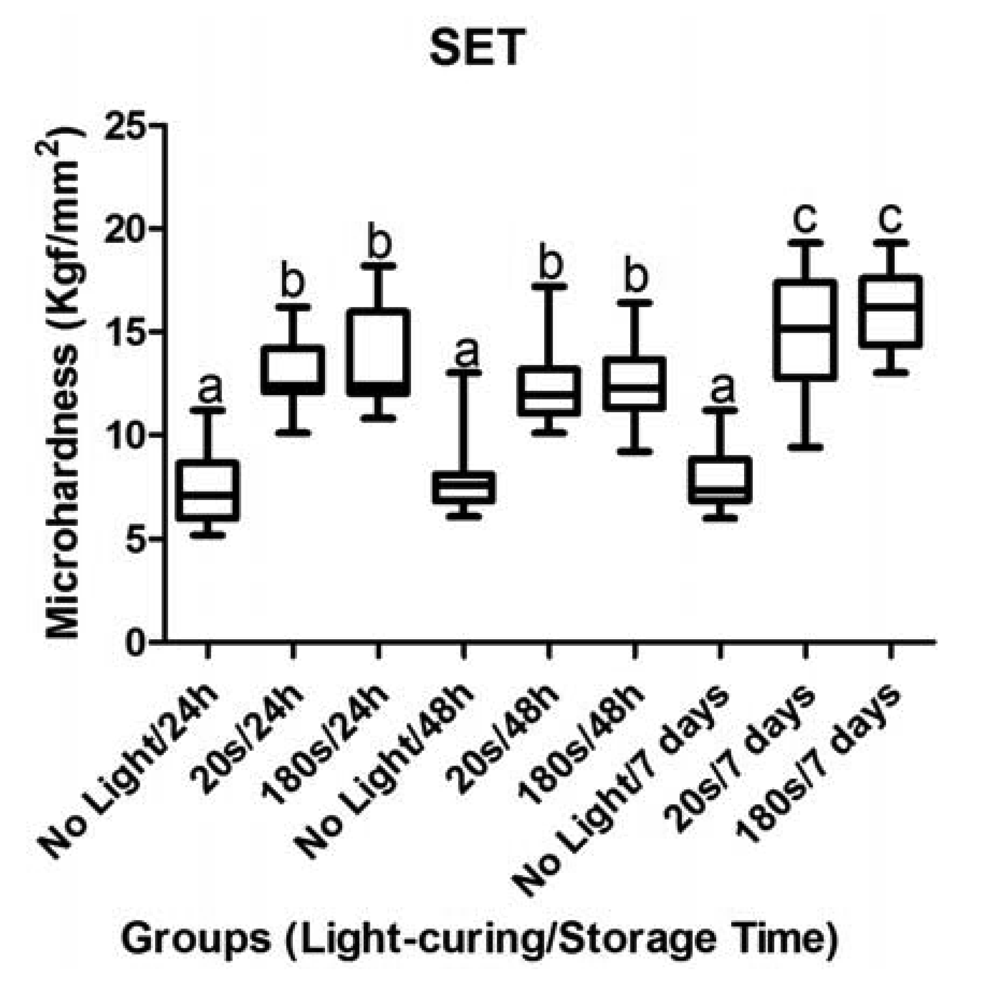
The SET resin cement (Figure 3) showed no significant difference to groups without light exposure for all storage times (p > 0.05). The cement analyzed 7 days after curing showed statistically significant difference for 24 and 48 h (p < 0.05).
The dental resin materials depend on many factors such as the properties of the monomer, polymer and the filler used, the photo-curing process, their concentration, the curing-light unit and their power density and exposure time [9-12].
Furthermore, physical properties of dental materials provide an indication of how the material will function under stress in the oral environment [13]. Microhardness test is widely used to examine dental resin material, analyzing their polymerization, the efficiency of the light units [14,15] and also related to its wear resistance and ability to maintain form stability [16]. This study was analyzed by microhardness test to eliminate any variation that could change the results, because the results of analyzing were only applied for cement. Some studies indicate that the RelyX Unicem (Self-adhesive, such as RelyX U100) contains phosphoric acid esters, which require wet surfaces for ionization and subsequent interaction with dentin and enamel and the values are better if pressure is maintained throughout the cementation procedure to include the self-curing period [17-19].
Resin cements setting by light and chemical activation (dual cured) resulted in the highest hardness values, even compared to resin cement setting only by chemical activation. This outcome might be attributed to the fact that the all resin cements studied are dual cure system, for which the photo-initiator was not sensitized. The chemical mode started the polymerization reaction, characterized by cross-linking formation, which could have reduced monomer mobility in the bulk mass, hindering the completion of cure [20].
Hardness of dental resin cements can be used as indicator of the degree of conversion [21,22], for which there is a correlation between the degree of conversion and hardness test [23]. In this study, the hardness value should not be compared between the dental cements used, because there is a variation in monomers composition, filler content and type.
The SET resin cement showed statistically significant difference 24 h after curing and after 7 days. Others resin cements were similar between them. The larger change of hardness probably happened within 24 h initial.
The mean values and standard deviations are shown in Table 1.
3. Experimental Section
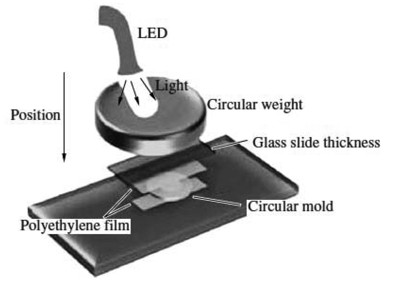
The cement materials used were RelyX ARC, RelyX U100, and SET (Tables 1 and 2). Precapsulated materials (SET, SDI) were prepared using a mixing unit (Ultramat 2, SDI) and the materials (RelyX ARC and RelyX U100) were dispensed, manually mixed, placed in molds. The metallic mold (fabricated in stainless steel) with central orifice (2 mm in diameter and 1 mm in thickness) was used. Five specimens of each group were prepared for each exposure time of light-curing and storage time evaluated. The resin cement was packed into the mold and a polyethylene film covered each side of the sample and a glass slide was placed on the top surface of the samples. The top surface was standardized with a circular weight (1 kg). The light tip of the light-curing unit was positioned on the glass slide, leaving the 0.55 mm of the material. Dual-cure samples were then photopolymerized with exposure times of 20 s and 180 s, using a LED polymerization unit with wavelength of 440 ˜ 480 nm and Light output was consistently 1,500 mW/cm2 (Radii Plus, SDI) (Figure 4). The power density were checked using a powermeter (Fieldmaster, Coherent Commercial Products Division, model number FM, set n° WX65, part number 33-0506 in USA) and the irradiance were calculated with this formula:
All samples were stored in darkness to prevent ambient light from causing additional light-curing polymerization. Specimens were stored in a 98 ± 2% humidity environment in a humidor at 37 ± 2 °C until being tested at 24 h, 48 h, and 7 days.
The Vickers hardness test was performed in a hardness testing machine, MMT-3 Microhardness Tester (Buehler Lake Bluff, Ilinois USA) equipped with Vickers diamond (VHN), which has a format of pyramid of 136° where the two diagonals are measured [8,9] using load of 50 gf (gram force) for 30 s. The surface was divided in quadrant and the diamond took place an impression for quadrant. The hardness data were submitted to the Analysis of Variance with two fixed criteria (two-way-ANOVA). The tests were performed at the level of 5%.
4. Conclusions
The values of hardening of the dual-cured resin cements improved after setting by light and chemical activation. The use of light-curing of dual-cured resin cements became important, within limitations of this study. However, further research is necessary.


| Storage time | Dental Materials | ||||||
|---|---|---|---|---|---|---|---|
| RelyX ARC | RelyX U100 | SET | |||||
| Mean | SD | Mean | SD | Mean | SD | ||
| No light/24 h | 10.86 | 1.88 | 9.17 | 2.94 | 7.68 | 3.50 | |
| 20 s/24 h | 19.99 | 3.18 | 16.18 | 3.41 | 12.93 | 2.88 | |
| 180 s/24 h | 19.35 | 3.72 | 16.83 | 4.60 | 13.33 | 2.61 | |
| No light/48 h | 11.49 | 3.00 | 8.37 | 2.42 | 7.40 | 1.87 | |
| 20 s/48 h | 18.33 | 2.01 | 13.54 | 2.93 | 11.64 | 1.06 | |
| 180 s/48 h | 18.25 | 3.38 | 16.17 | 2.02 | 11.62 | 2.52 | |
| No light/7 days | 11.64 | 1.46 | 11.88 | 4.35 | 7.75 | 1.55 | |
| 20 s/7 days | 18.96 | 2.66 | 15.92 | 2.74 | 14.32 | 3.75 | |
| 180 s/7 days | 17.60 | 1.61 | 18.51 | 1.62 | 16.00 | 4.77 | |
| Materials | Manufacturer | Composition | Shade | Recommended Light-curing Duration |
|---|---|---|---|---|
| RelyX ARC | 3M Dental products Div., St Paul, MN, USA | Paste A: Bis-GMA, tri-ethylene glycol dimethacrylate, zircon/silica filler, photoinitiators, amine, pigments Paste B: Bis-GMA, tri-ethylene glycol dimethacrylate, benzoic peroxide, zircon/silica filler | A2 | 20 s |
| RelyX U100 | 3M Dental products Div., St Paul, MN, USA | Paste A: Glass powder, silica calcium hydroxide, pigment, substituted pyrimidine, peroxy compound, initiator Paste B: Methacrylated, phosphoric esters, Dimethacrylates, Acetate, Stabilizers, Self-cure initiators, Light-cure initiators | A2 | 20 s |
| SET | SDI Limited. Bayswater, WA, AU | Methacrylated phosphoric esters, UDMA, photoinitiator, Fluoroaluminosilicate glass, pyrogenic silica | A2 | 20 s |
Acknowledgments
The authors would like to thank the CAPES—Brazil for financial support.
References
- Sinhoreti, M.A.; Manetta, I.P.; Tango, R.N.; Iriyama, N.T.; Consani, R.L.; Correr-Sobrinho, L. Effect of light-curing methods on resin cement Knoop hardness at different depths. Braz. Dent. J. 2007, 18, 305–308. [Google Scholar]
- Ferracane, J.L.; Stansbury, J.W.; Burke, F.J. Self-adhesive resin cements—chemistry, properties and clinical considerations. J. Oral Rehabil. 2010, 38, 295–314. [Google Scholar]
- Fabianelli, A.; Goracci, C.; Bertelli, E.; Davidson, C.L.; Ferrari, M. A clinical trial of Empress II porcelain inlays luted to vital teeth with a dual-curing adhesive system and a self-curing resin cement. J. Adhes. Dent. 2006, 8, 427–431. [Google Scholar]
- Rasetto, F.H.; Driscoll, C.F.; von Fraunhofer, J.A. Effect of light source and time on the polymerization of resin cement through ceramic veneers. J. Prosthodont. 2001, 10, 133–139. [Google Scholar]
- Radovic, I.; Monticelli, F.; Goracci, C.; Vulicevic, Z.R.; Ferrari, M. Self-adhesive resin cements: A literature review. J. Adhes. Dent. 2008, 10, 251–258. [Google Scholar]
- Caughman, W.F.; Chan, D.C.; Rueggeberg, F.A. Curing potential of dual-polymerizable resin cements in simulated clinical situations. J. Prosthet. Dent. 2001, 86, 101–106. [Google Scholar]
- Myers, M.L.; Caughman, W.F.; Rueggeberg, F.A. Effect of restoration composition, shade, and thickness on the cure of a photoactivated resin cement. J. Prosthodont. 1994, 3, 149–157. [Google Scholar]
- Darr, A.H.; Jacobsen, P.H. Conversion of dual cure luting cements. J. Oral Rehabil. 1995, 22, 43–47. [Google Scholar]
- Ilie, N.; Simon, A. Effect of curing mode on the micro-mechanical properties of dual-cured self-adhesive resin cements. Clin. Oral Investig. 2011. [Google Scholar] [CrossRef]
- Bandeca, M.C.; El-Mowafy, O.; Saade, E.G.; Rastelli, A.N.S.; Bagnato, V.S.; Porto-Neto, S.T. Effect of light-curing units on microleakage under dental composite resins. Laser Phys. 2009, 19, 1050–1055. [Google Scholar]
- Calixto, L.R.; Bandeca, M.C.; Silva, F.B.; Rastelli, A.N.S.; Porto-Neto, S.T.; Andrade, M.F. Curing depth of composite resin light cured by LED and halogen light-curing unit. Laser Phys. 2009, 19, 1867–1869. [Google Scholar]
- Queiroz, R.S.; Bandeca, M.C.; Calixto, L.R.; Saade, E.G.; Nadalin, M.R.; Andrade, M.F. Curing depth of composite resin light cured by LED and halogen light-curing unit. Laser Phys. 2009, 19, 1867–1869. [Google Scholar]
- Cobb, D.S.; Vargas, M.A.; Rundle, T. Physical properties of composites cured with conventional light or argon laser. Am. J. Dent. 1996, 9, 199–202. [Google Scholar]
- Kurachi, C.; Tuboy, A.M.; Magalhaes, D.V.; Bagnato, V.S. Hardness evaluation of a dental composite polymerized with experimental LED-based devices. Dent. Mater. 2001, 17, 309–315. [Google Scholar]
- Saade, E.G.; Bandeca, M.C.; Rastelli, A.N.S.; Bagnato, V.S.; Porto-Neto, S.T. Influence of pre-heat treatment and different light-curing units on Vickers hardness of a microhybrid composite resin. Laser Phys. 2009, 19, 1276–1281. [Google Scholar]
- Baharav, H.; Abraham, D.; Cardash, H.S.; Helft, M. Effect of exposure time on the depth of polymerization of a visible light-cured composite resin. J. Oral Rehabil. 1988, 15, 167–172. [Google Scholar]
- De Munck, J.; Vargas, M.; van Landuyt, K.; Hikita, K.; Lambrechts, P.; van Meerbeek, B. Bonding of an auto-adhesive luting material to enamel and dentin. Dent. Mater. 2004, 20, 963–971. [Google Scholar]
- Goracci, C.; Cury, A.H.; Cantoro, A.; Papacchini, F.; Tay, F.R.; Ferrari, M. Microtensile bond strength and interfacial properties of self-etching and self-adhesive resin cements used to lute composite onlays under different seating forces. J. Adhes. Dent. 2006, 8, 327–335. [Google Scholar]
- Mazzitelli, C.; Monticelli, F.; Osorio, R.; Casucci, A.; Toledano, M.; Ferrari, M. Effect of simulated pulpal pressure on self-adhesive cements bonding to dentin. Dent. Mater. 2008, 24, 1156–1163. [Google Scholar]
- Soh, M.S.; Yap, A.U. Influence of curing modes on crosslink density in polymer structures. J. Dent. 2004, 32, 321–326. [Google Scholar]
- Imazato, S.; McCabe, J.F.; Tarumi, H.; Ehara, A.; Ebisu, S. Degree of conversion of composites measured by DTA and FTIR. Dent. Mater. 2001, 17, 178–183. [Google Scholar]
- Keogh, P.; Ray, N.J.; Lynch, C.D.; Burke, F.M.; Hannigan, A. Surface microhardness of a resin composite exposed to a “first-generation” LED curing lamp, in vitro. Eur. J. Prosthodont. Restor. Dent. 2004, 12, 177–180. [Google Scholar]
- Ferracane, J.L. Correlation between hardness and degree of conversion during the setting reaction of unfilled dental restorative resins. Dent. Mater. 1985, 1, 11–14. [Google Scholar]
© 2011 by the authors; licensee MDPI, Basel, Switzerland. This article is an open access article distributed under the terms and conditions of the Creative Commons Attribution license (http://creativecommons.org/licenses/by/3.0/).
Share and Cite
Aguiar, T.C.; Saad, J.R.C.; Pinto, S.C.S.; Calixto, L.R.; Lima, D.M.; Silva, M.A.S.; Bandéca, M.C. The Effects of Exposure Time on the Surface Microhardness of Three Dual-Cured Dental Resin Cements. Polymers 2011, 3, 998-1005. https://doi.org/10.3390/polym3030998
Aguiar TC, Saad JRC, Pinto SCS, Calixto LR, Lima DM, Silva MAS, Bandéca MC. The Effects of Exposure Time on the Surface Microhardness of Three Dual-Cured Dental Resin Cements. Polymers. 2011; 3(3):998-1005. https://doi.org/10.3390/polym3030998
Chicago/Turabian StyleAguiar, Tatiana C., José R. C. Saad, Shelon C. S. Pinto, Luiz R. Calixto, Darlon M. Lima, Marcos A. S. Silva, and Matheus C. Bandéca. 2011. "The Effects of Exposure Time on the Surface Microhardness of Three Dual-Cured Dental Resin Cements" Polymers 3, no. 3: 998-1005. https://doi.org/10.3390/polym3030998