Article

Broad Spectrum Microbicidal Activity of Photocatalysis by TiO2

1
Department of Microbiology and Immunology, School of Medicine, Teikyo University, 2-11-1 Kaga, Itabashi-ku, Tokyo 173-8605, Japan
2
Kanagawa Academy of Science and Technology, 3-2-1 Sakado, Takatsu-ku, Kawasaki, Kanagawa 213-0012, Japan
3
Department of Urology, Graduate School of Medicine, Yokohama City University, 3-9, Fukuura, Kanazawa-ku, Yokohama, Kanagawa 236-0004, Japan
4
Division of Photocatalyst for Energy and Environment, Research Institute for Science and Technology, Tokyo University of Science, 1-3 Kagurazaka, Shinjuku-ku, Tokyo 162-8601, Japan
5
Research Center for Advanced Science and Technology, The University of Tokyo, 4-6-1 Komaba, Meguro-ku, Tokyo 153-8904, Japan
6
Department of Applied Chemistry, Faculty of Engineering, The University of Tokyo, 7-3-1 Hongo, Bunkyyoo-ku, Tok 113-8656, Japan
*
Author to whom correspondence should be addressed.
These authors contributed equally to this work.
Catalysts 2013, 3(1), 310-323; https://doi.org/10.3390/catal3010310
Submission received: 3 December 2012 / Revised: 25 January 2013 / Accepted: 6 March 2013 / Published: 21 March 2013
(This article belongs to the Special Issue Photocatalysts)

Abstract

:
Photocatalytically active titanium dioxide (TiO2) is widely used as a self-cleaning and self-disinfecting material in many applications to keep environments biologically clean. Several studies on the inactivation of bacteria and viruses by photocatalytic reactions have also been reported; however, only few studies evaluated the spectrum of the microbicidal activity with photocatalysis for various species. There is a need to confirm the expected effectiveness of disinfection by photocatalysis against multidrug-resistant bacteria and viruses. In this study, microbicidal activity of photocatalysis was evaluated by comparing the inactivation of various species of bacteria and viruses when their suspensions were dropped on the surface of TiO2-coated glass. Gram-positive bacteria, e.g., methicillin-resistant Staphylococcus aureus, vancomycin-resistant Enterococcus faecalis, and penicillin-resistant Streptococcus pneumoniae, were easily inactivated by photocatalysis, whereas some gram-negative bacteria, e.g., Escherichia coli and multidrug-resistant Pseudomonas aeruginosa, were gradually inactivated by photocatalysis. Influenza virus, an enveloped virus, was significantly inactivated by photocatalysis compared with feline calicivirus, a non-enveloped virus. The effectiveness of microbicidal activity by photocatalysis may depend on the surface structure. However, they are effectively inactivated by photocatalysis on the surface of TiO2-coated glass. Our data emphasize that effective cleaning and disinfection by photocatalysis in nosocomial settings prevents pathogen transmission.

1. Introduction

Infectious diseases caused by antimicrobial resistance bacteria are becoming one of the major problems during use of antibiotics worldwide [1,2]. Survival of these microbes on environmental surfaces and medical devices leads to nosocomial disease transmission and is thought to contribute to the 90,000 annual deaths from nosocomial pathogens [3,4,5]. Nosocomial infections are frequently associated with resistant organisms, including methicillin-resistant Staphylococcus aureus (MRSA), vancomycin-resistant Enterococcus spp. (VRE), penicillin-resistant Streptococcus pneumoniae (PRSP), Serratia marcescens, multidrug-resistant Acinetobacter baumannii (MDRA), and multidrug-resistant Pseudomonas aeruginosa (MDRP) [6,7]. MRSA and PRSA are also a prevalent cause of community-acquired infection [8]. Currently, new resistance mechanisms, e.g., the New Delhi metallo-β-lactamase-1 (NDM-1), have emerged among several gram-negative bacteria and spread worldwide [9]. Viable pathogens of these organisms in the environment have a significant role in nosocomial transmission, especially for immunocompromised patients.
Furthermore, viruses such as the influenza virus (IFV) and norovirus (NoV) spread across the world [10,11]. These viral species can be transmitted to susceptible hosts via contaminated inanimate surfaces. Control of viral contamination is important to prevent nosocomial transmission. Respiratory and enteric viruses have been reported to survive for days on surfaces, and viral infections are due to transfer from these surfaces to susceptible persons [12,13,14]. IFV and NoV can be transmitted by direct and indirect contact with contaminated surfaces and/or aerosol and is linked to the outbreaks in hospitals and long-term care facilities [15,16].
These infectious diseases are a threat to human health, and indeed, outbreaks and serious clinical cases have occurred [17]. Effective disinfection of healthcare equipment and surfaces is critical for preventing transmission of potential pathogens [18]. Therefore, new antibacterial and antiviral materials or new countermeasures against such microbes are urgently required to disinfect the surface.
Photocatalytically active titanium dioxide (TiO2) is widely used as a self-cleaning and self-disinfecting material in many applications to keep environments biologically clean [19,20,21]. Photocatalysis mainly uses a semiconductor such as TiO2 which can absorb UV light (k < 400 nm) and can photo-stimulate redox reactions on its surface producing Reactive Oxygen Species (ROS) such as hydroxyl radical (·OH), superoxide radical (·O2) [22] and singlet oxygen (1O2) [23], and the production of them may contribute to the biocidal activity with strong oxidative activity and destroy organic compounds [24].
This photocatalytic oxidizing power has been applied for the removal of several toxic substances from water and air [25,26,27]. Many photocatalytic disinfection studies in which TiO2 was used against bacteria and fungi have been reported [28,29]. Many products that utilize TiO2 photocatalysis have been commercialized in many countries. For example, TiO2-coated tiles have been used as self-cleaning and antibacterial materials [30]. While we have reported the virucidal activity of photocatalysis for IFV [31] and bacteriophages [32,33], few studies have addressed viruses [34]. Recently non-TiO2 photocatalysts (i.e., ZnO and ZnS) reported as significant progress archived in development were also observed their antimicrobial activity [35,36].
Thus, photocatalysis is expected to be applied by antimicrobial materials for many harmful pathogens. To use the superb photocatalytic effect of TiO2 is a conceptually feasible technology for this material, and its production at industrial scale is easy and inexpensive. However, there is no study comparing the disinfecting activity of photocatalysis for bacteria and virus using typical TiO2 sample in the same condition, and the effect on drug-resistant bacteria has also not been demonstrated. This study was designed to elucidate the antimicrobial spectrum and the microbicidal activity of photocatalysis on different bacteria species which were bacteria (i.e., gram-positive cocci and gram-negative rods included drug-resistant bacteria) and viruses (i.e., enveloped and non-enveloped viruses).

2. Results and Discussion

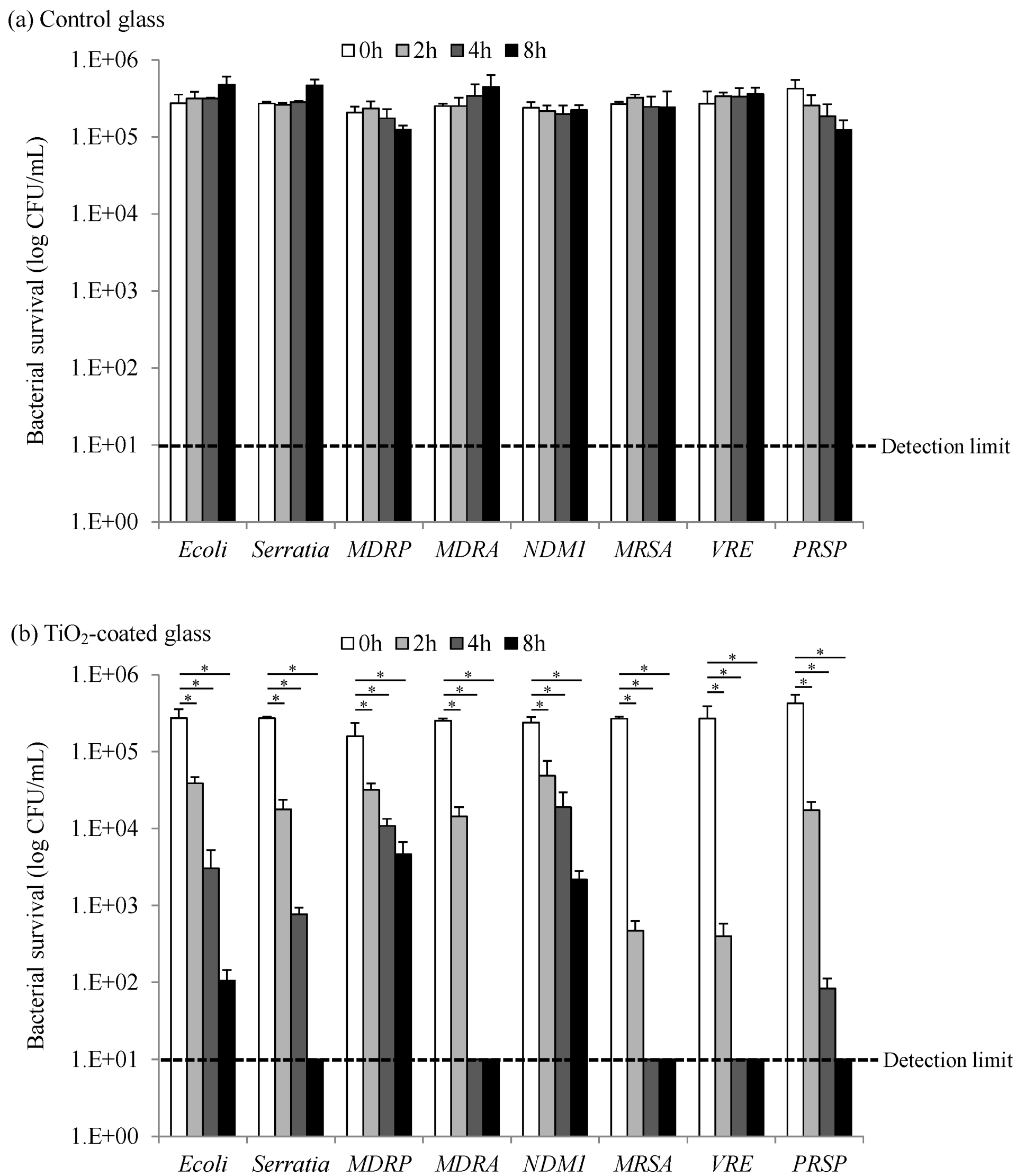
2.1. Bactericidal Activity of Photocatalysis by TiO2-Coated Glass
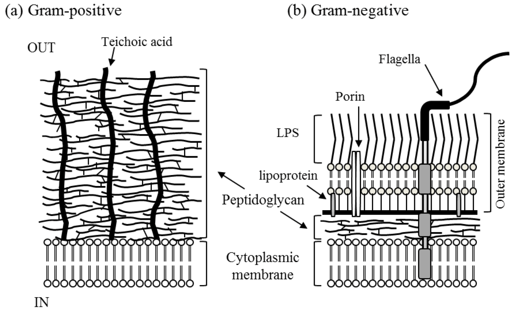
We investigated the bactericidal activity of photocatalysis against different bacteria species that cause an infectious disease at clinical sites. The tested antimicrobial-resistant bacteria species cause opportunistic infections by contact infection and it is very important to cleanse them in a clinical environment. Tested bacteria species on control glass were exposed to 0.1 mW/cm2 UVA; however, significant inactivation was not observed, it means the strains were stable under the condition (Figure 1). Photocatalytic inactivation of different bacteria species was evaluated with TiO2-coated glass under UVA light intensity of 0.1 mW/cm2. All bacteria species could be inactivated by treatment with TiO2 under UV irradiation in a time-dependent manner. We found that TiO2 exhibited a significantly better performance to reduce the number of surviving bacteria when compared with unirradiated (0 h) (Figure 1, p < 0.05). Viable bacterial counts of gram-positive cocci (GPC) such as MRSA, VRE, and PRSP were reduced; the log reduction value (LRV) of them was 1.4–2.8 logs after 2 h, respectively. The number of gram-negative rods (GNRs) such as Escherichia coli, S. marcescens, MDRP, MDRA, and NDM-1 producing Klebsiella pneumoniae gradually decreased, the LRV was between 0.8 and 1.5 logs after 2 h. It is interesting to note that there was a significantly different bactericidal activity between GPC and GNR. This finding suggests differences in the mode of antimicrobial action.
Figure 1. Photocatalytic inactivation of different bacteria with (a) control glass or (b) TiO2-coated glass under black-light illumination (UVA light intensity of 0.1 mW/cm2). The data shown in the figure are the average values of three experiments. Error bars: standard deviations of three replicate experiments. * p < 0.001, 2 h, 4 h, 8 h vs. 0 h exposed to 0.1 mW/cm2 UVA.
Catalysts 03 00310 g001
In particular, GPCs of MRSA, VRE, and PRSP were significantly decreased, i.e., number of surviving cells. They were inactivated below the detection limit (<1 log10 colony-forming units/mL) within 4 h. Many MRSA infections occur with a higher incidence rate in hospitals, healthcare facilities, or long-term care facilities. MRSA is transmitted by contact with a colonized carrier (someone carrying the infection on the skin but who is not infected), or a contaminated object, e.g., clothing and environmental surfaces. The bactericidal activity of photocatalysis would inactivate MRSA effectively at environmental surfaces in hospitals and could prevent the spread of nosocomial infections. The effectiveness of the bactericidal activity by photocatalysis could be one of the infection control strategies for preventing environment-associated infections in healthcare facilities. VRE and PRSP could also be inactivated by photocatalysis and it can be expected to prevent their spread as MRSA.
Except for MDRA, the number of surviving cells of GNRs gradually decreased compared to that of GPCs under UV irradiation with TiO2. The number of cells of MDRAs significantly decreased as the number of GPCs reached a value below the detection limit within 4 h. The number of cells of all tested GNRs decreased to below the detection limit after 16 h (data not shown). The difference in the bactericidal activity for GPC and GNR could be associated with their surface layer structure (Figure 2 and Table 1). The structure of S. aureus, as a representative GPC, is constituted of thick peptidoglycan layer, which covers more than 50%, usually almost 90%. The peptidoglycan layer in the bacterial cell wall has a crystal lattice structure formed from linear chains of two alternating amino sugars, N-acetylglucosamine and N-acetylmuramic acid. The structure of E. coli, as a representative GNR, consists of a thin peptidoglycan layer (which is much thinner than that in GPCs) and an outer membrane containing lipopolysaccharide (LPS) in its outer leaflet and phospholipids in the inner leaflet.
Figure 2. Structure of gram-positive and gram-negative bacteria: (a) Gram-positive bacteria have a thick wall composed of peptidoglycans; and (b) gram-negative bacteria have an outer membrane and a thin wall composed of peptidoglycans.
Catalysts 03 00310 g002
Table 1. Comparative characteristics of gram-positive and gram-negative bacteria.
CharacteristicGram-positiveGram-negative
Thickness of wall thick (20–80 nm)thin (10 nm)
Number of layers 12
Peptidoglycan content>50%10%–20%
Lipid and lipoprotein content 0%–3%58%
Lipopolysaccharide content 013%
The outer membrane of GNRs is unique due to the high content of lipids. The relative resistance of the cell wall of GNR to dissociation by detergents, disinfectants, and antiseptics is well documented [37,38]. It is proposed that the presence of large amounts of LPS and proteins, together with a very small amount of phospholipids in the outer leaflet of the outer membrane, is the decisive factor in the effective detergent resistance of this membrane [37]. GNRs are generally more resistant to disinfectants than GPCs due to outer membrane barrier, but the resistant mechanism is not well characterized. Although the microbicidal activity by photocatalysis has not been studied thoroughly and the mechanism of the bactericidal process is still unknown [27,39], there are some suggestions and evidences for the steps leading to cell inactivation. TiO2 photocatalysis generates various ROS as follows [24].
Reductive reactions:
O2 + e → O2
O2 + H+ ⇄ ·HO2
·HO2 + e + H+ → H2O2
Oxidative reactions:
OH + h+ → ·OH
These ROS can decompose organic compounds and extinguish cellular activity. Recent works attributed that the TiO2 photocatalytic action induced significant damage in cell membranes by ROS, followed by loss of essential functions such as respiratory activity and bacterial lysis leading to bacterial death [24,25,40]. Sunada et al. [41] demonstrated the TiO2 photocatalytic inactivation of E. coli endotoxin, which is a constituent of the outer membrane of GNRs. This finding suggests that the cell wall damage might take place prior to cytoplasmic membrane damage. After the outer membrane has been disordered and partially decomposed (Figure 2), ROS gain access to the cytoplasmic membrane, causing the cell to die. However, while the outer membrane protected GNRs, GPCs have a thick peptidoglycan layer instead of an outer membrane; these ROS would presumably easily penetrate the cytoplasmic membrane and attack the cell membrane directly. The difference in their structure may be of decisive influence on the survival abilities of bacteria.
Some GNRs have flagella, which are long filamentous protein structures projected from the cytoplasmic membrane, constituted of microtubules and surrounded by the cell membrane (Figure 2). Bacterial flagella provide mobility for capturing nutrients and avoiding harmful substances to survive [42]. In this study, MDRA is the exceptional strain among these tested GNR bacteria because it has does not have flagella surrounding the cell membrane and it is inactivated by photocatalysis within 4 h. It is suggested that the lack of flagella at the surface structure of MDRA may be part of the reason for higher bactericidal activity by photocatalysis. Alternatively, other structures such as capsule, pilus, and bacterial size and an antioxidative defense system [43] may be associated with the effectiveness of bactericidal activity. Further study is necessary to clarify the mechanism of the photocatalysis reaction to exercise a decisive influence on bactericidal activity by analyzing the effects against the cell membrane surface.

2.2. Virucidal Activity of Photocatalysis by TiO2-Coated Glass

We investigated the virucidal activity of photocatalysis against IFV and feline calicivirus FCV (a surrogate for NoV) using purified and adjusted suspensions. Tested viruses on control glass were also stable under the UVA light intensity of 0.1 mW/cm2 (Figure 3). As shown in Figure 3, photocatalytic inactivation of these viruses was evaluated with TiO2-coated glass under UVA light intensity of 0.1 mW/cm2. The viruses were inactivated by the treatment with TiO2 under UV irradiation in a time-dependent manner, as shown for bacteria. The experiments were relatively reproducible and this reduction was statistically significant when compared with unirradiated (0 h) (Figure 3, * p < 0.001). The LRV of IFV at 4 h was 3.6 and the titer decreased to below the detection limit within 8 h, while the LRV of FCV at 8 h was only 1.7. The FCV titer decreased to below the detection limit after 16 h. It was concluded that the FCV titer was insignificantly reduced by the photocatalysis effect.
Figure 3. Photocatalytic inactivation of influenza virus (IFV) and feline calicivirus (FCV) with (a) control glass or (b) TiO2-coated glass under black-light illumination (UVA light intensity of 0.1 mW/cm2). The data shown in the figure are the average values of three experiments. Error bars: standard deviations of three replicate experiments. * p < 0.001, 4 h, 8 h, 16 h vs. 0 h exposed to 0.1 mW/cm2 UVA.
Catalysts 03 00310 g003
These data demonstrate that IFV is faster inactivated by photocatalysis than FCV. The low virucidal activity of photocatalysis against the non-enveloped virus was not surprising considering previous studies on biocides. IFV, an enveloped virus, consists of a phospholipid bilayer membrane with glycoprotein spikes surrounding the capsid. FCV, a non-enveloped virus, has a capsid that consists of a protein and lacks an envelope that surrounds it. Although enveloped viruses are effectively eliminated by solvent/detergent treatment, non-enveloped viruses are not affected by this treatment [44]. Non-enveloped viruses, without a lipid coat, are resistant to most of the viral inactivation methods that have been described to date [45,46]. These data indicate that the difference in the effectiveness to enveloped and non-enveloped viruses by photocatalysis is the same as that for disinfectants. It is suggested that the virucidal activity of photocatalysis may depend on the viral envelope. Previously, we demonstrated that IFV decreased the infectability by damaging their constituent proteins prior to nucleic acid degradation with photocatalysis reaction [31]. The photocatalytic action of TiO2 may promote peroxidation of phospholipid components of the enveloped membrane, inducing significant damage in membranes as mode of inactivation that is observed for bacteria. The phospholipid content may influence the virucidal activity of photocatalysis. Further study is required to clarify the mechanism of virucidal activity by evaluating other viruses. Although there was a difference in an effect caused by IFV and FCV, TiO2 is a potential, environment-friendly antivirus agent.

3. Experimental Section

3.1. Bacteria Strains

Drug-resistant and clinically pathogenic strains of GPC and GNR were used as reference strains to evaluate the bactericidal activity by photocatalysis. They were donated from other researchers or purchased from ATCC (American Type Culture Collection, Manassas, VA, USA). They included the GPC strains MRSA ATCC 43300, VRE ATCC 51575, PRSP ATCC 700677, and the GNR strains E. coli ATCC 8739, S. marcescens ATCC 13880, MDRA, MDRP, and NDM-1 producing K. pneumoniae BAA-2146. MDRA and MDRP were isolated from a hospital in Japan and they were identified to be resistant to ciprofloxacin (minimum inhibitory concentration: MIC ≥ 4 mg/mL), imipenem (MIC ≥ 16 μg/mL), and amikacin (MIC ≥ 64 μg/mL). The profiles of these strains are listed in Table 2.
Table 2. Profiles of the organisms tested in this study.
SpeciesStructureTypes of Infections a
E. coliGram-negative rodDiarrhea, UTI, food poisoning, sepsis
S. marcescensGram-negative rodNosocomial infections, Pneumonia, UTI, and wound infections
MDRAGram-negative rodNosocomial infections, Pneumonia, UTI, VAP
MDRPGram-negative rodNosocomial infections, Pneumonia, UTI, VAP, Bacteremia, cystic fibrosis
NDM-1 producing K. pneumoniaeGram-negative rodNosocomial infections, Pneumonia, UTI, Bacteremia
MRSAGram-positive cocciNosocomial infections, skin infections, food poisoning, pneumonia, meningitis, bacteremia
VREGram-positive cocciNosocomial infections, UTI, meningitis, bacteremia
PRSPGram-positive cocciCommunity-acquired pneumonia, bacteremia, bacterial meningitis
IFVEnveloped virusRespiratory tract infections
FCVNon-enveloped virusRespiratory infection in cats
a UTI, Urinary tract infection; VAP, Ventilator Associated Pneumonia.
The strains except PRSP were cultivated twice consecutively on nutrient agar plates (Becton, Dickinson and Company, Franklin Lakes, NJ, USA) while PRSP was cultivated on 5% sheep blood agar plates at 37 °C for 18–20 h. Subsequently, the live cells were suspended in 1/500 nutrient broth solutions, which contained 6 mg of meat extract, 20 mg of peptone, and 10 mg of NaCl per liter (pH 7.0), and then diluted to approximately 107 colony-forming units per milliliter. All reagents and concentrations used were prepared in accordance with the JIS R 1702 standard method [47].

3.2. Virus Strains

The human IFV A/PR8/H1N1 strain and FCV strain F-9 ATCC VR-782 were used as reference strains to evaluate the virucidal activity by photocatalysis. They were purchased from ATCC. IFV was propagated by using 11-day-old embryonated chicken eggs and purified by using previously described methods [43]. FCV was propagated by using confluent layers of Crandell-Reese feline kidney (CRFK) cell cultures. The harvested samples were clarified and concentrated by a combination of depth and membrane filters (microfiltration with 0.45-μm polypropylene mesh and cross-flow polysulfone hollow-fiber ultrafiltration with a 100-kDa cut-off, GE Healthcare, Japan) [48]. Subsequently, the virus was purified by sucrose density gradient centrifugation using a linear sucrose gradient of 30%–70% [49]. Each individual experimental sample was adjusted to 0.1 mg/mL protein with phosphate-buffered saline, and 0.1 mg/mL of bovine serum albumin was added as stabilizing agent [50,51]. The virus titer of IFV and FCV was determined by using the plaque technique on confluent layers of Madin-Darby canine kidney (MDCK) cell or CRFK cell cultures grown in 12-well culture plates, respectively, as described before [49,52].

3.3. TiO2-Coated Glass

TiO2-coated glass (T3 sample, 50 mm × 50 mm) was obtained from TOTO Ltd., (Fukuoka, Japan) by employing a spin-coating process with TiO2 (obtained from Ishihara Sangyo Kaisha, Ltd., Osaka, Japan) and heat treatment at 150 °C. As mentioned previously, the T3 sample was used as test sample for the preparation of ISO27447:2009(E) [53] and JIS R 1702 [47] for evaluating the photocatalytic antibacterial activity. The phase space of this sample was smooth, the thickness was approximately 200 nm, and the specific surface area was 300 m2/g. Non-TiO2 coated glass was used as the control. The T3 sample and control glass plates were washed with sterilized water and then sterilized by UV exposure overnight or wiped with ethanol.

3.4. Photocatalytic Reaction

The photocatalytic inactivation of bacteria and virus was investigated by applying the test method for the evaluation of the antibacterial effect and the test method used in our previous study for the evaluation of the antivirus effect [31]. Overhead illumination by long-wavelength UV (UV-A) light was provided by using tubular 20-W black light fluorescent lamps (FL20S Black Light Blue (BLB); λ = 352–368 nm; Toshiba, Japan). Illumination conditions of 0.1 mW/cm2 were adjusted by changing the horizontal distance between the samples and lamp. UV-A intensity was analyzed by using an integrating actinometer for photocatalysis (C9536–01/H9958; Hamamatsu Photonics Co. Ltd., Hamamatsu, Japan). All experiments were performed in a light-tight box to prevent any influence of indoor light and sunlight.
The bacterial or viral suspension (100 μL) was dropped onto the T3 sample or control grass and equally distributed by covering it with adhesive film (VF-15, Kokuyo Co. Ltd., Osaka, Japan, 40 mm × 40 mm). The sample was then illuminated with UV-A irradiation. The photocatalytic reaction utilized light intensity in the range of 0.1 mW/cm2 at the sample surface, and the UV-A irradiation time was 0–8 h. After illumination for a certain period of time, the sample was immersed and washed with 10 mL of soybean-casein digest broth, which contained lecithin and polysorbate. The collected viable bacteria or virus cells were serially diluted in phosphate-buffered saline and evaluated by using the colony formation method or plaque assay, respectively. All experiments were repeated independently more than three times, and the average titer was determined. Statistical analysis was performed by the Bonferroni test using SPSS software (SPSS Inc., Chicago, IL, USA). p < 0.05 was considered statistically significant. All reagents and concentrations used were prepared in accordance with ISO27447:2009(E) [53] and JIS R 1702 [47], and with minor modifications for the concentrations of viruses as previously reported [31].

4. Conclusions

We have demonstrated broad-spectrum biocide efficacy of photocatalysis against both bacteria (GPC and GNR, including drug-resistant bacteria) and viruses (enveloped and non-enveloped virus). GPC such as MRSA and VRE were easily inactivated by photocatalysis, whereas some GNRs such as E. coli and MDRP were gradually inactivated by the photocatalysis. The difference in the bactericidal activity against GPC and GNR could be associated with the structure of their surface layer. IFV, an enveloped virus, was significantly faster inactivated by photocatalysis compared with FCV, a non-enveloped virus. These data indicated that the virucidal activity of photocatalysis may depend on the presence or absence of a viral envelope.
The LRV of representative species induced in the presence of the tested photocatalyst were found to decrease in the following order (LRV of the species at 4 h): MRSA (4.4) > IFV (3.6) > E. coli (2.1) > FCV (1.3). Photocatalysis has broad-spectrum microbicidal activity and the effects were found to be in the following order: GPC > enveloped virus > GNR > non-enveloped virus. Although there was a difference in the effect due to the surface structure, TiO2 is a potential, environmentally friendly microbicidal agent.
Photocatalysis would reduce the risk of contact infection spreading within the environmental surfaces at facilities, e.g., as measure against healthcare-associated infections at hospitals or healthcare facilities, and prevention of the spread of community-acquired infections at airports or day-care centers. Furthermore, because of the high level and broad-spectrum biocide efficacy of photocatalysis, it is considered a valuable biocide method for use in various areas, e.g., sewage disposal treatment systems and air purification systems [54].

Acknowledgments

We would like to thank Ishihara Sangyo Kaisha, LTD for the gift of TiO2. We also thank TOTO Ltd. for assistance in the preparation of the TiO2-coated samples. We thank Hiromi Yamamoto for excellent technical assistance. The original studies in our program were supported by the New Energy and Industrial Technology Development Organization (NEDO) of Japan.

Conflict of Interest

The authors declare no conflict of interest.

References

  1. Bratu, S.; Landman, D.; Haag, R.; Recco, R.; Eramo, A.; Alam, M.; Quale, J. Rapid spread of carbapenem-resistant Klebsiella pneumoniae in New York City: A new threat to our antibiotic armamentarium. Arch. Int. Med. 2005, 165, 1430–1435. [Google Scholar] [CrossRef]
  2. Livermore, D.M. Fourteen years in resistance. Int. J. Antimicrob. Agents 2012, 39, 283–294. [Google Scholar] [CrossRef]
  3. Burke, J.P. Infection control—A problem for patient safety. N. Engl. J. Med. 2003, 348, 651–656. [Google Scholar] [CrossRef]
  4. Lankford, M.G.; Collins, S.; Youngberg, L.; Rooney, D.M.; Warren, J.R.; Noskin, G.A. Assessment of materials commonly utilized in health care: Implications for bacterial survival and transmission. Am. J. Infect. Control 2006, 34, 258–263. [Google Scholar] [CrossRef]
  5. Neely, A.N.; Maley, M.P. Survival of enterococci and staphylococci on hospital fabrics and plastic. J. Clin. Microbiol. 2000, 38, 724–726. [Google Scholar]
  6. Kramer, A.; Schwebke, I.; Kampf, G. How long do nosocomial pathogens persist on inanimate surfaces? A systematic review. BMC Infect. Dis. 2006, 6, 130. [Google Scholar] [CrossRef]
  7. Weber, D.J.; Rutala, W.A. Use of germicides in the home and the healthcare setting: Is there a relationship between germicide use and antibiotic resistance? Infect. Control Hosp. Epidemiol. 2006, 27, 1107–1119. [Google Scholar] [CrossRef]
  8. Ramirez, J.A.; Anzueto, A.R. Changing needs of community-acquired pneumonia. J. Antimicrob. Chemother. 2011, 66, iii3–iii9. [Google Scholar] [CrossRef]
  9. Nordmann, P.; Naas, T.; Poirel, L. Global spread of carbapenemase-producing enterobacteriaceae. Emerg. Infect. Dis. 2011, 17, 1791–1798. [Google Scholar] [CrossRef]
  10. Fraser, C.; Donnelly, C.A.; Cauchemez, S.; Hanage, W.P.; van Kerkhove, M.D.; Hollingsworth, T.D.; Griffin, J.; Baggaley, R.F.; Jenkins, H.E.; Lyons, E.J.; et al. Pandemic potential of a strain of influenza A (H1N1): Early findings. Science 2009, 324, 1557–1561. [Google Scholar] [CrossRef]
  11. Hutson, A.M.; Atmar, R.L.; Estes, M.K. Norovirus disease: Changing epidemiology and host susceptibility factors. Trends Microbiol. 2004, 12, 279–287. [Google Scholar] [CrossRef]
  12. Boone, S.A.; Gerba, C.P. Significance of fomites in the spread of respiratory and enteric viral disease. Appl. Environ. Microbiol. 2007, 73, 1687–1696. [Google Scholar] [CrossRef]
  13. Sakaguchi, H.; Wada, K.; Kajioka, J.; Watanabe, M.; Nakano, R.; Hirose, T.; Ohta, H.; Aizawa, Y. Maintenance of influenza virus infectivity on the surfaces of personal protective equipment and clothing used in healthcare settings. Environ. Health Prev. Med. 2010, 15, 344–349. [Google Scholar] [CrossRef]
  14. Goldmann, D.A. Transmission of viral respiratory infections in the home. Pediatr. Infect. Dis. J. 2000, 19, S97–S102. [Google Scholar] [CrossRef]
  15. Wu, H.M.; Fornek, M.; Schwab, K.J.; Chapin, A.R.; Gibson, K.; Schwab, E.; Spencer, C.; Henning, K. A norovirus outbreak at a long-term-care facility: The role of environmental surface contamination. Infect. Control Hosp. Epidemiol. 2005, 26, 802–810. [Google Scholar]
  16. Infection Control in Health Care Facilities. Available online: http://www.cdc.gov/flu/professionals/infectioncontrol/ (accessed on 15 October 2012).
  17. Nakano, R.; Nakano, A.; Abe, M.; Inoue, M.; Okamoto, R. Regional outbreak of CTX-M-2 beta-lactamase-producing Proteus mirabilis in Japan. J. Med. Microbiol. 2012, 61, 1727–1735. [Google Scholar] [CrossRef]
  18. Guidelines for Environmental Infection Control in Health-Care Facilities. Recommendations of CDC and the Healthcare Infection Control Practices Advisory Committee (HICPAC); U.S. Department of Health and Human Services Centres for Disease Control and Prevention (CDC): Atlanta, GA, USA, 2003; pp. 1–42.
  19. Hashimoto, K.; Irie, H.; Fujishima, A. TiO2 photocatalysis: A historical overview and future prospects. Jpn. Soc. Appl. Phys. 2005, 44, 8269–8285. [Google Scholar] [CrossRef]
  20. Nakata, K.; Fujishima, A. TiO2 photocatalysis: Design and applications. J. Photochem. Photobiol. C 2012, 13, 169–189. [Google Scholar] [CrossRef]
  21. Nakata, K.; Ochiai, T.; Murakami, T.; Fujishima, A. Photoenergy conversion with TiO2 photocatalysis: New materials and recent applications. Electrochim. Acta 2012, 84, 103–111. [Google Scholar]
  22. Linsebigler, A.L.; Lu, G.; Yates, J.T. Photocatalysis on TiO2 surfaces: Principles, mechanisms, and selected results. Chem. Rev. 1995, 95, 735–758. [Google Scholar] [CrossRef]
  23. Daimon, T.; Hirakawa, T.; Kitazawa, M.; Suetake, J.; Nosaka, Y. Formation of singlet molecular oxygen associated with the formation of superoxide radicals in aqueous suspensions of TiO2 photocatalysts. Appl. Catal. A 2008, 340, 169–175. [Google Scholar] [CrossRef]
  24. Ishibashi, K.I.; Fujishima, A.; Watanabe, T.; Hashimoto, K. Generation and deactivation processes of superoxide formed on TiO2 film illuminated by very weak UV light in air or water. J. Phys. Chem. B 2000, 104, 4934–4938. [Google Scholar] [CrossRef]
  25. Wei, C.; Lin, W.Y.; Zainal, Z.; Williams, N.E.; Zhu, K.; Kruzic, A.P.; Smith, R.L.; Rajeshwar, K. Bactericidal activity of TiO2 photocatalyst in aqueous media: Toward a solar-assisted water disinfection system. Environ. Sci. Technol. 1994, 28, 934–938. [Google Scholar] [CrossRef]
  26. Ryu, H.; Gerrity, D.; Crittenden, J.C.; Abbaszadegan, M. Photocatalytic inactivation of cryptosporidium parvum with TiO2 and low-pressure ultraviolet irradiation. Water Res. 2008, 42, 1523–1530. [Google Scholar] [CrossRef]
  27. Ochiai, T.; Fujishima, A. Photoelectrochemical properties of TiO2 photocatalyst and its applications for environmental purification. J. Photochem. Photobiol. C 2012, 13, 247–262. [Google Scholar] [CrossRef]
  28. Sunada, K.; Watanabe, T.; Hashimoto, K. Bactericidal activity of copper-deposited TiO2 thin film under weak UV light illumination. Environ. Sci. Technol. 2003, 37, 4785–4789. [Google Scholar]
  29. Mitoraj, D.; Janczyk, A.; Strus, M.; Kisch, H.; Stochel, G.; Heczko, P.B.; Macyk, W. Visible light inactivation of bacteria and fungi by modified titanium dioxide. Photochem. Photobiol. Sci. 2007, 6, 642–628. [Google Scholar] [CrossRef]
  30. Fujishima, A.; Zhang, X.; Tryk, D. TiO2 photocatalysis and related surface phenomena. Surf. Sci. Rep. 2008, 63, 515–582. [Google Scholar] [CrossRef]
  31. Nakano, R.; Ishiguro, H.; Yao, Y.; Kajioka, J.; Fujishima, A.; Sunada, K.; Minoshima, M.; Hashimoto, K.; Kubota, Y. Photocatalytic inactivation of influenza virus by titanium dioxide thin film. Photochem. Photobiol. Sci. 2012, 11, 1293–1298. [Google Scholar] [CrossRef]
  32. Ishiguro, H.; Nakano, R.; Yao, Y.; Kajioka, J.; Fujishima, A.; Sunada, K.; Minoshima, M.; Hashimoto, K.; Kubota, Y. Photocatalytic inactivation of bacteriophages by TiO2-coated glass plates under low-intensity, long-wavelength UV irradiation. Photochem. Photobiol. Sci 2011, 10, 1825–1829. [Google Scholar] [CrossRef]
  33. Ishiguro, H.; Yao, Y.; Nakano, R.; Hara, M.; Sunada, K.; Hashimoto, K.; Kajioka, J.; Fujishima, A.; Kubota, Y. Photocatalytic activity of Cu2+/TiO2-coated cordierite foam inactivates bacteriophages and Legionella pneumophila. Appl. Catal. B 2013, 129, 56–61. [Google Scholar] [CrossRef]
  34. Gerrity, D.; Ryu, H.; Crittenden, J.; Abbaszadegan, M. Photocatalytic inactivation of viruses using titanium dioxide nanoparticles and low-pressure UV light. J. Environ. Sci. Health. A 2008, 43, 1261–1270. [Google Scholar] [CrossRef]
  35. Kavitha, T.; Gopalan, A.I.; Lee, K.P.; Park, S.Y. Glucose sensing, photocatalytic and antibacterial properties of graphene-ZnO nanoparticle hybrids. Carbon 2012, 50, 2994–3000. [Google Scholar] [CrossRef]
  36. Synnott, D.W.; Seery, M.K.; Hinder, S.J.; Michlits, G.; Pillai, S.C. Anti-bacterial activity of indoor-light activated photocatalysts. Appl. Catal. B 2013, 130–131, 106–111. [Google Scholar]
  37. Nixdorff, K.; Gmeiner, J.; Martin, H.H. Interaction of lipopolysaccharide with detergents and its possible role in the detergent resistance of the outer membrane of Gram-negative bacteria. Biochim. Biophys. Acta 1978, 510, 87–98. [Google Scholar] [CrossRef]
  38. McDonnell, G.; Russell, A.D. Antiseptics and disinfectants: Activity, action, and resistance. Clin. Microbiol. Rev. 1999, 12, 147–179. [Google Scholar]
  39. Blake, D.M.; Maness, P.C.; Huang, Z.; Wolfrum, E.J.; Huang, J.; Jacoby, W.A. Application of the photocatalytic chemistry of titanium dioxide to disinfection and the killing of cancer cells. Sep. Purif. Rev. 1999, 28, 1–50. [Google Scholar] [CrossRef]
  40. Maness, P.C.; Smolinski, S.; Blake, D.M.; Huang, Z.; Wolfrum, E.J.; Jacoby, W.A. Bactericidal activity of photocatalytic TiO2 reaction: Toward an understanding of its killing mechanism. Appl. Environ. Microbiol. 1999, 65, 4094–4098. [Google Scholar]
  41. Sunada, K.; Kikuchi, Y.; Hashimoto, K.; Fujishima, A. Bactericidal and detoxification effects of TiO2 thin film photocatalysts. Environ. Sci. Technol. 1998, 32, 726–728. [Google Scholar] [CrossRef]
  42. Macnab, R.M. How bacteria assemble flagella. Annu. Rev. Microbiol. 2003, 57, 77–100. [Google Scholar] [CrossRef]
  43. Ibáñez, J.A.; Litter, M.I.; Pizarro, R.A. Photocatalytic bactericidal effect of TiO2 on Enterobacter cloacae. J. Photochem. Photobiol. A 2003, 157, 81–85. [Google Scholar] [CrossRef]
  44. Horowitz, B.; Wiebe, M.E.; Lippin, A.; Stryker, M.H. Inactivation of viruses in labile blood derivatives. I. Disruption of lipid-enveloped viruses by tri(n-butyl)phosphate detergent combinations. Transfusion 1985, 25, 516–522. [Google Scholar]
  45. Beekes, M.; Mielke, M.; Pauli, G.; Baier, M.; Kurth, R. Aspects of risk assessment and risk management of nosocomial transmission of classical and variant Creutzfeldt-Jakob disease with special attention to German regulations. Contrib. Microbiol. 2004, 11, 117–135. [Google Scholar] [CrossRef]
  46. Rutala, W.A.; Weber, D.J.; Healthcare Infection Control Practices Advisory Committee (HICPAC). Guideline for Disinfection and Sterilization in Healthcare Facilities, 2008; Centres for Disease Control and Prevention (CDC): Atlanta, GA, USA, 2008. [Google Scholar]
  47. Shima, E.H. JIS R 1702, Fine Ceramics (Advanced Ceramics, Advanced Technical Ceramics)—Test Method for Antibacterial Activity of Photocatalytic Products under Photoirradiation and Efficacy; Japanese Standards Association: Tokyo, Japan, 2006. [Google Scholar]
  48. Kalbfuss, B.; Genzel, Y.; Wolff, M.; Zimmermann, A.; Morenweiser, R.; Reichl, U. Harvesting and concentration of human influenza A virus produced in serum-free mammalian cell culture for the production of vaccines. Biotechnol. Bioeng. 2007, 97, 73–85. [Google Scholar]
  49. Kreutz, L.C.; Seal, B.S.; Mengeling, W.L. Early interaction of feline calicivirus with cells in culture. Arch. Virol. 1994, 136, 19–34. [Google Scholar] [CrossRef]
  50. Van Regenmortel, M.H.V.; Fauquet, C.M.; Bishop, D.H.L. Virus Taxonomy. Classification and Nomenclature of Viruses; Academic Press: New York, NY, USA, 2000; pp. 835–849. [Google Scholar]
  51. Yachida, S.; Aoyama, S.; Takahashi, N.; Iritani, Y.; Katagiri, K. Plastic multiwell plates to assay avian infectious bronchitis virus in organ cultures of chicken embryo trachea. J. Clin. Microbiol. 1978, 8, 380–387. [Google Scholar]
  52. Tobita, K.; Sugiura, A.; Enomote, C.; Furuyama, M. Plaque assay and primary isolation of influenza A viruses in an established line of canine kidney cells (MDCK) in the presence of trypsin. Med. Microbiol. Immunol. 1975, 162, 9–14. [Google Scholar] [CrossRef]
  53. Fine Ceramics (Advanced Ceramics, Advanced Technical Ceramics)—Test Method for Antibacterial Activity of Semiconducting Photocatalytic Materials; Japan Industrial Standards Committee: Tokyo, Japan, 2009.
  54. Ochiai, T.; Nanba, H.; Nakagawa, T.; Masuko, K.; Nakata, K.; Murakami, T.; Nakano, R.; Hara, M.; Koide, Y.; Suzuki, T.; et al. Development of an O3-assisted photocatalytic water-purification unit by using a TiO2 modified titanium mesh filter. Catal. Sci. Technol. 2012, 2, 76. [Google Scholar] [CrossRef]

