<?xml version="1.0" encoding="UTF-8"?>
<!DOCTYPE article PUBLIC "-//NLM//DTD Journal Publishing DTD v2.3 20070202//EN" "journalpublishing.dtd">
<article xmlns:xlink="http://www.w3.org/1999/xlink" xml:lang="en" article-type="review-article">
<front>
<journal-meta>
<journal-id journal-id-type="publisher-id">Cancers</journal-id>
<journal-title>Cancers</journal-title>
<issn pub-type="epub">2072-6694</issn>
<publisher>
<publisher-name>Molecular Diversity Preservation International (MDPI)</publisher-name></publisher></journal-meta>
<article-meta>
<article-id pub-id-type="doi">10.3390/cancers3 010001</article-id>
<article-id pub-id-type="publisher-id">cancers-03-00001</article-id>
<article-categories>
<subj-group>
<subject>Review</subject></subj-group></article-categories>
<title-group>
<article-title>The Role of Apoptosis in the Pathology of Pancreatic Cancer</article-title></title-group>
<contrib-group>
<contrib contrib-type="author">
<name><surname>Samm</surname><given-names>Nicole</given-names></name></contrib>
<contrib contrib-type="author">
<name><surname>Werner</surname><given-names>Kristin</given-names></name></contrib>
<contrib contrib-type="author">
<name><surname>Rückert</surname><given-names>Felix</given-names></name></contrib>
<contrib contrib-type="author">
<name><surname>Saeger</surname><given-names>Hans Detlev</given-names></name></contrib>
<contrib contrib-type="author">
<name><surname>Grützmann</surname><given-names>Robert</given-names></name></contrib>
<contrib contrib-type="author">
<name><surname>Pilarsky</surname><given-names>Christian</given-names></name><xref ref-type="corresp" rid="c1-cancers-03-00001"><sup>*</sup></xref></contrib>
<aff id="af1-cancers-03-00001">Department of Visceral-, Thoracic-and Vascular-Surgery, University Hospital Dresden, Dresden, Germany; E-Mails: <email>sammynic82@yahoo.de</email> (N.S.); <email>Kristin.Werner@uniklinikum-dresden.de</email> (K.W.); <email>Felix.Rueckert@uniklinikum-dresden.de</email> (F.R.); <email>Hans-Detlev.Saeger@uniklinikum-dresden.de</email> (H.D.S.); <email>Robert.Gruetzmann@uniklinikum-dresden.de</email> (R.G.)</aff></contrib-group>
<author-notes>
<corresp id="c1-cancers-03-00001">
<label>*</label> Author to whom correspondence should be addressed; Tel.: +49 351 458 166 07; Fax: +49 351 449 210 303; E-Mail: <email>christian.pilarsky@uniklinikum-dresden.de</email>.</corresp></author-notes>
<pub-date pub-type="collection">
<year>2011</year></pub-date>
<pub-date pub-type="epub">
<day>23</day>
<month>12</month>
<year>2010</year></pub-date>
<volume>3</volume>
<issue>1</issue>
<fpage>1</fpage>
<lpage>16</lpage>
<history>
<date date-type="received">
<day>11</day>
<month>11</month>
<year>2010</year></date>
<date date-type="rev-recd">
<day>14</day>
<month>12</month>
<year>2010</year></date>
<date date-type="accepted">
<day>21</day>
<month>12</month>
<year>2010</year></date></history>
<permissions>
<copyright-statement>© 2010 by the authors; licensee MDPI, Basel, Switzerland.</copyright-statement>
<copyright-year>2010</copyright-year>
<license>
<p>This article is an open access article distributed under the terms and conditions of the Creative Commons Attribution license (http://creativecommons.org/licenses/by/3.0/).</p></license></permissions>
<abstract>
<p>Pancreatic cancer is a disease with high resistance to most common therapies and therefore has a poor prognosis, which is partly due to a lack of reaction to apoptotic stimuli. Signal transduction of such stimuli includes a death receptor-mediated extrinsic pathway as well as an intrinsic pathway linked to the mitochondria. Defects in apoptotic pathways and the deregulation of apoptotic proteins, such as Survivin, Bcl-2, Bcl-x<sub>L</sub> and Mcl-1, play decisive roles in the development of pancreatic cancer. Investigation of the molecular mechanism allowing tumors to resist apoptotic cell death would lead to an improved understanding of the physiology and the development of new molecular strategies in pancreatic cancer.</p></abstract>
<kwd-group>
<kwd>apoptosis</kwd>
<kwd>signal transduction pathway</kwd>
<kwd>pancreatic cancer</kwd></kwd-group></article-meta></front>
<body>
<sec sec-type="intro">
<label>1.</label>
<title>Introduction</title>
<p>Pancreatic cancer is one of the most malignant and aggressive types of cancer in humans and carries a very poor prognosis. With 40,000 new cases diagnosed in the United States each year, pancreatic cancer is the fourth and fifth leading cause of cancer-related death in the Western world for males and females, respectively [<xref ref-type="bibr" rid="b1-cancers-03-00001">1</xref>]. Approximately 95% of exocrine pancreatic cancer cases are ductal adenocarcinoma (PDAC) [<xref ref-type="bibr" rid="b2-cancers-03-00001">2</xref>]. In 65% of these cases, the tumor is located in the head of the pancreas; in 30%, in the corpus; and in 5%, in the tail of the organ [<xref ref-type="bibr" rid="b3-cancers-03-00001">3</xref>]. The delayed appearance of symptoms causes a late diagnosis; as a result, roughly 85% of patients show an organ overlapping growth of the tumor when the disease is first discovered, and only the remaining 15% of patients have an opportunity for curative surgical treatment. Despite improvements made in surgical techniques and pre- and postoperative care, less progress has been seen in improving the survival of patients with this type of cancer [<xref ref-type="bibr" rid="b3-cancers-03-00001">3</xref>]. The five-year survival rate after surgical resection is about 20% and is about 5% for all patients [<xref ref-type="bibr" rid="b1-cancers-03-00001">1</xref>]. Furthermore, uncontrolled proliferation, a high metastatic potential and resistance to most adjuvant therapies also contribute to the very poor prognosis of this disease. The response to oncology therapy options such as chemotherapy, radiotherapy and immunotherapy is not satisfying. Tumor development and progress of PDACs as well as resistance to most oncology therapies involve the absence of a reaction to apoptotic stimuli [<xref ref-type="bibr" rid="b4-cancers-03-00001">4</xref>].</p>
<p>Resistance to apoptosis and the ability to evade this process are two of the hallmarks of human cancer [<xref ref-type="bibr" rid="b5-cancers-03-00001">5</xref>]. This review will discuss the facts about apoptotic pathways and the deregulation of apoptotic proteins in pancreatic cancer to demonstrate the correlation between disease occurrence and defects in apoptotic mechanisms.</p></sec>
<sec>
<label>2.</label>
<title>Apoptosis—A General Overview</title>
<p>In 1842, Carl Vogt described the concept of natural cell death for the first time [<xref ref-type="bibr" rid="b6-cancers-03-00001">6</xref>]. Since 1972, Kerr, Wyllie and Curry have been linked to the word apoptosis and the development, progression and treatment of cancer. They published a paper in which they characterized and defined this form of cell death and gave birth to the term apoptosis [<xref ref-type="bibr" rid="b7-cancers-03-00001">7</xref>,<xref ref-type="bibr" rid="b8-cancers-03-00001">8</xref>]. The word apoptosis is of Greek origin and refers to the fall of leaves in autumn.</p>
<p>Apoptosis is an intrinsic cell suicide program. As a central regulation mechanism for tissue homeostasis, it is involved in the regulation of many physiological and pathophysiological processes such as the differentiation of the embryonic body figure, development of the nervous system, formation of the immune system and the homeostasis of the number of cells in proliferating tissue [<xref ref-type="bibr" rid="b9-cancers-03-00001">9</xref>,<xref ref-type="bibr" rid="b10-cancers-03-00001">10</xref>].</p>
<p>In contrast to necrosis, apoptosis takes place without any inflammatory reaction and is marked by cellular shrinking, condensation of the chromatin and ruffling of the plasma membrane with loss of contact to other cells of the cell assembly [<xref ref-type="bibr" rid="b11-cancers-03-00001">11</xref>,<xref ref-type="bibr" rid="b12-cancers-03-00001">12</xref>]. It is described as a breaking-up of the cell into apoptotic bodies. These bodies, which consist of cell organelles and nuclear material, are surrounded by an intact plasma membrane. Macrophages recognize the apoptotic cell fragments and abolish them by phagocytosis [<xref ref-type="bibr" rid="b13-cancers-03-00001">13</xref>].</p>
<p>
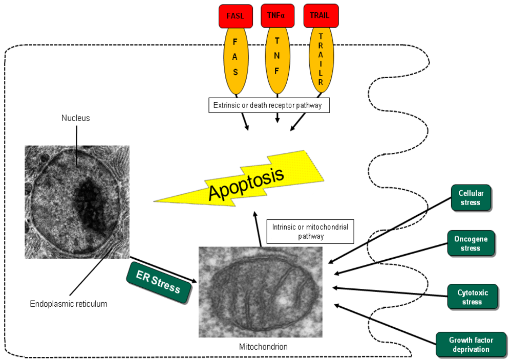
Different entry points to apoptosis have been described, such as the death receptor-mediated or extrinsic pathway and the mitochondrial intrinsic pathway [<xref ref-type="bibr" rid="b12-cancers-03-00001">12</xref>]. Another entry point for intrinsic apoptosis has been identified at the endoplasmic reticulum (ER) (<xref ref-type="fig" rid="f1-cancers-03-00001">Figure 1</xref>) [<xref ref-type="bibr" rid="b15-cancers-03-00001">15</xref>,<xref ref-type="bibr" rid="b16-cancers-03-00001">16</xref>]. Cell irritation, such as variation in calcium homeostasis or a collection of wrongly folded proteins, causes stress that initiates apoptosis by activation of the unfolded protein response or cleavage of the cargo receptor protein BAP31 [<xref ref-type="bibr" rid="b15-cancers-03-00001">15</xref>,<xref ref-type="bibr" rid="b17-cancers-03-00001">17</xref>,<xref ref-type="bibr" rid="b18-cancers-03-00001">18</xref>]. This mechanism causes the transfer of calcium from the endoplasmic reticulum into the mitochondria and the initiation of cytochrome c release [<xref ref-type="bibr" rid="b15-cancers-03-00001">15</xref>]. All pathways converge in the activation of the executive cell death enzymes, the effector caspases 3, 6 and 7. These enzymes conclude terminal apoptosis by cleaving the nuclear lamina, DNase inhibitors and cytoskeletal proteins.</p>
<p>These complex pathways are controlled and affected by an array of different pro- and anti-apoptotic factors, which are important in ensuring tissue balance [<xref ref-type="bibr" rid="b12-cancers-03-00001">12</xref>]. Altered expression levels and mutations that influence the activation and function of these pro- and anti-apoptotic genes influence cancer cell sensitivity to chemotherapy, radiotherapy, tumor development and progression [<xref ref-type="bibr" rid="b9-cancers-03-00001">9</xref>].</p></sec>
<sec>
<label>3.</label>
<title>The Extrinsic Pathway</title>
<p>
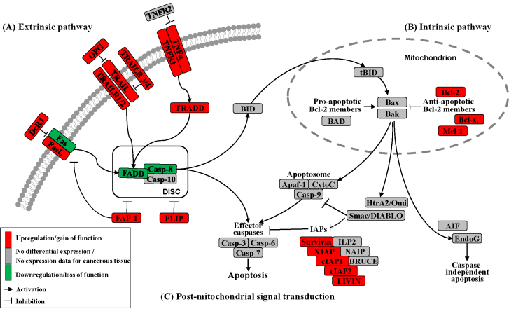
Apoptosis can be initiated by extrinsic but endogenous “death signals”. The receptors TNF (tumor necrosis factor), Fas (Apo-1, CD95) and TRAILR (TNF-related apoptosis-inducing ligand, Apo-2) are members of the TNF receptor superfamily and share a common internal death domain. Activation occurs by their natural ligands TNFα, FasL (Fas-ligand) and TRAIL [<xref ref-type="bibr" rid="b20-cancers-03-00001">20</xref>]. The interaction between receptor and ligand causes trimerization of the receptor followed by the activation and recruitment of FADD (Fas-associated death domain protein) and procaspases 8 and 10. This activation subsequently initiates the formation of the death-inducing signaling complex (DISC) (<xref ref-type="fig" rid="f2-cancers-03-00001">Figure 2</xref>) [<xref ref-type="bibr" rid="b21-cancers-03-00001">21</xref>]. The death domain that forms the DISC attaches to procaspase 8, which has low proteolytic activity. Through this connection to DISC, the local concentration of procaspase 8 increases, leading to an autoproteolytic cleavage and the release of active caspase 8. By cleavage next to their aspartate residues, the caspases activate each other, leading to a caspase cascade. Finally, the effector caspases 3, 6 and 7 conclude terminal apoptosis by cleaving the nuclear lamina, DNase inhibitors or cytoskeletal proteins [<xref ref-type="bibr" rid="b9-cancers-03-00001">9</xref>,<xref ref-type="bibr" rid="b22-cancers-03-00001">22</xref>,<xref ref-type="bibr" rid="b23-cancers-03-00001">23</xref>].</p></sec>
<sec>
<label>4.</label>
<title>The Death Receptor</title>
<p>The death receptor Fas is part of one of the main apoptotic cell death signaling pathways (<xref ref-type="fig" rid="f2-cancers-03-00001">Figure 2</xref>) [<xref ref-type="bibr" rid="b23-cancers-03-00001">23</xref>]. Altered expression levels of Fas or FasL have been found in many human cancers. Reports concerning Fas receptor expression in pancreatic cancer are conflicting. It has been demonstrated that Fas mRNA levels are elevated in pancreatic carcinomas [<xref ref-type="bibr" rid="b23-cancers-03-00001">23</xref>]. However, native pancreatic tumor cells show very little Fas receptor expression, though they express high levels of FasL and its antagonistic decoy receptor DcR3. Decoy receptors can also bind these death ligands, but because of their missing death domain, they do not transduce the apoptotic signal, thereby preventing this ligand from associating with the conventional signal receptor. Altogether, these data indicate that tumor cells, especially those from pancreatic carcinoma, can evade Fas-mediated apoptosis by downregulation of the Fas receptor [<xref ref-type="bibr" rid="b23-cancers-03-00001">23</xref>,<xref ref-type="bibr" rid="b24-cancers-03-00001">24</xref>]. This resistance is also closely linked to the expression of FAP-1 (Fas associated phosphatase-1) and several other intracellular proteins such as Bcl-2, Bcl-x<sub>L</sub> and FLIP [<xref ref-type="bibr" rid="b25-cancers-03-00001">25</xref>,<xref ref-type="bibr" rid="b26-cancers-03-00001">26</xref>].</p>
<p>The protein tyrosine phosphatase FAP-1 appears to be overexpressed in pancreatic cancer cells and protects these cells from Fas-mediated apoptosis by inhibiting the activation of caspase 8 (<xref ref-type="fig" rid="f2-cancers-03-00001">Figure 2</xref>) [<xref ref-type="bibr" rid="b26-cancers-03-00001">26</xref>,<xref ref-type="bibr" rid="b27-cancers-03-00001">27</xref>]. FAP-1 can interfere with the translocation of Fas to the cell surface. This interference causes a low receptor density and an interruption of receptor trimerization, which would be essential for DISC formation. In addition, FAP-1 may inhibit caspase 8 in a direct manner [<xref ref-type="bibr" rid="b20-cancers-03-00001">20</xref>,<xref ref-type="bibr" rid="b26-cancers-03-00001">26</xref>].</p>
<p>Another strong inhibitor of caspase 8 activation is FLIP (<xref ref-type="fig" rid="f2-cancers-03-00001">Figure 2</xref>). FADD-like ICE inhibitor proteins (FLIPs) are structural homologues of caspase 8 and compete with procaspase 8 for binding to FADD at the DISC. The proteins of this family are highly expressed in pancreatic carcinoma cells, resulting in a suppressed signal transduction [<xref ref-type="bibr" rid="b28-cancers-03-00001">28</xref>-<xref ref-type="bibr" rid="b30-cancers-03-00001">30</xref>]. In addition to high expression of FLIP, a low quantity of FADD was shown to be a factor important for resistance against FasL- and TRAIL-induced apoptosis in pancreatic carcinoma [<xref ref-type="bibr" rid="b20-cancers-03-00001">20</xref>,<xref ref-type="bibr" rid="b24-cancers-03-00001">24</xref>], which might cause the progression of malignant pancreatic carcinoma.</p>
<p>A second death receptor system recognizes the tumor necrosis factor α (TNFα) (<xref ref-type="fig" rid="f2-cancers-03-00001">Figure 2</xref>), an inflammatory cytokine with a large variety of biological functions such as regulation of cell death and survival, differentiation and inflammation. There are two different kinds of TNF receptors: TNF receptor 1 (TNFR1) is responsible for signal transduction, while TNF receptor 2 (TNFR2) is a decoy receptor [<xref ref-type="bibr" rid="b31-cancers-03-00001">31</xref>]. TNFR1 and TNFR2 are not overexpressed in pancreatic cancer; therefore, no participation in the mechanism of apoptosis resistance is anticipated [<xref ref-type="bibr" rid="b32-cancers-03-00001">32</xref>,<xref ref-type="bibr" rid="b33-cancers-03-00001">33</xref>].</p>
<p>A third extrinsic stimulus is provided by TRAIL (<xref ref-type="fig" rid="f2-cancers-03-00001">Figure 2</xref>). Identified in 1995, TRAIL has two death receptors, TRAIL-R1/DR4 and TRAIL-R2/DR5, and three antagonistic decoy receptors, TRAIL-R3/DcR1, TRAIL-R4/DcR2 and Osteoprotegrin (OPG) [<xref ref-type="bibr" rid="b34-cancers-03-00001">34</xref>]. TRAIL is normally expressed by natural killer cells in the immune system to combat tumorigenesis. In pancreatic cancer cells, TRAIL receptors as well as the decoy receptors TRAIL-R4 and OPG are highly expressed [<xref ref-type="bibr" rid="b35-cancers-03-00001">35</xref>], and it appears that the TRAIL system is functional in pancreatic cancer but is blocked at apoptotic pathways downstream [<xref ref-type="bibr" rid="b36-cancers-03-00001">36</xref>].</p>
<p>
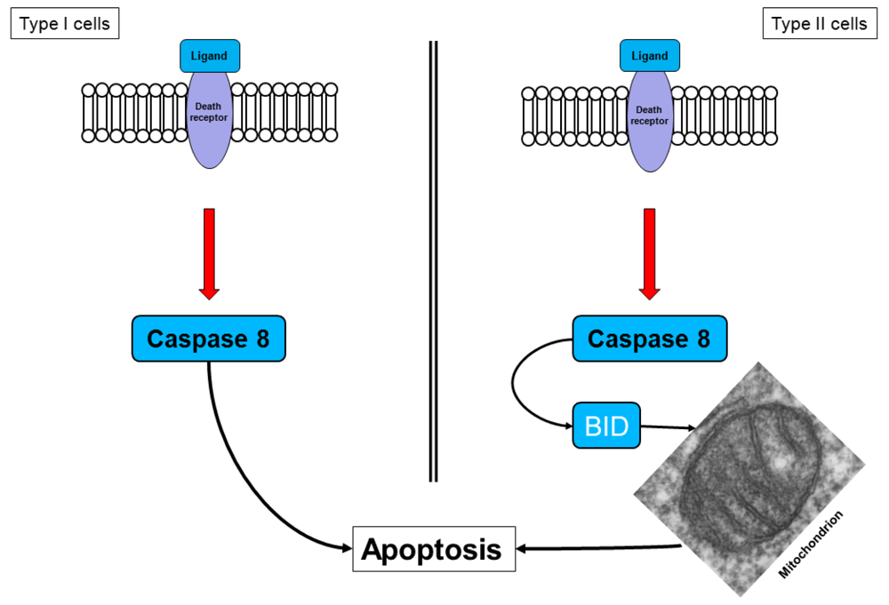
Two different cell types have been identified for the death receptor signaling pathway (<xref ref-type="fig" rid="f3-cancers-03-00001">Figure 3</xref>). In type I cells, the death receptor complex, together with the adaptor molecule FADD, recruits procaspase 8, which is then cleaved to the active caspase 8, thereby activating the effector caspases [<xref ref-type="bibr" rid="b37-cancers-03-00001">37</xref>]. This action implies that, in type I cells, the activated initiator caspases are sufficient to induce executioner caspases directly.</p>
<p>Pancreatic cancer cells are type II cells [<xref ref-type="bibr" rid="b27-cancers-03-00001">27</xref>,<xref ref-type="bibr" rid="b37-cancers-03-00001">37</xref>]. These cells have reduced receptor complex formation; thus, less procaspase 8 can be activated (<xref ref-type="fig" rid="f3-cancers-03-00001">Figure 3</xref>). Signal amplification is required for apoptosis induction. In these cells, caspase 8 cleaves BID (a Bcl-2 family member), which translocates to the mitochondrial membrane and induces the release of apoptotic factors [<xref ref-type="bibr" rid="b27-cancers-03-00001">27</xref>,<xref ref-type="bibr" rid="b37-cancers-03-00001">37</xref>]. The signal requires the enhancing effect of the mitochondria to induce apoptosis, a mechanism that is called the mitochondrial amplification loop [<xref ref-type="bibr" rid="b20-cancers-03-00001">20</xref>,<xref ref-type="bibr" rid="b22-cancers-03-00001">22</xref>].</p></sec>
<sec>
<label>5.</label>
<title>The Intrinsic Pathway</title>
<p>In the intrinsic or mitochondrial pathway of apoptosis, caspase activation is closely connected to the permeabilization of the outer mitochondrial membrane. This is mediated by pro-apoptotic members of the Bcl-2 family, mitochondrial lipids, proteins that control bioenergetic flux and components of the permeability transition pore [<xref ref-type="bibr" rid="b38-cancers-03-00001">38</xref>]. Numerous cytotoxic stimuli and pro-apoptotic signal-transducing molecules affect the permeability of the outer mitochondrial membrane (<xref ref-type="fig" rid="f1-cancers-03-00001">Figure 1</xref>). Trigger points or intracellular signals for the activation of the mitochondrial pathway include DNA damage, oxidative and cytotoxic stresses and ER stress [<xref ref-type="bibr" rid="b39-cancers-03-00001">39</xref>]. By disrupting the outer membrane, a collection of proteins normally found in the space between the inner and outer mitochondrial membranes are released; these proteins include cytochrome c, apoptosis inducing factor (AIF), SMAC/DIABLO, Omi/HtrA2 and endonuclease G [<xref ref-type="bibr" rid="b40-cancers-03-00001">40</xref>]. In the cytosol, these apoptotic proteins initiate cell death by promoting caspase activation [<xref ref-type="bibr" rid="b11-cancers-03-00001">11</xref>]. The release of cytochrome c triggers caspase 3 activation by formation of the apoptosome complex, which contains cytochrome c/Apaf-1/caspase 9 (<xref ref-type="fig" rid="f2-cancers-03-00001">Figure 2</xref>) [<xref ref-type="bibr" rid="b41-cancers-03-00001">41</xref>,<xref ref-type="bibr" rid="b42-cancers-03-00001">42</xref>]. The construction of the apoptosome and the release of cytochrome c seem to be unperturbed in pancreatic cancer cell lines [<xref ref-type="bibr" rid="b43-cancers-03-00001">43</xref>]. SMAC/DIABLO and Omi/HtrA2, which show normal expression levels in pancreatic carcinoma cells [<xref ref-type="bibr" rid="b44-cancers-03-00001">44</xref>], enable caspase activation by neutralizing important endogenous inhibitor of apoptosis proteins (IAPs) [<xref ref-type="bibr" rid="b45-cancers-03-00001">45</xref>-<xref ref-type="bibr" rid="b47-cancers-03-00001">47</xref>].</p>
<p>Most of the cell death in human cells is initiated by the intrinsic pathway and results from an unregulated increase in mitochondrial membrane permeability [<xref ref-type="bibr" rid="b48-cancers-03-00001">48</xref>]. Pathologic alterations of the intrinsic pathway in pancreatic cancer include intramitochondrial signal transduction, which is regulated by the Bcl-2 protein family [<xref ref-type="bibr" rid="b49-cancers-03-00001">49</xref>,<xref ref-type="bibr" rid="b50-cancers-03-00001">50</xref>]. It is the comparative balance of these pro- or anti-apoptotic members of this family that determines if activation of the intrinsic pathway occurs.</p>
<p>Links between the receptor (extrinsic) and the mitochondrial (intrinsic) pathway exist at different levels of the machinery. BID (BH3-interacting domain death agonist) is a pro-apoptotic member of the Bcl-2 protein family with a BH3 domain. Once cleaved by caspase 8, it translocates as “truncated BID” (tBID) to the mitochondrion where, along with other pro-apoptotic Bcl-2 proteins, it initiates the intrinsic pathway (<xref ref-type="fig" rid="f2-cancers-03-00001">Figure 2</xref>) [<xref ref-type="bibr" rid="b51-cancers-03-00001">51</xref>,<xref ref-type="bibr" rid="b52-cancers-03-00001">52</xref>]. Type II cells, which have low caspase 8 activation as seen in pancreatic carcinoma cells [<xref ref-type="bibr" rid="b37-cancers-03-00001">37</xref>], can achieve this mitochondrial amplification loop for an efficient transduction of the apoptotic signal [<xref ref-type="bibr" rid="b21-cancers-03-00001">21</xref>].</p>
<p>In summary, the death program can be initiated by different intracellular or extracellular stimuli that activate the common cell death machinery downstream. Both pathways result in activation of the effector caspases, which cleave proteins that are very important for the rigidity and function of the cells.</p></sec>
<sec>
<label>6.</label>
<title>Regulation of Apoptosis</title>
<p>The Bcl-2 gene was discovered in 1986 by Yoshide Tsujimoto at the junction of the hallmark t(14;18) chromosome translocation characteristic of human follicular lymphoma [<xref ref-type="bibr" rid="b53-cancers-03-00001">53</xref>]. It was named Bcl-2 for B-cell lymphoma 2 [<xref ref-type="bibr" rid="b54-cancers-03-00001">54</xref>].The expanding number of proteins related to Bcl-2 by sequence homology and participation in the control of the apoptotic machinery has led to the definition of a Bcl-2 family of proteins [<xref ref-type="bibr" rid="b55-cancers-03-00001">55</xref>].</p>
<p>The Bcl-2 family has two pro-apoptotic subgroups, the BAX-like subgroup (BAX, BAK and BOK) and the BH3-only subgroup (BAD, BIK, BID, BIM, BMF, HRK, NOXA and PUMA). The only anti-apoptotic group consists of Bcl-2, Bcl-x<sub>L</sub> and Mcl-1 (<xref ref-type="fig" rid="f2-cancers-03-00001">Figure 2</xref>) [<xref ref-type="bibr" rid="b22-cancers-03-00001">22</xref>,<xref ref-type="bibr" rid="b38-cancers-03-00001">38</xref>]. Bcl-2 family proteins interact with other molecules through an α-helical domain termed the BH-3 domain [<xref ref-type="bibr" rid="b48-cancers-03-00001">48</xref>]. The BH3-only proteins act as sensors of cellular stress, directly antagonizing anti-apoptotic Bcl-2 members and activating BAX-like proteins, ultimately leading to permeabilization of the outer mitochondrial membrane [<xref ref-type="bibr" rid="b56-cancers-03-00001">56</xref>,<xref ref-type="bibr" rid="b57-cancers-03-00001">57</xref>].</p>
<p>Bcl-2, an anti-apoptotic family member is located on the cytoplasmic side of the mitochondrial outer membrane, at the ER-membrane or on the nuclear cover. It registers damage to these compartments and prevents the release of cytochrome c from mitochondria in a number of different tissues [<xref ref-type="bibr" rid="b58-cancers-03-00001">58</xref>]. In a few human tumors, high expression of Bcl-2 has been found, but the expression in pancreatic cancer cells is normal or even decreased [<xref ref-type="bibr" rid="b57-cancers-03-00001">57</xref>,<xref ref-type="bibr" rid="b59-cancers-03-00001">59</xref>].</p>
<p>Bcl-x in human cells is present in two distinct isoforms. Bcl-x<sub>L</sub> is the longer form, acting in an anti-apoptotic manner. Bcl-x<sub>S</sub>, the shorter form, performs as an apoptosis promoter, in contrast to Bcl-x<sub>L</sub>. It has been shown that Bcl-x<sub>L</sub> prevents the release of cytochrome c from the mitochondria [<xref ref-type="bibr" rid="b48-cancers-03-00001">48</xref>]. In type II cells such as pancreatic cancer cells, the overexpressed Bcl-x<sub>L</sub> plays the most important role in protecting the cell from Fas-and TRAIL-mediated apoptosis [<xref ref-type="bibr" rid="b35-cancers-03-00001">35</xref>].</p>
<p>Another anti-apoptotic Bcl-2 family member that is highly overexpressed in pancreatic cancer cells is Mcl-1. High concentrations of this protein protect the cancer cells from hypoxia and oxidative stress during tumorigenesis [<xref ref-type="bibr" rid="b20-cancers-03-00001">20</xref>]. Fritsch <italic>et al.</italic> showed rapid and selective downregulation of Mcl-1 that preceded the activation of BAX, BAK and caspases [<xref ref-type="bibr" rid="b60-cancers-03-00001">60</xref>].</p>
<p>Of the pro-apoptotic members of the Bcl-2 family, Bax (Bcl-2 associated X protein) resides in the cytosol and translocates to mitochondria upon induction of apoptosis [<xref ref-type="bibr" rid="b61-cancers-03-00001">61</xref>]. Overexpression of Bax does not influence the apoptosis rate or expression of Bcl-2 and Bcl-x<sub>L</sub> in human pancreatic cancer cells transduced with a retroviral expression vector [<xref ref-type="bibr" rid="b62-cancers-03-00001">62</xref>]. In pancreatic cancer, Bak (Bcl-2 antagonist/killer protein) expression and apoptosis occur in regions of chronic inflammation surrounding the cancer cells but not in the tumor cells themselves, which may simplify accelerated growth and spread [<xref ref-type="bibr" rid="b63-cancers-03-00001">63</xref>]. Bad, a typical pro-apoptotic member of the Bcl-2 family, binds with its BH3 domain to both Bcl-2 and Bcl-x<sub>L</sub> and mediates the pro-apoptotic function of Bcl-x<sub>L</sub> [<xref ref-type="bibr" rid="b64-cancers-03-00001">64</xref>].</p>
<p>In summary, intramitochondrial signal transduction in pancreatic cancer cells is unbalanced towards the anti-apoptotic side [<xref ref-type="bibr" rid="b50-cancers-03-00001">50</xref>,<xref ref-type="bibr" rid="b65-cancers-03-00001">65</xref>,<xref ref-type="bibr" rid="b66-cancers-03-00001">66</xref>]. The deregulation of both the pro- and anti-apoptotic Bcl-2 proteins plays a crucial role both in the development, growth and expansion of pancreatic cancer and in the resistance to current therapy options [<xref ref-type="bibr" rid="b37-cancers-03-00001">37</xref>,<xref ref-type="bibr" rid="b55-cancers-03-00001">55</xref>].</p></sec>
<sec>
<label>7.</label>
<title>Main Mediators of Apoptosis</title>
<p>Caspases (Cysteine-dependent aspartate specific proteases) are proteases that contain a cysteine in their active center and are able to cleave proteins, particularly behind an aspartate residue [<xref ref-type="bibr" rid="b46-cancers-03-00001">46</xref>]. Based on their pro-apoptotic functions, the caspases have been divided into two groups. The initiator caspases 2, 8, 9 and 10 are at the beginning of the signal cascade and are involved in the initiation of apoptosis. The effector or downstream caspases 3, 6 and 7 cleave their substrates; this action leads to cell death because the caspases are able to degrade multiple substrates directly, including structural and regulatory proteins in the cell nucleus, the cytoplasm and the cytoskeleton (<xref ref-type="fig" rid="f2-cancers-03-00001">Figure 2</xref>) [<xref ref-type="bibr" rid="b67-cancers-03-00001">67</xref>].</p>
<p>In healthy cells, all of the caspases are constantly expressed in their inactive form as proenzymes in the cytoplasmic milieu. They are synthesized as a single chain of inactive zymogens composed of four domains: an N-terminal prodomain of variable length, a large subunit with a molecular weight of about 20 kDa, a small subunit and a linker region connecting these catalytic subunits [<xref ref-type="bibr" rid="b67-cancers-03-00001">67</xref>,<xref ref-type="bibr" rid="b68-cancers-03-00001">68</xref>]. Activation during progression of apoptosis happens by a proteolytic processing. Caspases initiate and execute cell death by inactivating anti-apoptotic proteins, shutting down DNA replication and repair and disrupting the cytoskeleton and nuclear lamina [<xref ref-type="bibr" rid="b37-cancers-03-00001">37</xref>].</p>
<p>Caspase 8 is essential for the extrinsic cell death pathway, which is initiated by TNF family members. Death receptors recruit the DISC upon binding specific TNF family ligands and trimerization. Several reports show that caspase 8 is mutated in diverse types of cancer, especially gastric cancer [<xref ref-type="bibr" rid="b69-cancers-03-00001">69</xref>-<xref ref-type="bibr" rid="b71-cancers-03-00001">71</xref>].</p>
<p>Caspase 9, the apical initiator caspase within the apoptosome-dependent cascade, is an almost ubiquitous protease. The activated apoptosome binds caspase 9 and activates the enzyme and caspase 3 downstream. It is expressed constitutively in a variety of fetal and adult normal or cancerous tissues [<xref ref-type="bibr" rid="b41-cancers-03-00001">41</xref>,<xref ref-type="bibr" rid="b72-cancers-03-00001">72</xref>,<xref ref-type="bibr" rid="b73-cancers-03-00001">73</xref>].</p>
<p>Caspase 3 is an effector caspase and can be activated by either the extrinsic or the intrinsic cell death pathway. It plays a central role in the execution phase of cell apoptosis. Several reports have focused on mutations of caspase 3 in a large group of different cancer types [<xref ref-type="bibr" rid="b74-cancers-03-00001">74</xref>,<xref ref-type="bibr" rid="b75-cancers-03-00001">75</xref>].</p>
<p>When the literature is analyzed for a correlation between these three main caspases in cancer development in general and pancreatic cancer in particular, it is clear that caspase expression is relatively normal in pancreatic cancer tissue. There is no evidence that a specific mutation of caspase 3, 8 or 9 leads to a higher incidence of pancreatic carcinoma [<xref ref-type="bibr" rid="b37-cancers-03-00001">37</xref>,<xref ref-type="bibr" rid="b68-cancers-03-00001">68</xref>].</p>
<p>Many different inhibitors and activators regulate caspase activation in a very strict manner. The oligomerization model explains that caspases exist as inactive monomers. The effector caspases bring them together, allowing for their intermolecular autoproteolytic activation. For example, to become functional, procaspase 8 requires association with its cofactor FADD, and procaspase 9 must interact with APAF-1 (<xref ref-type="fig" rid="f2-cancers-03-00001">Figure 2</xref>) [<xref ref-type="bibr" rid="b37-cancers-03-00001">37</xref>,<xref ref-type="bibr" rid="b76-cancers-03-00001">76</xref>].</p>
<p>In contrast to the normal expression of caspases in pancreatic cancer, effectors blocking caspase activation or function, such as FLIPs, show elevated expression in pancreatic cancer, leading to resistance to death receptor-mediated apoptosis [<xref ref-type="bibr" rid="b21-cancers-03-00001">21</xref>,<xref ref-type="bibr" rid="b37-cancers-03-00001">37</xref>].</p></sec>
<sec>
<label>8.</label>
<title>IAP Family Inhibitors of Apoptosis Proteins</title>
<p>The family of inhibitor of apoptosis proteins (IAPs) was discovered in 1993 in the baculovirus genome [<xref ref-type="bibr" rid="b10-cancers-03-00001">10</xref>]. There are eight human IAPs, including XIAP (X-linked inhibitor of apoptosis) and Survivin, which are direct inhibitors of caspases 3 and 7 and procaspase 9. Other members of this collective are cIAP 1 and 2 (cellular IAP1 and 2), ILP 2, ML-IAP, NAIP and BRUCE [<xref ref-type="bibr" rid="b45-cancers-03-00001">45</xref>,<xref ref-type="bibr" rid="b77-cancers-03-00001">77</xref>], which are involved in the signal transduction of human receptor complexes, for example TNFR2 (TNF receptor 2). Their expression is stimulated by growth factors and they are inhibited by mitochondrial proteins such as SMAC/DIABLO (second mitochondria-derived activator of caspase/direct IAP binding protein with low pI) (<xref ref-type="fig" rid="f2-cancers-03-00001">Figure 2</xref>). These proteins block caspases 3, 7 and 9 and lead to the ubiquitylation of caspases 3 and 7, which results in their degradation by the proteasome. IAPs are characterized by a 70–80 amino acid BIR domain (baculoviral IAP repeat), a carboxy-terminal zinc finger domain and one or more additional functional domains that are necessary for caspase interaction [<xref ref-type="bibr" rid="b78-cancers-03-00001">78</xref>].</p>
<p>Overexpression of XIAP, cIAP1, cIAP2 and Survivin has been demonstrated to suppress apoptosis, but the cellular function of the IAP family members is unclear [<xref ref-type="bibr" rid="b78-cancers-03-00001">78</xref>]. However, given their role in cellular homeostasis, it is not surprising that deregulation of IAP expression or function seems to be involved in a large number of cancer species [<xref ref-type="bibr" rid="b77-cancers-03-00001">77</xref>]. Indeed, there are data suggesting that altered expression of cIAP1, cIAP2, XIAP and Survivin play a role in the pathogenesis of pancreatic cancer [<xref ref-type="bibr" rid="b37-cancers-03-00001">37</xref>].</p>
<p>XIAP exerts an anti-apoptotic function by binding and inhibiting effector caspases such as caspases 3 and 7 and procaspase 9 [<xref ref-type="bibr" rid="b79-cancers-03-00001">79</xref>,<xref ref-type="bibr" rid="b80-cancers-03-00001">80</xref>]. It has been demonstrated that of all mammalian IAPs, XIAP is the only one that is truly a physiological inhibitor of caspases <italic>in vivo</italic> [<xref ref-type="bibr" rid="b81-cancers-03-00001">81</xref>]. XIAP targeting has been shown to be required not only for effective induction of apoptosis but also for potent suppression of long term survival; this finding has important implications for the development of experimental strategies directed toward IAP proteins in human cancer, especially pancreatic cancer [<xref ref-type="bibr" rid="b45-cancers-03-00001">45</xref>,<xref ref-type="bibr" rid="b57-cancers-03-00001">57</xref>,<xref ref-type="bibr" rid="b79-cancers-03-00001">79</xref>].</p>
<p>Survivin does not directly bind caspases but inhibits apoptosis by cooperative interactions with other partners <italic>in vivo</italic>. For example, such a partner for an IAP-IAP complex is XIAP [<xref ref-type="bibr" rid="b82-cancers-03-00001">82</xref>]. Survivin expression is turned off during fetal development and is absent from non-neoplastic adult human tissues. With 142 amino acids, Survivin is the smallest mammalian IAP, and its structure contains only a single BIR domain and lacks a carboxy-terminal RING finger domain [<xref ref-type="bibr" rid="b37-cancers-03-00001">37</xref>,<xref ref-type="bibr" rid="b83-cancers-03-00001">83</xref>]. Survivin is expressed in the G2/M phase of the cell cycle. Survivin is a conspicuous cancer gene that is overexpressed in almost every human tumor, including neuroblastoma and cancers of the lung, colon, breast and prostate, whereas it is largely undetectable or expressed at very low levels in normal human tissues [<xref ref-type="bibr" rid="b82-cancers-03-00001">82</xref>]. It has also been demonstrated that Survivin is frequently expressed in malignant pancreatic tumors [<xref ref-type="bibr" rid="b37-cancers-03-00001">37</xref>]. Survivin is a potent caspase inhibitor whose overexpression in cancer cells is implicated in the resistance to different apoptotic stimuli, including resistance to therapy—especially chemotherapy—as well as disseminated disease and an overall inappropriate disease outcome [<xref ref-type="bibr" rid="b37-cancers-03-00001">37</xref>,<xref ref-type="bibr" rid="b84-cancers-03-00001">84</xref>]. Survivin appears to be situated at the junction of cell death and cell division, leading to a checkpoint involved in cytokinesis while also suppressing apoptosis [<xref ref-type="bibr" rid="b57-cancers-03-00001">57</xref>,<xref ref-type="bibr" rid="b85-cancers-03-00001">85</xref>].</p>
<p>IAPs are regulated by a protein complex named SMAC/DIABLO [<xref ref-type="bibr" rid="b37-cancers-03-00001">37</xref>]. SMAC/DIABLO is synthesized as a precursor protein and is imported into the mitochondria by an N-terminal signal sequence. The aged form of SMAC/DIABLO is created via the cleavage of this signal [<xref ref-type="bibr" rid="b86-cancers-03-00001">86</xref>]. In case of cellular stress, SMAC/DIABLO is released into the cytosol from the intermembrane space. The pro-apoptotic effect is mediated by its interaction with the inhibitors of caspases [<xref ref-type="bibr" rid="b87-cancers-03-00001">87</xref>]. Release of SMAC/DIABLO from the mitochondria can be avoided by Bcl-2 [<xref ref-type="bibr" rid="b88-cancers-03-00001">88</xref>]. Several studies have illustrated that overexpression of SMAC/DIABLO sensitizes neoplastic cells to apoptotic death. These findings have resulted in the development of small molecules fused to an N-terminal signal sequence; these molecules imitate the function of SMAC/DIABLO as therapeutic agents to induce death or to increase the apoptotic effects of chemotherapeutic agents [<xref ref-type="bibr" rid="b89-cancers-03-00001">89</xref>-<xref ref-type="bibr" rid="b91-cancers-03-00001">91</xref>]. For pancreatic carcinoma in particular, however, there is no evidence that SMAC/DIABLO plays a decisive role in avoiding apoptosis.</p></sec>
<sec sec-type="conclusions">
<label>9.</label>
<title>Conclusions</title>
<p>Apoptosis avoidance is one of the hallmarks of pancreatic cancer that promotes formation, progression and resistance to treatment. In this review, we have demonstrated that there are numerous molecular defects at multiple levels of the apoptotic pathway that lead to apoptosis resistance; these defects include the deregulation of death receptors, negative regulation of post-receptor signaling, the anti-apoptotic imbalance of the intramitochondrial Bcl-2 proteins and the upregulation of IAPs such as XIAP and Survivin. All of these expression changes, as well as mutations in apoptotic proteins, are commonly found in pancreatic cancer cells and lead to tumor development, tumor growth and metastasis. This deregulation of the apoptotic machinery may explain why pancreatic cancers are resistant to most adjuvant therapies, including immuno-, chemo- and radiotherapy.</p>
<p>A few innovative cancer therapy approaches, such as small molecule inhibitors (SMI) of Bcl-2 family proteins, have been described [<xref ref-type="bibr" rid="b92-cancers-03-00001">92</xref>]. Another promising field of research is the design of TRAIL-based protocols that exploit the cytotoxicity of specific monoclonal antibodies to TRAILR1/R2 [<xref ref-type="bibr" rid="b93-cancers-03-00001">93</xref>].</p>
<p>Nevertheless, these therapies represent only a fraction of the potential for future treatments. Therapies in the future should take advantage of all that is known about apoptosis and its role in cancer genesis. There is a need to combine numerous strategies to adapt the sensitivity of pancreatic cancer cells to apoptosis without affecting normal cells to enhance the prognosis or even cure patients of this aggressive form of cancer.</p></sec></body>
<back>
<sec sec-type="display-objects">
<title>Figures and Table</title>
<fig id="f1-cancers-03-00001" position="float">
<label>Figure 1.</label>
<caption>
<p>Different entry points to apoptosis. The extrinsic or death receptor pathway is triggered by the interaction of the receptors Fas, TNF and TRAIL and their natural ligands. The intrinsic or mitochondrial pathway is activated by factors such as DNA damage, cytotoxic stimuli or cell stress. Another special entry point to intrinsic apoptosis begins in the endoplasmic reticulum, where internal variability within the cell, e.g., variation in calcium homeostasis or wrongly folded proteins, leads to the initiation of apoptosis [<xref ref-type="bibr" rid="b14-cancers-03-00001">14</xref>,<xref ref-type="bibr" rid="b15-cancers-03-00001">15</xref>].</p></caption>
<graphic xlink:href="cancers-03-00001f1.gif"/></fig>
<fig id="f2-cancers-03-00001" position="float">
<label>Figure 2.</label>
<caption>
<p>A map of the molecular mechanisms of apoptosis [<xref ref-type="bibr" rid="b19-cancers-03-00001">19</xref>]. Apoptosis pathways can be initiated by different stimuli. The left side shows death receptor ligation at the plasma membrane (<bold>A</bold>), and the right side shows activation at the mitochondria (<bold>B</bold>). Stimulation of the death receptor results in receptor aggregation and recruitment of the adaptor molecule FADD and caspase 8. Upon recruitment, caspase 8 becomes activated and initiates apoptosis by the direct cleavage of downstream effector caspases. The intrinsic pathway can be initiated in mitochondria by stress stimuli and is regulated by the balance of the action of pro-apoptotic and anti-apoptotic Bcl-2 protein family members. (<bold>C</bold>) Inhibitor of apoptosis proteins (IAPs) and other signal transduction molecules are able to diminish the effect of caspase 8 activation.</p></caption>
<graphic xlink:href="cancers-03-00001f2.gif"/></fig>
<fig id="f3-cancers-03-00001" position="float">
<label>Figure 3.</label>
<caption>
<p>Apoptosis mediated by death receptors in type I and type II cells. In type I cells, the quantity of initiator caspases is adequate to induce apoptosis directly, whereas in type II cells, the enhancing effect of mitochondria is necessary.</p></caption>
<graphic xlink:href="cancers-03-00001f3.gif"/></fig>
<table-wrap id="t1-cancers-03-00001" position="float">
<label>Table 1.</label>
<caption>
<p>Compendium of the death receptors with their adequate ligands and decoy receptors. The terms in parentheses are the genes' aliases.</p></caption>
<table frame="box" rules="all">
<thead>
<tr>
<th align="left" valign="top"><bold>Ligand</bold></th>
<th align="left" valign="top"><bold>Death receptor</bold></th>
<th align="left" valign="top"><bold>Decoy receptor</bold></th></tr></thead>
<tbody>
<tr>
<td align="left" valign="top">FASLG (TNFSF6)</td>
<td align="left" valign="top">FAS (TNFRSF6, Apo1)</td>
<td align="left" valign="top">TNFRSF6b (DcR3)</td></tr>
<tr>
<td align="left" valign="top">TNF (TNF-α)</td>
<td align="left" valign="top">TNFRSF1A (TNFR1)</td>
<td align="left" valign="top">TNFRSF1B (TNFR2)</td></tr>
<tr>
<td align="left" valign="middle" rowspan="3">TNFSF10 (TRAIL, Apo-2L)</td>
<td align="left" valign="top">TNFRSF10A (TRAILR1, DR4, Apo2)</td>
<td align="left" valign="top">TNFRSF10C (TRAILR3, DcR1)</td></tr>
<tr>
<td align="left" valign="top" rowspan="2">TNFRSF10B (TRAILR2, DR5)</td>
<td align="left" valign="top">TNFRSF10D (TRAILR4, DcR2)</td></tr>
<tr>
<td align="left" valign="top">TNFRSF11B (OPG)</td></tr></tbody></table></table-wrap></sec>
<ack>
<p>The illustrations were created with the assistance of Konrad Kamin. This work was supported by MeDDrive and the Wilhelm Sander Stiftung (2009.039.1).</p></ack>
<ref-list>
<title>References</title>
<ref id="b1-cancers-03-00001"><label>1.</label><citation citation-type="journal"><person-group person-group-type="author"><name><surname>Jemal</surname><given-names>A.</given-names></name><name><surname>Siegel</surname><given-names>R.</given-names></name><name><surname>Ward</surname><given-names>E.</given-names></name><name><surname>Hao</surname><given-names>Y.</given-names></name><name><surname>Xu</surname><given-names>J.</given-names></name><name><surname>Thun</surname><given-names>M.J.</given-names></name></person-group><article-title>Cancer statistics, 2009</article-title><source>CA Cancer J. Clin.</source><year>2009</year><volume>59</volume><fpage>225</fpage><lpage>249</lpage><pub-id pub-id-type="doi">10.3322/caac.20006</pub-id><pub-id pub-id-type="pmid">19474385</pub-id></citation></ref>
<ref id="b2-cancers-03-00001"><label>2.</label><citation citation-type="journal"><person-group person-group-type="author"><name><surname>Schneider</surname><given-names>G.</given-names></name><name><surname>Siveke</surname><given-names>J.T.</given-names></name><name><surname>Eckel</surname><given-names>F.</given-names></name><name><surname>Schmid</surname><given-names>R.M.</given-names></name></person-group><article-title>Pancreatic cancer: Basic and clinical aspects</article-title><source>Gastroenterology</source><year>2005</year><volume>128</volume><fpage>1606</fpage><lpage>1625</lpage><pub-id pub-id-type="doi">10.1053/j.gastro.2005.04.001</pub-id><pub-id pub-id-type="pmid">15887154</pub-id></citation></ref>
<ref id="b3-cancers-03-00001"><label>3.</label><citation citation-type="journal"><person-group person-group-type="author"><name><surname>Baer</surname><given-names>H.U.</given-names></name><name><surname>Wagner</surname><given-names>M.</given-names></name><name><surname>Sadowski</surname><given-names>C.</given-names></name><name><surname>Buchler</surname><given-names>M.W.</given-names></name></person-group><article-title>Pancreatic carcinoma: Surgical therapy</article-title><source>Ther. Umsch.</source><year>1996</year><volume>53</volume><fpage>394</fpage><lpage>400</lpage><pub-id pub-id-type="pmid">8685858</pub-id></citation></ref>
<ref id="b4-cancers-03-00001"><label>4.</label><citation citation-type="journal"><person-group person-group-type="author"><name><surname>Luqmani</surname><given-names>Y.A.</given-names></name></person-group><article-title>Mechanisms of drug resistance in cancer chemotherapy</article-title><source>Med. Princ. Pract.</source><year>2005</year><volume>14</volume><supplement>(Suppl. 1)</supplement><fpage>35</fpage><lpage>48</lpage><pub-id pub-id-type="pmid">16103712</pub-id></citation></ref>
<ref id="b5-cancers-03-00001"><label>5.</label><citation citation-type="journal"><person-group person-group-type="author"><name><surname>Hanahan</surname><given-names>D.</given-names></name><name><surname>Weinberg</surname><given-names>R.A.</given-names></name></person-group><article-title>The hallmarks of cancer</article-title><source>Cell</source><year>2000</year><volume>100</volume><fpage>57</fpage><lpage>70</lpage><pub-id pub-id-type="doi">10.1016/S0092-8674(00)81683-9</pub-id><pub-id pub-id-type="pmid">10647931</pub-id></citation></ref>
<ref id="b6-cancers-03-00001"><label>6.</label><citation citation-type="book"><person-group person-group-type="author"><name><surname>Vogt</surname><given-names>C.</given-names></name></person-group><source>Untersuchungen über die Entwicklungsgeschichte der Geburtshelferkröte (alytes obstetricians)</source><publisher-name>Jent und Gassman</publisher-name><publisher-loc>Solothurn, Switzerland</publisher-loc><year>1842</year></citation></ref>
<ref id="b7-cancers-03-00001"><label>7.</label><citation citation-type="journal"><person-group person-group-type="author"><name><surname>Kerr</surname><given-names>J.F.</given-names></name><name><surname>Wyllie</surname><given-names>A.H.</given-names></name><name><surname>Currie</surname><given-names>A.R.</given-names></name></person-group><article-title>Apoptosis: A basic biological phenomenon with wide-ranging implications in tissue kinetics</article-title><source>Br. J. Cancer</source><year>1972</year><volume>26</volume><fpage>239</fpage><lpage>257</lpage><pub-id pub-id-type="doi">10.1038/bjc.1972.33</pub-id><pub-id pub-id-type="pmid">4561027</pub-id></citation></ref>
<ref id="b8-cancers-03-00001"><label>8.</label><citation citation-type="journal"><person-group person-group-type="author"><name><surname>Kerr</surname><given-names>J.F.</given-names></name><name><surname>Winterford</surname><given-names>C.M.</given-names></name><name><surname>Harmon</surname><given-names>B.V.</given-names></name></person-group><article-title>Apoptosis. Its significance in cancer and cancer therapy</article-title><source>Cancer</source><year>1994</year><volume>73</volume><fpage>2013</fpage><lpage>2026</lpage><pub-id pub-id-type="doi">10.1002/1097-0142(19940415)73:8&lt;2013::AID-CNCR2820730802&gt;3.0.CO;2-J</pub-id><pub-id pub-id-type="pmid">8156506</pub-id></citation></ref>
<ref id="b9-cancers-03-00001"><label>9.</label><citation citation-type="journal"><person-group person-group-type="author"><name><surname>Fulda</surname><given-names>S.</given-names></name></person-group><article-title>Apoptosis pathways and their therapeutic exploitation in pancreatic cancer</article-title><source>J. Cell. Mol. Med.</source><year>2009</year><volume>13</volume><fpage>1221</fpage><lpage>1227</lpage><pub-id pub-id-type="doi">10.1111/j.1582-4934.2009.00748.x</pub-id><pub-id pub-id-type="pmid">19382915</pub-id></citation></ref>
<ref id="b10-cancers-03-00001"><label>10.</label><citation citation-type="journal"><person-group person-group-type="author"><name><surname>Cotter</surname><given-names>T.G.</given-names></name></person-group><article-title>Apoptosis and cancer: The genesis of a research field</article-title><source>Nat. Rev. Cancer</source><year>2009</year><volume>9</volume><fpage>501</fpage><lpage>507</lpage><pub-id pub-id-type="doi">10.1038/nrc2663</pub-id><pub-id pub-id-type="pmid">19550425</pub-id></citation></ref>
<ref id="b11-cancers-03-00001"><label>11.</label><citation citation-type="journal"><person-group person-group-type="author"><name><surname>Castedo</surname><given-names>M.</given-names></name><name><surname>Perfettini</surname><given-names>J.L.</given-names></name><name><surname>Roumier</surname><given-names>T.</given-names></name><name><surname>Andreau</surname><given-names>K.</given-names></name><name><surname>Medema</surname><given-names>R.</given-names></name><name><surname>Kroemer</surname><given-names>G.</given-names></name></person-group><article-title>Cell death by mitotic catastrophe: A molecular definition</article-title><source>Oncogene</source><year>2004</year><volume>23</volume><fpage>2825</fpage><lpage>2837</lpage><pub-id pub-id-type="doi">10.1038/sj.onc.1207528</pub-id><pub-id pub-id-type="pmid">15077146</pub-id></citation></ref>
<ref id="b12-cancers-03-00001"><label>12.</label><citation citation-type="journal"><person-group person-group-type="author"><name><surname>Hengartner</surname><given-names>M.O.</given-names></name></person-group><article-title>The biochemistry of apoptosis</article-title><source>Nature</source><year>2000</year><volume>407</volume><fpage>770</fpage><lpage>776</lpage><pub-id pub-id-type="doi">10.1038/35037710</pub-id><pub-id pub-id-type="pmid">11048727</pub-id></citation></ref>
<ref id="b13-cancers-03-00001"><label>13.</label><citation citation-type="journal"><person-group person-group-type="author"><name><surname>Van Cruchten</surname><given-names>S.</given-names></name><name><surname>Van Den Broeck</surname><given-names>W.</given-names></name></person-group><article-title>Morphological and biochemical aspects of apoptosis, oncosis and necrosis</article-title><source>Anat. Histol. Embryol.</source><year>2002</year><volume>31</volume><fpage>214</fpage><lpage>223</lpage><pub-id pub-id-type="doi">10.1046/j.1439-0264.2002.00398.x</pub-id><pub-id pub-id-type="pmid">12196263</pub-id></citation></ref>
<ref id="b14-cancers-03-00001"><label>14.</label><citation citation-type="journal"><person-group person-group-type="author"><name><surname>Rao</surname><given-names>R.V.</given-names></name><name><surname>Hermel</surname><given-names>E.</given-names></name><name><surname>Castro-Obregon</surname><given-names>S.</given-names></name><name><surname>del Rio</surname><given-names>G.</given-names></name><name><surname>Ellerby</surname><given-names>L.M.</given-names></name><name><surname>Ellerby</surname><given-names>H.M.</given-names></name><name><surname>Bredesen</surname><given-names>D.E.</given-names></name></person-group><article-title>Coupling endoplasmic reticulum stress to the cell death program. Mechanism of caspase activation</article-title><source>J. Biol. Chem.</source><year>2001</year><volume>276</volume><fpage>33869</fpage><lpage>33874</lpage><pub-id pub-id-type="doi">10.1074/jbc.M102225200</pub-id><pub-id pub-id-type="pmid">11448953</pub-id></citation></ref>
<ref id="b15-cancers-03-00001"><label>15.</label><citation citation-type="journal"><person-group person-group-type="author"><name><surname>Nawrocki</surname><given-names>S.T.</given-names></name><name><surname>Carew</surname><given-names>J.S.</given-names></name><name><surname>Pino</surname><given-names>M.S.</given-names></name><name><surname>Highshaw</surname><given-names>R.A.</given-names></name><name><surname>Dunner</surname><given-names>K.</given-names><suffix>Jr.</suffix></name><name><surname>Huang</surname><given-names>P.</given-names></name><name><surname>Abbruzzese</surname><given-names>J.L.</given-names></name><name><surname>McConkey</surname><given-names>D.J.</given-names></name></person-group><article-title>Bortezomib sensitizes pancreatic cancer cells to endoplasmic reticulum stress-mediated apoptosis</article-title><source>Cancer Res.</source><year>2005</year><volume>65</volume><fpage>11658</fpage><lpage>11666</lpage><pub-id pub-id-type="doi">10.1158/0008-5472.CAN-05-2370</pub-id><pub-id pub-id-type="pmid">16357177</pub-id></citation></ref>
<ref id="b16-cancers-03-00001"><label>16.</label><citation citation-type="journal"><person-group person-group-type="author"><name><surname>Ellgaard</surname><given-names>L.</given-names></name><name><surname>Helenius</surname><given-names>A.</given-names></name></person-group><article-title>Quality control in the endoplasmic reticulum</article-title><source>Nat. Rev. Mol. Cell. Biol.</source><year>2003</year><volume>4</volume><fpage>181</fpage><lpage>191</lpage><pub-id pub-id-type="pmid">12612637</pub-id></citation></ref>
<ref id="b17-cancers-03-00001"><label>17.</label><citation citation-type="journal"><person-group person-group-type="author"><name><surname>Nawrocki</surname><given-names>S.T.</given-names></name><name><surname>Carew</surname><given-names>J.S.</given-names></name><name><surname>Pino</surname><given-names>M.S.</given-names></name><name><surname>Highshaw</surname><given-names>R.A.</given-names></name><name><surname>Andtbacka</surname><given-names>R.H.</given-names></name><name><surname>Dunner</surname><given-names>K.</given-names><suffix>Jr.</suffix></name><name><surname>Pal</surname><given-names>A.</given-names></name><name><surname>Bornmann</surname><given-names>W.G.</given-names></name><name><surname>Chiao</surname><given-names>P.J.</given-names></name><name><surname>Huang</surname><given-names>P.</given-names></name><name><surname>Xiong</surname><given-names>H.</given-names></name><name><surname>Abbruzzese</surname><given-names>J.L.</given-names></name><name><surname>McConkey</surname><given-names>D.J.</given-names></name></person-group><article-title>Aggresome disruption: A novel strategy to enhance bortezomib-induced apoptosis in pancreatic cancer cells</article-title><source>Cancer Res.</source><year>2006</year><volume>66</volume><fpage>3773</fpage><lpage>3781</lpage><pub-id pub-id-type="doi">10.1158/0008-5472.CAN-05-2961</pub-id><pub-id pub-id-type="pmid">16585204</pub-id></citation></ref>
<ref id="b18-cancers-03-00001"><label>18.</label><citation citation-type="journal"><person-group person-group-type="author"><name><surname>Hitomi</surname><given-names>J.</given-names></name><name><surname>Katayama</surname><given-names>T.</given-names></name><name><surname>Eguchi</surname><given-names>Y.</given-names></name><name><surname>Kudo</surname><given-names>T.</given-names></name><name><surname>Taniguchi</surname><given-names>M.</given-names></name><name><surname>Koyama</surname><given-names>Y.</given-names></name><name><surname>Manabe</surname><given-names>T.</given-names></name><name><surname>Yamagishi</surname><given-names>S.</given-names></name><name><surname>Bando</surname><given-names>Y.</given-names></name><name><surname>Imaizumi</surname><given-names>K.</given-names></name><name><surname>Tsujimoto</surname><given-names>Y.</given-names></name><name><surname>Tohyama</surname><given-names>M.</given-names></name></person-group><article-title>Involvement of caspase-4 in endoplasmic reticulum stress-induced apoptosis and abeta-induced cell death</article-title><source>J. Cell. Biol.</source><year>2004</year><volume>165</volume><fpage>347</fpage><lpage>356</lpage><pub-id pub-id-type="doi">10.1083/jcb.200310015</pub-id><pub-id pub-id-type="pmid">15123740</pub-id></citation></ref>
<ref id="b19-cancers-03-00001"><label>19.</label><citation citation-type="journal"><person-group person-group-type="author"><name><surname>Werner</surname><given-names>K.</given-names></name><name><surname>Rueckert</surname><given-names>F.</given-names></name><name><surname>Saeger</surname><given-names>H.-D.</given-names></name><name><surname>Gruetzmann</surname><given-names>R.</given-names></name><name><surname>Pilarsky</surname><given-names>C.</given-names></name></person-group><article-title>Recent patents concerning therapeutic applications against the apoptosis resistance of pancreatic cancer</article-title><source>Recent Pat. DNA Gene seq.</source><year>2010</year><comment>submitted</comment></citation></ref>
<ref id="b20-cancers-03-00001"><label>20.</label><citation citation-type="journal"><person-group person-group-type="author"><name><surname>Hamacher</surname><given-names>R.</given-names></name><name><surname>Schmid</surname><given-names>R.M.</given-names></name><name><surname>Saur</surname><given-names>D.</given-names></name><name><surname>Schneider</surname><given-names>G.</given-names></name></person-group><article-title>Apoptotic pathways in pancreatic ductal adenocarcinoma</article-title><source>Mol. Cancer</source><year>2008</year><volume>7</volume><fpage>64</fpage><pub-id pub-id-type="doi">10.1186/1476-4598-7-64</pub-id><pub-id pub-id-type="pmid">18652674</pub-id></citation></ref>
<ref id="b21-cancers-03-00001"><label>21.</label><citation citation-type="journal"><person-group person-group-type="author"><name><surname>Nagata</surname><given-names>S.</given-names></name></person-group><article-title>Apoptosis by death factor</article-title><source>Cell</source><year>1997</year><volume>88</volume><fpage>355</fpage><lpage>365</lpage><pub-id pub-id-type="doi">10.1016/S0092-8674(00)81874-7</pub-id><pub-id pub-id-type="pmid">9039262</pub-id></citation></ref>
<ref id="b22-cancers-03-00001"><label>22.</label><citation citation-type="journal"><person-group person-group-type="author"><name><surname>Fulda</surname><given-names>S.</given-names></name><name><surname>Debatin</surname><given-names>K.M.</given-names></name></person-group><article-title>Extrinsic versus intrinsic apoptosis pathways in anticancer chemotherapy</article-title><source>Oncogene</source><year>2006</year><volume>25</volume><fpage>4798</fpage><lpage>4811</lpage><pub-id pub-id-type="doi">10.1038/sj.onc.1209608</pub-id><pub-id pub-id-type="pmid">16892092</pub-id></citation></ref>
<ref id="b23-cancers-03-00001"><label>23.</label><citation citation-type="journal"><person-group person-group-type="author"><name><surname>Kornmann</surname><given-names>M.</given-names></name><name><surname>Ishiwata</surname><given-names>T.</given-names></name><name><surname>Kleeff</surname><given-names>J.</given-names></name><name><surname>Beger</surname><given-names>H.G.</given-names></name><name><surname>Korc</surname><given-names>M.</given-names></name></person-group><article-title>Fas and fas-ligand expression in human pancreatic cancer</article-title><source>Ann. Surg.</source><year>2000</year><volume>231</volume><fpage>368</fpage><lpage>379</lpage><pub-id pub-id-type="doi">10.1097/00000658-200003000-00010</pub-id><pub-id pub-id-type="pmid">10714630</pub-id></citation></ref>
<ref id="b24-cancers-03-00001"><label>24.</label><citation citation-type="journal"><person-group person-group-type="author"><name><surname>Elnemr</surname><given-names>A.</given-names></name><name><surname>Ohta</surname><given-names>T.</given-names></name><name><surname>Yachie</surname><given-names>A.</given-names></name><name><surname>Kayahara</surname><given-names>M.</given-names></name><name><surname>Kitagawa</surname><given-names>H.</given-names></name><name><surname>Fujimura</surname><given-names>T.</given-names></name><name><surname>Ninomiya</surname><given-names>I.</given-names></name><name><surname>Fushida</surname><given-names>S.</given-names></name><name><surname>Nishimura</surname><given-names>G.I.</given-names></name><name><surname>Shimizu</surname><given-names>K.</given-names></name><name><surname>Miwa</surname><given-names>K.</given-names></name></person-group><article-title>Human pancreatic cancer cells disable function of fas receptors at several levels in fas signal transduction pathway</article-title><source>Int. J. Oncol.</source><year>2001</year><volume>18</volume><fpage>311</fpage><lpage>316</lpage><pub-id pub-id-type="pmid">11172597</pub-id></citation></ref>
<ref id="b25-cancers-03-00001"><label>25.</label><citation citation-type="journal"><person-group person-group-type="author"><name><surname>Ungefroren</surname><given-names>H.</given-names></name><name><surname>Voss</surname><given-names>M.</given-names></name><name><surname>Bernstorff</surname><given-names>W.V.</given-names></name><name><surname>Schmid</surname><given-names>A.</given-names></name><name><surname>Kremer</surname><given-names>B.</given-names></name><name><surname>Kalthoff</surname><given-names>H.</given-names></name></person-group><article-title>Immunological escape mechanisms in pancreatic carcinoma</article-title><source>Ann. N. Y. Acad. Sci.</source><year>1999</year><volume>880</volume><fpage>243</fpage><lpage>251</lpage><pub-id pub-id-type="doi">10.1111/j.1749-6632.1999.tb09529.x</pub-id><pub-id pub-id-type="pmid">10415870</pub-id></citation></ref>
<ref id="b26-cancers-03-00001"><label>26.</label><citation citation-type="journal"><person-group person-group-type="author"><name><surname>Ungefroren</surname><given-names>H.</given-names></name><name><surname>Kruse</surname><given-names>M.L.</given-names></name><name><surname>Trauzold</surname><given-names>A.</given-names></name><name><surname>Roeschmann</surname><given-names>S.</given-names></name><name><surname>Roeder</surname><given-names>C.</given-names></name><name><surname>Arlt</surname><given-names>A.</given-names></name><name><surname>Henne-Bruns</surname><given-names>D.</given-names></name><name><surname>Kalthoff</surname><given-names>H.</given-names></name></person-group><article-title>Fap-1 in pancreatic cancer cells: Functional and mechanistic studies on its inhibitory role in cd95-mediated apoptosis</article-title><source>J. Cell Sci.</source><year>2001</year><volume>114</volume><fpage>2735</fpage><lpage>2746</lpage><pub-id pub-id-type="pmid">11683408</pub-id></citation></ref>
<ref id="b27-cancers-03-00001"><label>27.</label><citation citation-type="journal"><person-group person-group-type="author"><name><surname>Scaffidi</surname><given-names>C.</given-names></name><name><surname>Fulda</surname><given-names>S.</given-names></name><name><surname>Srinivasan</surname><given-names>A.</given-names></name><name><surname>Friesen</surname><given-names>C.</given-names></name><name><surname>Li</surname><given-names>F.</given-names></name><name><surname>Tomaselli</surname><given-names>K.J.</given-names></name><name><surname>Debatin</surname><given-names>K.M.</given-names></name><name><surname>Krammer</surname><given-names>P.H.</given-names></name><name><surname>Peter</surname><given-names>M.E.</given-names></name></person-group><article-title>Two cd95 (apo-1/fas) signaling pathways</article-title><source>EMBO J.</source><year>1998</year><volume>17</volume><fpage>1675</fpage><lpage>1687</lpage><pub-id pub-id-type="doi">10.1093/emboj/17.6.1675</pub-id><pub-id pub-id-type="pmid">9501089</pub-id></citation></ref>
<ref id="b28-cancers-03-00001"><label>28.</label><citation citation-type="journal"><person-group person-group-type="author"><name><surname>Ruckert</surname><given-names>F.</given-names></name><name><surname>Dawelbait</surname><given-names>G.</given-names></name><name><surname>Winter</surname><given-names>C.</given-names></name><name><surname>Hartmann</surname><given-names>A.</given-names></name><name><surname>Denz</surname><given-names>A.</given-names></name><name><surname>Ammerpohl</surname><given-names>O.</given-names></name><name><surname>Schroeder</surname><given-names>M.</given-names></name><name><surname>Schackert</surname><given-names>H.K.</given-names></name><name><surname>Sipos</surname><given-names>B.</given-names></name><name><surname>Kloppel</surname><given-names>G.</given-names></name><name><surname>Kalthoff</surname><given-names>H.</given-names></name><name><surname>Saeger</surname><given-names>H.D.</given-names></name><name><surname>Pilarsky</surname><given-names>C.</given-names></name><name><surname>Grutzmann</surname><given-names>R.</given-names></name></person-group><article-title>Examination of apoptosis signaling in pancreatic cancer by computational signal transduction analysis</article-title><source>PLoS One</source><year>2010</year><volume>5</volume><fpage>e12243</fpage><pub-id pub-id-type="doi">10.1371/journal.pone.0012243</pub-id><pub-id pub-id-type="pmid">20808857</pub-id></citation></ref>
<ref id="b29-cancers-03-00001"><label>29.</label><citation citation-type="journal"><person-group person-group-type="author"><name><surname>Grutzmann</surname><given-names>R.</given-names></name><name><surname>Pilarsky</surname><given-names>C.</given-names></name><name><surname>Ammerpohl</surname><given-names>O.</given-names></name><name><surname>Luttges</surname><given-names>J.</given-names></name><name><surname>Bohme</surname><given-names>A.</given-names></name><name><surname>Sipos</surname><given-names>B.</given-names></name><name><surname>Foerder</surname><given-names>M.</given-names></name><name><surname>Alldinger</surname><given-names>I.</given-names></name><name><surname>Jahnke</surname><given-names>B.</given-names></name><name><surname>Schackert</surname><given-names>H.K.</given-names></name><name><surname>Kalthoff</surname><given-names>H.</given-names></name><name><surname>Kremer</surname><given-names>B.</given-names></name><name><surname>Kloppel</surname><given-names>G.</given-names></name><name><surname>Saeger</surname><given-names>H.D.</given-names></name></person-group><article-title>Gene expression profiling of microdissected pancreatic ductal carcinomas using high-density DNA microarrays</article-title><source>Neoplasia</source><year>2004</year><volume>6</volume><fpage>611</fpage><lpage>622</lpage><pub-id pub-id-type="doi">10.1593/neo.04295</pub-id><pub-id pub-id-type="pmid">15548371</pub-id></citation></ref>
<ref id="b30-cancers-03-00001"><label>30.</label><citation citation-type="journal"><person-group person-group-type="author"><name><surname>Pilarsky</surname><given-names>C.</given-names></name><name><surname>Ammerpohl</surname><given-names>O.</given-names></name><name><surname>Sipos</surname><given-names>B.</given-names></name><name><surname>Dahl</surname><given-names>E.</given-names></name><name><surname>Hartmann</surname><given-names>A.</given-names></name><name><surname>Wellmann</surname><given-names>A.</given-names></name><name><surname>Braunschweig</surname><given-names>T.</given-names></name><name><surname>Lohr</surname><given-names>M.</given-names></name><name><surname>Jesnowski</surname><given-names>R.</given-names></name><name><surname>Friess</surname><given-names>H.</given-names></name><name><surname>Wente</surname><given-names>M.N.</given-names></name><name><surname>Kristiansen</surname><given-names>G.</given-names></name><name><surname>Jahnke</surname><given-names>B.</given-names></name><name><surname>Denz</surname><given-names>A.</given-names></name><name><surname>Ruckert</surname><given-names>F.</given-names></name><name><surname>Schackert</surname><given-names>H.K.</given-names></name><name><surname>Kloppel</surname><given-names>G.</given-names></name><name><surname>Kalthoff</surname><given-names>H.</given-names></name><name><surname>Saeger</surname><given-names>H.D.</given-names></name><name><surname>Grutzmann</surname><given-names>R.</given-names></name></person-group><article-title>Activation of wnt signalling in stroma from pancreatic cancer identified by gene expression profiling</article-title><source>J. Cell. Mol. Med.</source><year>2008</year><volume>12</volume><fpage>2823</fpage><lpage>2835</lpage><pub-id pub-id-type="doi">10.1111/j.1582-4934.2008.00289.x</pub-id><pub-id pub-id-type="pmid">18298655</pub-id></citation></ref>
<ref id="b31-cancers-03-00001"><label>31.</label><citation citation-type="journal"><person-group person-group-type="author"><name><surname>Matsuzaki</surname><given-names>H.</given-names></name><name><surname>Schmied</surname><given-names>B.M.</given-names></name><name><surname>Ulrich</surname><given-names>A.</given-names></name><name><surname>Standop</surname><given-names>J.</given-names></name><name><surname>Schneider</surname><given-names>M.B.</given-names></name><name><surname>Batra</surname><given-names>S.K.</given-names></name><name><surname>Picha</surname><given-names>K.S.</given-names></name><name><surname>Pour</surname><given-names>P.M.</given-names></name></person-group><article-title>Combination of tumor necrosis factor-related apoptosis-inducing ligand (trail) and actinomycin d induces apoptosis even in trail-resistant human pancreatic cancer cells</article-title><source>Clin. Cancer Res.</source><year>2001</year><volume>7</volume><fpage>407</fpage><lpage>414</lpage><pub-id pub-id-type="pmid">11234897</pub-id></citation></ref>
<ref id="b32-cancers-03-00001"><label>32.</label><citation citation-type="journal"><person-group person-group-type="author"><name><surname>Bai</surname><given-names>J.</given-names></name><name><surname>Sui</surname><given-names>J.</given-names></name><name><surname>Demirjian</surname><given-names>A.</given-names></name><name><surname>Vollmer</surname><given-names>C.M.</given-names><suffix>Jr.</suffix></name><name><surname>Marasco</surname><given-names>W.</given-names></name><name><surname>Callery</surname><given-names>M.P.</given-names></name></person-group><article-title>Predominant bcl-xl knockdown disables antiapoptotic mechanisms: Tumor necrosis factor-related apoptosis-inducing ligand-based triple chemotherapy overcomes chemoresistance in pancreatic cancer cells <italic>in vitro</italic></article-title><source>Cancer Res.</source><year>2005</year><volume>65</volume><fpage>2344</fpage><lpage>2352</lpage><pub-id pub-id-type="doi">10.1158/0008-5472.CAN-04-3502</pub-id><pub-id pub-id-type="pmid">15781649</pub-id></citation></ref>
<ref id="b33-cancers-03-00001"><label>33.</label><citation citation-type="journal"><person-group person-group-type="author"><name><surname>Baran</surname><given-names>B.</given-names></name><name><surname>Bechyne</surname><given-names>I.</given-names></name><name><surname>Siedlar</surname><given-names>M.</given-names></name><name><surname>Szpak</surname><given-names>K.</given-names></name><name><surname>Mytar</surname><given-names>B.</given-names></name><name><surname>Sroka</surname><given-names>J.</given-names></name><name><surname>Laczna</surname><given-names>E.</given-names></name><name><surname>Madeja</surname><given-names>Z.</given-names></name><name><surname>Zembala</surname><given-names>M.</given-names></name><name><surname>Czyz</surname><given-names>J.</given-names></name></person-group><article-title>Blood monocytes stimulate migration of human pancreatic carcinoma cells <italic>in vitro</italic>: The role of tumour necrosis factor - alpha</article-title><source>Eur. J. Cell Biol.</source><year>2009</year><volume>88</volume><fpage>743</fpage><lpage>752</lpage><pub-id pub-id-type="doi">10.1016/j.ejcb.2009.08.002</pub-id><pub-id pub-id-type="pmid">19782426</pub-id></citation></ref>
<ref id="b34-cancers-03-00001"><label>34.</label><citation citation-type="journal"><person-group person-group-type="author"><name><surname>Satoh</surname><given-names>K.</given-names></name><name><surname>Kaneko</surname><given-names>K.</given-names></name><name><surname>Hirota</surname><given-names>M.</given-names></name><name><surname>Masamune</surname><given-names>A.</given-names></name><name><surname>Satoh</surname><given-names>A.</given-names></name><name><surname>Shimosegawa</surname><given-names>T.</given-names></name></person-group><article-title>Tumor necrosis factor-related apoptosis-inducing ligand and its receptor expression and the pathway of apoptosis in human pancreatic cancer</article-title><source>Pancreas</source><year>2001</year><volume>23</volume><fpage>251</fpage><lpage>258</lpage><pub-id pub-id-type="doi">10.1097/00006676-200110000-00005</pub-id><pub-id pub-id-type="pmid">11590320</pub-id></citation></ref>
<ref id="b35-cancers-03-00001"><label>35.</label><citation citation-type="journal"><person-group person-group-type="author"><name><surname>Hinz</surname><given-names>S.</given-names></name><name><surname>Trauzold</surname><given-names>A.</given-names></name><name><surname>Boenicke</surname><given-names>L.</given-names></name><name><surname>Sandberg</surname><given-names>C.</given-names></name><name><surname>Beckmann</surname><given-names>S.</given-names></name><name><surname>Bayer</surname><given-names>E.</given-names></name><name><surname>Walczak</surname><given-names>H.</given-names></name><name><surname>Kalthoff</surname><given-names>H.</given-names></name><name><surname>Ungefroren</surname><given-names>H.</given-names></name></person-group><article-title>Bcl-xl protects pancreatic adenocarcinoma cells against cd95- and trail-receptor-mediated apoptosis</article-title><source>Oncogene</source><year>2000</year><volume>19</volume><fpage>5477</fpage><lpage>5486</lpage><pub-id pub-id-type="doi">10.1038/sj.onc.1203936</pub-id><pub-id pub-id-type="pmid">11114725</pub-id></citation></ref>
<ref id="b36-cancers-03-00001"><label>36.</label><citation citation-type="journal"><person-group person-group-type="author"><name><surname>LeBlanc</surname><given-names>H.N.</given-names></name><name><surname>Ashkenazi</surname><given-names>A.</given-names></name></person-group><article-title>Apo2l/trail and its death and decoy receptors</article-title><source>Cell. Death Differ.</source><year>2003</year><volume>10</volume><fpage>66</fpage><lpage>75</lpage><pub-id pub-id-type="doi">10.1038/sj.cdd.4401187</pub-id><pub-id pub-id-type="pmid">12655296</pub-id></citation></ref>
<ref id="b37-cancers-03-00001"><label>37.</label><citation citation-type="journal"><person-group person-group-type="author"><name><surname>Westphal</surname><given-names>S.</given-names></name><name><surname>Kalthoff</surname><given-names>H.</given-names></name></person-group><article-title>Apoptosis: Targets in pancreatic cancer</article-title><source>Mol. Cancer</source><year>2003</year><volume>2</volume><fpage>6</fpage><pub-id pub-id-type="doi">10.1186/1476-4598-2-6</pub-id><pub-id pub-id-type="pmid">12605713</pub-id></citation></ref>
<ref id="b38-cancers-03-00001"><label>38.</label><citation citation-type="journal"><person-group person-group-type="author"><name><surname>Adams</surname><given-names>J.M.</given-names></name><name><surname>Cory</surname><given-names>S.</given-names></name></person-group><article-title>Bcl-2-regulated apoptosis: Mechanism and therapeutic potential</article-title><source>Curr. Opin. Immunol.</source><year>2007</year><volume>19</volume><fpage>488</fpage><lpage>496</lpage><pub-id pub-id-type="doi">10.1016/j.coi.2007.05.004</pub-id><pub-id pub-id-type="pmid">17629468</pub-id></citation></ref>
<ref id="b39-cancers-03-00001"><label>39.</label><citation citation-type="journal"><person-group person-group-type="author"><name><surname>Cory</surname><given-names>S.</given-names></name><name><surname>Adams</surname><given-names>J.M.</given-names></name></person-group><article-title>The bcl2 family: Regulators of the cellular life-or-death switch</article-title><source>Nat. Rev. Cancer</source><year>2002</year><volume>2</volume><fpage>647</fpage><lpage>656</lpage><pub-id pub-id-type="pmid">12209154</pub-id></citation></ref>
<ref id="b40-cancers-03-00001"><label>40.</label><citation citation-type="journal"><person-group person-group-type="author"><name><surname>Cory</surname><given-names>S.</given-names></name><name><surname>Huang</surname><given-names>D.C.</given-names></name><name><surname>Adams</surname><given-names>J.M.</given-names></name></person-group><article-title>The bcl-2 family: Roles in cell survival and oncogenesis</article-title><source>Oncogene</source><year>2003</year><volume>22</volume><fpage>8590</fpage><lpage>8607</lpage><pub-id pub-id-type="doi">10.1038/sj.onc.1207102</pub-id><pub-id pub-id-type="pmid">14634621</pub-id></citation></ref>
<ref id="b41-cancers-03-00001"><label>41.</label><citation citation-type="journal"><person-group person-group-type="author"><name><surname>Rodriguez</surname><given-names>J.</given-names></name><name><surname>Lazebnik</surname><given-names>Y.</given-names></name></person-group><article-title>Caspase-9 and apaf-1 form an active holoenzyme</article-title><source>Genes Dev.</source><year>1999</year><volume>13</volume><fpage>3179</fpage><lpage>3184</lpage><pub-id pub-id-type="doi">10.1101/gad.13.24.3179</pub-id><pub-id pub-id-type="pmid">10617566</pub-id></citation></ref>
<ref id="b42-cancers-03-00001"><label>42.</label><citation citation-type="journal"><person-group person-group-type="author"><name><surname>Campioni</surname><given-names>M.</given-names></name><name><surname>Santini</surname><given-names>D.</given-names></name><name><surname>Tonini</surname><given-names>G.</given-names></name><name><surname>Murace</surname><given-names>R.</given-names></name><name><surname>Dragonetti</surname><given-names>E.</given-names></name><name><surname>Spugnini</surname><given-names>E.P.</given-names></name><name><surname>Baldi</surname><given-names>A.</given-names></name></person-group><article-title>Role of apaf-1, a key regulator of apoptosis, in melanoma progression and chemoresistance</article-title><source>Exp. Dermatol.</source><year>2005</year><volume>14</volume><fpage>811</fpage><lpage>818</lpage><pub-id pub-id-type="doi">10.1111/j.1600-0625.2005.00360.x</pub-id><pub-id pub-id-type="pmid">16232302</pub-id></citation></ref>
<ref id="b43-cancers-03-00001"><label>43.</label><citation citation-type="journal"><person-group person-group-type="author"><name><surname>Gerhard</surname><given-names>M.C.</given-names></name><name><surname>Schmid</surname><given-names>R.M.</given-names></name><name><surname>Hacker</surname><given-names>G.</given-names></name></person-group><article-title>Analysis of the cytochrome c-dependent apoptosis apparatus in cells from human pancreatic carcinoma</article-title><source>Br. J. Cancer</source><year>2002</year><volume>86</volume><fpage>893</fpage><lpage>898</lpage><pub-id pub-id-type="doi">10.1038/sj.bjc.6600171</pub-id><pub-id pub-id-type="pmid">11953820</pub-id></citation></ref>
<ref id="b44-cancers-03-00001"><label>44.</label><citation citation-type="journal"><person-group person-group-type="author"><name><surname>Vogler</surname><given-names>M.</given-names></name><name><surname>Durr</surname><given-names>K.</given-names></name><name><surname>Jovanovic</surname><given-names>M.</given-names></name><name><surname>Debatin</surname><given-names>K.M.</given-names></name><name><surname>Fulda</surname><given-names>S.</given-names></name></person-group><article-title>Regulation of trail-induced apoptosis by xiap in pancreatic carcinoma cells</article-title><source>Oncogene</source><year>2007</year><volume>26</volume><fpage>248</fpage><lpage>257</lpage><pub-id pub-id-type="doi">10.1038/sj.onc.1209776</pub-id><pub-id pub-id-type="pmid">16832350</pub-id></citation></ref>
<ref id="b45-cancers-03-00001"><label>45.</label><citation citation-type="journal"><person-group person-group-type="author"><name><surname>Fulda</surname><given-names>S.</given-names></name></person-group><article-title>Inhibitor of apoptosis (iap) proteins: Novel insights into the cancer-relevant targets for cell death induction</article-title><source>ACS Chem. Biol.</source><year>2009</year><volume>4</volume><fpage>499</fpage><lpage>501</lpage><pub-id pub-id-type="doi">10.1021/cb900150g</pub-id><pub-id pub-id-type="pmid">19588916</pub-id></citation></ref>
<ref id="b46-cancers-03-00001"><label>46.</label><citation citation-type="journal"><person-group person-group-type="author"><name><surname>Shiozaki</surname><given-names>E.N.</given-names></name><name><surname>Shi</surname><given-names>Y.</given-names></name></person-group><article-title>Caspases, iaps and smac/diablo: Mechanisms from structural biology</article-title><source>Trends Biochem. Sci.</source><year>2004</year><volume>29</volume><fpage>486</fpage><lpage>494</lpage><pub-id pub-id-type="doi">10.1016/j.tibs.2004.07.003</pub-id><pub-id pub-id-type="pmid">15337122</pub-id></citation></ref>
<ref id="b47-cancers-03-00001"><label>47.</label><citation citation-type="journal"><person-group person-group-type="author"><name><surname>Vaux</surname><given-names>D.L.</given-names></name><name><surname>Silke</surname><given-names>J.</given-names></name></person-group><article-title>Htra2/omi, a sheep in wolf's clothing</article-title><source>Cell</source><year>2003</year><volume>115</volume><fpage>251</fpage><lpage>253</lpage><pub-id pub-id-type="doi">10.1016/S0092-8674(03)00851-1</pub-id><pub-id pub-id-type="pmid">14636553</pub-id></citation></ref>
<ref id="b48-cancers-03-00001"><label>48.</label><citation citation-type="journal"><person-group person-group-type="author"><name><surname>Tsujimoto</surname><given-names>Y.</given-names></name></person-group><article-title>Role of bcl-2 family proteins in apoptosis: Apoptosomes or mitochondria?</article-title><source>Genes Cells</source><year>1998</year><volume>3</volume><fpage>697</fpage><lpage>707</lpage><pub-id pub-id-type="doi">10.1046/j.1365-2443.1998.00223.x</pub-id><pub-id pub-id-type="pmid">9990505</pub-id></citation></ref>
<ref id="b49-cancers-03-00001"><label>49.</label><citation citation-type="journal"><person-group person-group-type="author"><name><surname>O'Neill</surname><given-names>J.</given-names></name><name><surname>Manion</surname><given-names>M.</given-names></name><name><surname>Schwartz</surname><given-names>P.</given-names></name><name><surname>Hockenbery</surname><given-names>D.M.</given-names></name></person-group><article-title>Promises and challenges of targeting bcl-2 anti-apoptotic proteins for cancer therapy</article-title><source>Biochim. Biophys. Acta</source><year>2004</year><volume>1705</volume><fpage>43</fpage><lpage>51</lpage><pub-id pub-id-type="pmid">15585172</pub-id></citation></ref>
<ref id="b50-cancers-03-00001"><label>50.</label><citation citation-type="journal"><person-group person-group-type="author"><name><surname>Shi</surname><given-names>X.</given-names></name><name><surname>Liu</surname><given-names>S.</given-names></name><name><surname>Kleeff</surname><given-names>J.</given-names></name><name><surname>Friess</surname><given-names>H.</given-names></name><name><surname>Buchler</surname><given-names>M.W.</given-names></name></person-group><article-title>Acquired resistance of pancreatic cancer cells towards 5-fluorouracil and gemcitabine is associated with altered expression of apoptosis-regulating genes</article-title><source>Oncology</source><year>2002</year><volume>62</volume><fpage>354</fpage><lpage>362</lpage><pub-id pub-id-type="doi">10.1159/000065068</pub-id><pub-id pub-id-type="pmid">12138244</pub-id></citation></ref>
<ref id="b51-cancers-03-00001"><label>51.</label><citation citation-type="journal"><person-group person-group-type="author"><name><surname>Billen</surname><given-names>L.P.</given-names></name><name><surname>Shamas-Din</surname><given-names>A.</given-names></name><name><surname>Andrews</surname><given-names>D.W.</given-names></name></person-group><article-title>Bid: A bax-like bh3 protein</article-title><source>Oncogene</source><year>2008</year><volume>27</volume><supplement>(Suppl. 1)</supplement><fpage>S93</fpage><lpage>S104</lpage><pub-id pub-id-type="doi">10.1038/onc.2009.47</pub-id><pub-id pub-id-type="pmid">19641510</pub-id></citation></ref>
<ref id="b52-cancers-03-00001"><label>52.</label><citation citation-type="journal"><person-group person-group-type="author"><name><surname>Bouillet</surname><given-names>P.</given-names></name><name><surname>Strasser</surname><given-names>A.</given-names></name></person-group><article-title>Bh3-only proteins - evolutionarily conserved proapoptotic bcl-2 family members essential for initiating programmed cell death</article-title><source>J. Cell Sci.</source><year>2002</year><volume>115</volume><fpage>1567</fpage><lpage>1574</lpage><pub-id pub-id-type="pmid">11950875</pub-id></citation></ref>
<ref id="b53-cancers-03-00001"><label>53.</label><citation citation-type="journal"><person-group person-group-type="author"><name><surname>Tsujimoto</surname><given-names>Y.</given-names></name><name><surname>Finger</surname><given-names>L.R.</given-names></name><name><surname>Yunis</surname><given-names>J.</given-names></name><name><surname>Nowell</surname><given-names>P.C.</given-names></name><name><surname>Croce</surname><given-names>C.M.</given-names></name></person-group><article-title>Cloning of the chromosome breakpoint of neoplastic b cells with the t(14;18) chromosome translocation</article-title><source>Science</source><year>1984</year><volume>226</volume><fpage>1097</fpage><lpage>1099</lpage><pub-id pub-id-type="doi">10.1126/science.6093263</pub-id><pub-id pub-id-type="pmid">6093263</pub-id></citation></ref>
<ref id="b54-cancers-03-00001"><label>54.</label><citation citation-type="journal"><person-group person-group-type="author"><name><surname>Tsujimoto</surname><given-names>Y.</given-names></name><name><surname>Croce</surname><given-names>C.M.</given-names></name></person-group><article-title>Analysis of the structure, transcripts, and protein products of bcl-2, the gene involved in human follicular lymphoma</article-title><source>Proc. Natl. Acad. Sci. USA</source><year>1986</year><volume>83</volume><fpage>5214</fpage><lpage>5218</lpage><pub-id pub-id-type="doi">10.1073/pnas.83.14.5214</pub-id><pub-id pub-id-type="pmid">3523487</pub-id></citation></ref>
<ref id="b55-cancers-03-00001"><label>55.</label><citation citation-type="journal"><person-group person-group-type="author"><name><surname>Ghiotto</surname><given-names>F.</given-names></name><name><surname>Fais</surname><given-names>F.</given-names></name><name><surname>Bruno</surname><given-names>S.</given-names></name></person-group><article-title>Bh3-only proteins: The death-puppeteer's wires</article-title><source>Cytometry A</source><year>2010</year><volume>77</volume><fpage>11</fpage><lpage>21</lpage><pub-id pub-id-type="pmid">19899133</pub-id></citation></ref>
<ref id="b56-cancers-03-00001"><label>56.</label><citation citation-type="journal"><person-group person-group-type="author"><name><surname>Evans</surname><given-names>J.D.</given-names></name><name><surname>Cornford</surname><given-names>P.A.</given-names></name><name><surname>Dodson</surname><given-names>A.</given-names></name><name><surname>Greenhalf</surname><given-names>W.</given-names></name><name><surname>Foster</surname><given-names>C.S.</given-names></name><name><surname>Neoptolemos</surname><given-names>J.P.</given-names></name></person-group><article-title>Detailed tissue expression of bcl-2, bax, bak and bcl-x in the normal human pancreas and in chronic pancreatitis, ampullary and pancreatic ductal adenocarcinomas</article-title><source>Pancreatology</source><year>2001</year><volume>1</volume><fpage>254</fpage><lpage>262</lpage><pub-id pub-id-type="doi">10.1159/000055820</pub-id><pub-id pub-id-type="pmid">12120204</pub-id></citation></ref>
<ref id="b57-cancers-03-00001"><label>57.</label><citation citation-type="journal"><person-group person-group-type="author"><name><surname>Ruckert</surname><given-names>F.</given-names></name><name><surname>Samm</surname><given-names>N.</given-names></name><name><surname>Lehner</surname><given-names>A.K.</given-names></name><name><surname>Saeger</surname><given-names>H.D.</given-names></name><name><surname>Grutzmann</surname><given-names>R.</given-names></name><name><surname>Pilarsky</surname><given-names>C.</given-names></name></person-group><article-title>Simultaneous gene silencing of bcl-2, xiap and survivin re-sensitizes pancreatic cancer cells towards apoptosis</article-title><source>BMC Cancer</source><year>2010</year><volume>10</volume><fpage>379</fpage><pub-id pub-id-type="doi">10.1186/1471-2407-10-379</pub-id><pub-id pub-id-type="pmid">20646298</pub-id></citation></ref>
<ref id="b58-cancers-03-00001"><label>58.</label><citation citation-type="journal"><person-group person-group-type="author"><name><surname>Green</surname><given-names>D.R.</given-names></name><name><surname>Reed</surname><given-names>J.C.</given-names></name></person-group><article-title>Mitochondria and apoptosis</article-title><source>Science</source><year>1998</year><volume>281</volume><fpage>1309</fpage><lpage>1312</lpage><pub-id pub-id-type="doi">10.1126/science.281.5381.1309</pub-id><pub-id pub-id-type="pmid">9721092</pub-id></citation></ref>
<ref id="b59-cancers-03-00001"><label>59.</label><citation citation-type="journal"><person-group person-group-type="author"><name><surname>Campani</surname><given-names>D.</given-names></name><name><surname>Esposito</surname><given-names>I.</given-names></name><name><surname>Boggi</surname><given-names>U.</given-names></name><name><surname>Cecchetti</surname><given-names>D.</given-names></name><name><surname>Menicagli</surname><given-names>M.</given-names></name><name><surname>De Negri</surname><given-names>F.</given-names></name><name><surname>Colizzi</surname><given-names>L.</given-names></name><name><surname>Del Chiaro</surname><given-names>M.</given-names></name><name><surname>Mosca</surname><given-names>F.</given-names></name><name><surname>Fornaciari</surname><given-names>G.</given-names></name><name><surname>Bevilacqua</surname><given-names>G.</given-names></name></person-group><article-title>Bcl-2 expression in pancreas development and pancreatic cancer progression</article-title><source>J. Pathol.</source><year>2001</year><volume>194</volume><fpage>444</fpage><lpage>450</lpage><pub-id pub-id-type="doi">10.1002/path.925</pub-id><pub-id pub-id-type="pmid">11523052</pub-id></citation></ref>
<ref id="b60-cancers-03-00001"><label>60.</label><citation citation-type="journal"><person-group person-group-type="author"><name><surname>Fritsch</surname><given-names>R.M.</given-names></name><name><surname>Schneider</surname><given-names>G.</given-names></name><name><surname>Saur</surname><given-names>D.</given-names></name><name><surname>Scheibel</surname><given-names>M.</given-names></name><name><surname>Schmid</surname><given-names>R.M.</given-names></name></person-group><article-title>Translational repression of mcl-1 couples stress-induced eif2 alpha phosphorylation to mitochondrial apoptosis initiation</article-title><source>J. Biol. Chem.</source><year>2007</year><volume>282</volume><fpage>22551</fpage><lpage>22562</lpage><pub-id pub-id-type="doi">10.1074/jbc.M702673200</pub-id><pub-id pub-id-type="pmid">17553788</pub-id></citation></ref>
<ref id="b61-cancers-03-00001"><label>61.</label><citation citation-type="journal"><person-group person-group-type="author"><name><surname>Finucane</surname><given-names>D.M.</given-names></name><name><surname>Bossy-Wetzel</surname><given-names>E.</given-names></name><name><surname>Waterhouse</surname><given-names>N.J.</given-names></name><name><surname>Cotter</surname><given-names>T.G.</given-names></name><name><surname>Green</surname><given-names>D.R.</given-names></name></person-group><article-title>Bax-induced caspase activation and apoptosis via cytochrome c release from mitochondria is inhibitable by bcl-xl</article-title><source>J. Biol. Chem.</source><year>1999</year><volume>274</volume><fpage>2225</fpage><lpage>2233</lpage><pub-id pub-id-type="doi">10.1074/jbc.274.4.2225</pub-id><pub-id pub-id-type="pmid">9890985</pub-id></citation></ref>
<ref id="b62-cancers-03-00001"><label>62.</label><citation citation-type="journal"><person-group person-group-type="author"><name><surname>Xu</surname><given-names>Z.W.</given-names></name><name><surname>Friess</surname><given-names>H.</given-names></name><name><surname>Buchler</surname><given-names>M.W.</given-names></name><name><surname>Solioz</surname><given-names>M.</given-names></name></person-group><article-title>Overexpression of bax sensitizes human pancreatic cancer cells to apoptosis induced by chemotherapeutic agents</article-title><source>Cancer Chemother. Pharmacol.</source><year>2002</year><volume>49</volume><fpage>504</fpage><lpage>510</lpage><pub-id pub-id-type="doi">10.1007/s00280-002-0435-5</pub-id><pub-id pub-id-type="pmid">12107556</pub-id></citation></ref>
<ref id="b63-cancers-03-00001"><label>63.</label><citation citation-type="journal"><person-group person-group-type="author"><name><surname>Graber</surname><given-names>H.U.</given-names></name><name><surname>Friess</surname><given-names>H.</given-names></name><name><surname>Zimmermann</surname><given-names>A.</given-names></name><name><surname>Korc</surname><given-names>M.</given-names></name><name><surname>Adler</surname><given-names>G.</given-names></name><name><surname>Schmid</surname><given-names>R.</given-names></name><name><surname>Buchler</surname><given-names>M.W.</given-names></name></person-group><article-title>Bak expression and cell death occur in peritumorous tissue but not in pancreatic cancer cells</article-title><source>J. Gastrointest. Surg.</source><year>1999</year><volume>3</volume><fpage>74</fpage><lpage>80</lpage><pub-id pub-id-type="doi">10.1016/S1091-255X(99)80012-2</pub-id><pub-id pub-id-type="pmid">10457328</pub-id></citation></ref>
<ref id="b64-cancers-03-00001"><label>64.</label><citation citation-type="journal"><person-group person-group-type="author"><name><surname>Adachi</surname><given-names>M.</given-names></name><name><surname>Imai</surname><given-names>K.</given-names></name></person-group><article-title>The proapoptotic bh3-only protein bad transduces cell death signals independently of its interaction with bcl-2</article-title><source>Cell Death Differ.</source><year>2002</year><volume>9</volume><fpage>1240</fpage><lpage>1247</lpage><pub-id pub-id-type="doi">10.1038/sj.cdd.4401097</pub-id><pub-id pub-id-type="pmid">12404123</pub-id></citation></ref>
<ref id="b65-cancers-03-00001"><label>65.</label><citation citation-type="journal"><person-group person-group-type="author"><name><surname>Miyamoto</surname><given-names>Y.</given-names></name><name><surname>Hosotani</surname><given-names>R.</given-names></name><name><surname>Wada</surname><given-names>M.</given-names></name><name><surname>Lee</surname><given-names>J.U.</given-names></name><name><surname>Koshiba</surname><given-names>T.</given-names></name><name><surname>Fujimoto</surname><given-names>K.</given-names></name><name><surname>Tsuji</surname><given-names>S.</given-names></name><name><surname>Nakajima</surname><given-names>S.</given-names></name><name><surname>Doi</surname><given-names>R.</given-names></name><name><surname>Kato</surname><given-names>M.</given-names></name><name><surname>Shimada</surname><given-names>Y.</given-names></name><name><surname>Imamura</surname><given-names>M.</given-names></name></person-group><article-title>Immunohistochemical analysis of bcl-2, bax, bcl-x, and mcl-1 expression in pancreatic cancers</article-title><source>Oncology</source><year>1999</year><volume>56</volume><fpage>73</fpage><lpage>82</lpage><pub-id pub-id-type="doi">10.1159/000011933</pub-id><pub-id pub-id-type="pmid">9885381</pub-id></citation></ref>
<ref id="b66-cancers-03-00001"><label>66.</label><citation citation-type="journal"><person-group person-group-type="author"><name><surname>Evan</surname><given-names>G.I.</given-names></name><name><surname>Vousden</surname><given-names>K.H.</given-names></name></person-group><article-title>Proliferation, cell cycle and apoptosis in cancer</article-title><source>Nature</source><year>2001</year><volume>411</volume><fpage>342</fpage><lpage>348</lpage><pub-id pub-id-type="doi">10.1038/35077213</pub-id><pub-id pub-id-type="pmid">11357141</pub-id></citation></ref>
<ref id="b67-cancers-03-00001"><label>67.</label><citation citation-type="journal"><person-group person-group-type="author"><name><surname>Ghavami</surname><given-names>S.</given-names></name><name><surname>Hashemi</surname><given-names>M.</given-names></name><name><surname>Ande</surname><given-names>S.R.</given-names></name><name><surname>Yeganeh</surname><given-names>B.</given-names></name><name><surname>Xiao</surname><given-names>W.</given-names></name><name><surname>Eshraghi</surname><given-names>M.</given-names></name><name><surname>Bus</surname><given-names>C.J.</given-names></name><name><surname>Kadkhoda</surname><given-names>K.</given-names></name><name><surname>Wiechec</surname><given-names>E.</given-names></name><name><surname>Halayko</surname><given-names>A.J.</given-names></name><name><surname>Los</surname><given-names>M.</given-names></name></person-group><article-title>Apoptosis and cancer: Mutations within caspase genes</article-title><source>J. Med. Genet.</source><year>2009</year><volume>46</volume><fpage>497</fpage><lpage>510</lpage><pub-id pub-id-type="doi">10.1136/jmg.2009.066944</pub-id><pub-id pub-id-type="pmid">19505876</pub-id></citation></ref>
<ref id="b68-cancers-03-00001"><label>68.</label><citation citation-type="journal"><person-group person-group-type="author"><name><surname>Thornberry</surname><given-names>N.A.</given-names></name><name><surname>Lazebnik</surname><given-names>Y.</given-names></name></person-group><article-title>Caspases: Enemies within</article-title><source>Science</source><year>1998</year><volume>281</volume><fpage>1312</fpage><lpage>1316</lpage><pub-id pub-id-type="doi">10.1126/science.281.5381.1312</pub-id><pub-id pub-id-type="pmid">9721091</pub-id></citation></ref>
<ref id="b69-cancers-03-00001"><label>69.</label><citation citation-type="journal"><person-group person-group-type="author"><name><surname>Ghavami</surname><given-names>S.</given-names></name><name><surname>Eshraghi</surname><given-names>M.</given-names></name><name><surname>Kadkhoda</surname><given-names>K.</given-names></name><name><surname>Mutawe</surname><given-names>M.M.</given-names></name><name><surname>Maddika</surname><given-names>S.</given-names></name><name><surname>Bay</surname><given-names>G.H.</given-names></name><name><surname>Wesselborg</surname><given-names>S.</given-names></name><name><surname>Halayko</surname><given-names>A.J.</given-names></name><name><surname>Klonisch</surname><given-names>T.</given-names></name><name><surname>Los</surname><given-names>M.</given-names></name></person-group><article-title>Role of bnip3 in tnf-induced cell death--tnf upregulates bnip3 expression</article-title><source>Biochim. Biophys. Acta</source><year>2009</year><volume>1793</volume><fpage>546</fpage><lpage>560</lpage><pub-id pub-id-type="doi">10.1016/j.bbamcr.2009.01.002</pub-id><pub-id pub-id-type="pmid">19321129</pub-id></citation></ref>
<ref id="b70-cancers-03-00001"><label>70.</label><citation citation-type="journal"><person-group person-group-type="author"><name><surname>Matsunaga</surname><given-names>T.</given-names></name><name><surname>Tsuji</surname><given-names>Y.</given-names></name><name><surname>Kaai</surname><given-names>K.</given-names></name><name><surname>Kohno</surname><given-names>S.</given-names></name><name><surname>Hirayama</surname><given-names>R.</given-names></name><name><surname>Alpers</surname><given-names>D.H.</given-names></name><name><surname>Komoda</surname><given-names>T.</given-names></name><name><surname>Hara</surname><given-names>A.</given-names></name></person-group><article-title>Toxicity against gastric cancer cells by combined treatment with 5-fluorouracil and mitomycin c: Implication in oxidative stress</article-title><source>Cancer Chemother. Pharmacol.</source><year>2010</year><volume>66</volume><fpage>517</fpage><lpage>526</lpage><pub-id pub-id-type="doi">10.1007/s00280-009-1192-5</pub-id><pub-id pub-id-type="pmid">19967538</pub-id></citation></ref>
<ref id="b71-cancers-03-00001"><label>71.</label><citation citation-type="journal"><person-group person-group-type="author"><name><surname>Huang</surname><given-names>X.</given-names></name><name><surname>Zhang</surname><given-names>Z.</given-names></name><name><surname>Jia</surname><given-names>L.</given-names></name><name><surname>Zhao</surname><given-names>Y.</given-names></name><name><surname>Zhang</surname><given-names>X.</given-names></name><name><surname>Wu</surname><given-names>K.</given-names></name></person-group><article-title>Endoplasmic reticulum stress contributes to vitamin e succinate-induced apoptosis in human gastric cancer sgc-7901 cells</article-title><source>Cancer Lett.</source><year>2010</year><volume>296</volume><fpage>123</fpage><lpage>131</lpage><pub-id pub-id-type="doi">10.1016/j.canlet.2010.04.002</pub-id><pub-id pub-id-type="pmid">20435408</pub-id></citation></ref>
<ref id="b72-cancers-03-00001"><label>72.</label><citation citation-type="journal"><person-group person-group-type="author"><name><surname>Zou</surname><given-names>H.</given-names></name><name><surname>Henzel</surname><given-names>W.J.</given-names></name><name><surname>Liu</surname><given-names>X.</given-names></name><name><surname>Lutschg</surname><given-names>A.</given-names></name><name><surname>Wang</surname><given-names>X.</given-names></name></person-group><article-title>Apaf-1, a human protein homologous to c. Elegans ced-4, participates in cytochrome c-dependent activation of caspase-3</article-title><source>Cell</source><year>1997</year><volume>90</volume><fpage>405</fpage><lpage>413</lpage><pub-id pub-id-type="doi">10.1016/S0092-8674(00)80501-2</pub-id><pub-id pub-id-type="pmid">9267021</pub-id></citation></ref>
<ref id="b73-cancers-03-00001"><label>73.</label><citation citation-type="journal"><person-group person-group-type="author"><name><surname>Soung</surname><given-names>Y.H.</given-names></name><name><surname>Lee</surname><given-names>J.W.</given-names></name><name><surname>Kim</surname><given-names>S.Y.</given-names></name><name><surname>Park</surname><given-names>W.S.</given-names></name><name><surname>Nam</surname><given-names>S.W.</given-names></name><name><surname>Lee</surname><given-names>J.Y.</given-names></name><name><surname>Yoo</surname><given-names>N.J.</given-names></name><name><surname>Lee</surname><given-names>S.H.</given-names></name></person-group><article-title>Mutational analysis of proapoptotic caspase-9 gene in common human carcinomas</article-title><source>APMIS</source><year>2006</year><volume>114</volume><fpage>292</fpage><lpage>297</lpage><pub-id pub-id-type="doi">10.1111/j.1600-0463.2006.apm_364.x</pub-id><pub-id pub-id-type="pmid">16689829</pub-id></citation></ref>
<ref id="b74-cancers-03-00001"><label>74.</label><citation citation-type="journal"><person-group person-group-type="author"><name><surname>Kischkel</surname><given-names>F.C.</given-names></name><name><surname>Lawrence</surname><given-names>D.A.</given-names></name><name><surname>Tinel</surname><given-names>A.</given-names></name><name><surname>LeBlanc</surname><given-names>H.</given-names></name><name><surname>Virmani</surname><given-names>A.</given-names></name><name><surname>Schow</surname><given-names>P.</given-names></name><name><surname>Gazdar</surname><given-names>A.</given-names></name><name><surname>Blenis</surname><given-names>J.</given-names></name><name><surname>Arnott</surname><given-names>D.</given-names></name><name><surname>Ashkenazi</surname><given-names>A.</given-names></name></person-group><article-title>Death receptor recruitment of endogenous caspase-10 and apoptosis initiation in the absence of caspase-8</article-title><source>J. Biol. Chem.</source><year>2001</year><volume>276</volume><fpage>46639</fpage><lpage>46646</lpage><pub-id pub-id-type="doi">10.1074/jbc.M105102200</pub-id><pub-id pub-id-type="pmid">11583996</pub-id></citation></ref>
<ref id="b75-cancers-03-00001"><label>75.</label><citation citation-type="journal"><person-group person-group-type="author"><name><surname>Soung</surname><given-names>Y.H.</given-names></name><name><surname>Lee</surname><given-names>J.W.</given-names></name><name><surname>Kim</surname><given-names>S.Y.</given-names></name><name><surname>Park</surname><given-names>W.S.</given-names></name><name><surname>Nam</surname><given-names>S.W.</given-names></name><name><surname>Lee</surname><given-names>J.Y.</given-names></name><name><surname>Yoo</surname><given-names>N.J.</given-names></name><name><surname>Lee</surname><given-names>S.H.</given-names></name></person-group><article-title>Somatic mutations of casp3 gene in human cancers</article-title><source>Hum. Genet.</source><year>2004</year><volume>115</volume><fpage>112</fpage><lpage>115</lpage><pub-id pub-id-type="pmid">15127291</pub-id></citation></ref>
<ref id="b76-cancers-03-00001"><label>76.</label><citation citation-type="journal"><person-group person-group-type="author"><name><surname>Wyllie</surname><given-names>A.H.</given-names></name></person-group><article-title>Apoptosis and carcinogenesis</article-title><source>Eur. J. Cell Biol.</source><year>1997</year><volume>73</volume><fpage>189</fpage><lpage>197</lpage><pub-id pub-id-type="pmid">9243179</pub-id></citation></ref>
<ref id="b77-cancers-03-00001"><label>77.</label><citation citation-type="journal"><person-group person-group-type="author"><name><surname>LaCasse</surname><given-names>E.C.</given-names></name><name><surname>Mahoney</surname><given-names>D.J.</given-names></name><name><surname>Cheung</surname><given-names>H.H.</given-names></name><name><surname>Plenchette</surname><given-names>S.</given-names></name><name><surname>Baird</surname><given-names>S.</given-names></name><name><surname>Korneluk</surname><given-names>R.G.</given-names></name></person-group><article-title>Iap-targeted therapies for cancer</article-title><source>Oncogene</source><year>2008</year><volume>27</volume><fpage>6252</fpage><lpage>6275</lpage><pub-id pub-id-type="doi">10.1038/onc.2008.302</pub-id><pub-id pub-id-type="pmid">18931692</pub-id></citation></ref>
<ref id="b78-cancers-03-00001"><label>78.</label><citation citation-type="journal"><person-group person-group-type="author"><name><surname>Deveraux</surname><given-names>Q.L.</given-names></name><name><surname>Reed</surname><given-names>J.C.</given-names></name></person-group><article-title>Iap family proteins--suppressors of apoptosis</article-title><source>Genes Dev.</source><year>1999</year><volume>13</volume><fpage>239</fpage><lpage>252</lpage><pub-id pub-id-type="doi">10.1101/gad.13.3.239</pub-id><pub-id pub-id-type="pmid">9990849</pub-id></citation></ref>
<ref id="b79-cancers-03-00001"><label>79.</label><citation citation-type="journal"><person-group person-group-type="author"><name><surname>Galban</surname><given-names>S.</given-names></name><name><surname>Duckett</surname><given-names>C.S.</given-names></name></person-group><article-title>Xiap as a ubiquitin ligase in cellular signaling</article-title><source>Cell Death Differ.</source><year>2010</year><volume>17</volume><fpage>54</fpage><lpage>60</lpage><pub-id pub-id-type="doi">10.1038/cdd.2009.81</pub-id><pub-id pub-id-type="pmid">19590513</pub-id></citation></ref>
<ref id="b80-cancers-03-00001"><label>80.</label><citation citation-type="journal"><person-group person-group-type="author"><name><surname>Mohr</surname><given-names>A.</given-names></name><name><surname>Albarenque</surname><given-names>S.M.</given-names></name><name><surname>Deedigan</surname><given-names>L.</given-names></name><name><surname>Yu</surname><given-names>R.</given-names></name><name><surname>Reidy</surname><given-names>M.</given-names></name><name><surname>Fulda</surname><given-names>S.</given-names></name><name><surname>Zwacka</surname><given-names>R.M.</given-names></name></person-group><article-title>Targeting of xiap combined with systemic mesenchymal stem cell-mediated delivery of strail ligand inhibits metastatic growth of pancreatic carcinoma cells</article-title><source>Stem Cells</source><year>2010</year><volume>28</volume><fpage>2109</fpage><lpage>2120</lpage><pub-id pub-id-type="doi">10.1002/stem.533</pub-id><pub-id pub-id-type="pmid">20882532</pub-id></citation></ref>
<ref id="b81-cancers-03-00001"><label>81.</label><citation citation-type="journal"><person-group person-group-type="author"><name><surname>Eckelman</surname><given-names>B.P.</given-names></name><name><surname>Salvesen</surname><given-names>G.S.</given-names></name><name><surname>Scott</surname><given-names>F.L.</given-names></name></person-group><article-title>Human inhibitor of apoptosis proteins: Why xiap is the black sheep of the family</article-title><source>EMBO Rep.</source><year>2006</year><volume>7</volume><fpage>988</fpage><lpage>994</lpage><pub-id pub-id-type="doi">10.1038/sj.embor.7400795</pub-id><pub-id pub-id-type="pmid">17016456</pub-id></citation></ref>
<ref id="b82-cancers-03-00001"><label>82.</label><citation citation-type="journal"><person-group person-group-type="author"><name><surname>Altieri</surname><given-names>D.C.</given-names></name></person-group><article-title>Survivin and iap proteins in cell-death mechanisms</article-title><source>Biochem. J.</source><year>2010</year><volume>430</volume><fpage>199</fpage><lpage>205</lpage><pub-id pub-id-type="doi">10.1042/BJ20100814</pub-id><pub-id pub-id-type="pmid">20704571</pub-id></citation></ref>
<ref id="b83-cancers-03-00001"><label>83.</label><citation citation-type="journal"><person-group person-group-type="author"><name><surname>Tamm</surname><given-names>I.</given-names></name><name><surname>Wang</surname><given-names>Y.</given-names></name><name><surname>Sausville</surname><given-names>E.</given-names></name><name><surname>Scudiero</surname><given-names>D.A.</given-names></name><name><surname>Vigna</surname><given-names>N.</given-names></name><name><surname>Oltersdorf</surname><given-names>T.</given-names></name><name><surname>Reed</surname><given-names>J.C.</given-names></name></person-group><article-title>Iap-family protein survivin inhibits caspase activity and apoptosis induced by fas (cd95), bax, caspases, and anticancer drugs</article-title><source>Cancer Res.</source><year>1998</year><volume>58</volume><fpage>5315</fpage><lpage>5320</lpage><pub-id pub-id-type="pmid">9850056</pub-id></citation></ref>
<ref id="b84-cancers-03-00001"><label>84.</label><citation citation-type="journal"><person-group person-group-type="author"><name><surname>Altieri</surname><given-names>D.C.</given-names></name></person-group><article-title>Validating survivin as a cancer therapeutic target</article-title><source>Nat. Rev. Cancer</source><year>2003</year><volume>3</volume><fpage>46</fpage><lpage>54</lpage><pub-id pub-id-type="doi">10.1038/nrc968</pub-id><pub-id pub-id-type="pmid">12509766</pub-id></citation></ref>
<ref id="b85-cancers-03-00001"><label>85.</label><citation citation-type="journal"><person-group person-group-type="author"><name><surname>Satoh</surname><given-names>K.</given-names></name><name><surname>Kaneko</surname><given-names>K.</given-names></name><name><surname>Hirota</surname><given-names>M.</given-names></name><name><surname>Masamune</surname><given-names>A.</given-names></name><name><surname>Satoh</surname><given-names>A.</given-names></name><name><surname>Shimosegawa</surname><given-names>T.</given-names></name></person-group><article-title>Expression of survivin is correlated with cancer cell apoptosis and is involved in the development of human pancreatic duct cell tumors</article-title><source>Cancer</source><year>2001</year><volume>92</volume><fpage>271</fpage><lpage>278</lpage><pub-id pub-id-type="doi">10.1002/1097-0142(20010715)92:2&lt;271::AID-CNCR1319&gt;3.0.CO;2-0</pub-id><pub-id pub-id-type="pmid">11466679</pub-id></citation></ref>
<ref id="b86-cancers-03-00001"><label>86.</label><citation citation-type="journal"><person-group person-group-type="author"><name><surname>Martinez-Ruiz</surname><given-names>G.</given-names></name><name><surname>Maldonado</surname><given-names>V.</given-names></name><name><surname>Ceballos-Cancino</surname><given-names>G.</given-names></name><name><surname>Grajeda</surname><given-names>J.P.</given-names></name><name><surname>Melendez-Zajgla</surname><given-names>J.</given-names></name></person-group><article-title>Role of smac/diablo in cancer progression</article-title><source>J. Exp. Clin. Cancer Res.</source><year>2008</year><volume>27</volume><fpage>48</fpage><pub-id pub-id-type="doi">10.1186/1756-9966-27-48</pub-id><pub-id pub-id-type="pmid">18822137</pub-id></citation></ref>
<ref id="b87-cancers-03-00001"><label>87.</label><citation citation-type="journal"><person-group person-group-type="author"><name><surname>Verhagen</surname><given-names>A.M.</given-names></name><name><surname>Vaux</surname><given-names>D.L.</given-names></name></person-group><article-title>Cell death regulation by the mammalian iap antagonist diablo/smac</article-title><source>Apoptosis</source><year>2002</year><volume>7</volume><fpage>163</fpage><lpage>166</lpage><pub-id pub-id-type="doi">10.1023/A:1014318615955</pub-id><pub-id pub-id-type="pmid">11865200</pub-id></citation></ref>
<ref id="b88-cancers-03-00001"><label>88.</label><citation citation-type="journal"><person-group person-group-type="author"><name><surname>Verhagen</surname><given-names>A.M.</given-names></name><name><surname>Ekert</surname><given-names>P.G.</given-names></name><name><surname>Pakusch</surname><given-names>M.</given-names></name><name><surname>Silke</surname><given-names>J.</given-names></name><name><surname>Connolly</surname><given-names>L.M.</given-names></name><name><surname>Reid</surname><given-names>G.E.</given-names></name><name><surname>Moritz</surname><given-names>R.L.</given-names></name><name><surname>Simpson</surname><given-names>R.J.</given-names></name><name><surname>Vaux</surname><given-names>D.L.</given-names></name></person-group><article-title>Identification of diablo, a mammalian protein that promotes apoptosis by binding to and antagonizing iap proteins</article-title><source>Cell</source><year>2000</year><volume>102</volume><fpage>43</fpage><lpage>53</lpage><pub-id pub-id-type="doi">10.1016/S0092-8674(00)00009-X</pub-id><pub-id pub-id-type="pmid">10929712</pub-id></citation></ref>
<ref id="b89-cancers-03-00001"><label>89.</label><citation citation-type="journal"><person-group person-group-type="author"><name><surname>Kashkar</surname><given-names>H.</given-names></name><name><surname>Seeger</surname><given-names>J.M.</given-names></name><name><surname>Hombach</surname><given-names>A.</given-names></name><name><surname>Deggerich</surname><given-names>A.</given-names></name><name><surname>Yazdanpanah</surname><given-names>B.</given-names></name><name><surname>Utermohlen</surname><given-names>O.</given-names></name><name><surname>Heimlich</surname><given-names>G.</given-names></name><name><surname>Abken</surname><given-names>H.</given-names></name><name><surname>Kronke</surname><given-names>M.</given-names></name></person-group><article-title>Xiap targeting sensitizes hodgkin lymphoma cells for cytolytic t-cell attack</article-title><source>Blood</source><year>2006</year><volume>108</volume><fpage>3434</fpage><lpage>3440</lpage><pub-id pub-id-type="doi">10.1182/blood-2006-05-021675</pub-id><pub-id pub-id-type="pmid">16868249</pub-id></citation></ref>
<ref id="b90-cancers-03-00001"><label>90.</label><citation citation-type="journal"><person-group person-group-type="author"><name><surname>Kashkar</surname><given-names>H.</given-names></name><name><surname>Haefs</surname><given-names>C.</given-names></name><name><surname>Shin</surname><given-names>H.</given-names></name><name><surname>Hamilton-Dutoit</surname><given-names>S.J.</given-names></name><name><surname>Salvesen</surname><given-names>G.S.</given-names></name><name><surname>Kronke</surname><given-names>M.</given-names></name><name><surname>Jurgensmeier</surname><given-names>J.M.</given-names></name></person-group><article-title>Xiap-mediated caspase inhibition in hodgkin's lymphoma-derived b cells</article-title><source>J. Exp. Med.</source><year>2003</year><volume>198</volume><fpage>341</fpage><lpage>347</lpage><pub-id pub-id-type="doi">10.1084/jem.20021279</pub-id><pub-id pub-id-type="pmid">12874265</pub-id></citation></ref>
<ref id="b91-cancers-03-00001"><label>91.</label><citation citation-type="journal"><person-group person-group-type="author"><name><surname>Arnt</surname><given-names>C.R.</given-names></name><name><surname>Chiorean</surname><given-names>M.V.</given-names></name><name><surname>Heldebrant</surname><given-names>M.P.</given-names></name><name><surname>Gores</surname><given-names>G.J.</given-names></name><name><surname>Kaufmann</surname><given-names>S.H.</given-names></name></person-group><article-title>Synthetic smac/diablo peptides enhance the effects of chemotherapeutic agents by binding xiap and ciap1 in situ</article-title><source>J. Biol. Chem.</source><year>2002</year><volume>277</volume><fpage>44236</fpage><lpage>44243</lpage><pub-id pub-id-type="doi">10.1074/jbc.M207578200</pub-id><pub-id pub-id-type="pmid">12218061</pub-id></citation></ref>
<ref id="b92-cancers-03-00001"><label>92.</label><citation citation-type="journal"><person-group person-group-type="author"><name><surname>Azmi</surname><given-names>A.S.</given-names></name><name><surname>Wang</surname><given-names>Z.</given-names></name><name><surname>Philip</surname><given-names>P.A.</given-names></name><name><surname>Mohammad</surname><given-names>R.M.</given-names></name><name><surname>Sarkar</surname><given-names>F.H.</given-names></name></person-group><article-title>Emerging bcl-2 inhibitors for the treatment of cancer</article-title><source>Expert Opin. Emerg. Drugs.</source><year>2010</year><pub-id pub-id-type="doi">10.1517/14728214.2010.515210</pub-id></citation></ref>
<ref id="b93-cancers-03-00001"><label>93.</label><citation citation-type="journal"><person-group person-group-type="author"><name><surname>Stadel</surname><given-names>D.</given-names></name><name><surname>Mohr</surname><given-names>A.</given-names></name><name><surname>Ref</surname><given-names>C.</given-names></name><name><surname>Macfarlane</surname><given-names>M.</given-names></name><name><surname>Zhou</surname><given-names>S.</given-names></name><name><surname>Humphreys</surname><given-names>R.</given-names></name><name><surname>Bachem</surname><given-names>M.</given-names></name><name><surname>Cohen</surname><given-names>G.</given-names></name><name><surname>Moller</surname><given-names>P.</given-names></name><name><surname>Zwacka</surname><given-names>R.</given-names></name><name><surname>Debatin</surname><given-names>K.M.</given-names></name><name><surname>Fulda</surname><given-names>S.</given-names></name></person-group><article-title>Trail-induced apoptosis is preferentially mediated via trail receptor 1 in pancreatic carcinoma cells and profoundly enhanced by xiap inhibitors</article-title><source>Clin. Cancer Res.</source><year>2010</year><volume>16</volume><fpage>5734</fpage><lpage>5749</lpage><pub-id pub-id-type="doi">10.1158/1078-0432.CCR-10-0985</pub-id><pub-id pub-id-type="pmid">20940278</pub-id></citation></ref></ref-list></back></article>
