Microfluidics is one of the emerging platforms for cell lysis on a micro scale. Microfluidics is the manipulation and handling of small volumes (nano- to picoliters) of liquid in microchannels. Due to the micro scale operation regime, microfluidics is well suited for application where the sample or sample volume is small. This lowers the cost of the analysis due to low consumption of reagents [
46]. Microfluidics also enables integration of different modules (or operations) into one device. For example, cells can be lysed and the intracellular products can directly be post processed (PCR or DNA isolation for diagnostics) inside the same device [
47,
48]. Although there have been a number of reviews on cell lysis in the past 10 years [
7,
8,
49], some of the recent developments in the field have not been reviewed. This review will focus on the recent developments from 2014 onwards and will briefly cover the developments from before, which have been extensively surveyed. Some of the macro scale techniques have been implemented in microfabricated devices for cell lysis. Techniques such as electrical lysis methods are applicable only in the micro scale. Microfluidic lysis technology can be broadly classified into six types. They include mechanical lysis, thermal lysis, chemical lysis, optical lysis, acoustic lysis and electrical lysis.
4.1. Mechanical Lysis
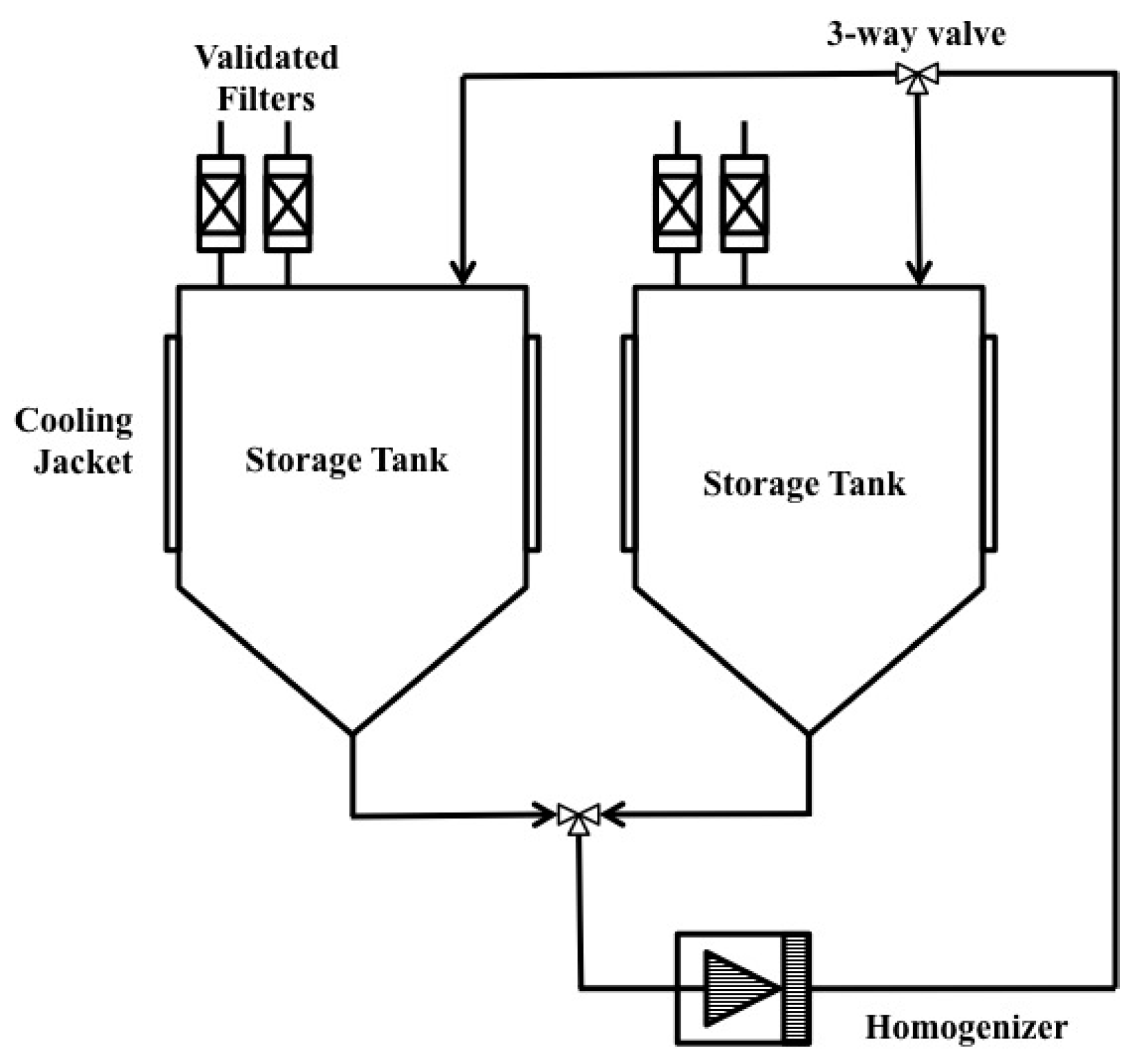
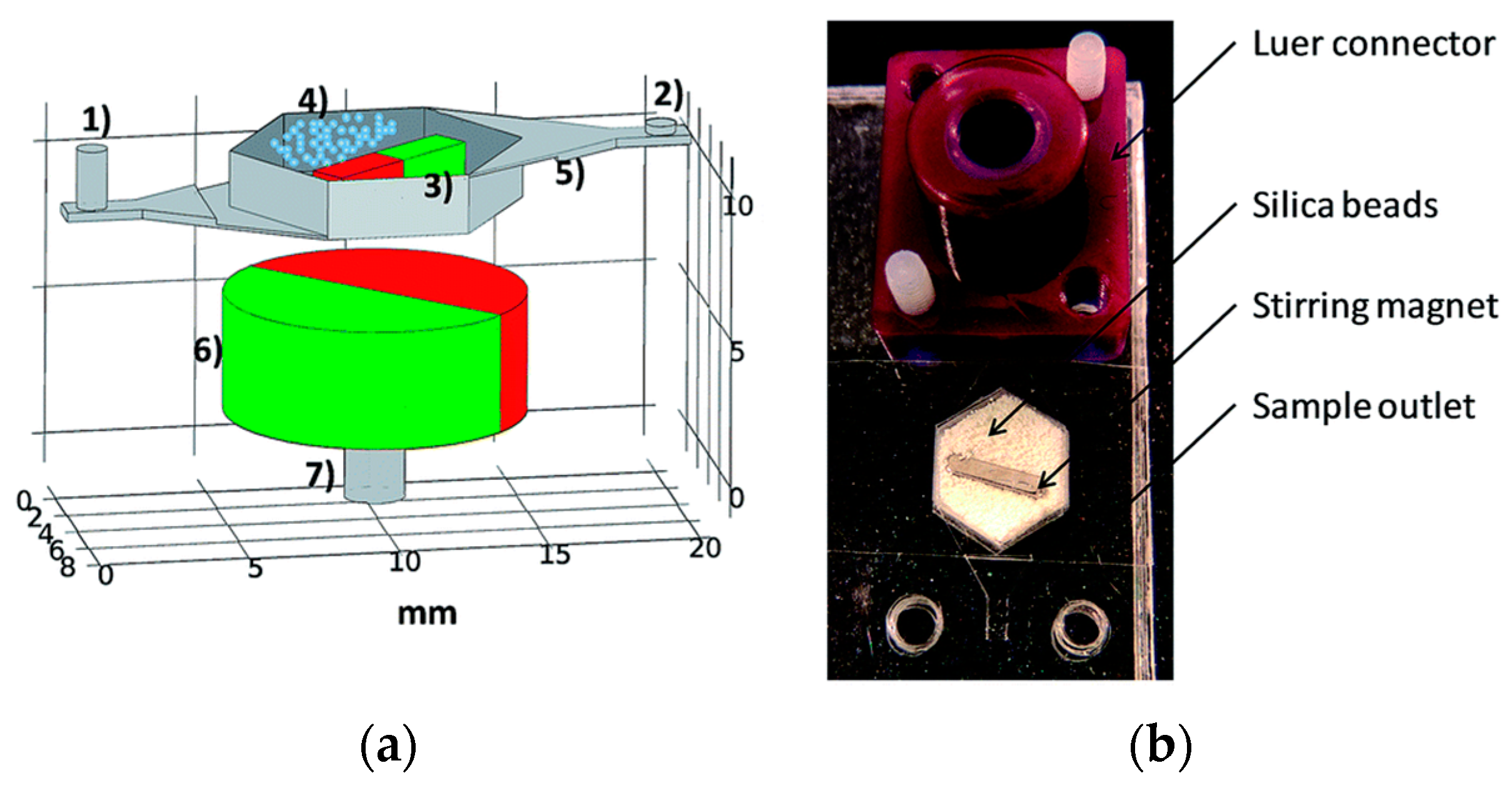
Mechanical lysis in microfluidics involves physically disrupting the cell membrane using shear or frictional forces and compressive stresses. Berasaluce et al. [
50] developed a miniaturized bead beating based method to lyse large cell volumes. Zirconium/silica beads were placed inside a cell lysis chamber along with a permanent magnet and actuation of an external magnetic field caused the motion of the beads inside the chamber.
Figure 7 shows the various components and device assembled for cell lysis.
Staphylococcus epidermidis cells were used in this study and they studied the effect of bead size, volume, flow rate and surfactant (Tween-20) on lysing efficiency. They found the optimum parameters achieved a 43% higher yield efficiency at a flow rate of 60 μL/min compared to off chip bead beating system.
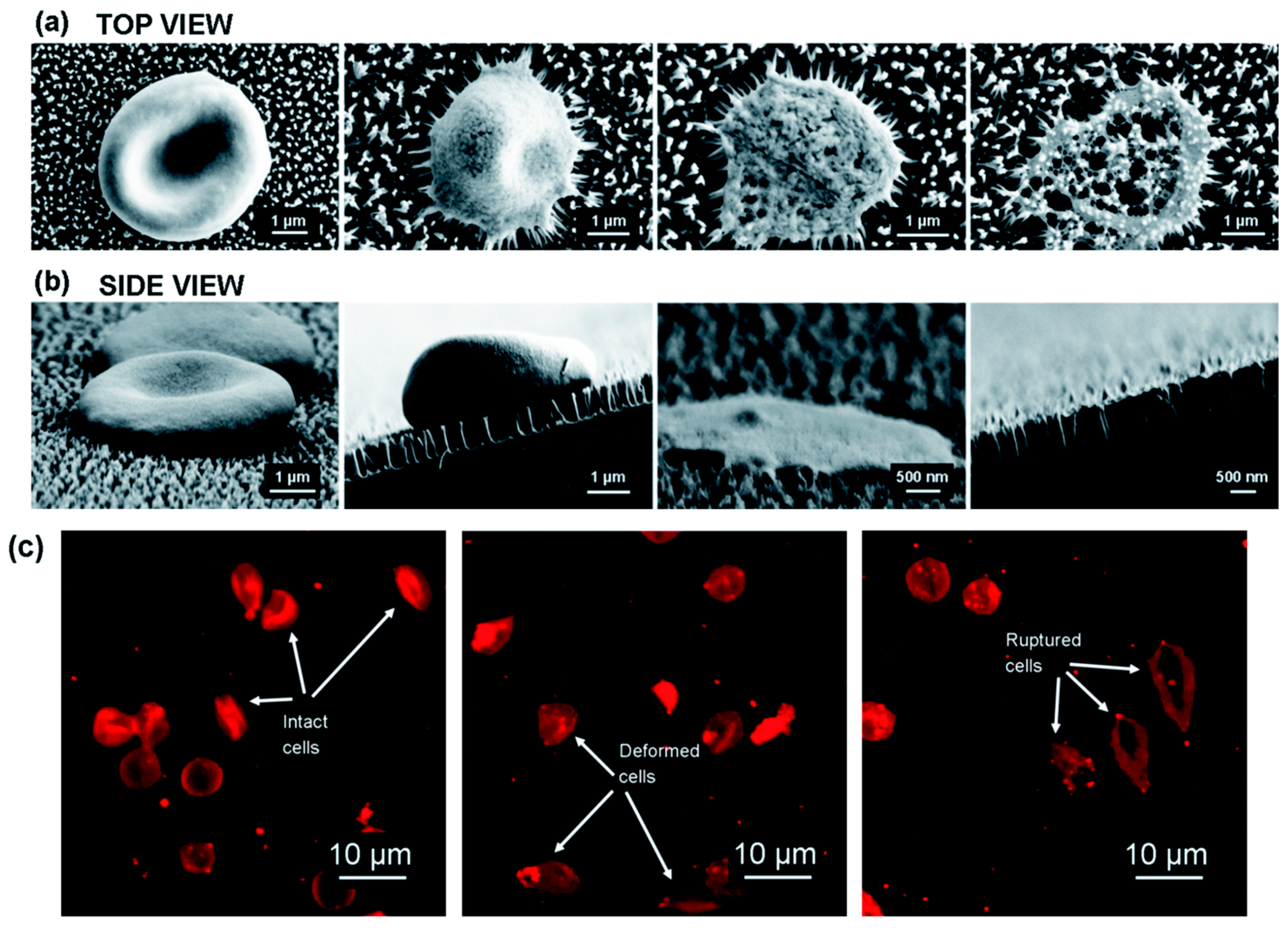
Pham et al. [
51] have recently used nanotechnology to fabricate black silicon nano pillars to lyse erythrocytes in about 3 min. They fabricated these nanopillar with ~12 nm tip diameter and 600 nm tall on silicon substrate using reactive ion etching technology. The authors showed that the interaction of erythrocytes cultured on nanopillar arrays causes stress induced cell deformation, rupture and lysis in about 3 min.
Figure 8 shows the interaction of erythrocytes with the nanostructures.
Mechanical lysis has been demonstrated by using nano-scale barb [
52]. When cells are forced through small opening, high shear forces cause rupture of the cell membrane. Similar principle has been used here where “nanoknives” were fabricated in the wall of microchannels by using modified deep reactive ion etching (DRIE). Distance between these sharp edges was 0.35 μm and width of the channel was 3 μm. The lysis section of this device consisted of an array of these “nanoknives” patterned on a microchannel as shown in
Figure 9b. Human promyelocytic leukemia cells (HL-60) were used to pass through this section at sufficient velocity. The addition of this “nanoknives” pattern increased the amount of lysis. This device was used to extract protein from inside the cell. It has been estimated that as much as 99% of the cell was lysed but, only 6% protein was released.
Alternatively, mechanical impingement through collision has also been used to lyse in the microscale [
53,
54,
55]. Cells were suspended in solution with glass beads and placed on the microfluidic compact disc (CD) device, which was then set to rotate at a very high velocity. The centrifugal force generated by the rotation, causes collision and friction between cells and beads, which results in cell lysis. Various kinds of cells including mammalian, bacteria and yeast have been lysed using this technique.
Though the efficiency of the mechanical lysis is very high, these disruption methods have some drawbacks in microscale application. Fabrication of these devices is complex as well as expensive and collecting the target materials from a complex mixture is very difficult.
4.3. Chemical Lysis
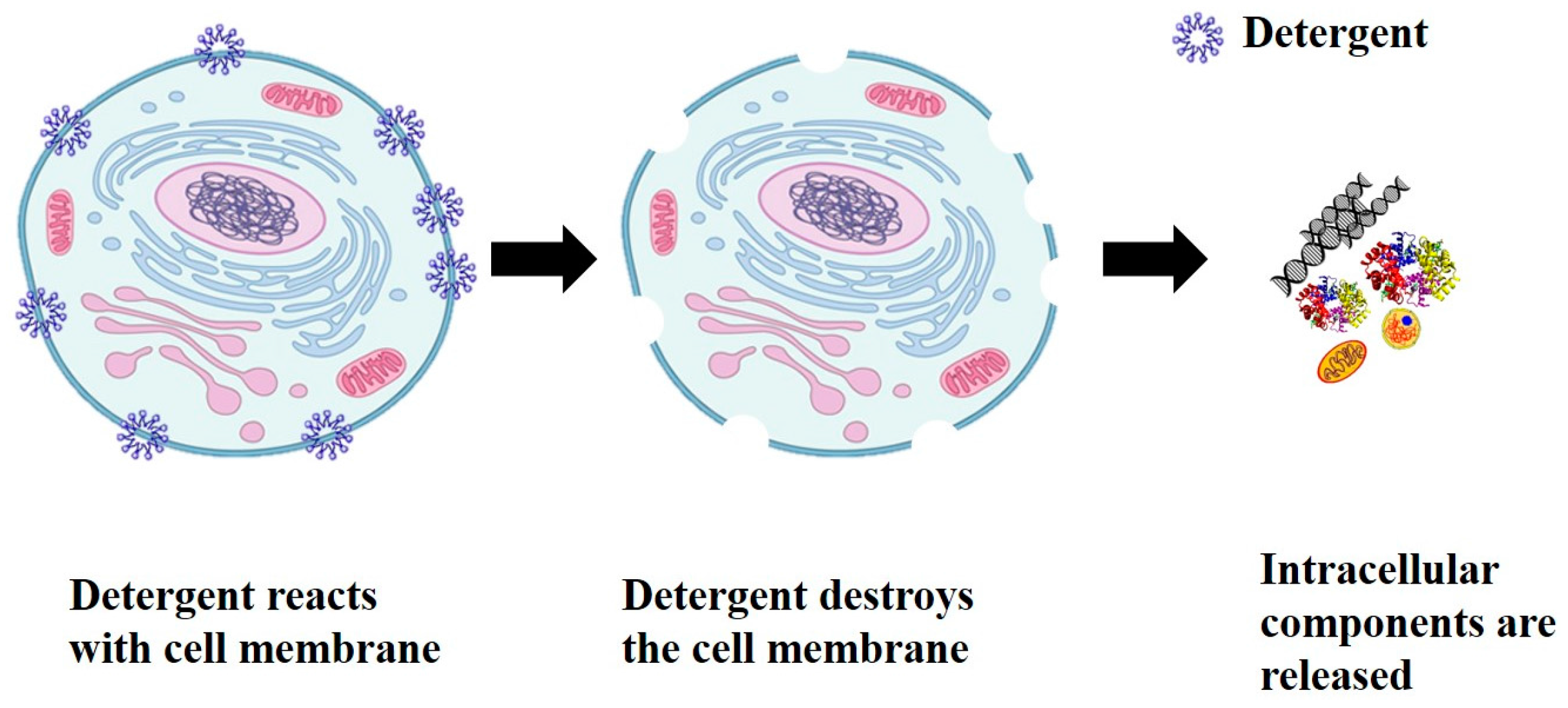
Chemical lysis methods use chemical reagents such as surfactants, lysis buffers and enzymes to solubilize lipids and proteins in the cell membrane to create pores and lyse cells. Although chemical and enzymatic methods are categorized separately in macro scale method, these two techniques are incorporated in the same group for micro scale cell lysis techniques. Buser et al. [
60] lysed gram-positive bacteria (Staphylococcus aureus) and RNA virus (respiratory syncytial virus) using a dried enzyme mixture (achromopeptidase). They were able to lyse in less than a minute and then used a disposable chemical heater to deactivate the lysis enzyme. They were able to amplify (off-chip) the lysate without purification and showed the proof of principle for a point of care device for diagnostics.
Kashyap et al. [
61] developed a microfluidic probe for selective local lysis of adherent cells (~300 cells) for nucleic acid analysis. Hall et al. [
62] used a device for cell lysis experiment, which had two supply wells and a pressure well. Mixing of cell and lysis solution was controlled by adjusting the pressure of the wells. Three different types of solution were used—Solution A containing only SDS (detergent based reagent), Solution B containing surfactant, Triton X-100, Tween-20 with enzyme such as lysozyme, protease, proteinase K and Solution C containing an antibiotic named polymyxin B. Gram-negative and gram-positive bacteria were used for lysis. It was concluded that detergent alone was not suitable for lysis, while Solution B, a mixture of chemical surfactants and biological reagents, can disintegrate the cell membrane and lyse various kinds of bacteria. However, polymyxin B can be potentially used in microfluidic cell lysis platform only for gram-negative bacteria.
Kim et al. [
63] also developed a microfluidic device with two inlets and outlets in order to develop an optimal lysis reagent for gram-negative bacteria. Heo et al. [
64] demonstrated a microfluidic based bioreactor which was capable of entrapping
E. coli by using hydrogel patches. Then the immobilized
E. coli was lysed by using SDS as it can penetrate hydrogel. Cell lysis was accomplished within 20 min. This device was capable of cell lysis using only SDS, however, the previous one could not due to lower exposure time in chemical environment. In another study, Sethu et al. [
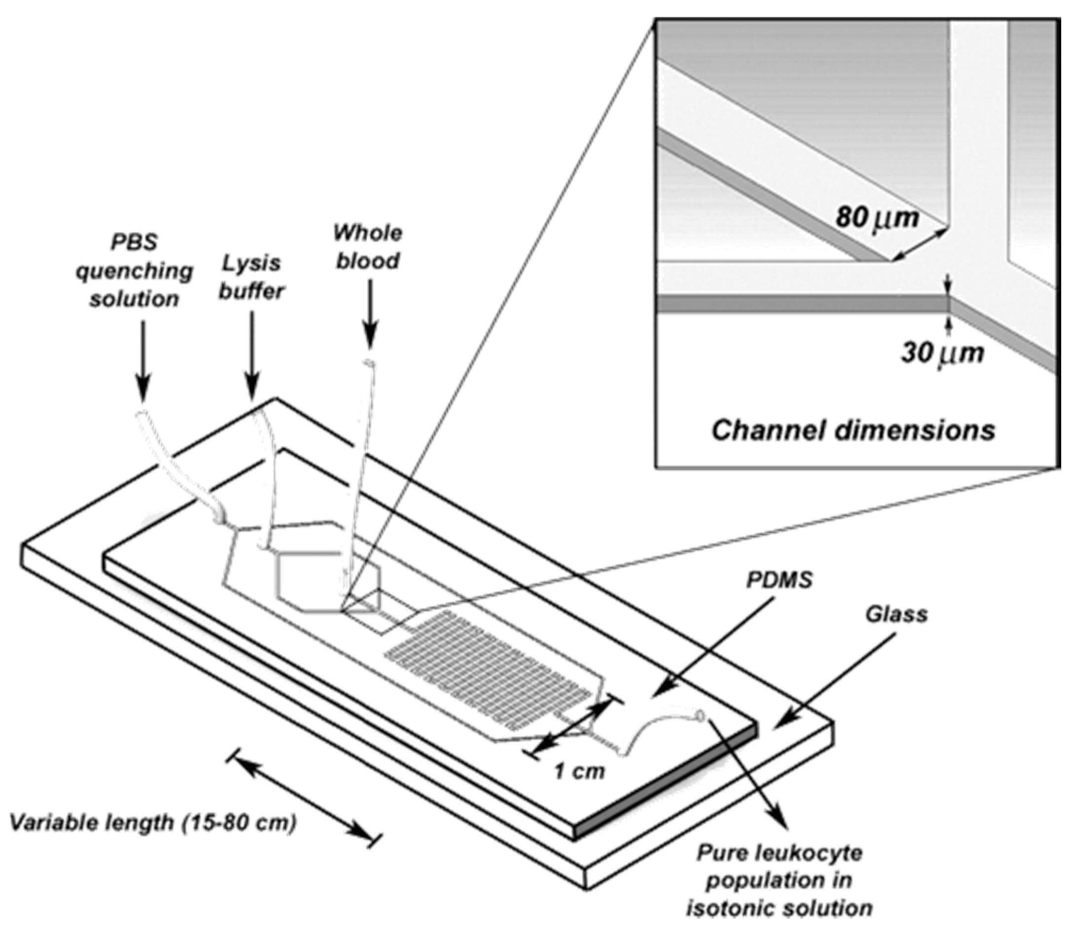
65] also developed a microfluidic chip (
Figure 10) to lyse Erythrocyte in order to isolate Leukocyte. One hundred-percent recovery was possible within 40 s. The device consists of three inlet reservoirs and one outlet reservoir. One inlet was used to flow the entire blood. Second inlet was used for lysis buffer containing mainly aluminum oxide and two side channels were connected with this inlet which converged to direct the entire blood into a narrow stream. This increases the surface contact between the lysis buffer and the cells. The mixture of cells and lysis buffer was then run through a long channel with a number of “U” turns to enhance the buffer. Finally, third inlet was used to flow the phosphate buffer in order to dilute the sample for restoring the physiological concentration [
66,
67].
Even though chemical lysis method is widely used in many microfluidic devices, this method requires an additional time consuming step for reagents delivery. Therefore, complex microfluidics structures including injection channels and micro-mixers to homogenize the samples are needed [
66,
68]. After lysis, these reagents might interfere with downstream assay as it is very hard to separate the target molecules [
69]. In addition, storage of these reagents is a problem which is why the device cannot be used for long time.
4.4. Optical Lysis
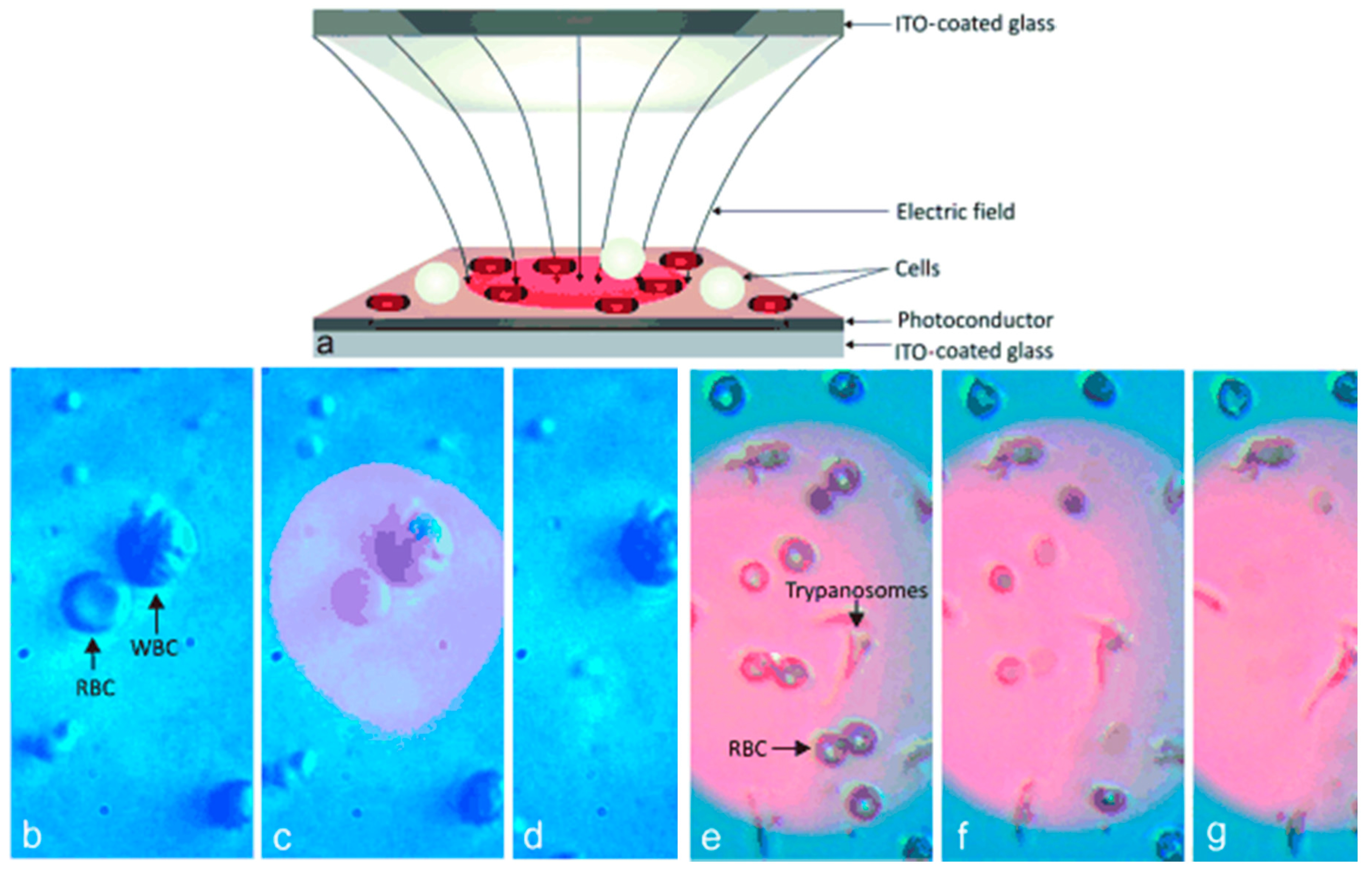
Optical lysis of cells involves the use of lasers and optically induced dielectrophoresis (ODEP) techniques to break open the cell membrane. In laser lysis, a shock wave created by a cavitation bubble, lysis the cell membrane. A focused laser pulse at the cell solution interface creates this cavitation bubble. In ODEP, a conductive electrode and a photoconductive layer (for example amorphous silicon) are formed on the top surface of glass slide. A non-uniform electric field is generated by shining light on the photoconductive layer which then generates a transmembrane potential across the cell membrane disrupting the cell membrane. Huang et al. [
70] developed an optically induced cell lysis microfluidic chip for lysing HEK293T cells and extracting intact nucleus. They report cell lysis and nucleus separation efficiency as 78% and 80% respectively using this device.
Kremer et al. [
71] lysed cells using an opto-electrical setup. They were able to lyse cells selected based on shape of the cell. They used ODEP to lyse red blood cells in a mixture of red and white blood cells. They developed a method that enabled shape-selectivity such that cells with a different geometry will lyse in a mixture of cell types. The cell with a different shape induces a non-uniform electric field which is used for lysis.
Figure 11 shows the schematic of the lysis chip and lysis of differently shaped cells.
Use of laser light to induce lysis has also been attempted in microfluidic devices. In one instance, optical lysis was induced by application of a nanosecond 532 nm laser pulse [
72] which generates a microplasma locally. The plasma collapses causing cavitation, bubble expansion and its collapse as described in previous section are the main reason for a laser induced cell lysis. Various types of cell lines such as rat basophilic leukemia (RBL) [
73], rat-kangaroo (Potorous tridactylis) epithelial kidney cells (PtK2) [
74], and murine interleukin-3 dependent pro-B (BAF-3) [
75] have been lysed by using this laser induced method. However, all these experiments had been done for single cell analysis. It has been found that when laser based lysis was incorporated with polydimethylsiloxane (PDMS) microchannel efficiency of lysis decreased [
75]. It was suggested that this may be due to the deformation of PDMS walls which dissipates the mechanical energy from the bubble collapse. For that reason, high energy was required.
Ultraviolet (UV) light array combined with titanium oxide has been used to lyse the cell [
76]. Titanium oxide possesses photolytic properties and excitation energy that falls within UV range. When titanium oxides are excited with UV light array, electrons in the valence band are excited to conduct ion band which results in electron–hole pairs. In aqueous environment, these electron–hole pairs react with surrounding molecules and generate free radicals such as OH, O and O
2−. These react with cell membrane and lyse the cell.
E. coli cells were lysed with the above technique. A primary disadvantage of ultraviolet lysis was that the time required to lyse the cell was very high (45 min).
4.5. Acoustic Lysis
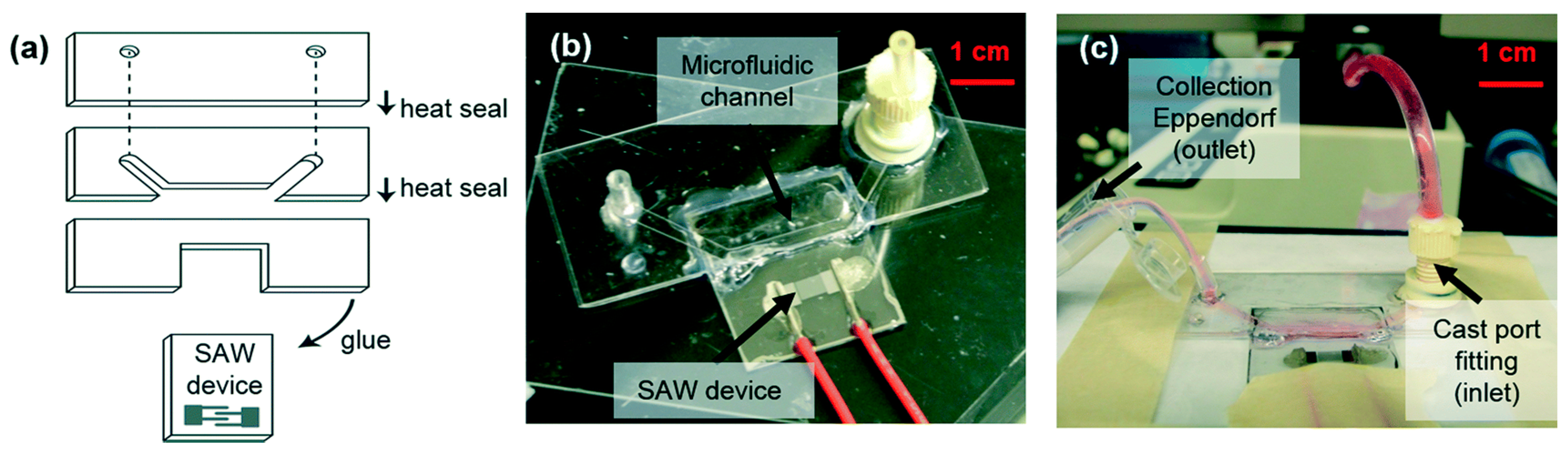
In acoustic lysis, a high energy sound wave is generated which is used for cell lysis. This surface acoustic wave (SAW) is produced on a piezoelectric substrate. An inter-digitated transducer (IDT) can be used to produce a SAW electrically with the wave propagating on the surface away from it. Taller et al. [
77] have used on chip surface acoustic wave lysis for detecting exosomal RNA for pancreatic cancer study. They achieved a lysis rate of 38% using this technique.
Figure 12 shows the fabricated device with the SAW transducer.
They report that the lysis of exosomes is possible due to the effects of acoustic radiation force and dielectric force acting on small particles [
78,
79]. The SAW device was fabricated using standard photolithography technology. Twenty pairs of titanium aluminum electrodes were patterned on top of piezoelectric lithium niobate substrate to form a single phase unidirectional SAW transducer. This transducer can generate SAW in only one direction. Raw media was exposed to SAW for 30 s at 1 W of power for lysing. The authors report that a lysis efficiency of 38% achieved using this method was sufficient for obtaining enough exosome RNA for detection.
Marentis et al. [
80] lysed the eukaryotic cell as well as bacteria by using sonication. This device consists of a microfluidic channel with integrated transducer. The channel was made on glass substrate and piezoelectric transducer was made by depositing zinc-oxide and gold on quartz substrate. The transducers were driven by a sinusoidal source in the 360-MHz range. Eighty-percent lysis of HL-60 and 50% lysis of Bacillus Subtilis spores were obtained by using this device. The temperature rise due to sonication was moderated by using ice pack and cold finger. Ultrasonic horn tip and liquid region are coupled in a microfluidic chip by increasing fluidic pressure in order to increase the efficiency of lysis [
81].
Reboud et al. [
82] have developed a disposable microfluidic chip to detect the rodent malaria parasite Plasmodium berghei in blood. They used SAW to lyse the red blood cells and parasitic cells in a drop of blood. They report a cell lysis efficiency of more than 99.8% using their device. Xueyong et al. [
83] have fabricated a SAW microfluidic device which can lyse red blood cells with high efficiency (95%).
However, sonication has limitations such as generation of heat, complex mechanism as well as expensive fabrication process. Due to this excessive heat generation denaturation of protein and excessive diffusion of the cell contents have been observed [
8,
84]. To reduce the operation time, cells were first treated with some weak detergent such as digitonin [
8,
85] before ultrasonic exposure. Digitonin weakened the cell membrane and facilitated lysis.
4.6. Electrical Lysis
In electrical method, cells are lysed by exposing them to a strong electric field. An electric field is applied across the cell membrane which creates a transmembrane potential. A potential higher than the threshold potential is required to form pores in the cell membrane. If the value of the potential is lower than the threshold potential, the pores can be resealed by the cell. On the other hand, a high enough potential can completely disintegrate the cell. At such high voltages, it is found that the electric field does not have any effect on the intracellular components [
86]. Electric field is the critical parameter to lyse the cell. As higher electric field is required for cell lysis, high voltage generator is required in order to generate this high electric field in macroscale. Thus, this method is not common in macroscale. However, in microscale due to small size of the devices, higher electric field can be obtained at lower voltage. For this reason and as a method for fast and reagentless procedure of lysis, electrical lysis has achieved substantial popularity in microfluidic community.
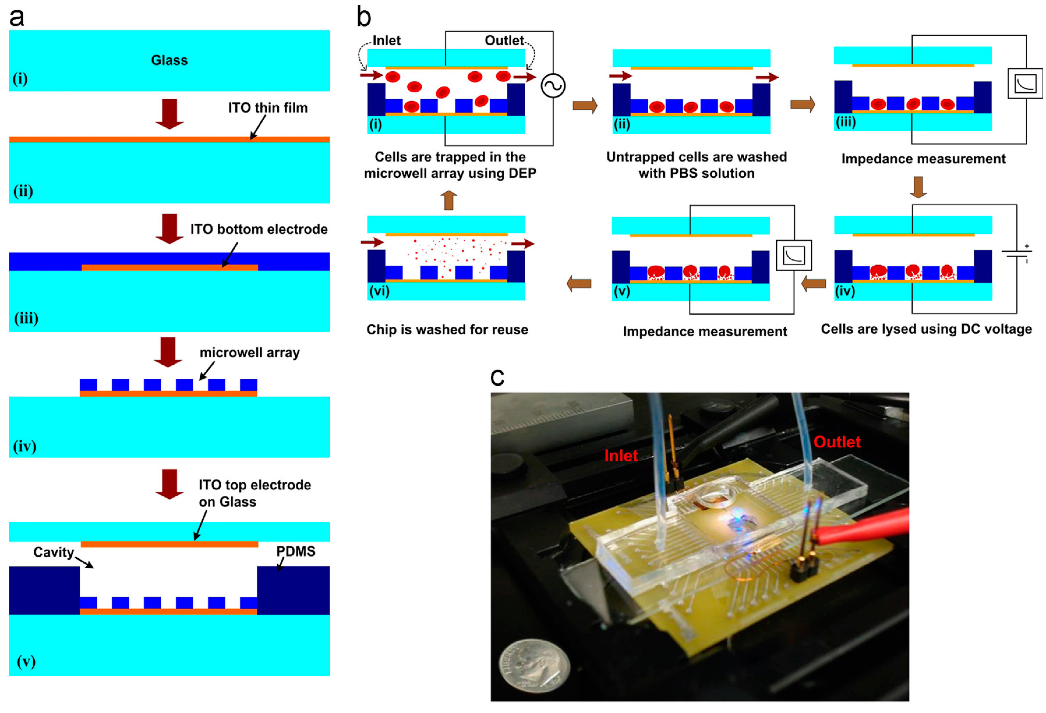
Ameri et al. [
87] used a direct current (DC) source to lyse cells in a microfluidic chip.
Figure 13 shows the fabrication and working principle of their chip. Their device consists of a glass slide coated with indium tin oxide coating patterned for electrodes. The 6400-Microwell arrays are fabricated using SU-8 polymer by photolithography technique. Inlet and outlet channels are created using PDMS polymer and is sealed using a glass slide with ITO electrode for impedance measurement. Red blood cells (10
7 cells/mL) are flown through the device at 20 μL/min and dielectrophoresis (DEP) is used to immobilize the cells into the microarray. A DC voltage of 2 V for 10 s was applied to the cell for lysis. The lysis process was monitored using impedance measurement before and after lysis and a decrease in impedance suggested a complete lysis of cells. They report a lysis efficiency of 87% in their device. The authors proposed a device for cell lysis by electric fields and optical free monitoring of the lysis process on a microfluidic platform which could have potential use in the medical diagnostic field.
Jiang et al. [
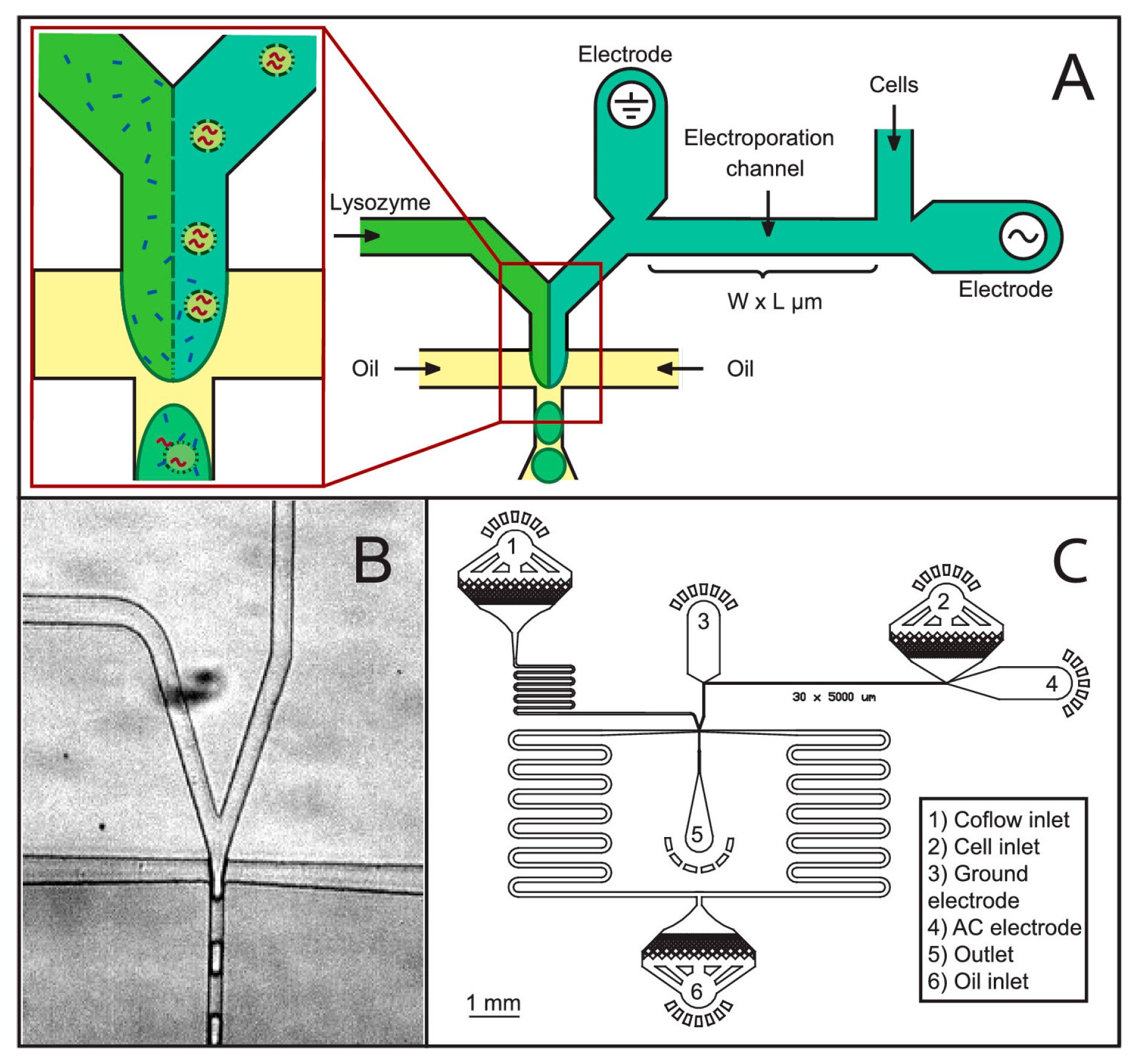
88] developed a low cost microfluidic device for cell lysis using electric fields. They applied a 10 V square pulse to lyse cells at 50% efficiency. They report a device which had the capability to lyse cells at a much lower voltage compared to a commercially available electropolator device which operated at 1000 V to lyse 200 μL of PK15 cells. They observed bubble formation in their device during cell lysis due to joule heating effect. De Lange et al. [
89] have lysed cells in droplets using electric fields. They demonstrated a robust new technique for detergent free cell lysis in droplets. In their device, electric field was applied to lyse bacteria immediately before merging the cell stream with lysozyme and encapsulating the mixture in droplets. They report that with lysozyme alone the lysis efficiency is poor (less than 50%) but when combined with electric fields they were able to obtain up to 90% cell lysis efficiency.
Figure 14 shows their microfluidic device for cell lysis in droplets. The authors suggest that their device could be used in applications where use of cell lysis detergents could hinder the cell analysis such as binding assays or studying the chemical activity of proteins and in mass spectroscopy studies where chemical lysis agents can hamper the results.
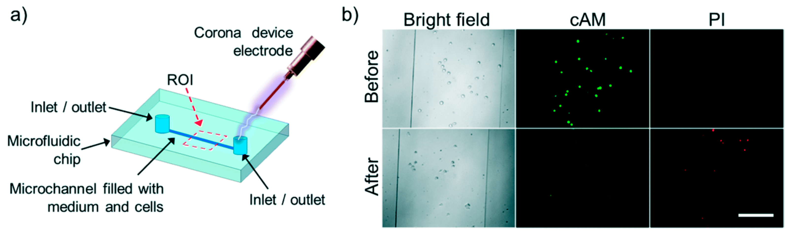
Escobedo et al. [
90] showed electrical lysis of cells inside a microfluidic chip using a hand held corona device. They were able to lyse baby hamster kidney cells (BHK), enhanced green fluorescent protein human-CP cells (eGFP HCP) 116 and non-adherent K562 leukemia cells completely inside a microfluidic channel. A metal electrode was embedded inside the channel which was used to discharge 10 to 30 kV to lyse the cells in less than 300 ms. Lysis was assessed by observing before and after images of cells using bright field and high speed microscope and also by cell-viability fluorescence probes. They also report no bubble formation during lysis indicating no joule heating effect thereby making this method suitable for analyzing sensitive proteins and intracellular components.
Figure 15 shows the setup and results of the study.
Besant et al. [
91] detected mRNA molecules of
E. coli by electrochemical lysis technique. They applied a potential of 20 V, which initiated the cell lysis by producing hydroxide ions from water at cathode to break down bacterial membranes. The sensor electrodes were placed 50 μm away which was enough to detect the mRNA molecules in 10 min. They reported lysis and detection of
E. coli mRNA at concentrations as low as 0.4 CFU/μL in 2 min which was relevant for clinical application in both sensitivity and time.
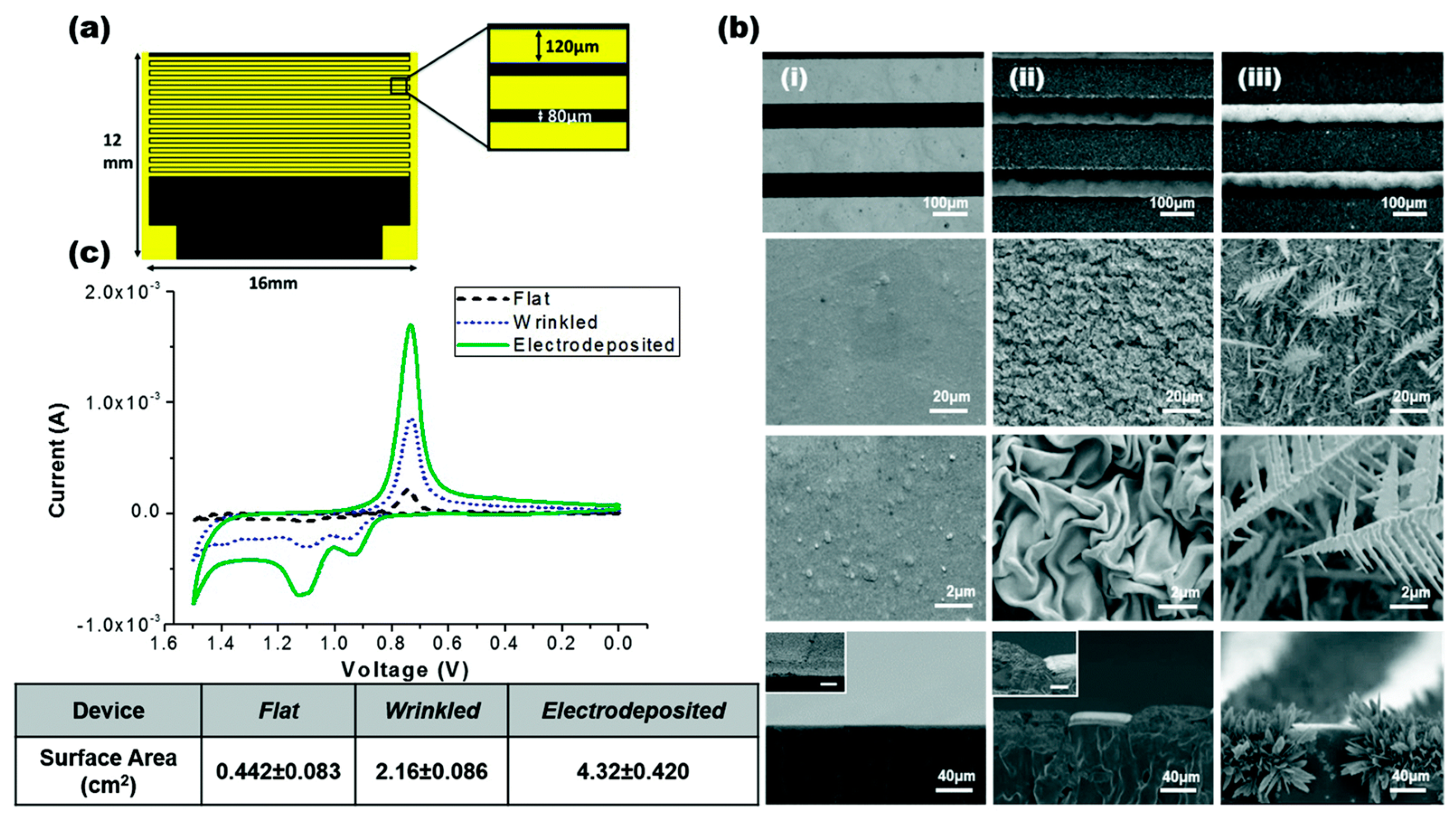
Gabardo et al. [
92] developed a low cost and easy method to fabricate multi-scale 3D electrodes that could be used for bacterial lysis using a combination of electrical and electrochemical means. These micron-sized electrodes can be rapidly prototyped using craft cutting, polymer induced wrinkling and electro-deposition techniques. They report that these tunable electrodes performed better as compared to lithographically prepared electrodes. They were able to successfully extract nucleic acids extracted from lysed bacteria on a microfluidic platform. They reported 95% lysis efficiency at 4 V using their electrodes.
Figure 16 shows the device and electrode structures.
Li et al. [
93] developed a double nano-electrode electrical cell lysis device to lyse single neuronal cells. Similarly, Wassermann et al. [
94] showed cell specific lysis of up to 75% of the total human blood cells using SiO
2 passivated electrical cell lysis electrodes at an applied voltage of 8–20 V. Ma et al. [
95] reported a 10–20-fold increase in mRNA extracted from
M. smegmatis using electrical lysis in a microfluidic platform as compared to a commercial bead beading instrument. They used a 4000–8000 V/cm field intensity to lyse the bacteria with long pulses (5 s). They report that their device can be effective for mRNA release from hard to lyse cells.
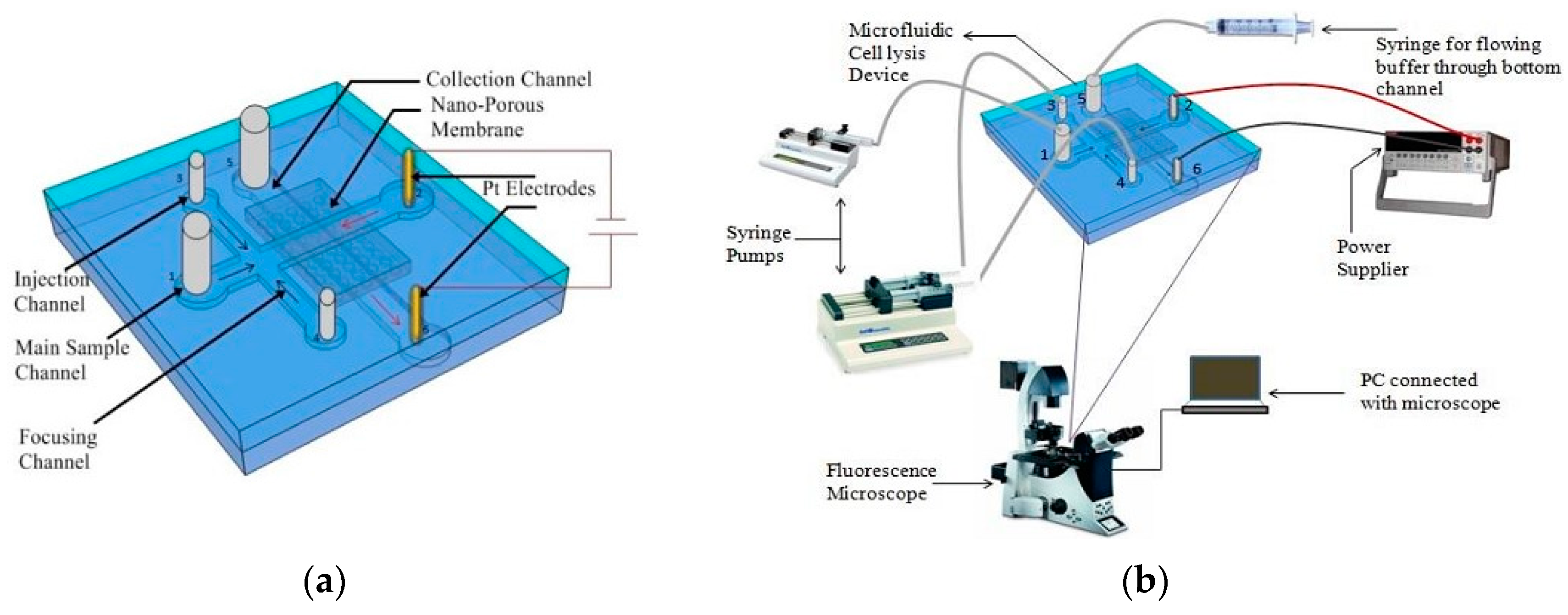
Islam et al. [
96] showed the proof of concept of a simple microfluidic device for electrical lysis of larger volumes of sample. They used a nanoporous membrane sandwiched between two microfluidic channels to trap and lyse
E. coli bacteria by applying 300 V. They report a lysis efficiency of 90% in less than 3 min.
Figure 17 shows the schematic of the device used for lysis in their study.
Different types of voltages such as alternating current (AC) [
97,
98], DC pulses [
99,
100,
101] and continuous DC voltages [
102] have been used in order to lyse the cells. Along with electric field, exposure time of cells within that electric field is also an important parameter for cell lysis. It has been found that cells can be lysed by using higher electric field for short period of time as well as lower electric field for long period of time [
103]. For that reason, AC and DC pulses of a higher electric field are needed as compared to a continuous DC electric field. As the electric field depends on the distance between the electrodes, microfabricated electrodes have been used during AC or DC pulses. An overview of different electrical lysis devices and the characteristics of the designed system is presented in
Table 4.
Lu et al. [
104] developed a microfluidic electroporation platform in order to lyse human HT-29 cell. Microfabricated saw-tooth electrode array was used in order to intensify the electric field periodically along the channel. Seventy-four-percent efficiency was obtained for an operational voltage of 8.5 V. However, this mode of lysis is not suitable for bacteria due their sizes and shapes. Compared to mammalian cell, high electric field and longer exposure is needed to lyse bacteria. Rosa [
105] developed a chip to lyse bacteria consisting of an array of circular gold electrodes. DC pulses were used and lysis with 17% efficiency was achieved by using an operational voltage 300 V. This efficiency was increased up to 80% after adding enzyme with cell solution. In 2006, Wang et al. [
107] proposed application of continuous DC voltage along the channel for cell lysis. The device consists of a single channel with uniform depth and variable width. Since the electric field is inversely proportional to width of the channel, high electric field can be obtained at the narrow section of the channel. Thus, lysis occurs into a predetermined portion of the device. Exposure time of the cell to the electric field can be tuned by changing the length of this narrow section. The configuration of the device was optimized and lysis of complete
E. coli bacteria was possible at 930 V. Complete disintegration of cell membrane was observed when the electric field was higher than 1500 V/cm. This device was very simple and did not need any microfabricated electrodes. Pt wires were used as electrodes. Only a power generator was needed to operate it. However, bubble generation and Joule heating issue could not be completely eliminated. Similar kind of device was used by Lee [
102] where the length and width of the narrow section was modified in order to lyse mammalian cell. Bao et al. [
108] also developed a device to lyse
E. coli by using DC pulses. Release of intracellular materials was observed when the electric field was higher than 1000 V/cm.
In conclusion, electrical method offers a simple, fast and reagent less lysis procedure to lyse various kinds of cells. This method is also suitable for selective lysis and is compatible with other downstream assays such as amplification and separation. Although requirement of high voltage is a problem in this procedure, it can be overcome by decreasing the gap between electrodes through microfabrication. However, heat generation and formation of bubble is a major problem for electric lysis method.


