Review

Antigenotoxic Studies of Different Substances to Reduce the DNA Damage Induced by Aflatoxin B1 and Ochratoxin A

1
Instituto de Ciencias de la Salud, Universidad Autónoma del Estado de Hidalgo., Ex-Hacienda de la Concepción. Tilcuautla. Pachuca de Soto, Hidalgo. CP 42080, México
2
Laboratorio de Genética, Escuela Nacional de Ciencias Biológicas, I.P.N., Av. Wilfrido Massieu. Unidad A. López Mateos. Zacatenco. Col Lindavista. D.F. CP 07738, México
3
Laboratorio de Toxicología Preclínica, Escuela Nacional de Ciencias Biológicas, I.P.N., Av. Wilfrido Massieu. Unidad A. López Mateos. Zacatenco. Col Lindavista. D.F. CP 07738, México
*
Author to whom correspondence should be addressed.
Toxins 2010, 2(4), 738-757; https://doi.org/10.3390/toxins2040738
Submission received: 21 March 2010 / Revised: 8 April 2010 / Accepted: 13 April 2010 / Published: 19 April 2010

Abstract

:
Mycotoxins are produced mainly by the mycelial structure of filamentous fungi, or more specifically, molds. These secondary metabolites are synthesized during the end of the exponential growth phase and appear to have no biochemical significance in fungal growth and development. The contamination of foods and feeds with mycotoxins is a significant problem for the adverse effects on humans, animals, and crops that result in illnesses and economic losses. The toxic effect of the ingestion of mycotoxins in humans and animals depends on a number of factors including intake levels, duration of exposure, toxin species, mechanisms of action, metabolism, and defense mechanisms. In general, the consumption of contaminated food and feed with mycotoxin induces to neurotoxic, immunosuppressive, teratogenic, mutagenic, and carcinogenic effect in humans and/or animals. The most significant mycotoxins in terms of public health and agronomic perspective include the aflatoxins, ochratoxin A (OTA), trichothecenes, fumonisins, patulin, and the ergot alkaloids. Due to the detrimental effects of these mycotoxins, several strategies have been developed in order to reduce the risk of exposure. These include the degradation, destruction, inactivation or removal of mycotoxins through chemical, physical and biological methods. However, the results obtained with these methods have not been optimal, because they may change the organoleptic characteristics and nutritional values of food. Another alternative strategy to prevent or reduce the toxic effects of mycotoxins is by applying antimutagenic agents. These substances act according to several extra- or intracellular mechanisms, their main goal being to avoid the interaction of mycotoxins with DNA; as a consequence of their action, these agents would inhibit mutagenesis and carcinogenesis. This article reviews the main strategies used to control AFB1 and ochratoxin A and contains an analysis of some antigenotoxic substances that reduce the DNA damage caused by these mycotoxins.

Graphical Abstract

1. Introduction

Mycotoxins are structurally diverse groups composed mainly of small molecular weight compounds. These compounds are produced mainly by the mycelial structure of filamentous fungi, or more specifically, the molds. Mycotoxins are secondary metabolites synthesized during the end of the exponential phase of growth and appear to have no biological significance with respect to mould growth/development or competitiveness, but when ingested by higher vertebrates and other animals cause diseases called mycotoxicoses [1,2]. The metabolites are found in a wide range of countries, feeds and foods [3]. They are toxic to mammals, poultry, and fish [2,4,5]. The toxic effect of mycotoxin ingestion in both humans and animals depends on a number of factors including intake levels, the toxicity of the compound, duration of exposure (acute or chronic), the body weight of the individual, the presence of other mycotoxins (synergistic effects), mechanisms of action, metabolism, and defense mechanisms [1,6,7]. Metabolism and defense mechanisms are important factors in understanding mycotoxin toxicity in specific species or individual animals. Specificity of such mechanisms is well demonstrated in the significant difference between ruminants and non ruminants in handling mycotoxins. Ruminants have generally been more resistant to the adverse effects of mycotoxins. This is because the rumen microbiota is capable of degrading mycotoxins [8]. Starting with the discovery of the aflatoxins in the early 1960s, the isolation of mycotoxins from food has led to the identification of over 100 toxigenic fungi and more than 400 mycotoxins [1,9]. These toxins account for millions of dollars annually in losses worldwide in human and animals health, and condemned agricultural products. Different factors contributing to the presence or production of mycotoxins in foods or feeds include storage, environmental, and ecological conditions. Often times most factors are beyond human control [8].
Nowadays, mycotoxins with carcinogenic potential in experimental animal models include aflatoxins, ochratoxin, sterigmatocystin, fumonisin, zearalenone, citrinin, patulin, and luteoskyrin [9]. These carcinogenic mycotoxins are DNA damaging agents, with the only exception of fumonisins [10]. They may produce cancer by interference with signal transduction pathways [11].
Many cereals, oil seeds, tree nuts, and dehydrated fruits are susceptible to fungus contamination and mycotoxin production. Under laboratory conditions at least 300 mycotoxins produced by fungal culture broth have been chemically characterized. Fortunately, only about 20 mycotoxins are known to be present in food at significant levels. These toxins are mainly produced by five genera of fungi: Aspergillus, Penicillium, Fusarium, Alternaria, and Claviceps [12].
This review attempts to briefly summarize the current available data of the main biological properties and mechanisms of DNA damage caused by aflatoxin B1 and ochratoxin A. It also reviews the main strategies used to control of these mycotoxins.

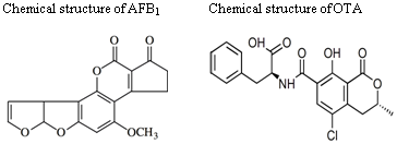
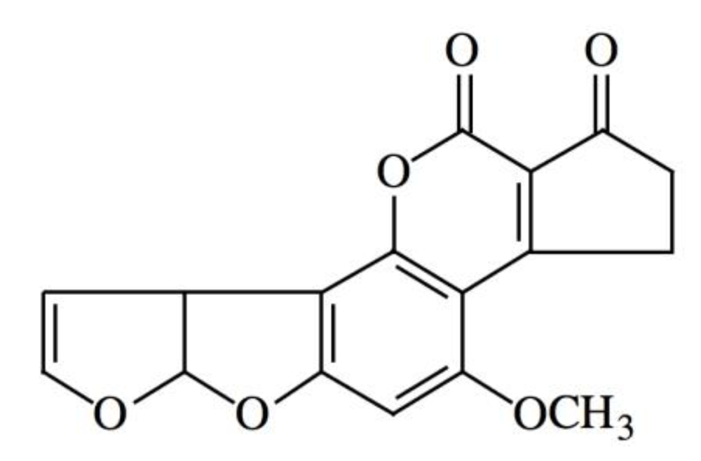
2. Generalities of Aflatoxins

Aflatoxins were first isolated some 40 years ago after outbreaks of disease and death in turkeys and of cancer in rainbow trout fed on rations formulated from peanut and cottonseed meals. These secondary metabolites are a group of closely related difuranocoumarin compounds produced by several strains of filamentous fungi, mainly by Aspergillus flavus and Aspergillus parasiticus [12,13]. Aflatoxins may produce considerable economic losses by attacking different stages of sowing and industrialization of different agricultural and dairy products. They can contaminate a great number of crops used for human and animal consumption, for example, corn, peanut, sorghum, rice, wheat, and nut as well as various milk-made products [14]. However, the range of contaminated products differs depending on the country: for example, in Japan, aflatoxins were detected in about 50% of peanut butter and bitter chocolate samples, while their presence was not found in corn products; in contrast, a study in China reported contamination of 70% of corn products [15,16]. In Mexico and other countries, corn is a cereal used as the main component of several meals. Studies have shown that in the cultivation or storage processes of this grain there exist different levels of aflatoxin contamination, particularly with aflatoxin B1 (AFB1) (Figure 1) [14,17]. This chemical is the most potent natural mutagen and carcinogen known and is usually the major aflatoxin produced by toxigenic strains. It is also the best studied: in a large percentage of the papers published, the term aflatoxin can be construed to mean aflatoxin B1 [2].

3. DNA Damage by Aflatoxin B1 (AFB1)

In regards to its genotoxic effects, AFB1 has been evaluated through in vitro and in vivo systems which have shown an increase in the rate of DNA adducts, histidine revertants, chromosomal aberrations, micronucleus and sister chromatid exchanges [18,19,20,21,22]. AFB1 is a powerful carcinogen for humans and many animal species, including rodents, non-human primates, and fish [5,23]. The main target of this carcinogen is the liver, although tumors may also develop in other organs, such as the lungs, kidney and colon [9].
There are substantial differences in species susceptibility. Moreover, within a given species, the magnitude of the response is influenced by age, sex, weight, diet, exposure to infectious agents, and the presence of other mycotoxins and pharmacologically active substances. Diverse studies on aflatoxin toxicity have been conducted, mostly concerning laboratory models or agriculturally important species [2,24]. Cytochrome P450 enzymes convert aflatoxin B1 to the reactive 8,9-epoxide form (AFBO), which is capable of binding to both DNA and proteins [24]. Mechanistically, the AFBO metabolite binds covalently to guanine to form the N7-guanine-AFBO adduct, an event which could cause the diverse expressions that characterize the AFB1 genotoxicity [24,25]. Moreover, aflatoxin B1-DNA adducts can result in GC to TA transversions. A reactive glutathione S-transferase system found in the cytosol and microsomes catalyzes the conjugation of activated aflatoxins with reduced glutathione, leading to the excretion of aflatoxin [26]. Variation in the level of the glutathione transferase system as well as variations in the cytochrome P450 system are thought to contribute to the differences observed in interspecies aflatoxin susceptibility [24,27].

3.1. Antimutagenic Strategies Used to Control the AFB1 Damage

There is some evidence suggesting that there may be a potential risk that AFB1 can cause cancer. Therefore since 1993, The International Agency for Research on Cancer (IARC) has classified it as a high potential carcinogenic agent (Class I) [28]. The potential risk has promoted the evaluation of norms that regulate the quantity of AFB1 in foods.
One of the first authors that evaluated these norms was Labuza, who in 1983, examined three aspects of mycotoxins in food in the USA: (a) relevant laws, (b) the Food and Drug Administration (FDA) guidelines with respect to the law and (c) the courts interpretation of these laws. Also presented several cases of regulations applied to interstate shipments of corn. FDA analyzes raw agricultural products for aflatoxin through the compliance program which objectives are: the collection and analysis of food and feeds to determine regulatory levels; to remove from interstate commerce food that contains violative aflatoxin levels and to determine potential problems and control measures. Nevertheless, no information on the regulatory issues dealing with mycotoxins in international trade of corn was presented. Furthermore, the USA was one the first countries to introduce legislation regulating on aflatoxin levels in food and feed [29]. Labuza’s publication leads to the adoption, expansion, and changes in regulations concerning mycotoxins in many countries. An international inquiry on mycotoxins was initiated by the National Institute for Public Health and the Environment. Dutch embassies around the world were requested to gather up-to-date information about mycotoxin regulations from local authorities. At least 99 countries had regulations concerning mycotoxins in food and feed in 2003. All of them have at least regulatory limits for aflatoxin B1 contents and specific regulations exist for other mycotoxins as well. The maximum tolerated levels for aflatoxin B1 in food are from 1 to 20 µg/kg. The most frequently occurring limit is 4 µg/kg, and this limit is applied in the 29 countries which follow the harmonized regulations of the European Free Trade Association (EFTA) and the European Union (EU). The 20 µg/kg limit is applied in 17 countries: half of them are in Latin America, United States and Africa [30]. Although security standards have been implemented, the damage caused by AFB1 is still important and serious. In our country, the contamination caused by this toxin can affect food and sweets made of corn, cereals and oilseeds. As well as meat of chicken, pork and other animal-derived products (milk, eggs or cheese) [31,32,33].
Figure 1. Chemical structure of aflatoxin B1 (AFB1).
Toxins 02 00738 g001
Several strategies have been developed in order to reduce the risk of aflatoxin exposure. These include the degradation, destruction, inactivation or removal of mycotoxins through chemical and physical methods (Table 1). However, the results obtained with these methods have not been optimal, because they change the organoleptic characteristics and nutritional values of food [34,35,36]. Another alternative strategy to prevent or reduce the genotoxic effects of AFB1 is by applying antimutagenic agents. These substances act according to different mechanisms being their main goal to avoid the interaction of AFB1 with DNA. In general, the antigenotoxic agents may act blocking the mutation and cancer initiation in the extracellular environment or in nontarget cells (inhibition of uptake of mutagens/carcinogens, inhibition of the endogenous formation of carcinogens, or favoring the adsorption of protective agents) as well as by intracellular mechanisms such as the inhibition of mutation and cancer initiation in target cells or the tumor promotion. As a consequence of their action, these agents would inhibit mutagenesis and carcinogenesis [37].
Table 1. Possible strategies to avoid mycotoxin contamination of agricultural products.
MethodsTechniqueExampleReference
PhysicalInactivation by heatVapor pressure [38]
Microwave treatment[39]
Nixtamalization[40]
Inactivation by radiationUltraviolet light [39]
Radiation gamma[1,41]
Elimination by adsorbent substancesZeolites[1]
Bentonites[39]
Aluminosilicates[42]
ChemicalExtraction by solvents organicsEthanol 95%[43]
Acetone 90%[39]
Chemical destructionHexane-ethanol[43]
Hydrogen peroxide[44]
Ammonium hydroxyde[45,46]
Methylamine[39]
Sodium hypochlorite[44]

3.2. In Vitro Studies

In 1994 Singh et al. studied the effect of piperine on the cytotoxicity of aflatoxin B1 (AFB1) in rat hepatoma cells H4IIEC3/G-(H4IIE) and demonstrated that AFB1 inhibited the growth of H4IIE cells. Likewise, piperine reduced the AFB1-induced formation of micronuclei in a concentration-dependent manner [47]. In another study about the protective effect of food additives by Soni et al. (1997) it was demonstrated that all the additives studied had an important inhibition against mutation, the most effective was turmeric and curcumin that inhibited mutation frequency by more than 80% at concentrations of 2 µg/plate in Salmonella tester strains TA 98 and TA 100 [48]. In 1996 Loarca-Piña used the Salmonella microsuspension assay to examine the antimutagenicity of ellagic acid (EA) against this potent mycotoxin using tester strains TA98 and TA100, the results of the sequential incubation studies support the hypothesis that one mechanism of inhibition could involve the formation of a chemical complex between EA and AFB1 [49]. On the other hand, in 1992 Decoudu et al. studied the effect of vitamin A dietary intake on in vitro and showed the relationship between vitamin A and AFB1 [42]. It demonstrated the activities of metabolizing enzymes which specifically activate or deactivate AFB1 and has decreased significantly in vitamin A-deficient animals [47,48,49,50,51].

3.3. In Vivo Studies

Considering the above results, the next stage has been to confirm the effectiveness of antimutagenic agents in eukaryotic organisms, particularly in mammals. Nowadays, these studies have been used as experimental models on mice and rats (Table 2). Those studies began in the nineties, and they evaluated the antimutagenic activity of some vitamins (thiamine, riboflavin, niacin and folic acid), extract of coffee and some carotenoids. Cytogenetic tests have been used (micronucleus assay) and evaluation of adducts and DNA breaks [50,52,53,54]. In general, the results have been favorable, showing different inhibitions of genotoxicity, which depend on the dose, route of administration and the exposure time.
Since 1993, our laboratory has evaluated the potential of ammonium hydroxide used to reduce the frequency of micronuclei (MN) and sister chromatid exchange (SCEs) in mice fed with AFB1 contaminated corn. The micronucleus intracytoplasmic of chromatin corpuscles formed from the breakup of acentric chromosome fragments or chromosomes with an anaphase lag. SCEs are chromosomal damage referred to the production of homologous segments exchanges between sister chromatids in any chromosome and both are useful parameters to detect DNA damage.
The experiment lasted eight weeks. During four weeks the animals were fed with a balanced diet and corn. One animal of this group was intoxicated by AFB1 contaminated diet (15 ppb). AFB1 and ammonia was added to other group and only ammonia was added to the last. In the last four weeks of the experiment, the animals were only fed with a balanced diet and uncontaminated maize [55]. The MN were quantified every week with a erythrocyte normochromic of peripheral blood and the results showed a significant reduction in MN since the first week of treatment, reaching a maximum treatment effect of the 60% in the fourth week. The SCEs were obtained from bone marrow and quantified in the fourth and eighth weeks. An inhibition of 55% was observed in the fourth week. At the end of the following four weeks the groups treated with AFB1 and AFB1 plus ammonia showed incomplete recovery of genotoxic damage. In this experiment the antigenotoxic effect of ammonium was attributed to the production of the corresponding hydroxy acid.
Table 2. Summary of various antigenotoxic agents used in mice or rats treated with AFB1.
YearBiological modelAntimutagenType of study ObservationInhibition (%)Reference
1991MiceCoffeAcuteMicronucleus60[53]
1992RatVitamin ASubchronicSingle cell electrophoresis50[50]
1992MiceVitamins: thiamine, riboflavin, niacin, and folic acidSubchronicMicronucleus60[54]
1993MiceAmmonium hydroxydeSubchronicMicronucleus and Sister chromatid exchanges (SCEs)60[55]
1998RatCarotenoids Quantification of adducts65[52]
1998MiceS. cerevisiaeSubchronicMicronucleus70[22]
2007MiceMannanSubchronicMicronucleus50[56]
2009MiceMannanAcutesingle cell electrophoresis60[25]

3.4. Antimutagenic Effect of Yeast

Probiotics are organisms and substances that contribute to intestinal microbial balance. They have the capacity to capture microorganisms pathogens. Among them are the lactobacilli and yeasts [57]. Based on the usefulness of the model used with ammonium and considering that probiotics have shown adsorbent, immunostimulants and antigenotóxic effects, our laboratory continued studies using Saccharomyces cerevisiae (Sc) as antimutagen.
The experiment was similar to the ammonium one, except that its total duration was of nine weeks (six of treatment with the mutagen and antimutagen and three weeks of recovery with a diet free of these compounds). In this case, the antimutagen agent consisted of a 0.3% suspension of viable organisms of Saccharomyces cerevisiae. The frequency of MN was only observed at weeks 3, 6, and 9. The results showed a significant reduction since the third week on (50% approximately) and in the sixth week the inhibition reached 70%. A spontaneous recovery was observed in animals treated with AFB1 This recovery was of 60% in the last period. Furthermore, the use of probiotics reflected a recovery of 50% in the weight of the mice damaged by AFB1 (Figure 2). The antigenotoxic effect observed in our experiment is attributed to the Sc adsorbent capacity, particularly due to a chemical interaction between the mycotoxin and the components of the cell wall of yeast [22].
Figure 2. Saccharomyces cerevisiae (Sc) inhibitory effect on micronucleus frequency induced by aflatoxin B1 (AFB1) [58].
Toxins 02 00738 g002

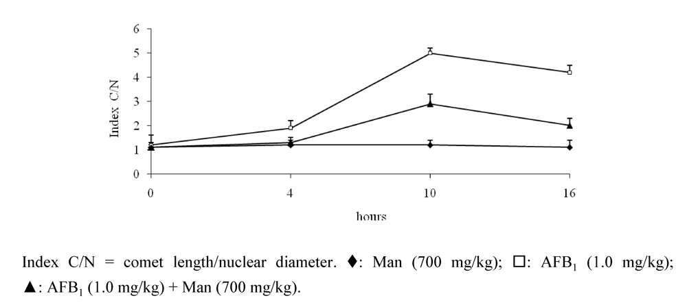
3.5. Protective Effect Caused by Constituents of Yeast Cell Wall against the DNA Damage Induced by AFB1

The yeast cell wall consists mainly of homopolysaccharides (mannans and glucans) and a minor proportion of heteropolysaccharides (glucomannans, galactomannans and xilomannans), proteins, chitin and lipids. There is evidence of the antimutagenic capacity of these oligosaccharides, specifically mannan (mannose with links α-1,6 and ramifications α-1,2 and α-1,3) and glucans (glucoses with links α-1,6 and ramifications β-1,2 and β-1,3) against antineoplastic compounds such as cyclophosphamide and mitomycin C [59,60,61].
Taking into account these antecedents and the properties mentioned above, our current researches are directed to determine the antigenotoxic capacity of mannan, glucan and glucomann and to decide if the mechanism is related to the formation of a molecular complex that diminishes the adsorption of the mutagenic agent.
Recently, we completed a study of eight weeks with mannan following the model mentioned above: in the first period (four weeks), the animals consumed a balanced diet. They were fed with maize: (a) without AFB1; (b) with mannan (500 mg/kg); (c) with AFB1 (0.25 mg/kg), and (d) mannan (50, 250 and 500 mg/kg) plus AFB1 (0.25 mg/kg). In the last four weeks the mice consumed a diet free of antimutagen and mycotoxin. The evaluation of MN was made every week and showed a significant inhibition after the third week in mice treated with the highest dose of mannan, reaching the maximum antigenotoxic effect (50%) in the fourth week (Figure 3).
During the last four weeks, the animals showed a spontaneous recovery of approximately 70% [56]. To demonstrate directly the protection of mannan to DNA, we made a study using the single cell electrophoresis technique (comet assay) in hepatocytes of mice. Comet assay is a technique that can be applied practically to any cell suspension to determine breaks in the strings of DNA in individuals cells [62]. In our case, the procedure detected single-strand breaks. This experiment, unlike previous ones, was an acute one. The compounds were administered orally and the quantification of DNA damage was performed at 4, 10 and 16 hours. We included a control group treated with 700 mg/kg of mannan, a control group treated with 1.0 mg/kg of AFB1, and three groups in which we combined the mycotoxin (1.0 mg/kg) and mannan (100, 400 and 700 mg/kg). The results showed that the highest dose of mannan inhibited the genotoxicity caused by the mycotoxin since the first time, and the highest antigenotoxic effect was between 10 and 16 hours (approximately 60%) (Figure 4) [25]. These results corroborate the data previously obtained by SCEs and MN, and support the hypothesis of a possible chemical interaction between the compounds, which would cause a molecular complex that could be eliminated through the intestinal via without damaging the DNA. A partial damage was observed and it may be related to an amount of AFB1 absorbed and few adducts formed. Our hypothesis, suggests that this interaction would occur between the oligosaccharide hydroxide and the carbonyl group of AFB1, so, we actually analyzed this possibility by using the technique of attenuated total reflectance (ATR) in the intestine of animals treated with the same compounds. The incidence of a light beam of infrared frequency would show the functional groups of the probable chemical complex, which will be identified in the corresponding spectrum.
Figure 3. Mannan (Man) inhibitory effect on DNA damage on micronucleus frequency induced by aflatoxin B1 (AFB1) [58].
Toxins 02 00738 g003
Figure 4. Mannan inhibitory effect on DNA damages induced by AFB1 in mice hepatocytes [58].
Toxins 02 00738 g004

4. Generalities about the Ochratoxins

Ochratoxins are a group of structurally related metabolites that are produced by Aspergillus ochraceus and related species, as well as Penicillium viridicatum and other Penicillium species [63,64,65]. The main mycotoxin in this group is ochratoxin A (OTA) which also appears to be the only one of major carcinogenic significance. Ochratoxin B (OB), the decholoro derivate of OTA, is essentially non-toxic. Ochratoxin A was discovered as a metabolite of Aspergillus ochraceus in 1965 during a large screen of fungal metabolites that was designed specifically to identify new mycotoxins. Shortly thereafter, it was isolated from a commercial corn sample in the United States and recognized as a potent nephrotoxin [2]. Chemically, OTA comprises a polyketide-derived dihydroisocoumarin moiety linked via its 12-carboxyl group by an amide bond to L-β-phenylalanine (Figure 5). As with other mycotoxins, the substrate on which the molds grow as well as the moisture level, temperature (cold weather with low temperatures of 5 °C), and presence of competitive microflora interact to influence the level of toxin produced. The presence of OTA in several plants and animal products has been extensively reported [66,67,68,69,70]. OTA contamination is typically associated with grains stored in the mild weather of Europe and North America. This mycotoxin has been found in barley, oats, rye, wheat, coffee beans, and other plant products, with barley having a particularly high likelihood of contamination. There is also concern that ochratoxin may be present in certain wines, especially those from grapes contaminated with Aspergillus carbonarius [2]. The kidneys are the most susceptible organs to be contaminated by OTA. It can cause both acute and chronic kidney lesions. It principally operates in the first part of the proximal tubules in the kidney and induces a defect in the anion transport mechanism on the brush border of the proximal convoluted tubular cells and basolateral membranes [12,33].

5. DNA Damage Caused by OTA

Pfohl-Leszkowicz and Manderville [33] reviewed the toxicology of OTA and observed that following oxidative metabolism, forms a DNA-reactive quinone that can form guanine-specific DNA adducts. These adducts provide an important source of mutation. Very potent mutagenic effects can be shown in S. typhimurium TA 1535, 1538, and 98, providing certain preincubation protocols and types of metabolic activation are used. Ochratoxin A is genotoxic in Escherichia coli by means of induction of the SOS DNA repair activity. It is also mutagenic in NIH 3T3 cells expressing selected P450 cytochrome and carrying a shuttle vector containing the bacterial lacZ gene as a reporter gene [28]. An increase in sister chromatid exchange rates has been observed in CHO cells in presence of S9 mixture and in peripheral human lymphocytes cultured in a conditioned medium. Therefore, it induced DNA single-strand breaks in cultured mouse and CHO cells. Mammalian cells has been treated with a dose range of this toxin that would accumulate in single-strand DNA breaks, as revealed by the alkaline single-cell gel electrophoresis. This effect is greatly enhanced by the addition of an external metabolizing system in the form of a S9 mix from rat liver [33]. OTA induced a weak positive response for induction of unscheduled DNA synthesis in primary hepatocytes from ACI C3H strain mice and ACI strain rats. Dietary OTA induced renal adenomas and human hepatocellular carcinomas on mice and rats. Carcinogenic effects on humans are suspected because of the high incidence of kidney, pelvis, ureter, and urinary bladder carcinomas among patients suffering from Balkan endemic nephropathy. In regions with Balkan endemic nephropathy, high levels of OTA were found in human blood [2].
Figure 5. Chemical structure of ochratoxin A (OTA).
Toxins 02 00738 g005

6. Ochratoxin A Decontamination Methods

Different studies in animal models have demonstrated that ochratoxin A is nephrotoxic, hepatotoxic, neurotoxic, immunotoxic, teratogenic, mutagenic and genotoxic [64,65,68,71,72]. Since 2002, the OTA has been classified by the International Agency for Research on Cancer as a carcinogenic agent for humans included in the group 2B [28,65,68]. Different strategies have been implemented to control and decontaminate food. In general, these methods have been classified as physical, chemical and biological (Table 3). In the case of the physical methods, milling is one of the most used because it reduces approximately 66% of the levels of OTA during the production of white wheat fluor [1]. Also, washing and brushing have shown good results in reducing the concentration of OTA below the permissible limits (Table 4) in sausage for human consumption [73]. It is important to remember that the permissible levels of OTA are different from country to country.
Table 3. Decontamination methods for ochratoxin A.
MethodsTechniqueExampleReference
PhysicalMechanical removalMilling[1]
Washing and brushing[73]
ChemicalPesticidesDinocap, Penconazole[1]
FungicidesIproidine, Azoxystrobin[1]
Essential oilsOregano, Mint, Basil, Sage[1]
Gas treatmentOzone[74]
AntifungalsFusapyrone[1]
BiologicalSporesConidia[75]
BiodegradationAcinetobacter, Lactobacillus, Streptococcus thermophilus, and Bifidobacterium[1,76,77]
On the other hand, the chemical methods have been directed to the use of pesticides (like iproidine and azoxystrobin), because these substances can reduce by up to 80% the development of ochratoxin A produced by A. ochraceus. Essential oils of spices such as oregano, mint, basil, and sage have been effective used against damage produced by OTA. For example, oregano and mint can inhibit the development of this mycotoxin after 21 days of treatment, at concentrations of 100 ppm. There is evidence that treatment with ozone (O3) slightly decreases levels of OTA [1,74]. Several reports of OTA biodegradation have been published. Streptococcus, thermophilus Bifidobacterium and yogurt bacteria have completely reduced ochratoxin A levels in milk samples containing 0.05 and 0.1mg/L. Some strains of A. fumigates, A. japonicus, and A. niger have also been reported to be able to degrade OTA in products such as ochratoxin α [1,75,76,77,78].
Table 4. Permissible limits of ochratoxin A in several foods recommended in the European Union.
FoodPermissible limit (μg/kg)
Coffee beans1.0–5.0
Instant coffee0.8–10
Cereals4.0–5.0
Table wine2.0
Grape juice2.0
Food for infants and childrens0.5
Sausages3.0
Dried fruits0.2
Beer3.0

6.1. Antimutagenic Strategies for the Control of Ochratoxin A Damage

Like decontamination methods for aflatoxins, the strategies to control the toxic effects of ochratoxin A have not been optimal, because they modify the nutritional values of food. Once again, the use of antimutagenic agents is another alternative to decrease this toxicity. The study is focused on analyzing the capacity of some substances to reduce the oxidative stress induced by the ochratoxin A.
Many reports have suggested the potential role for oxidative stress in OTA toxicity and carcinogenesis. In cell culture, OTA increases the DNA oxidation (generation of 8-OH-guanine) was correlated with a production of reactive oxygen species (ROS). Considering the role of ROS in chemically induced carcinogenesis, the ability of OTA to induce oxidative damage in cells may play an important role in OTA-induced carcinogenicity. Chemopreventive strategies designed to limit the several toxic effects caused by this mycotoxin are important public health goals in reducing the incidence of OTA induced neoplastic diseases [79].

6.2. In Vitro Studies

In vitro studies related to antimutagenesis began in the nineties. Initially, the experiments evaluated the possibility of success in microbial cultures and mammalian cell lines (Table 5). One of the first studies was conducted by Baudrimont et al. who analized the prevention of lipid peroxidation induced by OTA in Vero cells in culture by aspartame (L-aspartyl-L-phenylalanine methyl ester) a structural analogue of OTA and phenylalanine, piroxicam, a non steroidal anti-inflammatory drug and superoxide dismutase (SOD) + catalase (endogenous oxygen radical scavengers). The study found that in the presence of SOD + catalase, the malonaldehyde (MDA) production induced by OTA was significantly decreased. SOD and catalase, when applied prior to the mycotoxin, seemed to prevent lipid peroxidation more efficiently than piroxicam and aspartame. These molecules also partially prevented the OTA-induced leakage of MDA in the culture medium [80]. Another study was conducted by Turbic et al. These authors suggested that specific strains of lactic acid bacteria possessing antimutagenic properties can be used to remove the mutagenic contaminants of food, such as OTA [81]. More recently, studies with polyphenolic compounds (such as chrysin, quercetin, genistein, biochanin A), catechins, and rosmarinic acid have shown the same capacity [79,82,83,84].

6.3. In Vivo Studies

There is a variety of studies about the antimutagenic capacity of several substances. However, there are few experiments made with animals which demonstrate the potential protection of substances used against the damage produced by OTA. One of the earliest studies was made by Obrecht-Pflumio et al. (1996). These authors evaluated the protection capability of indomethacin and aspirin tested against the genotoxicity of ochratoxin A, particularly in the urinary bladder and kidney. The study showed that after a single oral administration of OTA to mice (2 mg/kg body weight) there existed a high level of DNA adducts detected in the urinary bladder. The study also demonstrated that two inhibitors of the prostaglandin H synthase, indomethacin and aspirin, administered before OTA treatment, dramatically reduced the amounts of DNA adducts, particularly in the urinary bladder and kidney. This suggests that protaglandin H synthase in the metabolism of OTA activates metabolites which react with DNA [85]. Recent studies (Table 6) are focused on analyzing substances in dietary honey, aspartame, Inula crithmoides extract, indomethacin, aspirin, and melatonin, and they generally measure glutathione (GSH), glutathione reductase (GR), glutathione peroxidase (GSPx), superoxide dismutase (SOD), catalase (CAT), glutathione-S-transferase (GST), lipid peroxidation (LPO), DNA adducts, and frequency of micronucleus [85,86,87,88,89,90].
Table 5. Principal antimutagenic studies in vitro.
AntimutagenCell line or strains of studyInhibition%Reference
Aspartame, phenylalanine, and piroxicamVero cells50[79]
Lactobacillus rhamnosus 76[81]
Rosmarinic acidHuman hepatoma cells (Hep G2)40[84]
Chrysin, quercetin genistein, and biochanin ACaco-2-cells70[83]
Epigallocathechin gallate, and epicatechin gallatePig kidney cells (LLC-PK1)80[79]
Gallic acid, vanillic acid, protocatechuic acid, caffeic acid, chlorogenic acid, and 4-hydroxybenzoic acidOchratoxigenic Aspergilli strains50–60[82]
Table 6. Summary of recent in vivo studies.
YearBiological modelEvaluation parametersChemopreventive substanceConclusionReference
1996MiceQuantification of aductsIndomethacin and aspirinThese substances reduce the amounts of DNA adducts, particularly in the urinary bladder and kidney. This suggests a role of protaglandin H synthase in the metabolism of OTA leading to active metabolites which react with DNA[85]
1998RatsQuantification of aductsAspartameThe molecular mechanism mediating the preventive effect of Aspartame is the delivery of phenylalanine by cleavage of the peptide and also the direct effect of the peptide on the bending capacity and transport of the toxin[90]
2001RatsLPO, GSH, GR, GSPx, SOD, CAT, and GSTMelatonin (Mel)Mel has a protective effect against OTA toxicity through an inhibition of the oxidative damage and stimulation of GST activities[88]
2004RatsLPO, GSPx, CAT, and SODMelatonin (Mel)Mel decreased the OTA-induced damage to support the antioxidant defense system and/or with free radical scavenger action [87]
2006MiceColonic probiotic bacteria, colon enzyme glucuronidases, and chromosomal aberrationsDietary honeySubstituting sugars with honey in processed food can inhibit the harmful and genotoxic effects of mycotoxins, and improve the gut microflora[86]
2008RatsMicronucleusInula crithmoides extractThe extract alone was successful in counteracting the oxidative stress and protect against the cytotoxicity produced by OTA[89]

7. Conclusions

Finally, the information contained in this review suggests the probable application of different substances as an alternative method to avoid the toxicity produced by the AFB1 and the OTA in animals that have been fed with food contaminated with these mycotoxins. If the mycotoxicoses are reduced in animals dedicated to human consumption, the possibility of developing any neoplasy would diminish. This possibility is sustained by the information obtained from the substances that were analyzed as chemo-preventive and/or antigenotoxic and has contributed to develop the idea that cancer is susceptible of being detained in the predisplasic and displasic stages, that is to say, when the genetic changes are still reversible. Therefore, it is important and convenient to expand the surveys in order to confirm the antigenotoxic power of the substances analyzed in this review. The evaluation of other genotoxic parameters such as the sister chromatid exchanges, gene mutations, and single cell electrophoresis would be among them. Besides, it is necessary to establish clearly the anti-genotoxic action mechanisms of these and other substances.

References

  1. Kabak, B.; Dobson, A.D.; Var, I. Strategies to prevent mycotoxin contamination of food and animal feed: a review. Crit. Rev. Food Sci. Nutr. 2006, 46, 593–619. [Google Scholar]
  2. Bennett, J.W.; Klich, M. Mycotoxins. Clin. Microbiol Rev. 2003, 16, 497–516. [Google Scholar]
  3. Murphy, P.A.; Hendrich, S.; Landgren, C. Scientific status summary: Food mycotoxins-an update. J. Food Sci. 2006, 71, R51–R65. [Google Scholar]
  4. Girish, C.K.; Smith, T.K. Impact of feed-borne mycotoxins on avian cell-mediated and humoral immune responses. World Mycotoxin J. 2008, 1, 105–121. [Google Scholar]
  5. Santacroce, M.P.; Conversano, M.C.; Casalino, E.; Lai, O.; Zizzadoro, C.; Centoducati, G.; Crescenzo, G. Aflatoxins in aquatic species: metabolism, toxicity and perspectives. Rev. Fish Biol. Fish. 2008, 18, 99–130. [Google Scholar] [CrossRef]
  6. Kuiper-Goodman, T. Risk assessment to humans of mycotoxins in animal-derived food products. Vet. Hum. Toxicol. 1991, 33, 325–333. [Google Scholar]
  7. Abdel-Wahhab, M.A.; Kholif, A.M. Mycotoxins in animal feeds: prevention strategies. Asian J. Anim. Sci. 2008, 2, 7–25. [Google Scholar]
  8. Hussein, H.S.; Brasel, J.M. Toxicity, metabolism, and impact of mycotoxins on humans and animals. Toxicology 2001, 167, 101–134. [Google Scholar]
  9. Wang, J.S.; Groopman, J.D. DNA damage by mycotoxins. Mutat. Res. 1999, 424, 167–181. [Google Scholar]
  10. Gelderblom, W.C.A.; Snyman, S.D.; Abel, S.; Lebepe-Mazur, S.; Smuts, C.M.; van der Westhuitzen, L.; Marasas, W.F.O.; Victor, T.C.; Knasmuller, S.; Huber, W. Hepatotoxicity andcarcinogenicity of fumonisins in rats: a review regarding mechanistic implications for establising risk in humans. In Fumonisins in Food. Advances in Experimental Medicine and Biology; Jackson, L.S., DeVries, J.W., Bullerman, L.B., Eds.; Plenum: New York, NY, USA, 1995; Volume 392, pp. 279–296. [Google Scholar]
  11. WHO. Evaluation of certain mycotoxins in food. Fifty-sixth report of the Joint FAO/WHO Expert Committee on Food Additives, Technical Report Series 906. World Health Organization: Geneva, Switzerland, 2002. [Google Scholar]
  12. Steyn, P.S. Mycotoxins, general view, chemistry and structure. Toxicol. Lett. 1995, 82-83, 843–851. [Google Scholar] [CrossRef] [PubMed]
  13. Williams, J.H.; Phillips, T.D.; Jolly, P.E.; Stiles, J.K.; Jolly, C.M.; Aggarwal, D. Human aflatoxicosis in developing countries: a review of toxicology, exposure, potential health consequences, and interventions. Am. J. Clin. Nutr. 2004, 80, 1106–1122. [Google Scholar]
  14. Cleveland, T.E.; Dowd, P.E.; Desjardins, A.E.; Bhatnagar, D.; Coty, P.J. United States Department of Agriculture-Agricultural Research Service. Research on Preharvest Prevention of Mycotoxins and Mycotoxigenic Fungi in US Crops. Pest. Manag. Sci. 2003, 59, 629–642. [Google Scholar] [PubMed]
  15. Kumagai, S.; Nakajima, M.; Tabata, S.; Ishikuro, E.; Tanaka, T.; Norizuki, H.; Itoh, Y.; Aoyama, K.; Fujita, K.; Kai, S.; Sato, T.; Yoshiike, N.; Sugita-Konishi, Y. Aflatoxin and ochratoxin. A contamination of retail foods and intake of these mycotoxins in Japan. Food Add. Contam. Part A Chem. Anal. Control Expo Risk Assess. 2008, 25, 1101–1106. [Google Scholar]
  16. Wang, J.; Liu, X.M. Contamination of aflatoxins in different kinds of foods in China. Biomed. Environ. Sci. 2007, 20, 483–487. [Google Scholar]
  17. Caldas, E.D.; Silva, S.C.; Oliveira, J.N. Aflatoxins and ochratoxin A in food and risks to human health. Rev. Saude Publ. 2002, 36, 319–323. [Google Scholar] [CrossRef]
  18. El-Zawahri, M.M.; Morad, M.M.; Khishin, A.F. Mutagenic effect of aflatoxin G1 in comparison with B1. J. Environ. Pathol. Toxicol. Oncol. 1990, 10, 45–51. [Google Scholar]
  19. Anwar, W.A.; Khalil, M.M.; Wild, C.P. Micronuclei, chromosomal aberrations and aflatoxin-albumin adducts in experimental animals after exposure to aflatoxin B1. Mutat. Res. 1994, 322, 61–67. [Google Scholar]
  20. Neal, G.E. Genetic implications in the metabolism and toxicity of mycotoxins. Toxicol. Lett. 1995, 82-83, 861–867. [Google Scholar] [CrossRef] [PubMed]
  21. Witt, K.L.; Knapton, A.; Wehr, C.M.; Hook, G.J.; Mirsalis, J.; Shelby, M.D.; MacGregor, J.T. Micronucleated erythrocyte frequency in peripheral blood of B6C3F(1) mice from short-term, prechronic, and chronic studies of the ntp carcinogenesis bioassay program. Environ. Mol. Mutagen. 2000, 36, 163–194. [Google Scholar]
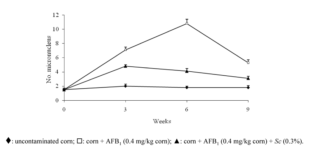
  22. Madrigal-Santillán, E.; Madrigal-Bujaidar, E.; Márquez-Márquez, R.; Reyes, A. Antigenotoxic effect of Saccharomyces cerevisiae on the damage produced in mice fed with aflatoxin B(1) contaminated corn. Food Chem. Toxicol. 2006, 44, 2058–2063. [Google Scholar]
  23. Kimura, M.; Lehmann, K.; Gopalan-Kriczky, P.; Lotlikar, P.D. Effect of diet on aflatoxin B1-DNA binding and aflatoxin B1-induced glutathione S-transferase placental form positive hepatic foci in the rat. Exp. Mol. Med. 2004, 36, 351–357. [Google Scholar]
  24. Eaton, D.L.; Gallagher, E.P. Mechanisms of aflatoxin carcinogenesis. Ann. Rev. Pharmacol. Toxicol. 1994, 34, 135–172. [Google Scholar]
  25. Madrigal-Santillán, E.; Morales-González, J.A.; Sánchez-Gutiérrez, M.; Reyes-Arellano, A.; Madrigal-Bujaidar, E. Investigation on the Protective Effect of alpha-Mannan against the DNA Damage Induced by Aflatoxin B(1) in Mouse Hepatocytes. Int. J. Mol. Sci. 2009, 10, 395–406. [Google Scholar]
  26. Raj, H.G.; Prasanna, H.R.; Mage, P.; Lotlikar, P.D. Effect of purified rat and hamster hepatic glutathione S-transferases on the microsome mediated binding of aflatoxin B1 to DNA. Cancer Lett. 1986, 33, 1–9. [Google Scholar]
  27. Eaton, D.L.; Ramsdel, H.S. Species and diet related differences in aflatoxin biotransformation. In Handbook of Applied Mycology, Mycotoxins in Ecological Systems; Bhatnagar, D., Lillehoj, E.B., Arora, D.K., Eds.; Marcel Dekker, Inc.: New York, NY, USA, 1992; Volume 5, pp. 157–182. [Google Scholar]
  28. IARC. Evaluation of carcinogen risks to humans. Some naturally occurring substances: food, items and constituents, heterocyclic aromatic amines and mycotoxins. IARC Monogr. Eval. Carcinog. Risks Hum. 1993, 56, 489–521. [Google Scholar] [PubMed]
  29. Labuza, T.P. Regulation of Mycotoxins. J. Food Prot. 1983, 46, 260–265. [Google Scholar]
  30. Guzmán-de-Peña, D.; Peña-Cabriales, J.J. Regulatory considerations of aflatoxin contamination of food in Mexico. Rev. Latinoam. Microbiol. 2005, 47, 160–164. [Google Scholar]
  31. Díaz, G.J.; Perilla, N.S.; Rojas, Y. Ocurrence of aflatoxins in selected colombians foods. Mycotoxin Res. 2001, 17, 15–20. [Google Scholar]
  32. Tanaka, K.; Sago, Y.; Zheng, Y.; Nakagawa, H.; Kushiro, M. Mycotoxins in rice. Int. J. Food Microbiol. 2007, 119, 59–66. [Google Scholar] [CrossRef] [PubMed]
  33. Ferguson, L.R.; Philpott, M. Nutrition and mutagenesis. Annu. Rev. Nutr. 2008, 28, 313–329. [Google Scholar]
  34. Ellis, W.; Smith, J.; Simpson, B.; Oldham, J. Aflatoxin in food: Ocurrence, biosynthesis, effects on organism, detection and methods of control. Crit. Rev. Food Sci. Nutr. 1991, 30, 403–439. [Google Scholar]
  35. Robb, J. Mycotoxins: Contamination and descontamination. Feed Mix. 1993, 1, 18–23. [Google Scholar]
  36. Cazzaniga, D.; Basilico, J.C.; González, R.J.; Torres, R.L.; de Greef, D.M. Mycotoxins inactivation by extrusion cooking of corn fluor. Lett. Appl. Microbiol. 2001, 33, 144–147. [Google Scholar]
  37. De Flora, S.; Ferguson, L.R. Overview of mechanisms of cancer chemopreventive agents. Mutat. Res. 2005, 591, 8–15. [Google Scholar]
  38. Park, D.L. Perspectives on mycotoxin decontamination procedures. Food Addit. Contam. 1993, 10, 49–60. [Google Scholar]
  39. Samarajeewa, U.; Sen, A.C.; Cohen, M.D.; Wei, C.I. Detoxification of aflatoxin in foods and feeds by physical and chemical methods. J. Food Prot. 1990, 53, 489–501. [Google Scholar]
  40. Elias-Orozco, R.; Castellanos-Nava, A.; Gaytán-Martínez, M.; Figueroa-Cárdenas, J.D.; Loarca-Piña, G. Comparison of nixtamalization and extrusion processes for a reduction in aflatoxin content. Food Addit. Contam. 2002, 19, 878–885. [Google Scholar]
  41. Shahin, A.A.; Aziz, N.H. Influence of gamma rays and sodium chloride on aflatoxin production by Aspergillus flavus. Microbios 1997, 90, 163–175. [Google Scholar]
  42. Kubena, L.F.; Harvey, N.E.; Phillips, T.D.; Clement, A.B. Effect of hidrated sodium calcium aluminosilicate on aflatoxicosis in broiler chicks. Poult. Sci. 1993, 72, 651–653. [Google Scholar]
  43. Stahr, H.M.; Obioha, W.O. Detoxification of aflatoxin contaminated corn by methanol extraction. Vet. Hum. Toxicol. 1982, 24, 16–17. [Google Scholar]
  44. Mukendi, N.; Rollmann, B.; de Meester, C. Detoxification of aflatoxin B1 by different chemical methods and evaluation of the effectiveness of the treatments applied. J. Pharm. Belg. 1991, 46, 182–188. [Google Scholar]
  45. Dvorák, M. Possibilities of chemical detoxification of aflatoxin. Vet. Med. (Praha) 1990, 35, 37–42. [Google Scholar] [PubMed]
  46. Norred, W.P.; Voss, K.A.; Bacon, C.W.; Riley, R.T. Effectiveness of ammonia treatment in detoxification of fumonisin-contaminated corn. Food Chem. Toxicol. 1991, 29, 815–819. [Google Scholar]
  47. Singh, J.; Reen, R.K.; Wiebel, F.J. Piperine, a major ingredient of black and long peppers, protects against AFB1-induced cytotoxicity and micronuclei formation in H4IIEC3 rat hepatoma cells. Cancer Lett. 1994, 86, 195–200. [Google Scholar]
  48. Soni, K.B.; Lahiri, M.; Chackradeo, P.; Bhide, S.V.; Kuttan, R. Protective effect of food additives on aflatoxin-induced mutagenicity and hepatocarcinogenicity. Cancer Lett. 1997, 115, 129–133. [Google Scholar]
  49. Loarca-Piña, G.; Kuzmicky, P.A.; Gonzalez de Mejia, E.; Kado, N.Y.; Hsieh, D.P. Antimutagenicity of ellagic acid against aflatoxin B1 in the Salmonella microsuspencion assay. Mutat. Res. 1996, 17, 15–21. [Google Scholar]
  50. Decoudu, S.; Cassand, P.; Daubeze, M.; Frayssient, C.; Narbonne, J.F. Effect of vitamin A dietary intake on in vitro and in vivo activation of aflatoxin B1. Mutat. Res. 1992, 269, 269–278. [Google Scholar]
  51. Kusamran, W.R.; Tepsuwan, A.; Kupradinum, P. Antimutagenic and anticarcinogenic potentials of some Thai vegetables. Mutat. Res. 1998, 402, 247–258. [Google Scholar]
  52. Gradelet, S.; Le Bon, A.M.; Berges, R.; Suschetet, M.; Astorg, P. Dietary carotenoids inhibit aflatoxin B1-induced liver preneoplastic foci and DNA damage in rat: role of the modulation of aflatoxin B1 metabolism. Carcinogenesis 1998, 19, 403–411. [Google Scholar]
  53. Abraham, S.K. Inhibitory effects of coffe on the genotoxicity of carcinogens in mice. Mutat. Res. 1991, 262, 109–114. [Google Scholar]
  54. Lim-Sylianco, C.Y.; Sylianco, W.L. Antigenotoxic effects of B vitamins. Asian Food J. 1992, 7, 100–104. [Google Scholar]
  55. Márquez-Márquez, R.; Madrigal-Bujaidar, E.; Tejada de Hernández, I. Genotoxic evaluation of ammonium inactivated aflatoxin B1 in mice fed with contaminated corn. Mutat. Res. 1993, 299, 1–8. [Google Scholar] [CrossRef] [PubMed]
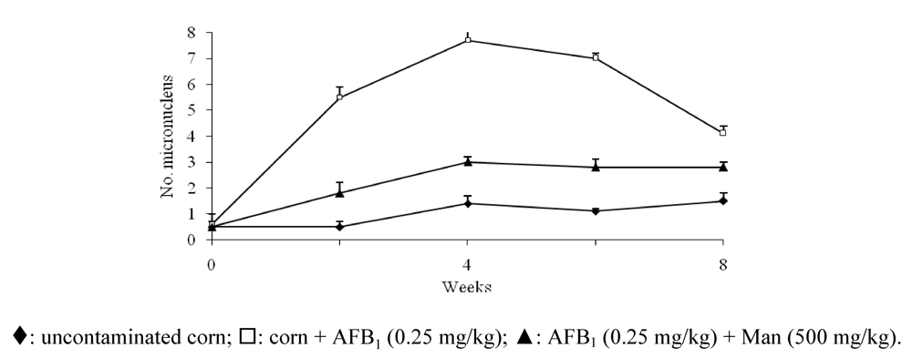
  56. Madrigal-Santillán, E.; Alvarez-González, I.; Márquez-Márquez, R.; Velázquez-Guadarrama, N.; Madrigal-Bujaidar, E. Inhibitory effect of mannan on the toxicity produced in mice fed aflatoxin B1 contaminated corn. Arch. Environ. Contam. Toxicol. 2007, 53, 466–472. [Google Scholar]
  57. Girard, I.D.; Dawson, K.A. Effect of yeast culture on the growth of representative ruminal bacteria. J. Anim. Sci. 1994, 77, 300–304. [Google Scholar]
  58. Madrigal-Bujaidar, E.; Madrigal-Santillán, E.; Pages, N.; Kogan, G.; Chamorro, C. Etude de L’antigenotoxicite de diferentes substances pour reduire la toxicite de L’aflatoxine B1 chez la souris. In Toxines et recherchers biomédicales; Francoise, G.-P., Cassian, B., Simone, P.-D., Sauviat, M.-P., Eds.; Reencontres en Toxinologie, Elsevier: Paris, France, 2002; pp. 123–132. [Google Scholar]
  59. Chorvatovicová, D.; Machova, E.; Sandula, J.; Kogan, G. Protective effect of the yeast glucomannan against cyclophosphamide-induced mutagenicity. Mutat. Res. 1999, 444, 117–122. [Google Scholar]
  60. Kogan, G.; Machova, E.; Chorvatovicová, D.; Slovakova, D.; Soltes, L.; Sandula, J. Chitin-glucan complex of Aspergillus niger and its derivates: antimutagenic, antiinfective and antiviral activity. In Advances in Chitin Science; Domard, A., Roberts, G.A., Eds.; Jackes André Publisher V II: Lyon, France, 1998; pp. 640–647. [Google Scholar]
  61. Aizawa, K.; Matsumoto, T.; Tsukadi, K.; Ito, A.; Satu, H.; Suzuki, S.; Suzuki, M. Antitumor effect of a Baker´s yeast mannan-mitomycin C conjugate against mouse hepatoma, MHI34, in vivo and in vitro. Int. J. Immunopharmacol. 1989, 11, 191–195. [Google Scholar]
  62. Tice, R.R.; Agurell, E.; Anderson, D.; Burlinson, B.; Hartmann, A.; Kobayashi, H.; Miyamae, Y.; Rojas, E.; Ryu, J.C.; Sasaky, Y.F. Single cell gel/comet assay: guidelines for in vitro and in vivo genetic toxicology testing. Environ. Mol. Mutagen 2000, 35, 206–221. [Google Scholar]
  63. Pohland, A.C. Mycotoxins in review. Food Addit. Contam. 1993, 10, 17–28. [Google Scholar]
  64. Favilla, M.; Pascale, M.; Ricelli, A.; Evidente, A.; Amalfitano, C.; Altomare, C. Inhibition of species of the Aspergillus section nigri and Ochratoxin A production in grapes by fusapyrone. Appl. Environ. Microbiol. 2008, 74, 2248–2253. [Google Scholar] [CrossRef] [PubMed]
  65. Duarte-Vogel, S.; Villamil-Jimenez, L.C. Micotoxinas en la salud pública. Rev. Salud Pública. 2006, 8, 129–135. [Google Scholar] [CrossRef]
  66. Bragulat, M.R.; Martínez, E.; Castellá, G.; Cabañes, F.J. Ochratoxin A and citrinin producing species of the genus Penicillium from feedstuffs. Int. J. Food Microbiol. 2008, 15, 43–48. [Google Scholar]
  67. Visconti, A.; Perrone, G.; Cozzi, G.; Solfrizzo, M. Managing ochratoxin A risk in the grape-wine food chain. Food Addit. Contam. Part A Chem. Anal. Control Expo. Risk Assess. 2008, 25, 193–202. [Google Scholar]
  68. Magnoli, C.E.; Astoreca, A.L.; Chiacchiera, S.M.; Dalcero, A.M. Occurrence of ochratoxin A and ochratoxigenic mycoflora in corn and corn based foods and feeds in some South American countries. Mycopathologia 2007, 163, 249–260. [Google Scholar]
  69. Serra Bonvehí, J. Occurrence of ochratoxin A in cocoa products and chocolate. J. Agric. Food Chem. 2004, 52, 6347–6352. [Google Scholar] [CrossRef] [PubMed]
  70. Walker, R. Risk assessment of ochratoxin: current views of the European Scientific Committee on Food, the JECFA and the Codex Committee on Food Additives and Contaminants. Adv. Exp. Med. Biol. 2002, 504, 249–255. [Google Scholar] [PubMed]
  71. Arbillaga, L.; Azqueta, A.; Expeleta, O.; Lopez de Cerfin, A. Oxidative DNA damage by ochratoxin A in the HK2 human kidney cell line: evidence of the relationship with cytotoxicity. Mutagenesis 2007, 22, 35–42. [Google Scholar]
  72. Marin-Kuan, M.; Cavin, C.; Delatour, T.; Schilter, B. Ochratoxin A carcinogenicity involves a complex network of epigenetic mechanisms. Toxicon 2008, 52, 195–202. [Google Scholar]
  73. Iacumin, L.; Chiesa, L.; Boscolo, D.; Manzano, M.; Cantoni, C.; Orlic, S.; Corni, G. Moulds and ochratoxin A on surfaces of artisanal and industrial dry sausages. Food Microbiol. 2009, 26, 65–70. [Google Scholar]
  74. McKenzie, K.S.; Sarr, A.B.; Mayura, K.; Bailey, R.H.; Miller, D.R.; Rogers, T.D.; Norred, W.P.; Voss, K.A.; Plattner, R.D.; Kubena, L.F.; Phillips, T.D. Oxidative degradation and detoxification of mycotoxins using a novel source of ozone. Food Chem. Toxicol. 1997, 35, 807–820. [Google Scholar] [CrossRef] [PubMed]
  75. Bejaoui, H.; Mathieu, F.; Taillander, P.; Lebrihi, A. Conidia of black aspergilla as new biological adsorbents for ochratoxin A in grapes juices and musts. J. Agric. Food Chem. 2005, 53, 8224–8229. [Google Scholar]
  76. Abrunhosa, L.; Serra, R.; Venancio, A. Biodegradation of ochratoxin A by fungi isolated from grapes. J. Agric. Food Chem. 2002, 50, 7493–7496. [Google Scholar]
  77. Varga, J.; Rigo, K.; Téren, J. Degradation of ochratoxin A by Aspergillus species. Int. J. Food Microbiol. 2000, 59, 1–7. [Google Scholar]
  78. Petchkongkaew, A.; Taillandier, P.; Gasaluck, P.; Lebrihi, A. Isolation of bacillus spp. From Thai fermented soybean (Thua-nao): screening for aflatoxin B1 and ochratoxin A detoxification. J. Appl. Microbiol. 2008, 104, 1495–1502. [Google Scholar] [PubMed]
  79. Costa, S.; Utan, A.; Cervellati, R.; Speroni, E.; Guerra, M.C. Catechins: natural free-radical scavengers against ochratoxin A-induced cell damage in a pig kidney cell line (LLC-PK1). Food Chem. Toxicol. 2007, 45, 1910–1917. [Google Scholar]
  80. Baudrimont, I.; Ahouandjivo, R.; Creppy, E.E. Prevention of lipid peroxidation induced by ochratoxin A in Vero cells in culture by several agents. Chem. Biol. Interact. 1997, 104, 29–40. [Google Scholar] [CrossRef] [PubMed]
  81. Turbic, A.; Ahokas, J.T.; Haskard, C.A. Selective in vitro binding of dietary mutagens, individually or in combination, by lactic acid bacteria. Food Addit. Contam. 2002, 19, 144–152. [Google Scholar]
  82. Palumbo, J.D.; O´Keeffe, T.L. Inhibition of ochratoxin A production and growth of Aspergillus species by phenolic antioxidant compounds. Mycopathologia 2007, 164, 241–248. [Google Scholar]
  83. Sergent, T.; Garsou, S.; Schaut, A.; De Saeger, S.; Pussemier, L.; Van Peteghem, C.; Larondelle, Y.; Schneider, Y.J. Differential modulation of ochratoxin A absorption across Caco-2-cells by dietary polyphenols, used at realistic intestinal concentrations. Toxicol. Lett. 2005, 159, 60–70. [Google Scholar] [CrossRef] [PubMed]
  84. Renzulli, C.; Galvano, F.; Pierdomenico, L.; Speroni, E.; Guerra, M.C. Effects of rosmarinic acid against aflatoxin B1 and ochratoxin A induced cell damage in a human hepatoma cell line (Hep G2). J. Appl. Toxicol. 2004, 24, 289–296. [Google Scholar] [CrossRef] [PubMed]
  85. Obrecht-Pflumio, S.; Grosse, Y.; Pfohl-Leszkowicz, A.; Dirheimer, G. Protection by indomethacin and aspirin genotoxicity of ochratoxin A, particulary in the urinary bladder and kidney. Arch. Toxicol. 1996, 70, 244–248. [Google Scholar]
  86. Ezz El-Arab, A.M.; Girgis, S.M.; Hegazy, E.M.; Abd El-Khalek, A.B. Effect of dietary honey on intestinal microflora and toxicity of mycotoxins in mice. BMC Complement. Altern. Med. 2006, 50, 799–804. [Google Scholar]
  87. Soyoz, M.; Ozcelik, N.; Kilinc, I.; Altuntas, I. The effects of ochratoxin A on lipid peroxidation and antioxidant enzymes: a protective role of melatonin. Cell. Biol. Toxicol. 2004, 20, 213–219. [Google Scholar]
  88. Abdel-Raheim, M.A.; Hussein, A.A. Melatonin reduces oxidative stress induced by ochratoxin A in rat liver and kidney. Comp. Biochem. Physiol. Part C. 2001, 130, 305–313. [Google Scholar]
  89. Abdel-Wahhab, M.A.; Abdel-Azim, S.H.; El-Nekeety, A.A. Inula crithmoides extract protects against ochratoxin A-induced oxidative stress, clastogenic, and mutagenic alterations in male rats. Toxicon 2008, 52, 566–573. [Google Scholar]
  90. Creppy, E.E.; Baudrimont, I.; Anne-Marie. How aspartame prevents the toxicity of ochratoxin A. J. Toxicol Sci. 1998, 23, 165–172. [Google Scholar] [PubMed]

