
Molecular Characterization of Two Totiviruses from the Commensal Yeast Geotrichum candidum


1
Botany and Microbiology Department, Faculty of Science, Damietta University, Damietta 34517, Egypt
2
The New Zealand Institute for Plant and Food Research Limited, Auckland 1025, New Zealand
3
School of Biological Sciences, The University of Auckland, Auckland 1010, New Zealand
*
Author to whom correspondence should be addressed.
Viruses 2023, 15(11), 2150; https://doi.org/10.3390/v15112150
Submission received: 13 September 2023
/
Revised: 16 October 2023
/
Accepted: 20 October 2023
/
Published: 25 October 2023
(This article belongs to the Collection Mycoviruses)
Abstract
:Mycoviruses can infect many of the major taxa of fungi including yeasts. Mycoviruses in the yeast fungus Geotrichum candidum are not well studied with only three G. candidum-associated viral species characterized to date, all of which belong to the Totiviridae genus Totivirus. In this study, we report the molecular characteristics of another two totiviruses co-infecting isolate Gc6 of G. candidum. The two totiviruses were tentatively named Geotrichum candidum totivirus 2 isolate Gc6 (GcTV2-Gc6) and Geotrichum candidum totivirus 4 isolate Gc6 (GcTV4-Gc6). Both viruses have the typical genome organization of totiviruses comprising two ORFs encoding capsid protein (CP) and RNA-dependent RNA polymerase (RdRp) at the N and C termini, respectively. The genomes of GcTV2-Gc6 and GcTV4-Gc6 are 4592 and 4530 bp long, respectively. Both viruses contain the—frameshifting elements and their proteins could be expressed as a single fusion protein. GcTV2-Gc6 is closely related to a totivirus isolated from the same host whereas GcTV4-Gc6 is related to insect-associated totiviruses. The phylogenetic analysis indicated that GcTV2-Gc6 and GcTV4-Gc6 belong to two different sister clades, I-A and I-B, respectively. It is interesting that all viruses identified from G. candidum belong to the genus Totivirus; however, this might be due to the lack of research reporting the characterization of mycoviruses from this fungal host. It is possible that the RNA interference (RNAi) mechanism cannot actively suppress totivirus accumulation in G. candidum Gc6.
Keywords:
mycoviruses; dsRNA; Totivirus; Geotrichum; high-throughput sequencing; siRNA; RNA interference; usRNA1. Introduction
The ascomycetous yeast Geotrichum candidum is a common, dimorphic member of the Saccharomycetes class [1]. It is known to infect plants and cause post-harvest rot in a variety of fruit and vegetable crops [2]. It supports the growth of Penicillium camemberti through the metabolism of lactate and suppresses the growth of undesired organisms, and hence, is used by the commercial cheesemaking industry as a ripening culture [3,4]. Moreover, it is a commensal parasite able to infect human skin, respiratory tracts, and gastrointestinal tracts [5,6].
Mycoviruses, viruses that infect and replicate in fungi, have been reported in several fungi belonging to all phyla of true fungi [7] as well as oomycetes [8]. Although mycovirus infections are mostly associated with symptomless infections, they may impact their fungal hosts in several ways [9,10,11,12]. Mycoviruses with three genome types (single-stranded RNA (ssRNA), double-stranded RNA (dsRNA), and ssDNA) have been reported with ssDNA mycoviruses being less common [7,9,13,14]. According to the International Committee on the Taxonomy of Viruses (ICTV), dsRNA mycoviruses are classified within the families Alternaviridae, Amalgaviridae, Birnaviridae, Chrysoviridae, Curvulaviridae, Megabirnaviridae, Partitiviridae, Polymycoviridae, Quadriviridae, Spinareoviridae, and Totiviridae, and the genus Botybirnavirus (https://ictv.global/taxonomy, accessed on 10 September 2023). Members of Totiviridae family are classified into five genera: Giardiavirus, Trichomonasvirus, Leishmaniavirus, Victorivirus, and Totivirus [15]. The known host range of Totiviridae members and totivirus-like genomes has expanded over recent years, with the increased utilization of next-generation sequencing technologies, to include insects, arthropods, fish, crab, and plants [16,17,18,19,20]. Historically, virus-like particles (VLPs) and dsRNAs have been detected in G. candidum without further biological or molecular identification of these associated VLPs or dsRNAs [21,22]. Recently, four totiviruses were detected and characterized from G. candidum isolates from Pakistan [23]. The current study reports the characterization of two mycoviruses from a different isolate of the same fungus. Interestingly, the two mycoviruses were also classified into the Totiviridae family, indicating that totiviruses are common in G. candidum.
2. Materials and Methods
2.1. Isolation, Maintenance, and Identification of Isolate Gc6
Isolate Gc6 was isolated along with other fungal species from a soil sample collected from Amberely, New Zealand, and grown on potato dextrose agar (PDA) using the soil-plate method [24]. A pure culture was obtained, and DNA was extracted using a Zymo DNA Fungal/Bacterial Miniprep Kit as described by the manufacturer. The identity of isolate Gc6 was determined by amplifying and sequencing the non-coding internal transcribed spacer (ITS) region of the fungal ribosomal DNA (rDNA) using the primer pair ITS4/ITS5 [25]. Throughout the course of this study, isolate Gc6 was maintained and cultured on PDA plates.
2.2. Purification and Visualization of dsRNA
Potato dextrose broth (PDB) media was inoculated with freshly grown fungal mycelial plugs and incubated at 25 °C for 5 days. Two grams of fungal mycelia was harvested and used to survey for the presence of dsRNA using a method based on the selective binding of dsRNA to cellulose powder in the presence of 16.5% ethanol as previously described [26]. Purified dsRNA was treated with DNase and RNase in a high salt buffer [27], separated on an ethidium bromide-prestained 1% (w/v) agarose gel in Tris–acetate EDTA (TAE) buffer, and visualized on a UV transilluminator.
2.3. Sequencing, Bioinformatics, and Sequence and Phylogenetic Analyses of dsRNA
The purified dsRNA fraction was used as a template for a reverse transcription polymerase chain reaction (RT-PCR) to generate random cDNA as described by Khalifa and Pearson [28]. The RT-PCR products were purified using a Gel/PCR DNA purification kit (Geneaid Biotech Ltd., New Taipei City, Taiwan) and sequenced using an Illumina HiSeq2000 at Macrogen Inc. (Seoul, Republic of Korea). Illumina short reads were quality checked using FastQC and based on the quality check reports, Illumina reads were filtered and trimmed based on quality. The reads were then de novo assembled using Geneious Prime 2022.1.1 software (Biomatters, New Zealand) set to medium sensitivity and default parameters. The assembled contigs were identified using Blastx against the GenBank non-redundant (nr) database. To determine the sequence of dsRNA termini and obtain full-length sequences of the viral contigs, the T4L adapter (5′-PO4-CCCGTCGTTTGCTGGCTCTTT-NH2-3′) was ligated to the 3′ end of the dsRNAs using T4 RNA ligase enzyme. The T4L-ligated dsRNAs were purified using a PCR clean-up and gel extraction kit (GeneDireX, Taoyuan City, Taiwan) following the manufacturer’s instructions, and used as a template for RT-PCR amplification of the terminal sequences using the T4LC primer (5′-AAAGAGCCAGCAAACGACGGG-3′) together with dsRNA specific primers designed based on the sequences obtained by Illumina sequencing (Supplementary Materials Table S1). RT-PCR products corresponding to the terminal sequences were purified and Sanger sequenced by Macrogen Inc. (Seoul, Republic of Korea).
Open reading frames (ORFs) were detected using the ORF finder tool (https://www.ncbi.nlm.nih.gov/orffinder/, accessed on 1 May 2023). Viral protein sequences were aligned using MAFFT [29] in order to identify conserved motifs. For the phylogenetic analysis, related protein sequences were retrieved from the NCBI database (accessed in April 2023) and aligned using MAFFT. Based on the obtained alignments, MEGA-X 10.2.5 software [30] was used to construct maximum-likelihood (ML) phylogenetic trees using the best fit models. A le–Gascuel model with a gamma distribution and some sites set to be evolutionary invariable (LG + G + I) and General Reverse Transcriptase + Freq with a gamma distribution (rtREV + G + F) model with 1000 bootstrap replicates were used for RNA-dependent RNA polymerase (RdRp) and capsid protein (CP) ML phylogenetic trees construction, respectively.
2.4. Small RNA (sRNA) Purification and Sequencing
G. candidum Gc6 was grown on cellophane-covered PDA Petri dishes for 5 days at 25 °C. The sRNA fraction was purified from grown mycelia using a mirPremier™ microRNA Isolation Kit (Sigma-Aldrich, Burlington, MA, USA). Purified microRNAs were run on an agarose gel to confirm their integrity and their concentration and purity were determined using a NanoDrop spectrophotometer. An aliquot of 20 µL of purified microRNAs at a concentration of 369 ng/µL was sent to Marogen Inc. (Seoul, Republic of Korea) for library construction and sequencing. A library of sRNA was prepared using a Truseq small RNA kit and sequenced using Illumina sequencing on one lane of a HiSeq2000 platform with a read length of 50 bp SR. For library preparation, RNA 3′ adapter (RA3) and RNA 5′ adapter (RA5) were ligated to RNAs using a T4 RNA ligase enzyme. Adapter-ligated libraries were reverse transcribed using SuperScript II reverse transcriptase and 2 primers that anneal to the adapter ends. Following amplification, the library was run on a high-sensitivity DNA chip for a quality check (size, purity, and concentration). cDNA constructs were gel purified in preparation for subsequent cluster generation. Constructs representing different sRNA species such as miRNAs, siRNAs, and Piwi-interacting RNAs were normalized to 2 nM using Tris-HCl 10 mM, pH 8.5, and sequenced. Sequencing data were obtained in FASTQ format. The obtained Illumina reads were trimmed before being mapped against the full sequences of dsRNAs obtained from G. candidum Gc6 using Bowtie2 with default parameters [31]. As a control, the obtained reads were also mapped against the G. candidum reference genome LMA-244_clib (GenBank: GCA_013365045.1).
3. Results and Discussion
The taxonomy of fungal viruses has grown extensively in the past few years due to the increasing interest in mycoviruses and their associated effects on the one hand, and with the practicability of high-throughput sequencing (HGS) technologies and bioinformatic software on another hand [32]. Below, we report the molecular characteristics of two dsRNA mycoviruses from the ascomycete G. candidum.
3.1. Identity of the Host Fungus
Soil is a rich source of different types of microorganisms that may be infected with viruses. Since these viruses affect the metabolic activity of their hosts, they play a pivotal role in the functionality of the soil ecosystems [33]. Fungi are one of the main soil microbial components that can harbor a wide range of viruses causing variable effects on their hosts starting from symptomless to severely debilitating infections [7]. Isolate Gc6 was isolated from a soil sample on PDA using standard isolation methods. A pure culture was obtained (Figure 1a), and the fungal identity was determined by sequencing the ITS region. The nucleotide sequence of the amplified fragment shared nucleotide (nt) sequence identities greater than 98% with the corresponding ITS regions of several G. candidum isolates (Supplementary materials Figure S1). Therefore, Gc6 was considered an isolate of G. candidum fungus.
3.2. Presence and Sequencing of dsRNA Associated with G. candidum Isolate Gc6
Electrophoretic analysis of the dsRNA purified from G. candidum Gc6 revealed the presence of a single nucleic acid band with an estimated molecular size of about 4.5 kb. This band resisted DNase treatment and RNase treatment in a high salt buffer, confirming its double-stranded nature (Figure 1b). Yeast fungi have been long known as hosts of mycoviruses since the discovery of a killer phenotype associated with Saccharomyces-virus killer systems [34]. In G. candidum, uncharacterized dsRNAs and virus-like particles have been discovered in the past [21,22]. Recently, three species belonging to the family Totiviridae have been characterized at the molecular level [23].
The purified dsRNA from G. candidum isolate Gc6 was subjected to random cDNA synthesis and sequencing, and its composition and identity were determined. De novo assembly of the Illumina short reads resulted in the creation of three long contiguous sequences 4519, 2447 and 743 nt in length that shared identities with totiviral sequences based upon initial Blastx analysis. The assembly was completed and validated, and the full-length sequences of the Gc6 dsRNAs were obtained using RACE-sequencing. The Gc6-dsRNAs were found to be 4592 (dsRNA1: contig 1) and 4530 (dsRNA2: contig 2 and 3) nt in length. Blastx searches of dsRNA1 and dsRNA2 full-length sequences revealed up to 94.15 and 89.24% identities compared to RdRp and 97.50% and 91.69% identities compared to CP sequences of viruses in the family Totiviridae, respectively (Supplementary materials Tables S2 and S3). Therefore, the apparently single dsRNA segment purified from isolate Gc6 consists of two co-migrating segments representing the genomes of two totiviruses. The closest fungal virus to dsRNA1 was Geotrichum candidum totivirus 2 (GcTV2) and therefore, dsRNA1 was considered as an isolate of the same virus (GcTV2-Gc6). dsRNA2 shared the highest identity with an insect totivirus, and the closest fungal totivirus shared RdRp and CP aa sequence identities of 48.05 and 41.86% with dsRNA2. Hence, dsRNA2 was given a new tentative name, Geotrichum candidum totivirus 4 (GcTV4-Gc6). The sequences of GcTV2-Gc6 and GcTV4-Gc6 were deposited to GenBank under accession numbers OR250782 and OR250783, respectively. Infection of a single fungal isolate with multiple related or unrelated mycoviruses has been previously reported in several fungal species [23,28,35,36,37,38,39,40,41,42]. Mycoviruses previously discovered from G. candidum as well as those described in the current study were found to cause co-infections in one host isolate.
3.3. Genome Organization and Analysis of Geotrichum candidum Totiviruses
The Totiviridae family includes viruses with a single linear, uncapped dsRNA genome segment of approximately 4.6–7.0 kilobase pair (kbp) in length [15]. The genomes of its members consist of two large, sometimes overlapping, ORFs with the potential to encode major CPs of about 70–100 kilodalton (kDa) (5′ ORF) and an RdRp (3′ ORF). The GcTV2-Gc6 genome has a GC content of 45.5% and has the potential to code for two long open ORFs that are separated by a 326 nt long intergenic region (IR). ORF1 in frame three is 2043 nt long and is located following a 29 nt long 5′ untranslated region (UTR). It starts at nt position 30, ends at an UAA termination codon at nt position 2072, and encodes a 680 amino acid (aa) long protein with an estimated molecular mass of 76.6 kDa. ORF2 in frame two precedes a 37 nt long 3′ UTR and has a length of 2157 nt. It starts at an AUG codon (nt position 2399), terminates at an UAA codon (nt position 4555), and encodes a 718 aa long protein with an estimated molecular weight of 82.06 kDa.
The GcTV4-Gc6 genome has a GC content of 42.6% and consists of 2097 (ORF1) and 2373 (ORF2) nt long ORFs with the potential to encode proteins with sizes of 698 and 790 aa and estimated molecular weights of 78.6 and 90.11 kDa, respectively. ORF1 and ORF2 start at nt positions 6 and 2123 with an AUG codon and end at nt positions 2102 and 4495, respectively. The two ORFs are separated by a 20 nt long IR and the genome has 5 and 35 nt long UTRs at the 5′ and 3′ termini, respectively. The genome organization of GcTV2-Gc6 and GcTV4-Gc6 is similar to genomes of other G. candidum totiviruses [23] and is typical for those of yeast totiviruses coding for two ORFs [43].
Blastp searches of putative proteins translated from ORF1 and ORF2 (for both GcTV2-Gc6 and GcTV4-Gc6) against the nr protein sequences database returned several hits for CP and RdRp of totiviruses, respectively. In most totiviruses, ORF1 and ORF2 are expressed as a single fusion protein due to the existence of a −1 ribosomal frameshifting mechanism. A slippery site where the frameshifting occurs is required near the stop codon of ORF1 and represented by the canonical sequence XXXYYYZ where X is A/G/C/U, Y is A/U, and Z is A/C/U [44]. A pseudoknot secondary structure preceding the slippery site is required to pause the ribosome and increase the efficiency of frameshifting [45]. Putative slippery sites were found at the 3′ terminus of ORF1 in both viruses. This was represented by the nt stretches GGGUUUA (nt positions 1958–1964) and UUUUUUA (nt positions 1943–1949) for GcTV2-Gc6 and GcTV4-Gc6, respectively. The slippery site in GcTV2-Gc6 is identical to that of the Saccharomyces cerevisiae virus L-A (ScV-L-A) [46]. In GcTV2-Gc6, the slippery site and an H-type pseudoknot at nt positions 1969–2023 are separated by a 4 nt spacer (Figure 1c). Similarly, the slippery site of GcTV4-Gc6 was followed by a 27 nt long spacer and an H-type pseudoknot at nt positions 1977–1997 (Figure 1c). Although the presence of −1 frameshifting signature does not necessarily reflect its functionality [47], the CP and RdRp genes of GcTV2-Gc6 and GcTV4-Gc6 could be expressed as fusion proteins with sizes of 1508 and 1496 aa, respectively.
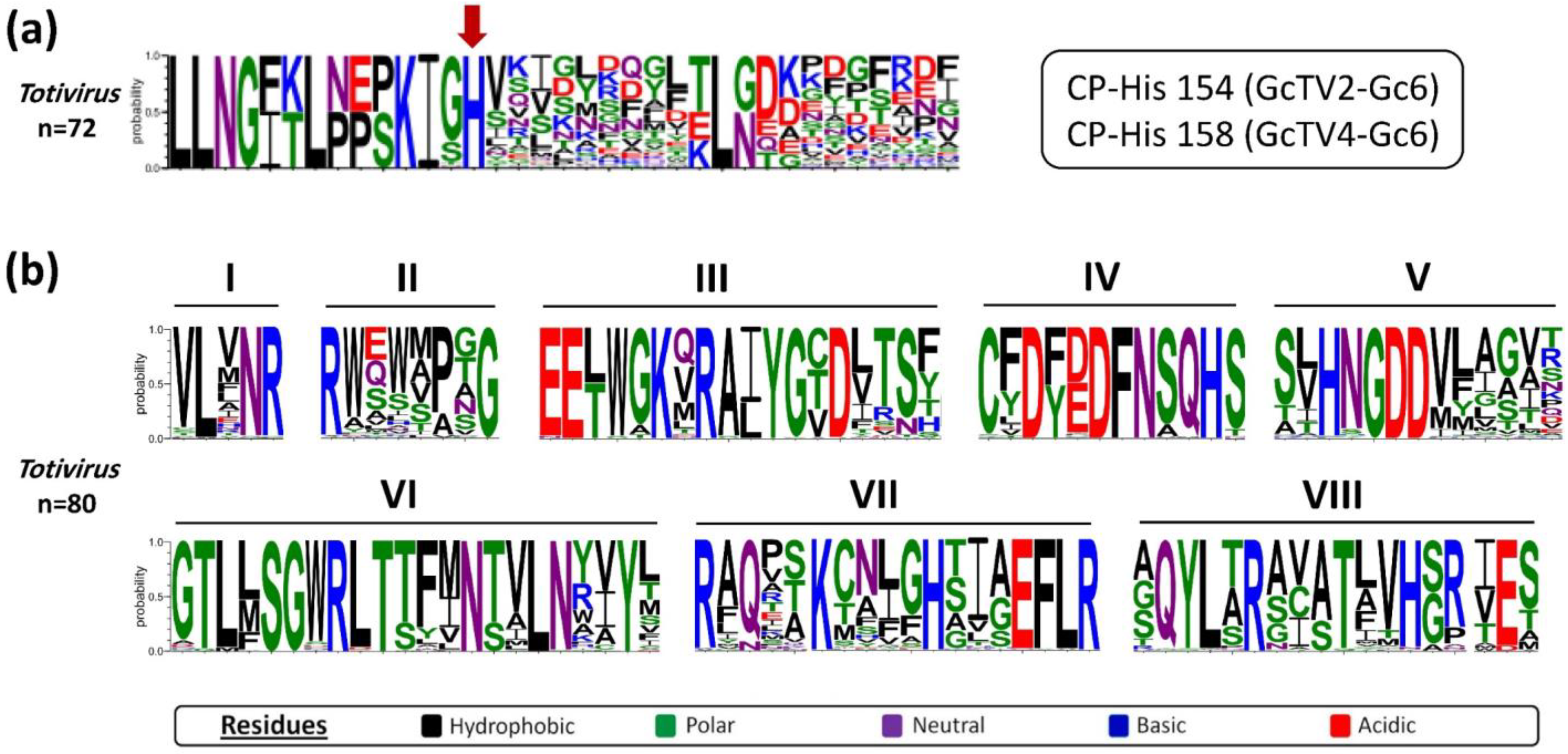
The RNA molecules synthesized by totiviral RdRps are not capped at their 5′ termini which makes them unrecognizable by their host translation machinery [48,49]. Therefore, it is established that totiviruses such as ScV-L-A and ScV-L-BC can de-cap their host’s mRNAs and transfer their caps to the totiviral transcripts using a unique cap-snatching mechanism that requires a certain histidine aa residue in the capsid protein [50,51]. Multiple aa sequence alignments of GcTV2-Gc6 and GcTV4-Gc6 CPs with other totiviral CP sequences revealed the presence of the conserved histidine aa at positions 154 and 158 of the translated protein (Figure 2a), respectively. This is analogous to the histidine residues found at aa positions 154, 15,6 and 159 of CPs of ScV-L-A [50], ScV-L-BC [51], red clover powdery mildew-associated totiviruses (RPaTVs) [52], and Trichoderma koningiopsis totivirus 1 (TkTV1) [53], respectively. Moreover, the aa sequence alignment between RdRps of GcTV2-Gc6, GcTV4-Gc6, and other totiviruses verified the presence of an RT-like superfamily domain (pfam02123) with eight conserved motifs (Figure 2b) including the GDD motif which is highly conserved among RdRp sequences of dsRNA viruses [54].
3.4. Phylogenetic Analysis
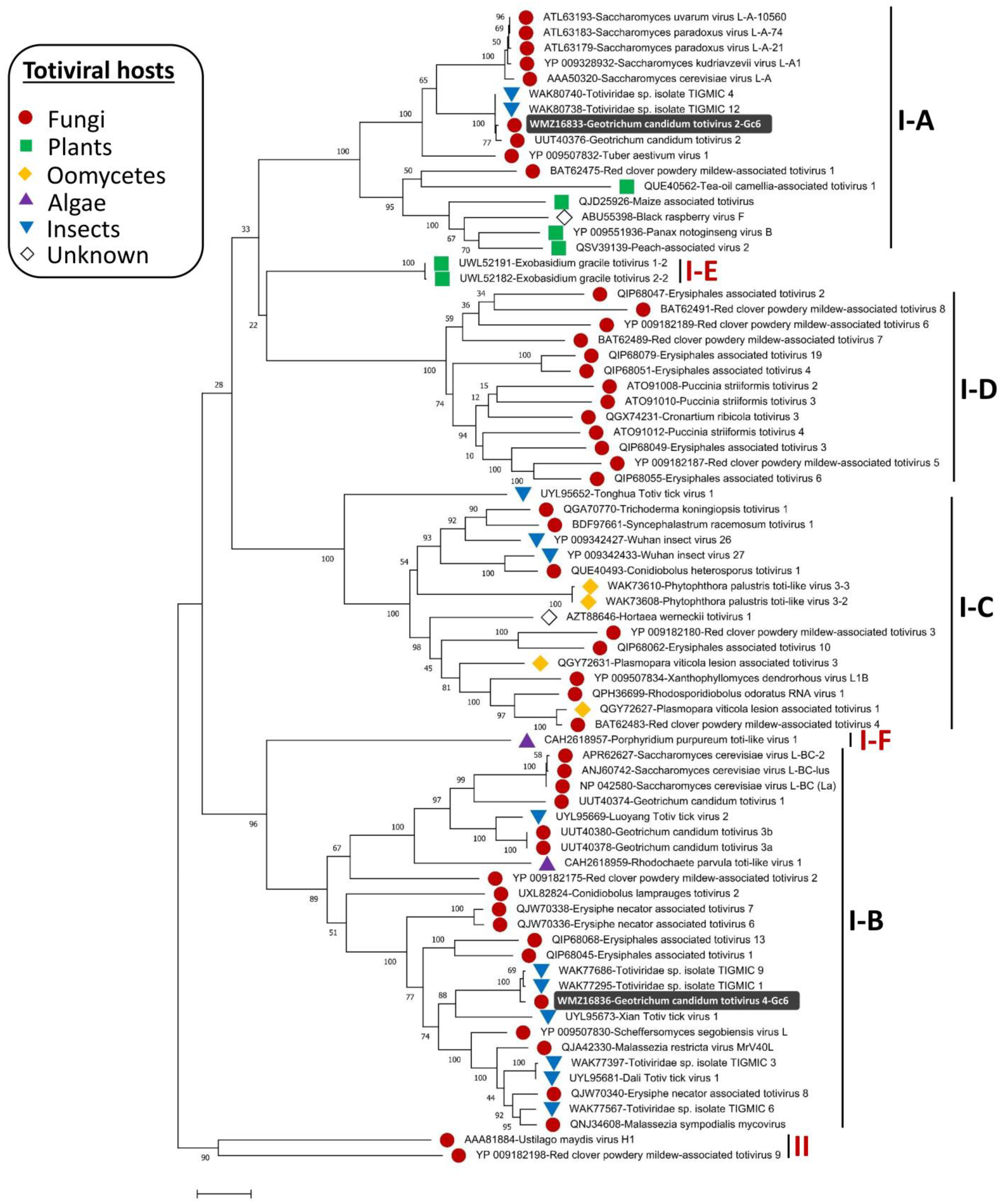
The phylogenetic structure of the genus Totivirus was determined, and members were found distributed in two major groups (groups I and II). Members in the first clade (group I) were further separated into four sister clades (I-A to I-D) [52]. The current phylogenetic status of the Totivirus genus has been re-determined in this study based on the increasing diversity of the discovered totiviral genomes, partially due to the advent of HGS. The phylogenetic trees constructed based on multiple alignments of RdRp and CP genes (Figure 3 and Figure 4) revealed that two additional sister clades (I-E and I-F) were formed in group I. The appearance of these two sister clades is taxonomically sensible since subclade I-E includes plant totiviruses exclusively and the sister clade I-A comprises totiviruses from a range of hosts including plant totiviruses. Moreover, subclade I-F that hosts totiviral members from algae is more related to subclade I-B where a related algal totivirus is found. Fungal totiviruses are distributed among subclades I-A to I-D among which, subclade I-D has no totiviruses from other hosts. G. candidum totiviruses are distributed among subclades I-A and I-B [23] which have totiviruses from multiple hosts including insects.
GcTV2-Gc6 was placed together with GcTV2-E1 [23] in subclade I-A and is most closely related to yeast and insect totiviruses. The closest viruses to GcTV4-Gc6 were isolated from insects (accession numbers ON812606 and ON812795). The existence of related fungal and insect totiviruses has previously been reported, reflecting the existence of common ancestors and possible horizontal virus transmission [52,53]. Previous reports suggested that unknown totiviruses from a wide range of different hosts may exist [52] and this was supported by the presence of closely related fungal and insect totiviruses such as TkTV1, Wuhan insect virus 26 (WIV26), and WIV27 [53]. As shown in Figure 3, this suggestion was confirmed, and it is believed that the diversity of totiviruses will expand further.
3.5. Are G. candidum Totiviruses Targets for the Host’s RNA Interference (RNAi) System?
Viral infections are repelled by their host’s defense machinery through RNAi which is a sequence-specific post-transcriptional gene silencing induced by dsRNA [55]. Viral dsRNA is common during the replication of all viral genome types. It is the replicative form during the replication of ssRNA viruses or duplex regions in complementary stretches of their genomes as well as in the transcripts of DNA viruses. Since the nature of dsRNA viral genomes is double-stranded, it is likely that the viral genome itself has the ability to trigger RNAi [55,56,57]. Therefore, the accumulation of G. candidum totivirus-derived short interfering RNAs (siRNAs) within G. candidum Gc6 implies that the host’s RNAi machinery is actively attempting to suppress viral replication and accumulation. A total small RNA (sRNA) fraction was purified from G. candidum Gc6, sequenced, and mapped against the genomes of GcTV2-Gc6 and GcTV4-Gc6 to obtain the viral-derived sRNAs. As a control to confirm the validity of the sRNA sequencing, sRNAs were also mapped against the LMA-244_clib reference genome of the G. candidum fungus. A ratio of 99.847% of the obtained reads had a phred quality score of over 20. Populations of 94,044 and 111,335 short reads, representing ~0.27% and 0.32% of G. candidum Gc6 sRNAs, were mapped to the GcTV2 and GcTV4 genomes from G. candidum Gc6, respectively. A minority of the assembled short reads perfectly matched the corresponding sequences in the totiviral genomes (2097 (2.23%) and 841 (0.75%) reads for GcTV2 and GcTV4, respectively). Mostly, the reads had a single mismatch to the genomes of either virus. Some reports indicate that mismatches in siRNA prevent gene silencing while others report that this can be tolerated [58,59]. The profile of siRNA mutations against three plant viruses was revealed and found to be due to viral or cellular RdRps [60]. Unlike viral-derived sRNAs, approximately 23% (7,630,682) of the short reads mapped to the fungal reference genome perfectly matched their corresponding sequences. This confirms that the prevalence of virus-derived short reads with one mismatch is not due to sequencing errors.
Diverse types of sRNAs have been identified in RNA-virus-infected hosts such as siRNAs, unusually small RNAs (usRNAs), and Piwi-interacting RNAs (piRNAs) [61,62]. Virus-derived usRNAs range in length from 13 to 19 nt and were abundantly identified among sRNA populations of human and viral origin [63,64]. usRNAs are shorter than the canonical length of functional miRNAs and are considered neglected intermediate degradation molecules. However, previous reports indicate that usRNAs derived from miRNA and tRNA could probably function in gene regulation [64]. Among the detected totivirus-derived small RNAs from G. candidum Gc6, reads with the common siRNA length (20–24 nt) were not abundant. The longest sRNA segment detected was 20 nt in length, mapped to GcTV2-Gc6, and was represented by only 10 sequences. The rest of the sequenced sRNAs were shorter, ranging from 10 to 19 nt for either virus (Figure 5, Table 1). Therefore, the sequenced totivirus-derived sRNAs from G. candidum Gc6 may represent populations of usRNAs, mostly with a single mismatch in their sequence. To eliminate the possibility that the failure to detect viral-derived sRNAs with the common siRNA length was not due to sequencing issues, comparisons were made between the number and length of viral-derived and host-derived sRNAs (Table 1). About 51.5% of the host-derived reads ranged in length between 20 and 24 nt, confirming the validity of the sequencing process and data obtained for the viral usRNAs.
Although it was clearly shown that RNA silencing is likely active in G. candidum isolate Gc6, the RNAi pathway is absent in some eukaryotes including Saccharomyces cerevisiae. Studies have shown that the loss of the RNAi machinery during evolution is beneficial to the yeast fungi as this helps in maintaining the killer virus system in the host fungus. Experimental restoration of the RNAi pathway caused a decreased ability to maintain the killer virus system, resulting in increased sensitivity to toxins from RNAi-deficient killer virus-containing cells [65]. Similarly, scarcity of viral-derived siRNAs with a functional length in G. candidum isolate Gc6 might be a result of an evolutionary mechanism to lose the RNAi pathway against viral infection and hence beneficial to the host fungi.
In summary, the inability to detect siRNAs, the abundance of usRNAs with mismatches, and the successful accumulation of totiviral genomes as shown by the dsRNA and electrophoresis data suggests that the host–defense machinery is not fully able to suppress the replication of G. candidum viruses and/or the totiviruses manage to counteract the RNAi mechanisms by expressing viral suppressors of RNAi (VSRs). Although VSR expression has been previously reported for several mycoviruses [57], our speculations regarding the ability of G. candidum viruses to express VSRs and the functional viability of their usRNAs need to be confirmed in future studies.
4. Conclusions
In this study, two members of the family Totiviridae were characterized from the commensal yeast G. candidum. Their genome organization is similar to that of many other totiviruses whose genes have the potential to be expressed as a fusion protein through a −1 frameshifting mechanism. A phylogenetic analysis placed the two viruses into two different sub-clades of the Totivirus genus. To our knowledge, this is the first report of mycovirus-derived usRNAs. The G. candidum–totivirus system may provide an easy model to study the role of usRNAs in fungi and other organisms. It can also be used to further reveal if G. candidum is deficient in RNAi as is the case for S. cerevisiae.
Supplementary Materials
The following supporting information can be downloaded at: https://www.mdpi.com/article/10.3390/v15112150/s1, Figure S1: Neighbor-joining phylogenetic tree constructed based on partial ITS-5.8s rRNA gene sequence alignment of isolate Gc6 and other members of Saccharomycotina. Table S1: Primers used for the RT-PCR amplifications of terminal sequences of the two dsRNAs (GcTV2-Gc6 and GcTV4-Gc6). Each primer was used in combination with primer T4LC; Table S2: Results of Blastx search (accessed in October 2023) for dsRNA1 (Geotrichum candidum totivirus 2 isolate Gc6) sequence (top 50 hits are shown); Table S3: Results of Blastx search (accessed in October 2023) for dsRNA2 (Geotrichum candidum totivirus 4 isolate Gc6) sequence (top 50 hits are shown).
Author Contributions
Conceptualization, M.E.K. and R.M.M.; methodology, M.E.K. and R.M.M.; validation, M.E.K.; formal analysis, M.E.K.; investigation, M.E.K.; resources, R.M.M.; data curation, M.E.K. and R.M.M.; writing—original draft preparation, M.E.K.; writing—review and editing, R.M.M.; visualization, M.E.K.; supervision, R.M.M.; project administration, R.M.M.; funding acquisition, R.M.M. All authors have read and agreed to the published version of the manuscript.
Funding
This research was funded by the Plant Biosecurity Cooperative Research Council and the New Zealand Institute for Plant and Food Research Limited, including the Growing Futures Rejuvenating Crop Ecosystems programme.
Institutional Review Board Statement
Not applicable.
Informed Consent Statement
Not applicable.
Data Availability Statement
GcTV2 and GcTV4 genome sequences are available in GenBank under the accession numbers OR250782 and OR250783, respectively.
Acknowledgments
The authors thank Nick Waipara and Mark Bullians as well as the journal’s reviewers for their helpful comments that have improved this manuscript.
Conflicts of Interest
The authors declare no conflict of interest.
References
- Alper, I.; Frenette, M.; Labrie, S. Ribosomal DNA polymorphisms in the yeast Geotrichum candidum. Fungal Biol. 2011, 115, 1259–1269. [Google Scholar] [CrossRef]
- Kim, Y.-K.; Kim, T.-S.; Shim, H.-S.; Park, K.-S.; Yeh, W.-H.; Hong, S.-J.; Shim, C.-K.; Kim, J.-S.; Park, J.-H.; Han, E.-J.; et al. First Report of Sour Rot on Post-harvest Oriental Melon, Tomato, Cucumber, Potato, Pumpkin and Carrot Caused by Geotrichum candidum. Res. Plant Dis. 2011, 17, 232–234. [Google Scholar] [CrossRef]
- Aziza, M.; Couriol, C.; Amrane, A.; Boutrou, R. Evidences for synergistic effects of Geotrichum candidum on Penicillium camembertii growing on cheese juice. Enzyme Microb. Technol. 2005, 37, 218–224. [Google Scholar] [CrossRef]
- Boutrou, R.; Kerriou, L.; Gassi, J.-Y. Contribution of Geotrichum candidum to the proteolysis of soft cheese. Int. Dairy J. 2006, 16, 775–783. [Google Scholar] [CrossRef]
- Bonifaz, A.; Vázquez-González, D.; Macías, B.; Paredes-Farrera, F.; Hernández, M.A.; Araiza, J.; Ponce, R.M. Oral geotrichosis: Report of 12 cases. J. Oral Sci. 2010, 52, 477–483. [Google Scholar] [CrossRef] [PubMed]
- Vasei, M.; Imanieh, M.H. Duodenal colonization by Geotrichum candidum in a child with transient low serum levels of IgA and IgM. APMIS 1999, 107, 681–684. [Google Scholar] [CrossRef] [PubMed]
- Pearson, M.N.; Beever, R.E.; Boine, B.; Arthur, K. Mycoviruses of filamentous fungi and their relevance to plant pathology. Mol. Plant Pathol. 2009, 10, 115–128. [Google Scholar] [CrossRef]
- Xu, Z.; Khalifa, M.E.; Frampton, R.A.; Smith, G.R.; McDougal, R.L.; MacDiarmid, R.M.; Kalamorz, F. Characterization of a Novel Double-Stranded RNA Virus from Phytophthora pluvialis in New Zealand. Viruses 2022, 14, 247. [Google Scholar] [CrossRef]
- Khalifa, M.E.; MacDiarmid, R.M. A Mechanically Transmitted DNA Mycovirus Is Targeted by the Defence Machinery of Its Host, Botrytis cinerea. Viruses 2021, 13, 1315. [Google Scholar] [CrossRef]
- Nuss, D.L. Hypovirulence: Mycoviruses at the fungal–plant interface. Nat. Rev. Microbiol. 2005, 3, 632–642. [Google Scholar] [CrossRef]
- Tipper, D.J.; Schmitt, M.J. Yeast dsRNA viruses: Replication and killer phenotypes. Mol. Microbiol. 1991, 5, 2331–2338. [Google Scholar] [CrossRef]
- Márquez, L.M.; Redman, R.S.; Rodriguez, R.J.; Roossinck, M.J. A Virus in a Fungus in a Plant: Three-Way Symbiosis Required for Thermal Tolerance. Science (80-) 2007, 315, 513–515. [Google Scholar] [CrossRef] [PubMed]
- Li, P.; Wang, S.; Zhang, L.; Qiu, D.; Zhou, X.; Guo, L. A tripartite ssDNA mycovirus from a plant pathogenic fungus is infectious as cloned DNA and purified virions. Sci. Adv. 2020, 6, aay9634. [Google Scholar] [CrossRef]
- Yu, X.; Li, B.; Fu, Y.; Jiang, D.; Ghabrial, S.A.; Li, G.; Peng, Y.; Xie, J.; Cheng, J.; Huang, J.; et al. A geminivirus-related DNA mycovirus that confers hypovirulence to a plant pathogenic fungus. Proc. Natl. Acad. Sci. USA 2010, 107, 8387–8392. [Google Scholar] [CrossRef]
- Wickner, R.B.; Ghabrial, S.A.; Nibert, M.L.; Patterson, J.L.; Wang, C.C. Family Totiviridae. In Virus Taxonomy: Classification and Nomenclature of Viruses: Ninth Report of the International Committee on Taxonomy of Viruses; King, A.M.Q., Adams, M.J., Carstens, E.B., Lefkowits, E.J., Eds.; Elsevier Academic Press: London, UK, 2012; pp. 639–650. [Google Scholar]
- Alvarez-Quinto, R.A.; Espinoza-Lozano, R.F.; Mora-Pinargote, C.A.; Quito-Avila, D.F. Complete genome sequence of a variant of maize-associated totivirus from Ecuador. Arch. Virol. 2017, 162, 1083–1087. [Google Scholar] [CrossRef] [PubMed]
- Colmant, A.M.G.; Etebari, K.; Webb, C.E.; Ritchie, S.A.; Jansen, C.C.; van den Hurk, A.F.; Bielefeldt-Ohmann, H.; Hobson-Peters, J.; Asgari, S.; Hall, R.A. Discovery of new orbiviruses and totivirus from Anopheles mosquitoes in Eastern Australia. Arch. Virol. 2017, 162, 3529–3534. [Google Scholar] [CrossRef] [PubMed]
- Huang, Y.; Guo, X.; Zhang, S.; Zhao, Q.; Sun, Q.; Zhou, H.; Zhang, J.; Tong, Y. Discovery of two novel totiviruses from Culex tritaeniorhynchus classifiable in a distinct clade with arthropod-infecting viruses within the family Totiviridae. Arch. Virol. 2018, 163, 2899–2902. [Google Scholar] [CrossRef] [PubMed]
- Mor, S.K.; Phelps, N.B.D. Molecular detection of a novel totivirus from golden shiner (Notemigonus crysoleucas) baitfish in the USA. Arch. Virol. 2016, 161, 2227–2234. [Google Scholar] [CrossRef]
- Zhao, M.; Xu, L.; Bowers, H.; Schott, E.J. Characterization of Two Novel Toti-Like Viruses Co-infecting the Atlantic Blue Crab, Callinectes sapidus, in Its Northern Range of the United States. Front. Microbiol. 2022, 13, 855750. [Google Scholar] [CrossRef]
- Mor, H.; Steinlauf, R.; Barash, I. Viruslike Particles and Double-Stranded RNA in Geotrichum candidum, the Causal Agent of Citrus Sour Rot. Phytopathology 1984, 74, 921. [Google Scholar] [CrossRef]
- Matte, O.; Chabalier, C.; Ratomahenina, R.; Bossy, J.P.; Galzy, P. Isolation of a double-stranded RNA and a virus-like particle from Geotrichum candidum. J. Basic Microbiol. 1991, 31, 447–452. [Google Scholar] [CrossRef]
- Khan, H.A.; Kondo, H.; Shahi, S.; Bhatti, M.F.; Suzuki, N. Identification of novel totiviruses from the ascomycetous fungus Geotrichum candidum. Arch. Virol. 2022, 167, 2833–2838. [Google Scholar] [CrossRef]
- Warcup, J.H. The Soil-Plate Method for Isolation of Fungi from Soil. Nature 1950, 166, 117–118. [Google Scholar] [CrossRef]
- White, T.J.; Bruns, T.; Lee, S.; Taylor, J. Amplification and direct sequencing of fungal ribosomal RNA genes for phylogenetics. In PCR Protocols; Elsevier: Amsterdam, The Netherlands; Academic Press: San Diego, CA, USA, 1990; pp. 315–322. [Google Scholar] [CrossRef]
- Valverde, R.A.; Nameth, S.T.; Jordan, R.L. Analysis of double-stranded RNA for plant virus diagnosis. Plant Dis. 1990, 74, 255–258. [Google Scholar]
- Howitt, R.L.J.; Beever, R.E.; Pearson, M.N.; Forster, R.L.S. Presence of double-stranded RNA and virus-like particles in Botrytis cinerea. Mycol. Res. 1995, 99, 1472–1478. [Google Scholar] [CrossRef]
- Khalifa, M.E.; Pearson, M.N. Molecular characterization of three mitoviruses co-infecting a hypovirulent isolate of Sclerotinia sclerotiorum fungus. Virology 2013, 441, 22–30. [Google Scholar] [CrossRef] [PubMed]
- Katoh, K.; Misawa, K.; Kuma, K.; Miyata, T. MAFFT: A novel method for rapid multiple sequence alignment based on fast Fourier transform. Nucleic Acids Res. 2002, 30, 3059–3066. [Google Scholar] [CrossRef] [PubMed]
- Kumar, S.; Stecher, G.; Li, M.; Knyaz, C.; Tamura, K. MEGA X: Molecular Evolutionary Genetics Analysis across Computing Platforms. Mol. Biol. Evol. 2018, 35, 1547–1549. [Google Scholar] [CrossRef]
- Langmead, B.; Salzberg, S.L. Fast gapped-read alignment with Bowtie 2. Nat. Methods 2012, 9, 357–359. [Google Scholar] [CrossRef]
- Dolja, V.V.; Koonin, E.V. Metagenomics reshapes the concepts of RNA virus evolution by revealing extensive horizontal virus transfer. Virus Res. 2018, 244, 36–52. [Google Scholar] [CrossRef] [PubMed]
- Sutela, S.; Poimala, A.; Vainio, E.J. Viruses of fungi and oomycetes in the soil environment. FEMS Microbiol. Ecol. 2019, 95, fiz119. [Google Scholar] [CrossRef] [PubMed]
- Bevan, E.A.; Herring, A.J.; Mitchell, D.J. Preliminary Characterization of Two Species of dsRNA in Yeast and their Relationship to the “Killer” Character. Nature 1973, 245, 81–86. [Google Scholar] [CrossRef]
- Baeza, M.; Bravo, N.; Sanhueza, M.; Flores, O.; Villarreal, P.; Cifuentes, V. Molecular characterization of totiviruses in Xanthophyllomyces dendrorhous. Virol. J. 2012, 9, 140. [Google Scholar] [CrossRef] [PubMed]
- Herrero, N.; Zabalgogeazcoa, I. Mycoviruses infecting the endophytic and entomopathogenic fungus Tolypocladium cylindrosporum. Virus Res. 2011, 160, 409–413. [Google Scholar] [CrossRef]
- Khalifa, M.E.; Pearson, M.N. Molecular characterisation of an endornavirus infecting the phytopathogen Sclerotinia sclerotiorum. Virus Res. 2014, 189, 303–309. [Google Scholar] [CrossRef] [PubMed]
- Khalifa, M.E.; Pearson, M.N. Molecular characterisation of novel mitoviruses associated with Sclerotinia sclerotiorum. Arch. Virol. 2014, 159, 3157–3160. [Google Scholar] [CrossRef] [PubMed]
- Kim, J.W.; Choi, E.Y.; Lee, J.I. Genome Organization and Expression of the Penicillium stoloniferum Virus F. Virus Genes 2005, 31, 175–183. [Google Scholar] [CrossRef]
- Osaki, H.; Nomura, K.; Matsumoto, N.; Ohtsu, Y. Characterization of double-stranded RNA elements in the violet root rot fungus Helicobasidium mompa. Mycol. Res. 2004, 108, 635–640. [Google Scholar] [CrossRef]
- Park, Y.; James, D.; Punja, Z.K. Co-infection by two distinct totivirus-like double-stranded RNA elements in Chalara elegans (Thielaviopsis basicola). Virus Res. 2005, 109, 71–85. [Google Scholar] [CrossRef]
- Preisig, O.; Wingfield, B.D.; Wingfield, M.J. Coinfection of a Fungal Pathogen by Two Distinct Double-Stranded RNA Viruses. Virology 1998, 252, 399–406. [Google Scholar] [CrossRef]
- Icho, T.; Wickner, R.B. The Double-stranded RNA Genome of Yeast Virus L-A Encodes Its Own Putative RNA Polymerase by Fusing Two Open Reading Frames. J. Biol. Chem. 1989, 264, 6716–6723. [Google Scholar] [CrossRef]
- Bekaert, M.; Rousset, J.-P. An Extended Signal Involved in Eukaryotic −1 Frameshifting Operates through Modification of the E Site tRNA. Mol. Cell 2005, 17, 61–68. [Google Scholar] [CrossRef] [PubMed]
- Rice, N.R.; Stephens, R.M.; Burny, A.; Gilden, R.V. The gag and pol genes of bovine leukemia virus: Nucleotide sequence and analysis. Virology 1985, 142, 357–377. [Google Scholar] [CrossRef] [PubMed]
- Brierley, I. Ribosomal frameshifting on viral RNAs. J. Gen. Virol. 1995, 76, 1885–1892. [Google Scholar] [CrossRef]
- Jamal, A.; Sato, Y.; Shahi, S.; Shamsi, W.; Kondo, H.; Suzuki, N. Novel Victorivirus from a Pakistani Isolate of Alternaria alternata Lacking a Typical Translational Stop/Restart Sequence Signature. Viruses 2019, 11, 577. [Google Scholar] [CrossRef] [PubMed]
- Fujimura, T.; Esteban, R. Yeast Double-stranded RNA Virus L-A Deliberately Synthesizes RNA Transcripts with 5′-Diphosphate. J. Biol. Chem. 2010, 285, 22911–22918. [Google Scholar] [CrossRef]
- Rowley, P.A.; Ho, B.; Bushong, S.; Johnson, A.; Sawyer, S.L. XRN1 Is a Species-Specific Virus Restriction Factor in Yeasts. PLoS Pathog. 2016, 12, e1005890. [Google Scholar] [CrossRef]
- Fujimura, T.; Esteban, R. Cap-snatching mechanism in yeast L-A double-stranded RNA virus. Proc. Natl. Acad. Sci. USA 2011, 108, 17667–17671. [Google Scholar] [CrossRef]
- Fujimura, T.; Esteban, R. Cap Snatching in Yeast L-BC Double-stranded RNA Totivirus. J. Biol. Chem. 2013, 288, 23716–23724. [Google Scholar] [CrossRef]
- Kondo, H.; Hisano, S.; Chiba, S.; Maruyama, K.; Andika, I.B.; Toyoda, K.; Fujimori, F.; Suzuki, N. Sequence and phylogenetic analyses of novel totivirus-like double-stranded RNAs from field-collected powdery mildew fungi. Virus Res. 2016, 213, 353–364. [Google Scholar] [CrossRef]
- Khalifa, M.E.; MacDiarmid, R.M. A Novel Totivirus Naturally Occurring in Two Different Fungal Genera. Front. Microbiol. 2019, 10, 02318. [Google Scholar] [CrossRef] [PubMed]
- Routhier, E.; Bruenn, J.A. Functions of Conserved Motifs in the RNA-Dependent RNA Polymerase of a Yeast Double-Stranded RNA Virus. J. Virol. 1998, 72, 4427–4429. [Google Scholar] [CrossRef] [PubMed]
- Gammon, D.B.; Mello, C.C. RNA interference-mediated antiviral defense in insects. Curr. Opin. Insect Sci. 2015, 8, 111–120. [Google Scholar] [CrossRef] [PubMed]
- Mongelli, V.; Saleh, M.-C. Bugs Are Not to Be Silenced: Small RNA Pathways and Antiviral Responses in Insects. Annu. Rev. Virol. 2016, 3, 573–589. [Google Scholar] [CrossRef]
- Rodriguez Coy, L.; Plummer, K.M.; Khalifa, M.E.; MacDiarmid, R.M. Mycovirus-encoded suppressors of RNA silencing: Possible allies or enemies in the use of RNAi to control fungal disease in crops. Front. Fungal Biol. 2022, 3, 965781. [Google Scholar] [CrossRef]
- Zeng, Y.; Cullen, B.R. Sequence requirements for micro RNA processing and function in human cells. RNA 2003, 9, 112–123. [Google Scholar] [CrossRef]
- Elbashir, S.M.; Martinez, J.; Patkaniowska, A.; Lendeckel, W.; Tuschl, T. Functional anatomy of siRNAs for mediating efficient RNAi in Drosophila melanogaster embryo lysate. EMBO J. 2001, 20, 6877–6888. [Google Scholar] [CrossRef]
- Nigam, D.; LaTourrette, K.; Garcia-Ruiz, H. Mutations in virus-derived small RNAs. Sci. Rep. 2020, 10, 9540. [Google Scholar] [CrossRef]
- Usme-Ciro, J.A.; Campillo-Pedroza, N.; Almazán, F.; Gallego-Gomez, J.C. Cytoplasmic RNA viruses as potential vehicles for the delivery of therapeutic small RNAs. Virol. J. 2013, 10, 185. [Google Scholar] [CrossRef]
- Kolliopoulou, A.; Santos, D.; Taning, C.N.T.; Wynant, N.; Vanden Broeck, J.; Smagghe, G.; Swevers, L. PIWI pathway against viruses in insects. WIREs RNA 2019, 10, 1555. [Google Scholar] [CrossRef]
- Hess, A.M.; Prasad, A.N.; Ptitsyn, A.; Ebel, G.D.; Olson, K.E.; Barbacioru, C.; Monighetti, C.; Campbell, C.L. Small RNA profiling of Dengue virus-mosquito interactions implicates the PIWI RNA pathway in anti-viral defense. BMC Microbiol. 2011, 11, 45. [Google Scholar] [CrossRef] [PubMed]
- Li, Z.; Kim, S.W.; Lin, Y.; Moore, P.S.; Chang, Y.; John, B. Characterization of Viral and Human RNAs Smaller than Canonical MicroRNAs. J. Virol. 2009, 83, 12751–12758. [Google Scholar] [CrossRef] [PubMed]
- Drinnenberg, I.A.; Fink, G.R.; Bartel, D.P. Compatibility with Killer Explains the Rise of RNAi-Deficient Fungi. Science 2011, 333, 1592. [Google Scholar] [CrossRef] [PubMed]
Figure 1.
(a) Colony morphology of Geotrichum candidum isolate Gc6 grown on PDA media. (b) Agarose gel electrophoresis of dsRNAs purified from G. candidum Gc6. Purified dsRNA fraction was treated with DNase and RNase in a high salt buffer before electrophoretic separation. M: 1 kb plus DNA marker (Invitrogen). (c) Schematic representation of the genome organization of G. candidum Gc6 totiviruses. Coding regions are indicated by colored boxes whereas untranslated regions are indicated by short lines at both termini. The potential −1 frameshifting site (−1FS) is indicated by black arrows and its three components (slippery heptamer, spacer, and pseudoknot) are shown as an inset in each totiviral genome schematic representation. EFE refers to estimated free energy values for the H-type pseudoknots.

Figure 2.
(a) Partial multiple amino acid (aa) sequence alignment of the CP sequences of totiviruses including those of G. candidum. The conserved histidine residue required for cap snatching is indicated by a red arrow. (b) aa sequence alignments showing the conserved motifs (I–VIII) of RNA-dependent RNA polymerase (RdRp) sequences of G. candidum totiviruses and other members of the genus Totivirus.

Figure 3.
Maximum likelihood phylogenetic tree based on multiple alignments of RNA-dependent RNA polymerase amino acid (aa) sequences of Geotrichum candidum totivirus 2 (GcTV2-Gc6), GcTV4-Gc6, and other members of the genus Totivirus. The phylogenetic tree was constructed using MEGA-X software and LG + G + I as the best evolutionary model with 1000 bootstrap replicates.

Figure 4.
Maximum likelihood phylogenetic tree based on multiple alignments of capsid protein (CP) amino acid (aa) sequences of Geotrichum candidum totivirus 2 (GcTV2-Gc6), GcTV4-Gc6, and other members of the genus Totivirus. The phylogenetic tree was constructed using MEGA-X software and rtREV + G + F as the best evolutionary model with 1000 bootstrap replicates.

Figure 5.
Length distribution in log scale of small RNAs (sRNAs) mapped against Geotrichum candidum totivirus 2 (GcTV2-Gc6) and GcTV4-Gc6.
Figure 5.
Length distribution in log scale of small RNAs (sRNAs) mapped against Geotrichum candidum totivirus 2 (GcTV2-Gc6) and GcTV4-Gc6.


Table 1.
Size distribution of small RNA (sRNA) sequencing reads mapped to the genomes of GcTV2-Gc6, GcTV4-Gc6, and G. candidum reference genome LMA-244_clib (GenBank: GCA_013365045.1). Numbers of sRNA reads with 0 and 1 mismatches are presented. ND: not detected.
Table 1.
Size distribution of small RNA (sRNA) sequencing reads mapped to the genomes of GcTV2-Gc6, GcTV4-Gc6, and G. candidum reference genome LMA-244_clib (GenBank: GCA_013365045.1). Numbers of sRNA reads with 0 and 1 mismatches are presented. ND: not detected.
| sRNA Length (nts) | Total Viral | Total Fungal | Perfect Match | 1 Mismatch | |||||
|---|---|---|---|---|---|---|---|---|---|
| GcTV2 | GcTV4 | GcTV2 | GcTV4 | G. candidum | GcTV2 | GcTV4 | G. candidum | ||
| 10 | 56,987 | 75,858 | 579,036 | 1450 | 545 | 579,020 | 55,537 | 75,313 | 16 |
| 11 | 15,616 | 26,851 | 439,532 | 522 | 232 | 436,820 | 15,094 | 26,619 | 2712 |
| 12 | 7309 | 4859 | 980,610 | 99 | 43 | 866,277 | 7210 | 4816 | 114,333 |
| 13 | 12,614 | 2410 | 1,247,012 | 21 | 8 | 646,920 | 12,593 | 2402 | 600,092 |
| 14 | 615 | 695 | 2,423,441 | 5 | 8 | 257,021 | 610 | 687 | 2,166,420 |
| 15 | 369 | 386 | 2,374,600 | ND | 5 | 243,264 | 369 | 381 | 2,131,336 |
| 16 | 47 | 154 | 1,670,491 | ND | ND | 262,674 | 47 | 154 | 1,407,817 |
| 17 | 133 | 43 | 3,120,220 | ND | ND | 301,826 | 133 | 43 | 2,818,394 |
| 18 | 337 | 49 | 1,246,059 | ND | ND | 337,262 | 337 | 49 | 908,797 |
| 19 | 7 | 30 | 2,103,211 | ND | ND | 602,221 | 7 | 30 | 1,500,990 |
| 20 | 10 | ND | 12,694,084 | ND | ND | 1,312,581 | 10 | ND | 11,381,503 |
| 21 | ND | ND | 1,503,777 | ND | ND | 535,043 | ND | ND | 968,734 |
| 22 | ND | ND | 1,025,590 | ND | ND | 351,560 | ND | ND | 674,030 |
| 23 | ND | ND | 1,181,768 | ND | ND | 269,011 | ND | ND | 912,757 |
| 24 | ND | ND | 1,518,337 | ND | ND | 233,035 | ND | ND | 1,285,302 |
| 25 | ND | ND | 240,986 | ND | ND | 121,743 | ND | ND | 119,243 |
| 26 | ND | ND | 151,291 | ND | ND | 79,791 | ND | ND | 71,500 |
| 27 | ND | ND | 109,991 | ND | ND | 64,998 | ND | ND | 44,993 |
| 28 | ND | ND | 87,651 | ND | ND | 51,255 | ND | ND | 36,396 |
| 29 | ND | ND | 49,420 | ND | ND | 23,245 | ND | ND | 26,175 |
| 30 | ND | ND | 39,603 | ND | ND | 19,653 | ND | ND | 19,950 |
| >30 | ND | ND | 77,785 | ND | ND | 35,462 | ND | ND | 42,323 |
| Total | 94,044 | 111,335 | 34,864,495 | 2097 | 841 | 7,630,682 | 91,947 | 110,494 | 27,233,813 |
Disclaimer/Publisher’s Note: The statements, opinions and data contained in all publications are solely those of the individual author(s) and contributor(s) and not of MDPI and/or the editor(s). MDPI and/or the editor(s) disclaim responsibility for any injury to people or property resulting from any ideas, methods, instructions or products referred to in the content. |
© 2023 by the authors. Licensee MDPI, Basel, Switzerland. This article is an open access article distributed under the terms and conditions of the Creative Commons Attribution (CC BY) license (https://creativecommons.org/licenses/by/4.0/).
Share and Cite
MDPI and ACS Style
Khalifa, M.E.; MacDiarmid, R.M. Molecular Characterization of Two Totiviruses from the Commensal Yeast Geotrichum candidum. Viruses 2023, 15, 2150. https://doi.org/10.3390/v15112150
AMA Style
Khalifa ME, MacDiarmid RM. Molecular Characterization of Two Totiviruses from the Commensal Yeast Geotrichum candidum. Viruses. 2023; 15(11):2150. https://doi.org/10.3390/v15112150
Chicago/Turabian StyleKhalifa, Mahmoud E., and Robin M. MacDiarmid. 2023. "Molecular Characterization of Two Totiviruses from the Commensal Yeast Geotrichum candidum" Viruses 15, no. 11: 2150. https://doi.org/10.3390/v15112150
Note that from the first issue of 2016, this journal uses article numbers instead of page numbers. See further details here.