Review

Potential of Electrospun Nanofibers for Biomedical and Dental Applications

1
Department of Restorative Dentistry, College of Dentistry, Taibah University, Madina Munawwarah 41311, Saudi Arabia
2
Department of Restorative Dental Sciences, Al Farabi Colleges, King Abdullah Road, Riyadh 11313, Saudi Arabia
3
School of Metallurgy and Materials, University of Birmingham, Birmingham B15 2TT, UK
4
Department of Biology, Faculty of Science, University of Isfahan, Isfahan 81746-73441, Iran
5
Department of Biomedical Engineering, College of Engineering, King Faisal University, Al-Hofuf 31982, Saudi Arabia
6
School of Dentistry, Riyadh College of Dentistry and Pharmacy, P.O. Box 84891, Riyadh 11313, Saudi Arabia
7
Department of Medical Engineering, University of Bradford, Bradford BD7 1DP, UK
8
Department of Biomedical Engineering, King Faisal University, Al-Hofuf 31982, Saudi Arabia
9
Department of Chemistry, Chemical Biology and Biomedical Engineering, Stevens Institute of Technology, Hoboken, NJ 07030, USA
*
Author to whom correspondence should be addressed.
Materials 2016, 9(2), 73; https://doi.org/10.3390/ma9020073
Submission received: 29 November 2015 / Revised: 6 January 2016 / Accepted: 18 January 2016 / Published: 26 January 2016
(This article belongs to the Special Issue Electrospun Materials)

Abstract

:
Electrospinning is a versatile technique that has gained popularity for various biomedical applications in recent years. Electrospinning is being used for fabricating nanofibers for various biomedical and dental applications such as tooth regeneration, wound healing and prevention of dental caries. Electrospun materials have the benefits of unique properties for instance, high surface area to volume ratio, enhanced cellular interactions, protein absorption to facilitate binding sites for cell receptors. Extensive research has been conducted to explore the potential of electrospun nanofibers for repair and regeneration of various dental and oral tissues including dental pulp, dentin, periodontal tissues, oral mucosa and skeletal tissues. However, there are a few limitations of electrospinning hindering the progress of these materials to practical or clinical applications. In terms of biomaterials aspects, the better understanding of controlled fabrication, properties and functioning of electrospun materials is required to overcome the limitations. More in vivo studies are definitely required to evaluate the biocompatibility of electrospun scaffolds. Furthermore, mechanical properties of such scaffolds should be enhanced so that they resist mechanical stresses during tissue regeneration applications. The objective of this article is to review the current progress of electrospun nanofibers for biomedical and dental applications. In addition, various aspects of electrospun materials in relation to potential dental applications have been discussed.

1. Introduction

Nanofibers remain an important division of biomaterials due to a wide range of biomedical applications [1]. The fabrication of nanofibers has attracted a lot of researchers due to unique properties required for biomedical applications for example availability of greater surface area for cellular interaction [2], protein absorption and binding sites to cell receptors [3]. Nanofibers can facilitate packing of maximum volume fraction by controlling fibers alignment and orientation hence improving the material strength [2]. The material properties such as surface morphology, porosity and geometry can be tailored or functionalized for certain applications, for example, bioactive agents for biomedical applications [4].
In order to fabricate fibers nanofibers, different techniques have been used for example, phase separation [5,6,7], nanofiber seeding [8] template synthesis [9,10], self-assembly [11,12] and electrospinning [13,14,15,16,17,18,19,20,21]. Amongst these techniques, electrospinning is a resourceful and cost effective technique that can be used to synthesize continuous nanofibers [1]. This technique can be used for soluble or fusible polymers alone or polymers can be modified with additives such as particles or enzymes to get the desired properties [20]. The resultant ultrafine fibers exhibit many interesting features, e.g., high surface area, tailorable porosity in the range of submicron to nanoscale and greater potential for surface functionalization [20,22,23,24]. In addition, electrospun fibers are considered an excellent candidate for a variety of biomedical purposes, e.g., wound dressings, drug delivery and tissue engineering scaffolds [21]. Electrospinning has been used for several biopolymers and blended biopolymers with synthetic polymers to obtain nanofibers [20]. Additionally, electrospinning can be used for fabricating polymer composite fibers by blending additives such as particles, antimicrobials or enzymes to get the desired properties [20].
Considering these benefits, electrospinning has gained a remarkable popularity for various disciplines hence, projecting a sharp rise in scientific publications in recent years (Table 1). There are relatively few electrospinning studies involving oral and dental sciences. The keywords “electrospinning” and “oral dental electrospinning” searched only 47 publications; 39 published in last five years (2011–2015) and only 8 published during 2005–2010. The aim of this article is to review the current progress of electrospun nanofibers for biomedical and dental applications. In addition, various aspects of electrospun materials in relation to potential dental applications have been discussed.
Table 1. Number of peer reviewed scientific papers published on “electrospinning” in recent years.
YearsElectrospinningOral/Dental Electrospinning
Topic SearchTitle SearchTopic Search
20052961141
20064822040
20076232590
200810473731
200911834422
201014315074
201118455793
2012210262712
201333776398
2014611779310
201552336006
Search was carried on using the keywords “electrospinning” and “oral dental electrospinning” in topic and title search options of ISI Web of Knowledge database for particular publication years.

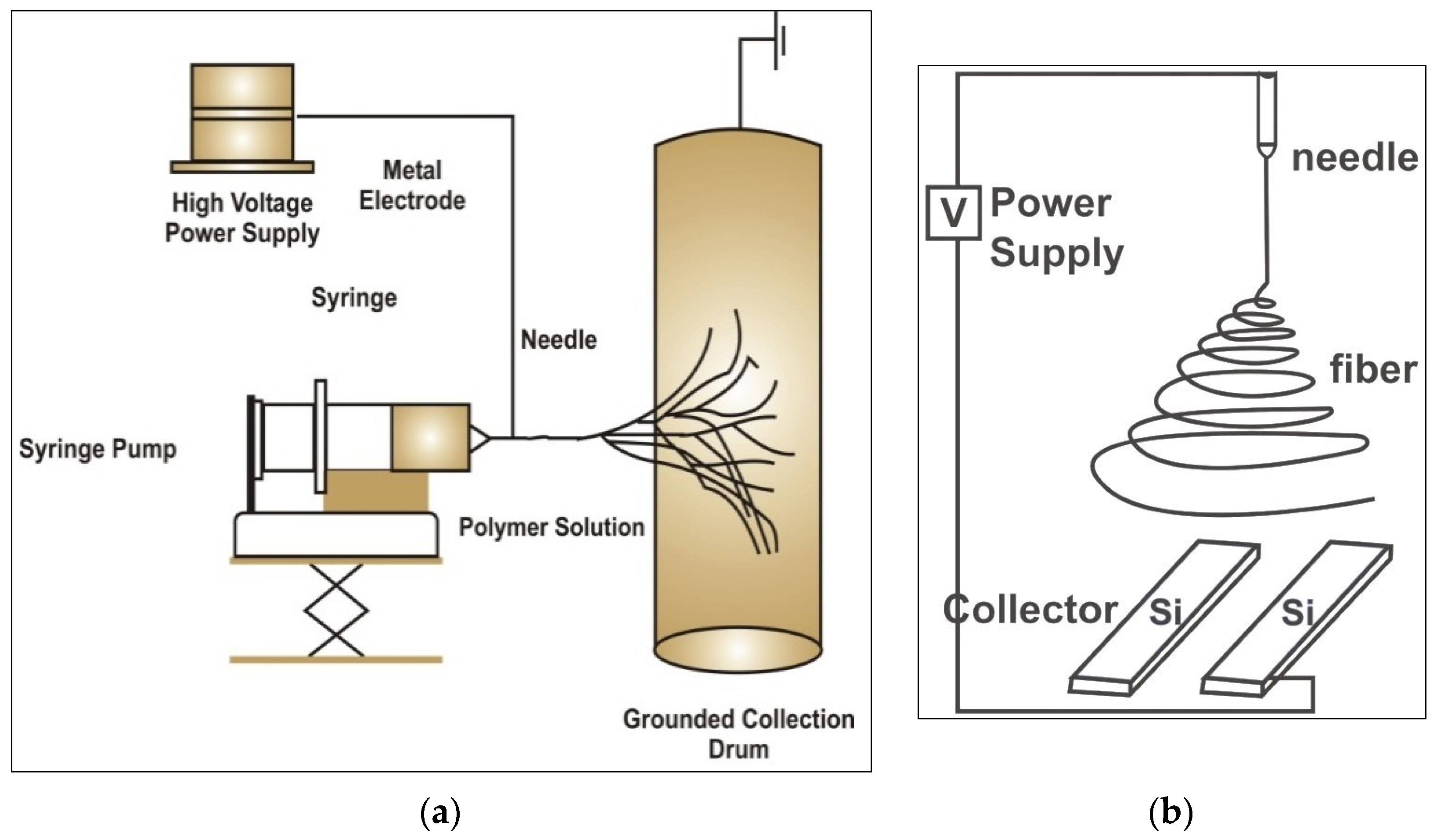
2. Basic Principle and Technique

The electrospinning technique involves the introduction of a strong potential difference between a polymer solution flowing through a capillary tip towords a metallic collector [25]. A typical electrospinning setup only requires a high voltage power supply, a syringe, a flat tip needle and a conducting collector (Figure 1a) [17]. However, the basic equipment can be modified for various applications such as using dual needle syringe (to make blended fibers), or rotating mandrel collectors (to make hollow tube like materials). Conventionally, electrospun materials have unwoven arrangement of nanofibers. Electrospinning with two strips of electrodes (Figure 1b) can be used to collect aligned fibers [26].
Figure 1. Schematic presentation of electrospinning process (a) a typical electrospinning equipment and its components (b) modifications of collector for aligning electrospun nanofibers.
Materials 09 00073 g001
The potential voltage difference between the polymer solution and the collection plate, electrostatic forces overcome the solution surface tension to pull a jet of charged fluid that splits into nanofibers that fall towards the collection plate and solidify [27]. The polymer jet splits into multiple nanofibers that are deposited at the collector. The solvent evaporates as the jet is electrospun and leaving dry nanofibers on the collector [28].

3. Factors Affecting Electrospinning

Electrospinning is able to produce continuous nanofibers from a wide range of materials. However, there are many parameters (processing, physical, systemic and solution) which affect the fiber morphology and properties of electrospun fibers [29]. A list of key factors affecting electro-spun fibers is listed in Table 2 [25].
Table 2. List of variable parameter affecting the characteristics of electro-spun fibers.
Process ParametersSystemic ParametersSolution ParametersPhysical Parameters
VoltagePolymer typeViscosityHumidity
Flow rateMolecular weightConcentrationTemperature
Collection platePolymerConductivityAir velocity
DistanceArchitectureDielectric constant-
AngleSolvent usedSurface tension-
Motion-Charge of jet-

3.1. Solution Related Parameters

The solution properties are important; it should have an optimal low surface tension and high enough charge density and viscosity so that collapse of the jet into droplets can be prevented before the solvent evaporates [30]. Polymer characteristics such as molecular weight, concentration, solution viscosity, surface tension and solution conductivity influence the nanofiber morphology and properties. Molecular weight represents the polymer chain length that in turn influences the entanglements; hence higher molecular weight results in viscous solutions compared to lower molecular weight. These entanglements prevent the jet from premature splitting during the process. Low viscous polymer solution jet breaks up into small droplets or creates beaded fibers [31]. Viscous solutions enhance chain entanglements and results in bead free uniform fibers. If, however, the viscosity is too high, it will be difficult to pump the solution through the capillary and the solution may dry up or drip at the tip [29].
Surface tension decreases the surface area of the solution and forces it to form a spherical droplet. In case of low concentration, high ratio of solvent molecules have greater tendency to assemble and form a spherical shape or bead formation [31]. In order to get bead free uniform fibers, low surface tension solvents should be used. In case of higher conductivity solutions (containing ions), the jet carries heavy amount of electrostatic charge. For example, adding a tiny fraction of salt or polyelectrolyte to electrospinning solution can increase the jet stretching and assists in forming smooth fibers in place of beaded fibers [32].

3.2. Polymer Concentration

The solution concentration below the threshold value will result in droplets formation instead of fibers. High solution concentrations result in viscous solutions and may lead to processing problems. For example, higher viscosity resists jet elongation and thinning and results in larger fiber diameter [33]. A previous study explored the relationship of polyethylene oxide (PEO) solutions viscosity and bead formation. Their results indicated that solution viscosity is linked to the bead size and density. Viscous solution resulted in less spherical and more spindle-like beads followed by nanofiber formation with occasional bead defects [31].

3.3. Processing Conditions

Processing conditions such as voltage, distance of collector, flow rate, needle guage and type of collector may affect the electrospinning process. High voltage induces required charges on the solution to cause the jet to emerge from the needle. Higher voltage accelerates more volume of electrospinning solution with relatively smaller Taylor cone [25]. Amount of solution available between the needle and electrospinning target is determined by the feed rate. The increase in voltage results in more stretching of solution and increased diameter due to the increase in feed rate. Increased feed rate may also cause fusing of fibers due to improper evaporation of solvent before the fiber is collected. The reduction in the distance causes shorter flight time for the jet. So the jet may not have enough time to solidify and result in fusing of fibers. Diameter of the orifice also has an effect. Smaller internal diameter reduces the clogging due to less exposure time of the jet to the environment. Reduction in needle internal diameter increases the surface tension of the solution corresponding to smaller droplet. This causes the acceleration of the jet to decrease. So jet gets more flight time before deposition and has more stretching and elongation; this results in smaller diameter fibers.
The above parameters are the major factors affecting the fiber morphology and web properties in electrospinning. Another factor is the design of the collector. Regular electrospinning yields randomly aligned nanofibers. Control on the geometry of deposition of fiber or getting other desired fiber patterns can be achieved with change in design of collectors. One of these is parallel bars with a gap between the two that leads to aligned nanofibers (Figure 2a). Li et al. [26] used this set-up for producing aligned fiber bundles. The fibers suspended across the nonmetallic part remain charged and align parallel due to repulsion between the electrospun and upcoming nanofibers [26,34].
Figure 2. Fabrication of three dimensional (3D) electrospun scaffolds using regenerated natural Bombyx mori silk; (a) modification of electrospinning collector; (b) electrospun scaffold; (c) 3D natural silk electrospun using oval shape collector.
Materials 09 00073 g002

3.4. Effect of Voltage

Increasing the applied voltage would discharge the polymer jet with stronger repulsion, causing it to undergo higher levels of drawing stress [33]. As a result there is a decrease in fiber diameter and therefore, the fiber diameter distribution would become increasingly higher, making the control of the process more difficult. An optimal voltage is required to initiate the polymer jet from the Taylor cone apex [35]. The applied voltage had a significant effect on droplet shape prior to jet formation. Higher voltage results in an increased flowrate of solution and faster electrospinning [25].

3.5. Volumetric Flow Rate

In order to stabilize the Taylor cone, the flow rate needs to be adjusted in a correct range. Vacuum usually form due to slow flow rate in the needle, causing the Taylor cone to disappear and temporarily stop the electrospinning process. Faster flow may buildup solution at the needle tip. As flow rate increases, the surface charge density decreases therefore the rate of charge withdrawal into the solution is dependent upon the residence time of ions in contact with the needle. The solution flow rate affects various features of nanofibers such as diameter, porosity, and geometry [25]. A constant and stable flow-rate is required to minimize the bead formation in electrospun materials [36]. Slow flow-rate reduced the diameter of electrospun nanofibers [37]. In addition, slow flow rate resulted in less number of beads and smaller diameter compared to faster flow rate [38].

3.6. Distance of Collector

It follows a negative power relationship as increasing the distance allows bending instabilities and whipping action to elongate and decreases the diameter of the polymer jet. There is a negative exponential relationship with surface charge density. Increasing gap distance drops the surface charge density. As the distance between the charged solution and collector increases, the magnitude of the electric field between the two decreases, forming fewer charged ions [39,40]. Another process parameter is the diameter of the needle tip as fiber diameter is reported to increase with a greater needle tip diameter [41,42]. In contrast, lack of correlation between needle diameter and resulting fiber diameter has been reported [43].

3.7. Effect of Conductivity

High conductivity enables polymer solutions to carry greater charge compared to low conductivity. Hence, high conductivity is yields greater tensile forces correspondingly to applied voltage and reduction in nanofiber diameter [44,45,46]. Fong et al. [31] examined the effect of sodium chloride to a polymer for electrospun nanofiber fabrication and reported a higher net charge density of the electrospinning jet. The increased charge density results in the formation of smooth and uniform nanofibers [31]. Zong and coworkers [45] explored the effects of adding salts to poly-DL-lactic acid (PDLLA) solutions and electrospun smooth, bead-free and fine diameter nanofibers. Alternatively, conductivity of polymer solution can be enhanced using surfactants [47,48]. Modifications using surfactants revealed similar results fabricating uniform and smaller diameter nanofibers [49,50].

3.8. Effects of Solvent

Solubility and boiling point of the solvent are important factors for choosing a solvent before electrospinning. Volatile solvents are ideal options due to rapid evaporation and dehydration of the nanofibers [51]. A very low boiling points favors rapid evaporation should be avoided to prevent the obstruction or occlusion of needle orifice prior to electrospinning. In contrast, high boiling points solvents may not dehydrate completely prior to hitting the target resulting in flat ribbon shape fibers instead of round fibers [50,52]. The volatility of the solvent may affect the microscopic features of electrospun fibers including porosity, shape and size hence, particular care must be taken during the evaluation and selection of electrospinning solvents [50].

4. Properties of Electrospun (ES) Materials

The electrospun materials may have unique chemical and physical properties distinguishing them from scaffold prepared using other fabrication techniques. Perhaps one of the most apparent advantages of electrospun scaffolds is ability to mimic extra-cellular matrix (ECM). It has been observed that cells seeded on highly porous electrospun meshes proliferate and differentiate at a higher rate when compared to less porous scaffolds [53]. Furthermore, in vitro studies conducted on nano-porous electrospun scaffolds strongly suggest that cells exhibit higher cellular adhesion with decreasing pore size and a higher pore density [54]. However, some studies suggest that fibers such as poly-L-lactic acid (PLLA), due to their hydrophobic nature, may have a detrimental effect on cellular adhesion. This can be overcome by spraying hydrophilic surfactants on such fibers [55]. Fiber orientation also plays a part in controlling the cellular growth. It has been seen that although osteoblast proliferation is somewhat comparable on aligned and random fibers, a higher calcium production has been detected when the cells are seeded on aligned fibers [56]. The generalized properties of electrospun tissue engineering scaffolds have been discussed here.

4.1. Physical Properties

Electrospun nanofibers can be fabricated in a range of diameters from micro to nanometers based on electrospinning process variables and modifications. The microscopic features of nanofiber are highly dependent on fiber morphology, diameter and surface area [57]. Nanofiber diameter is inversely proportional to surface area. The surface area facilitates cellular attachment and migration. Similarly, electrospun fiber reinforced epoxy composite materials showed improved toughness compared to unmodified resin composites [58]. Fibers also can attach to the surface in different orientation such as random, aligned or many specific shapes and patterns according to the underneath attaching surface. In tissue engineering, various electrospun nanofibers with various architectures and patterns employed for various tissues such as skin, bone and cornea [59,60]. Electrospun nanofibers may have infinite length and a random network of various lines corresponding to the longitudinal axes of fibers [61]. Pore size also plays a crucial role in cell attachment and cell infiltration in tissue engineering applications. Fiber diameter is an excellent indication of degradation in electrospun nanofibers [62].

4.2. Mechanical Properties

Mechanical properties of electrospun nanofibers are important as their applications in products should provide long life durability and structural integrity. Traditional testing methods can be applied for tensile testing of electrospun materials however, modelling and validating their behavior that requires the characterization of single a fiber is challenging. Mechanical properties of electrospun materials play a vital role and required to support cell growth and stability [63,64]. The poor mechanical properties and inability to manipulate certain mechanical properties for specific applications are real challenges for currently available electrospun materials. In order to improve the mechanical and handling properties of electrospun nanofibers, a number of techniques are employed. Cross-linking agents can be used to increase the tensile and flexural strengths of fibers [65]. Furthermore, scaffolds cross-linked with genepin display a better morphology after being immersed in water in addition to enhanced mechanical properties [66]. Conformational changes in polymers may be linked to mechanical properties. For example, β-sheet conformation in natural silk materials has better mechanical properties compared to α-helix conformation [67,68,69]. Further research is required to understand and improve the mechanical properties.

4.3. Biological Properties

Cell attachment to biomaterials especially electrospun nanofibers is crucial for tissue engineering applications. The major advantage of nanofibers is that they favor cell attachment because of higher surface area to absorb proteins and promoting binding sites [70]. Human embryonic stem cells showed promising results for cellular attachment while cultivated on polyurethane electrospun scaffolds [71]. Deshpande et al. [72] showed an excellent epithelial cell attachment to poly(lactic-co-glycolic acid) (PLGA) electrospun microfibers for cornea tissue engineering. Recently, a research group represented high cell attachment to Poly carpolactone (PCL)/Collagen electrospun nanofibers for skin tissue engineering [59]. Physical properties (pore size, volume) significantly affected the cell proliferation [73] and must be controlled during the assessment of biological properties. In addition to materials, cell electrospinning has been suggested as a valuable tool for functionalization of scaffolds for tissue engineering applications [74,75]. Electrospun fibers containing living cells for scaffold applications have been reported [75]. This approach can be used to fabricate a variety of biological (cellular) scaffolds using various cell lines and solvents. The final biological properties (such as cell count, type and medium) can be tailored depending on the type of target tissues and applications.

4.4. Chemical Properties

In the biological environment, degradable electrospun fibers are disintegrated chemically by enzymes such as lysozyme [76]. It is important to consider that there must not be any biocompatibility issues from broken down chemicals. In terms of tissue regeneration applications, the biodegradation rate should be controlled to match with the pace of tissue regeneration. The chemical properties of electrospun fibers is influenced by two main factors: hydrophilicity and chemical composition of the fibers. The electrospun fibers composed of copolymers have a reduced hydrophobicity and degradation compared to homopolymers [77]. Hence, altering the polymer chemistry may be an effective way to control the degradation rate of the fibers. As observed by You et al., the crystallinity of polyglycolide, polylactide, and poly (lactide-co-glycolide) was decreased corresponding to in vitro degradation [78]. This accounts for the progress decrease in their mechanical properties. Indeed, the breakdown products of non-electrospun scaffolds such as poly-L-lactic acid (PLLA) and PLGA also account for the inflammatory response observed clinically [76]. However, there are insufficient randomized clinical trials to prove that similar reactions may be associated with electrospun scaffolds.

5. Electrospun Nanofibers for Dental Applications

The major application of electrospun materials remain for tissue engineering and regeneration of oral and dental tissues. The electrospinning is an excellent technique for fabricating tissue engineering scaffolds [4,17,52,79]. A variety of materials including natural polymers (silk, collagen, chitosan), synthetic polymers (polyvinyl alcohol, polydioxanone) and nanocomposites (hydroxyapatite blends) have been electrospun for tissue engineering of oral and dental tissues (Table 3). In addition, these materials have been used for biomaterials applications such as modifications of implant surfaces, restorative nanocomposites and drug delivery. The typical approach for dental tissue regeneration using electrospun scaffolds is shown schematically in Figure 3. The progress and potentials of electrospun nanomaterials for dental applications has been discussed.
Figure 3. Schematic presentation of using electrospinning scaffolds for tissue engineering of various oral and dental tissues.
Materials 09 00073 g003
Table 3. Potential and progress of electrospun materials for dental applications.
Applications in DentistryMaterial(s) ElectrospunReferences
Tooth regenerationPolyvinyl alcohol (PVA) Polydiaxonone (PDS)[80,81,82]
Guided tissue regenerationCollagen, Poly (lactide-co-glycolide) (PLGA) Poly-L-Lactic Acid (PLLA) Poly carpolactone (PCL) Polyethylene oxide (PEO), Silk[83,84,85,86,87,88]
Caries preventionChitosan[89]
Reinforcement of resin compositesPolyvinyl alcohol (PVA), Polyacrylnitrile, Polystyrene, Nylon[90,91,92,93,94]
Implant modificationPLGA, Collagen[95]
Cartilage regenerationPCL Polyethylene oxide (PEO), Chitosan[96,97,98,99,100]
Drug deliveryPoly(ethylene-co-vinylacetate), Poly(lactic acid) (PLLA), Poly (lactide-co-glycolide) (PLGA)[77,101,102,103,104,105,106]
Wound and mucosal repairPoly-L-(lactic acid), Poly (lactide-co-glycolide) (PLGA), chitin, chitosan, silk fibroin, collagen[107,108,109,110,111]

5.1. Regeneration of Pulp Dentin Complex

Various pathological processes such as dental caries and trauma can result in the loss of dental tissues. Furthermore, various forms of pulp therapies are aimed at regenerating the roots of teeth (apexogenesis) that have undergone trauma. Although various medicaments such as calcium hydroxide, ferric sulphate and mineral trioxide aggregate are used as regenerative materials aimed at regenerating pulpal and radicular dentin, some cases still result in internal resorption of teeth [112,113]. In order to improve and speed up the results of pulp therapy, electrospun scaffolds have been studied that show the potential to induce odontoblast regeneration. Kim et al. produced electrospun scaffolds of polyvinyl alcohol and hydroxyapatite (HA) which could possess dentin regenerative properties [80]. Moreover, electrospun meshes of PCL have strongly shown potential for promoting odontogenic differentiation and growth as suggested by increased turnover of collagen I and other proteins when tested in vitro with human pulpal cells [81].
Endodontic therapy (root canal treatment) also requires delivery of drugs into the root canal and pulp chamber to eradicate the pathological microflora [114]. Bottino et al. produced electrospun scaffolds of polydiaxonone (PDS) in which antibiotics (metronidazole and ciprofloxacin) were incorporated in the solution. It was observed that these scaffolds were able to deliver the antibiotics more effectively and required a lower dose against pathogenic bacteria including Porphyromonas gingivalis and Enterococcus faecalis compared to drugs delivered via pastes [79]. Electrospinning has made it possible to produce bioactive 3D scaffolds made of PDS and halloysite aluminosilicate clay with the potential to regenerate pulp dentin complex by delivering agents such as antimicrobial drugs and growth factors [82]. Perhaps the biggest advantage of electrospinning is its ability to produce complex geometry of fibers to suit the specific application. The ultimate goal of regenerative endodontics is to regenerate the complex dentino-pulpal histology along with restoring the mechanical and physical attributes of the tooth. It is hoped that in the next decade electrospun fibers, along with injectable scaffolds and stem cells allowing optimum regeneration.

5.2. Guided tissue Regeneration for Periodontium

Untreated periodontal disease can lead to periodontal loss and eventual loss of teeth [115]. Regeneration of lost periodontal tissues had always been a challenge for clinicians. However, since advent of the guided tissue regeneration (GTR) membrane, it has been possible to regenerate lost bone by placing a barrier membrane between the gingival epithelium and the underlying periodontal bone [116]. Traditionally, non-resorbable materials such as expanded polytetrafluoriethylene (ePTFE) were used as GTR membranes but they had the disadvantage of requiring a secondary surgical procedure to remove the membrane which often carried a risk of infection and patient discomfort [76,116]. More recently synthetic and natural biodegradable materials such as collagen, Poly-L-Lactic Acid (PLLA) and PCL have been used as GTR membranes which degrade in the periodontal tissues thus avoiding the need of a second procedure to remove them [76,117,118,119]. Lately, electrospinning has been used to produce fibrous and porous electrospun biodegradable scaffolds as GTR membranes.
One of main advantages of electrospinning is its ability to produce fibers of different orientations and size for fibrous scaffolds for tissue regeneration [120,121]. Research indicates that these fibers are effective as tissue regenerative scaffolds because of their ability to mimic the fibrous extra-cellular matrix (ECM) of the human tissues such as bone and cartilage [122]. Indeed, it has been observed that a higher degree of fiber-orientation makes it possible to accelerate proliferation of fibroblasts. This has been attributed to an increased surface area and porosity of electrospun scaffolds [123]. Furthermore, changing the fiber orientation also makes it possible to “control” the direction of cellular proliferation as it has been that cells tend to proliferate in the direction of the fiber orientation [124,125].
The aforementioned properties of fibrous scaffolds can be taken advantage of if they are employed as periodontal GTR scaffolds. Many biodegradable materials have been electrospun and revealed the potential to function as GTR scaffolds [76,116]. Electrospun collagen nanofibers have the potential for GTR scaffolds applications [40]. Additionally, collagen fibers have the potential to allow differentiation of human bone marrow-derived mesenchymal stem cells (MSCs) [125]. However, to date, no studies have attempted to ascertain the mechanical properties of electrospun collagen fibers. Research has also been conducted to produce scaffolds composed of collagen blended with PCL, PEO, PLGA and PLLA [122,126]. One of the major disadvantages of collagen is that, due to its animal origins, there are ethical issues and concerns of cross-infection. Hence, the use of collagen scaffolds could be limited in quite a few demographics.
PCL is another material that has been electrospun to produce bone regenerative scaffolds [127]. It can be blended with collagen or other biodegradable polymers such as gelatin with enhanced tissue regenerative properties [128,129]. Moreover, biomimetic and osseoconductive materials such as nano-sized hydroxyapatite (nano-HA) crystals can be incorporated to PCL-PLA fibers to produce composite scaffolds [130]. Additionally, incorporation of nano-HA crystals not only increases the osteogenic potential of these scaffolds but it has also been suggested that these scaffolds have mechanical properties superior to those made of PCL alone [131]. Another exciting prospect of using electrospun scaffolds is their ability to function as carriers of growth factors and drugs such as bone morphogenic protein-2 (BMP2) and antibiotics which can enhance bone regeneration and prevent periodontal infections [83,84].
Although synthetic degradable polymers have been extensively studied to ascertain their periodontal regenerative properties, their major drawback is the production of acidic breakdown products resulting in inflammation at the site of implantation [76]. Hence, along with collagen, several natural polymers have been probed for GTR applications. Chitosan, a derivative of chitin which is a polymer present in the shells of crustaceans, can be electrospun to produce highly porous and fibrous scaffolds [85,86,87]. In order to improve the spinning ability and handling properties, chitosan was blended with PEO [132]. More recently, drug-incorporated and releasing chitosan-PEO fibrous scaffolds have been produced [29]. Natural silk is another example of degradable materials that has been electrospun for GTR and related applications [133,134]. Electrospun scaffolds of silk fibroin have shown promising results while human periodontal ligament (PDL) are seeded on their surface [15,17,18,135,136].
During the last few years, the idea of functionally graded membrane (FGM) has emerged [116,137]. This principle aims to produce a multilayered guided tissue regenerative membrane in which each layer has a specific function and physical properties, very much akin to the natural human tissues [138]. These layers can contain drugs and various growth factors which be released into the surrounding environment to enhance the regeneration of multiple tissues at the same time [139,140]. It has been speculated that electrospun fibers can form part of these FGMs [137]. Although electrospinning has added exciting new prospects to the field of guided tissue and bone regeneration, much more needs to be explored to validate the use of electrospun scaffolds in the clinical settings. For instance, more research is required to explore the mechanical properties of these scaffolds. More importantly, an adequate number of randomized clinical trials are required to prove their clinical efficacy.

5.3. Caries Prevention

Dental caries (tooth decay) not only lead to loss of tooth tissues but also have systemic ramifications. Conventionally, topical fluoride regimens in the form of mouthwashes, dentifrices [141,142,143,144] or fluoride-containing restorations [145,146] and oral hygiene measures have been used to prevent caries. More recently, electrospun mats having anti-caries properties have been studied. Non-toxic mucoadhesive chitosan fibers containing mangosteen extract have shown antibacterial activity against cariogenic pathogens including Streptococcus mutans and Streptococcus sanguinis [89]. These types of mats could be beneficial for individuals who are unable to self-administer oral hygiene protocols and can be used synergistically with existing methods.

5.4. Modification of Resin Composites

Like majority of polymeric materials, resin composites can be modified by addition of nano-sized fillers or nano-sized fibers [147,148,149]. Nanofibers produced by electrospinning have been incorporated to produce fiber-reinforced composites (FRCs). It has been observed that incorporation of nano-tube reinforced PVA fibers to resin composites can significantly improve the mechanical properties such as elastic modulus of resin composites [90]. However, dispersion of fibers leads to a decreased modulus due to weaker bonding between the fiber and resin phases. Electrospun polyacrylonitrile and polyamide containing nano-diamonds have shown to increase the mechanical properties of polymeric composites when fused with each other [91] that can be used as a means to reinforce dental composites. Similarly, incorporation of electrospun polystyrene fibers to epoxy has also been shown to improve the mechanical properties of the polymers [92]. Electrospun carbon nanotubes and nylon fibers have been successfully used to reinforce resin composites [93]. Production of self-healing nanofiber-reinforced resin composites holds an exciting prospect in increasing the marginal integrity and sealability of resin composites [94]. In addition to improving the mechanical and physical properties of resin composites and dental polymers, incorporation of electrospun fibers could also be used to produce bioactive composites and add anti-cariogenic properties to restorative materials. However, much needs to be learned about the bonding between the fibers and the composites as well as the biological and in vitro implications of these materials.

5.5. Implant Surface Modification

Dental implant is a surgical device that is in direct contact with the bone (i.e., osseointegrated) and holds removable or fixed prosthodontic and orthodontic appliances [150,151]. Several materials (such as titanium and its alloys) have been used for dental implants. Recently, zirconia and reinforced polymers such as polyetheretherketone have also been used as dental implants [148,149,152,153,154,155,156]. To make the implant surface more bioactive and osseoconductive, several surface treatment methods have been employed [157,158]. However, many of these processes alter the mechanical or physical surface properties of the dental implant and ultimately leading to poor success rate [159,160]. To overcome these deleterious effects of surface modifications, several methods have been postulated but most of them are time consuming [158]. Electrospinning is an alternative attractive option that can be used to coat the implant surface using bioactive materials. In addition, electrospun nanofiber coatings have the benefits of greater surface area for the attachment of fibroblasts. Titanium dental implants coated by PLGA/collagen/nano-hydroxyapatite (n-HA) nanofibers significantly enhanced cellular proliferation on the surface and keeping water contact angles as low as 0° in addition to accelerated mineralization [95]. However, more studies are needed to investigate how well an electrospun coating adheres to a dental implant surface in comparison to conventional methods.

5.6. Cartilage Regeneration

Like elsewhere in the body, extensive trauma or pathologies in the head and neck region can result in the loss of not only the bone but also cartilage and ligament. Scaffolds hold potential to be a power adjunct tool to conventional surgical options. Electrospun PCL nanofibers have the potential to accelerate the proliferation of animal and human chondrocytes when tested in vitro [99,100]. PCL can be electrospun with fibrin to produce scaffolds of a combination of fibers having diameters in the range of both, nano- and micrometers to increase cellular infiltration [98]. Similarly, chitosan fibers have also shown potential to function as scaffolds for cartilage regeneration and they also possess mechanical properties superior to those of foams and hydrogels [97]. More recently a scaffold constructed of PCL, chitosan and PEO nanofibers has been used to successfully as a scaffold for chondrogenesis in vitro [96].

5.7. Drug Delivery

Like all other fields of surgery, dentistry requires preoperative and postoperative administration of drugs such as analgesics and antibiotics. As discussed above, electrospun scaffolds can be used as drug delivery devices to minimize the systemic dosage. Drugs such as antimicrobial agents, analgesics and anti-inflammatory regimens have been carried using electrospun scaffolds [77,105,106]. More recent applications of electrospun scaffolds include implantable drugs and growth factor-releasing scaffolds that help repair surgical sites by preventing infection and/or increase the rate of osseointegration [77,101,102,103,104,105,106]. PLLA fibers containing nanodiamonds loaded with growth factors not only possess better mechanical properties than unmodified fibers but can also carry and deliver growth factors and drugs to prevent infection, reduce inflammation and accelerate bone regeneration [161]. Furthermore, because the nanodiamonds can be made fluorescent, such scaffolds can be used to study and monitor guided tissue regeneration at a cellular level by using various imaging techniques [162,163].

5.8. Repair of Wounds and Oral Mucosa

Electrospun fibrous mats have been extensively researched as wound dressings capable of imparting anti-bacterial attributes to the wound as well as regenerative properties [107,108]. Such wound dressings can be used as media to deliver analgesics and antibiotics such topical anesthesia and which can decrease the amount of systemic administration of these drugs needed and hence help in decreasing their many unwanted adverse effects. Similarly, in dentistry, electrospun mats can used to deliver topical anesthesia and antibiotics to surgical or traumatic wounds [107]. In addition to wound repair, electrospun fibers could also be used as dressings for oral mucosal lesions such as ulcers or surgical wounds to relieve patients discomfort [109]. Polymers such as chitin and PLLA have been observed to function as effective scaffolds for proliferation and differentiations for human mucosal cells [109,110]. More recently, electrospun silk fibroin have also shown similar potential to human dermal matrices when tested against rat mucosal cells in vitro [111]. Although more studies are definitely required to ascertain the future of fibrous scaffolds and dressings for oral mucosal abrasions, these materials hold great promise in managing various mucosal ailments.

6. Limitations of Electrospinning

It is evident from the above discussion that electrospinning is a versatile technique that has fabricated unique materials for various biomedical and dental applications. However, there are a few limitations hindering the progress of its applications. Majority of biomedical and dental applications involve tissue engineering or regeneration hence, material’s ability to facilitate cell attachment and infiltration is very important. The randomly unwoven nature of electrospun mats and pore size does not provide ideal structure for cell infiltration [164,165]. Reducing the fiber dimeter increases the surface area however reducing the pore that may affect the cellular infiltration [57]. On the other hand, electrospinning is not a suitable technique for fabricating micron size or larger diameter fibers [1]. The fiber morphology can be altered however not very well controlled and is further complicated by the involvement of multiple electrospinning parameters. It has low fiber production efficiency [166].
Majority of tissue engineering applications required 3D scaffolds [164]. It is challenging to electrospun 3D scaffolds with precise dimensions and morphology [1]. Authors have attempted to electrospun 3D silk scaffolds successfully however, remain unable to control the dimensions and morphology precisely (Figure 2). In addition, there are a few technical challenges, for example, the rate of electrospinning. In order to electrospinning a couple of grams of polymer, it may take several hours. However, recent research suggested that incorporating salt solution (such as NaCl) results in thick meshes of 3D electrospun fibers [167]. Another way of using electrospun fibers and at the same time overcoming their mechanical shortcomings is making them components of multi-layered functionally graded membranes as proposed by Bottino et al. [103]. Additionally, incorporation of nanosized particles such as nanodiamonds has also been effective in increasing the mechanical properties of electrospun scaffolds [168].
Compared to cast membranes, electrospun materials are weaker mechanically. Detaching electrospun mats from the target substrate without damaging is a challenging task. Materials to be electrospun must be dissolved in a solvent of desired properties. The electrospinning solvents may alarm additional issues such as biocompatibility, pungent smell. Polymeric nature of materials restricts this technique for low concentration (~30% or below) solutions [169]. Another concern is the toxicity of the solvents and cross linking agents [170]. Although, relatively safer crosslinking agents such as genepin have been used recently, in vivo biocompatibility of these materials has yet to be evaluated extensively [171]. Hence, it is recommended that the electrospinning process should be carried in well-ventilated fume-cupboards with optimal conditions of humidity and temperature. Authors also recommend storing the polymer solutions in adequate conditions prior to electrospinning. It has been observed by the authors that chitosan-PEO solutions are very difficult to spin after being stored for 72 h. Therefore, the polymer solutions should be spun as soon as possible after being prepared Environmental conditions such as temperature and humidity also affect the electrospinning process [172].
Another important issue is the safety of the technical staff. Special precautions should be taken in order to avoid being electrocuted by the high voltage supply used for electrospinning. Therefore, insulating gloves and apparel in addition to protective masks should be worn. For tissue engineering and biological applications, the sterilization of materials is essentials. Special measures for sterilization must be considered to avoid any damages to the delicate nanofibers. These limitations are critical and may jeopardize the practical applications of electrospun nanomaterials. In order to translate electrospinning products from laboratory to clinical applications, further research is required to understand materials better and address these limitations.

7. Conclusions

There is no doubt that the electrospinning has gained popularity in recent years for bio-dental applications mainly for tissue engineering scaffolds. The progress of oral and dental tissue engineering is promising for the regeneration of oral tissues such as dentin, enamel, pulp, mucosa [173]. Various polymer and composite materials have been electrospun to fabricate scaffolds for tissue regeneration of dental tissues including dentin, periodontium, oral mucosa, bone and cartilage. The materials aspects of electrospun nanofibers such as fabrication, properties and functioning have already been explored in detail and suggested positive outcome for intended biomedical applications. The progress of electrospun materials for various oral applications is promising however there is lack of in vivo and clinical studies. There are a number of challenges (discussed in Section 6) that need to be resolved for further progress. Extensive research involving multiple disciplines (material scientists, chemists, engineers and health care professionals) is needed to translate the basic research to clinical trials and practical applications. It is very much expected that most of the limitations of electrospinning (Section 6) will be addressed in the near future and dragging electrospun materials for practical and clinical applications.

Author Contributions

Muhammad Zafar outlined the review, performed literature search, wrote the manuscript and responsible for the correspondence. Shariq Najeeb performed literature search and helped in compiling the wide information related to topic and data presentation. Zohaib Khurshid and Sana Zohaib wrote a part of the manuscript, developed idea for designing figures. Masoud Vazirzadeh and Bilal Najeeb helped in writing the manuscript and developed the idea of the topic. Farshid Sefat provided guidelines for the preparation of the manuscript, technical editing for all correction and proof reading.

Conflicts of Interest

The authors declare no conflict of interest.

References

  1. Zhang, Y.; Lim, C.T.; Ramakrishna, S.; Huang, Z.M. Recent development of polymer nanofibers for biomedical and biotechnological applications. J. Mater. Sci. Mater. Med. 2005, 16, 933–946. [Google Scholar] [CrossRef] [PubMed]
  2. Bosworth, L.A.; Downes, S. Electrospinning for Tissue Regeneration; Bosworth, L., Downes, S., Eds.; Woodhead Publishing in Materials; Woodhead Publishing: Oxford, UK, 2011; p. 409. [Google Scholar]
  3. Stevens, M.M.; George, J.H. Exploring and Engineering the Cell Surface Interface. Science 2005, 310, 1135–1138. [Google Scholar] [CrossRef] [PubMed]
  4. Liang, D.; Hsiao, B.S.; Chu, B. Functional electrospun nanofibrous scaffolds for biomedical applications. Adv. Drug Deliv. Rev. 2007, 59, 1392–1412. [Google Scholar] [CrossRef] [PubMed]
  5. Smith, L.A.; Ma, P.X. Nano-fibrous scaffolds for tissue engineering. Colloids Surf. B Biointerfaces 2004, 39, 125–131. [Google Scholar] [CrossRef] [PubMed]
  6. Ma, P.X.; Zhang, R. Synthetic nano-scale fibrous extracellular matrix. J. Biomed. Mater. Res. 1999, 46, 60–72. [Google Scholar] [CrossRef]
  7. Yang, F.; Murugan, R.; Ramakrishna, S.; Wang, X.; Ma, Y.X.; Wang, S. Fabrication of nano-structured porous PLLA scaffold intended for nerve tissue engineering. Biomaterials 2004, 25, 1891–1900. [Google Scholar] [CrossRef] [PubMed]
  8. Zhang, X.; Goux, W.J.; Manohar, S.K. Synthesis of polyaniline nanofibers by “nanofiber seeding”. J. Am. Chem. Soc. 2004, 126, 4502–4503. [Google Scholar] [CrossRef] [PubMed]
  9. Martin, C.R. Template synthesis of electronically conductive polymer nanostructures. Acc. Chem. Res. 1995, 28, 61–68. [Google Scholar] [CrossRef]
  10. Lakshmi, B.B.; Patrissi, C.J.; Martin, C.R. Sol-Gel Template Synthesis of Semiconductor Oxide Micro-and Nanostructures. Chem. Mater. 1997, 9, 2544–2550. [Google Scholar] [CrossRef]
  11. Hartgerink, J.D.; Beniash, E.; Stupp, S.I. Self-assembly and mineralization of peptide-amphiphile nanofibers. Science 2001, 294, 1684–1688. [Google Scholar] [CrossRef] [PubMed]
  12. Niece, K.L.; Hartgerink, J.D.; Donners, J.J.J.M.; Stupp, S.I. Self-assembly combining two bioactive peptide-amphiphile molecules into nanofibers by electrostatic attraction. J. Am. Chem. Soc. 2003, 125, 7146–7147. [Google Scholar] [CrossRef] [PubMed]
  13. Amiraliyan, N.; Nouri, M.; Kish, M.H. Electrospinning of silk nanofibers. I. An investigation of nanofiber morphology and process optimization using response surface methodology. Fibers Polym. 2009, 10, 167–176. [Google Scholar] [CrossRef]
  14. Wadbua, P.; Promdonkoy, B.; Maensiri, S.; Siri, S. Different properties of electrospun fibrous scaffolds of separated heavy-chain and light-chain fibroins of Bombyx mori. Int. J. Biol. Macromol. 2010, 46, 493–501. [Google Scholar] [CrossRef] [PubMed]
  15. Cao, H.; Chen, X.; Huang, L.; Shao, Z. Electrospinning of reconstituted silk fiber from aqueous silk fibroin solution. Mater. Sci. Eng. C 2009, 29, 2270–2274. [Google Scholar] [CrossRef]
  16. Kang, M.; Chen, P.; Jin, H. Preparation of multiwalled carbon nanotubes incorporated silk fibroin nanofibers by electrospinning. Curr. Appl. Phys. 2009, 9, S95–S97. [Google Scholar] [CrossRef]
  17. Zhang, X.; Reagan, M.R.; Kaplan, D.L. Electrospun silk biomaterial scaffolds for regenerative medicine. Adv. Drug Deliv. Rev. 2009, 61, 988–1006. [Google Scholar] [CrossRef] [PubMed]
  18. Zhou, J.; Cao, C.; Ma, X. A novel three-dimensional tubular scaffold prepared from silk fibroin by electrospinning. Int. J. Biol. Macromol. 2009, 45, 504–510. [Google Scholar] [CrossRef] [PubMed]
  19. Silva, S.S.; Maniglio, D.; Motta, A.; Mano, J.F.; Reis, R.L.; Migliaresi, C. Genipin-modified silk-fibroin nanometric nets. Macromol. Biosci. 2008, 8, 766–774. [Google Scholar] [CrossRef] [PubMed]
  20. Greiner, A.; Wendorff, J.H. Electrospinning: A fascinating method for the preparation of ultrathin fibres. Angew. Chem. Int. Ed. 2007, 46, 5670–5703. [Google Scholar] [CrossRef] [PubMed]
  21. Jeong, L.; Lee, K.Y.; Park, W.H. Effect of solvent on the characteristics of electrospin regenerated silk fibroin nanofibers. Key Eng. Mater. 2007, 342–343, 813–816. [Google Scholar] [CrossRef]
  22. Bao, W.; Zhang, Y.; Yin, G.; Wu, J. The structure and property of the electrospinning silk fibroin/gelatin blend nanofibers. E Polym. 2008, 8, 1131–1139. [Google Scholar]
  23. Soffer, L.; Wang, X.; Mang, X.; Kluge, J.; Dorfmann, L.; Kaplan, D.L.; Leisk, G. Silk-based electrospun tubular scaffolds for tissue-engineered vascular grafts. J. Biomater. Sci. Polym. Ed. 2008, 19, 653–664. [Google Scholar] [CrossRef] [PubMed]
  24. Meechaisue, C.; Wutticharoenmongkol, P.; Waraput, R.; Huangjing, T.; Ketbumrung, N.; Pavasant, P.; Supaphol, P. Preparation of electrospun silk fibroin fiber mats as bone scaffolds: A preliminary study. Biomed. Mater. 2007, 2, 181–188. [Google Scholar] [CrossRef] [PubMed]
  25. Deitzel, J.M.; Kleinmeyer, J.; Harris, D.; Beck Tan, N.C. The effect of processing variables on the morphology of electrospun nanofibers and textiles. Polymer 2001, 42, 261–272. [Google Scholar] [CrossRef]
  26. Li, D.; Wang, Y.; Xia, Y. Electrospinning of polymeric and ceramic nanofibers as uniaxially aligned arrays. Nano Lett. 2003, 3, 1167–1171. [Google Scholar] [CrossRef]
  27. Subbiah, T.; Bhat, G.; Tock, R.; Parameswaran, S.; Ramkumar, S. Electrospinning of nanofibers. J. Appl. Polym. Sci. 2005, 96, 557–569. [Google Scholar] [CrossRef]
  28. Dersch, R.; Liu, T.; Schaper, A.; Greiner, A.; Wendorff, J. Electrospun nanofibers: Internal structure and intrinsic orientation. J. Polym. Sci. Part A Polym. Chem. 2003, 41, 545–553. [Google Scholar] [CrossRef]
  29. Ramakrishna, S.; Fujihara, K.; Teo, W.; Lim, T.; Ma, Z. Electrospinning process. In An Introduction to Electrospinning and Nanofibers; World Scientific Publishing: Singapore, Singapore, 2005; pp. 135–137. [Google Scholar]
  30. Ioannis, S.C. Novel nanocomposites and nanoceramics based on polymer nanofibers using electrospinning process—A review. J. Mater. Process. Technol. 2005, 167, 283–293. [Google Scholar]
  31. Fong, H.; Chun, I.; Reneker, D.H. Beaded nanofibers formed during electrospinning. Polymer 1999, 40, 4585–4592. [Google Scholar] [CrossRef]
  32. Moghe, A.K. Core-Sheath Differentially Biodegradable Nanofiber Structures for Tissue Engineering. Ph.D. Thesis, North Carolina State University, Raleigh, NC, USA, 2008. [Google Scholar]
  33. James, C.M. Assessment of Electrospinning as an in-House Fabrication Technique for Blood Vessel Mimic Cellular Scaffolding. Master's Thesis, California Polytechnic State University, San Luis Obispo, CA, USA, 2009. [Google Scholar]
  34. Tan, E.; Goh, C.; Sow, C.; Lim, C. Tensile test of a single nanofiber using an atomic force microscope tip. Appl. Phys. Lett. 2005, 86, 073115. [Google Scholar] [CrossRef]
  35. Taylor, G. Electrically Driven Jets. Proc. R. Soc. Lond. A Math. Phys. Sci. 1969, 313, 453–475. [Google Scholar] [CrossRef]
  36. Zeleny, J. The role of surface instability in electrical discharges from drops of alcohol and water in air at atmospheric pressure. J. Frankl. Inst. 1935, 219, 659–675. [Google Scholar] [CrossRef]
  37. Garg, K.; Bowlin, G.L. Electrospinning jets and nanofibrous structures. Biomicrofluidics 2011, 5, 013403. [Google Scholar] [CrossRef] [PubMed]
  38. Megelski, S.; Stephens, J.S.; Chase, D.B.; Rabolt, J.F. Micro-and nanostructured surface morphology on electrospun polymer fibers. Macromolecules 2002, 35, 8456–8466. [Google Scholar] [CrossRef]
  39. Pena, T.R. Preparation and Characterization of Electrospun Poly(D,L-lactide-co-glycolide) Scaffolds for Vascular Tissue Engineering and the Advancement of an In Vitro Blood Vessel Mimic. Master's Thesis, California Polytechnic State University, San Luis Obispo, CA, USA, 2009; p. 152. [Google Scholar]
  40. Matthews, J.A.; Wnek, G.E.; Simpson, D.G.; Bowlin, G.L. Electrospinning of collagen nanofibers. Biomacromolecules 2002, 3, 232–238. [Google Scholar] [CrossRef] [PubMed]
  41. Tong, H.W.; Wang, M. Effects of Processing Parameters on the Morphology and Size of Electrospun PHBV Micro-and Nano-Fibers. Key Eng. Mater. 2007, 334–335, 1233–1236. [Google Scholar] [CrossRef]
  42. Jeun, J.; Kim, Y.; Lim, Y.; Choi, J.; Jung, C.; Kang, P.; Nho, Y. Electrospinning of Poly(L-lactide-co-D,L-lactide). J. Ind. Eng. Chem. 2007, 13, 592–596. [Google Scholar]
  43. Macossay, J.; Marruffo, A.; Rincon, R.; Eubanks, T.; Kuang, A. Effect of needle diameter on nanofiber diameter and thermal properties of electrospun poly(methyl methacrylate). Polym. Adv. Technol. 2007, 18, 180–183. [Google Scholar] [CrossRef]
  44. Baumgarten, P.K. Electrostatic spinning of acrylic microfibers. J. Colloid Interface Sci. 1971, 36, 71–79. [Google Scholar] [CrossRef]
  45. Zong, X.; Kim, K.; Fang, D.; Ran, S.; Hsiao, B.S.; Chu, B. Structure and process relationship of electrospun bioabsorbable nanofiber membranes. Polymer 2002, 43, 4403–4412. [Google Scholar] [CrossRef]
  46. Huang, L.; Nagapudi, K.; Apkarian, R.P.; Chaikof, E.L. Engineered collagen–PEO nanofibers and fabrics. J. Biomater. Sci. Polym. Ed. 2001, 12, 979–993. [Google Scholar] [CrossRef] [PubMed]
  47. Jung, Y.H.; Kim, H.Y.; Lee, D.R.; Park, S.Y.; Khil, M.S. Characterization of PVOH nonwoven mats prepared from surfactant-polymer system via electrospinning. Macromol. Res. 2005, 13, 385–390. [Google Scholar] [CrossRef]
  48. Lin, T.; Wang, H.; Wang, H.; Wang, X. The charge effect of cationic surfactants on the elimination of fibre beads in the electrospinning of polystyrene. Nanotechnology 2004, 15, 1375–1381. [Google Scholar] [CrossRef]
  49. Zeng, J.; Xu, X.; Chen, X.; Liang, Q.; Bian, X.; Yang, L.; Jing, X. Biodegradable electrospun fibers for drug delivery. J. Controlled Release 2003, 92, 227–231. [Google Scholar] [CrossRef]
  50. Sill, T.J.; von Recum, H.A. Electrospinning: Applications in drug delivery and tissue engineering. Biomaterials 2008, 29, 1989–2006. [Google Scholar] [CrossRef] [PubMed]
  51. Pillay, V.; Dott, C.; Choonara, Y.E.; Tyagi, C.; Tomar, L.; Kumar, P.; du Toit, L.C.; Ndesendo, V.M. A review of the effect of processing variables on the fabrication of electrospun nanofibers for drug delivery applications. J. Nanomater. 2013, 2013, 1–22. [Google Scholar] [CrossRef]
  52. Lannutti, J.; Reneker, D.; Ma, T.; Tomasko, D.; Farson, D. Electrospinning for tissue engineering scaffolds. Mater. Sci. Eng. C 2007, 27, 504–509. [Google Scholar] [CrossRef]
  53. McCann, J.T.; Li, D.; Xia, Y. Electrospinning of nanofibers with core-sheath, hollow, or porous structures. J. Mater. Chem. 2005, 15, 735–738. [Google Scholar] [CrossRef]
  54. Kwon, I.K.; Kidoaki, S.; Matsuda, T. Electrospun nano-to microfiber fabrics made of biodegradable copolyesters: Structural characteristics, mechanical properties and cell adhesion potential. Biomaterials 2005, 26, 3929–3939. [Google Scholar] [CrossRef] [PubMed]
  55. Cheng, Q.; Lee, B.L.; Komvopoulos, K.; Yan, Z.; Li, S. Plasma surface chemical treatment of electrospun poly(L-lactide) microfibrous scaffolds for enhanced cell adhesion, growth, and infiltration. Tissue Eng. Part A 2013, 19, 1188–1198. [Google Scholar] [CrossRef] [PubMed]
  56. Ma, J.; He, X.; Jabbari, E. Osteogenic differentiation of marrow stromal cells on random and aligned electrospun poly(L-lactide) nanofibers. Ann. Biomed. Eng. 2011, 39, 14–25. [Google Scholar] [CrossRef] [PubMed]
  57. Eichhorn, S.J.; Sampson, W.W. Statistical geometry of pores and statistics of porous nanofibrous assemblies. J. R. Soc. Interface 2005, 2, 309–318. [Google Scholar] [CrossRef] [PubMed]
  58. Jong-Sang, K.; Reneker, D.H. Mechanical properties of composites using ultrafine electrospun fibers. Polym. Compos. 1999, 20, 124–131. [Google Scholar]
  59. Mahjour, S.B.; Fu, X.; Yang, X.; Fong, J.; Sefat, F.; Wang, H. Rapid creation of skin substitutes from human skin cells and biomimetic nanofibers for acute full-thickness wound repair. Burns 2015, 41, 1764–1774. [Google Scholar] [CrossRef] [PubMed]
  60. Sefat, F.; Denyer, M.; Youseffi, M. Imaging via widefield surface plasmon resonance microscope for studying bone cell interactions with micropatterned ECM proteins. J. Microsc. 2011, 241, 282–290. [Google Scholar] [CrossRef] [PubMed]
  61. Miles, R.E. Random Polygons Determined by Random Lines in a Plane, II. Proc. Natl. Acad. Sci. USA 1964, 52, 1157–1160. [Google Scholar] [CrossRef] [PubMed]
  62. Sefat, F.; McKean, R.; Deshpande, P.; Ramachandran, C.; Hill, C.J.; Sangwan, V.S.; Ryan, A.J.; MacNeil, S. Production, sterilisation and storage of biodegradable electrospun PLGA membranes for delivery of limbal stem cells to the cornea. Procedia Eng. 2013, 59, 101–116. [Google Scholar] [CrossRef]
  63. Mauck, R.L.; Baker, B.M.; Nerurkar, N.L.; Burdick, J.A.; Li, W.; Tuan, R.S.; Elliott, D.M. Engineering on the straight and narrow: The mechanics of nanofibrous assemblies for fiber-reinforced tissue regeneration. Tissue Eng. Part B Rev. 2009, 15, 171–193. [Google Scholar] [CrossRef] [PubMed]
  64. McManus, M.C.; Boland, E.D.; Koo, H.P.; Barnes, C.P.; Pawlowski, K.J.; Wnek, G.E.; Simpson, D.G.; Bowlin, G.L. Mechanical properties of electrospun fibrinogen structures. Acta Biomater. 2006, 2, 19–28. [Google Scholar] [CrossRef] [PubMed]
  65. Barnes, C.P.; Pemble, C.W.; Brand, D.D.; Simpson, D.G.; Bowlin, G.L. Cross-linking electrospun type II collagen tissue engineering scaffolds with carbodiimide in ethanol. Tissue Eng. 2007, 13, 1593–1605. [Google Scholar] [CrossRef] [PubMed]
  66. Panzavolta, S.; Gioffrè, M.; Focarete, M.L.; Gualandi, C.; Foroni, L.; Bigi, A. Electrospun gelatin nanofibers: Optimization of genipin cross-linking to preserve fiber morphology after exposure to water. Acta Biomater. 2011, 7, 1702–1709. [Google Scholar] [CrossRef] [PubMed]
  67. Wang, M.; Jin, H.J.; Kaplan, D.L.; Rutledge, G.C. Mechanical properties of electrospun silk fibers. Macromolecules 2004, 37, 6856–6864. [Google Scholar] [CrossRef]
  68. Xue, Y.F.; He, C.L.; Mo, X.M. Electrospun spider nanofibers: The influence of ethanol on the structure and properties. In Proceedings of the 2007 International Forum on Biomedical Textile Materials, Shanghai, China, 30 May–2 June 2007; pp. 178–181.
  69. Min, B.M.; Jeong, L.; Lee, K.Y.; Park, W.H. Regenerated silk fibroin nanofibers: Water vapor-induced structural changes and their effects on the behavior of normal human cells. Macromol. Biosci. 2006, 6, 285–292. [Google Scholar] [CrossRef] [PubMed]
  70. Mijovic, B.; Trcin, M.; Agic, A.; Zdraveva, E.; Bujic, M.; Spoljaric, I.; Kosec, V. Study on cell adhesion detection onto biodegradable electrospun PCL scaffolds. JFBI 2012, 5, 33–40. [Google Scholar] [CrossRef]
  71. Carlberg, B.; Axell, M.Z.; Nannmark, U.; Liu, J.; Kuhn, H.G. Electrospun polyurethane scaffolds for proliferation and neuronal differentiation of human embryonic stem cells. Biomed. Mater. 2009, 4, 045004. [Google Scholar] [CrossRef] [PubMed]
  72. Deshpande, P.; Ramachandran, C.; Sefat, F.; Mariappan, I.; Johnson, C.; McKean, R.; Hannah, M.; Sangwan, V.S.; Claeyssens, F.; Ryan, A.J. Simplifying corneal surface regeneration using a biodegradable synthetic membrane and limbal tissue explants. Biomaterials 2013, 34, 5088–5106. [Google Scholar] [CrossRef] [PubMed]
  73. Lowery, J.L.; Datta, N.; Rutledge, G.C. Effect of fiber diameter, pore size and seeding method on growth of human dermal fibroblasts in electrospun poly (ɛ-caprolactone) fibrous mats. Biomaterials 2010, 31, 491–504. [Google Scholar] [CrossRef] [PubMed]
  74. Jayasinghe, S.N. Cell electrospinning: A novel tool for functionalising fibres, scaffolds and membranes with living cells and other advanced materials for regenerative biology and medicine. Analyst 2013, 138, 2215–2223. [Google Scholar] [CrossRef] [PubMed]
  75. Townsend-Nicholson, A.; Jayasinghe, S.N. Cell electrospinning: A unique biotechnique for encapsulating living organisms for generating active biological microthreads/scaffolds. Biomacromolecules 2006, 7, 3364–3369. [Google Scholar] [CrossRef] [PubMed]
  76. Sheikh, Z.; Najeeb, S.; Khurshid, Z.; Verma, V.; Rashid, H.; Glogauer, M. Biodegradable Materials for Bone Repair and Tissue Engineering Applications. Materials 2015, 8, 5744–5794. [Google Scholar] [CrossRef]
  77. Kim, K.; Luu, Y.K.; Chang, C.; Fang, D.; Hsiao, B.S.; Chu, B.; Hadjiargyrou, M. Incorporation and controlled release of a hydrophilic antibiotic using poly(lactide-co-glycolide)-based electrospun nanofibrous scaffolds. J. Controlled Release 2004, 98, 47–56. [Google Scholar] [CrossRef] [PubMed]
  78. You, Y.; Min, B.; Lee, S.J.; Lee, T.S.; Park, W.H. In vitro degradation behavior of electrospun polyglycolide, polylactide, and poly(lactide-co-glycolide). J. Appl. Polym. Sci. 2005, 95, 193–200. [Google Scholar] [CrossRef]
  79. Bottino, M.C.; Kamocki, K.; Yassen, G.H.; Platt, J.A.; Vail, M.M.; Ehrlich, Y.; Spolnik, K.J.; Gregory, R.L. Bioactive nanofibrous scaffolds for regenerative endodontics. J. Dent. Res. 2013, 92, 963–969. [Google Scholar] [CrossRef] [PubMed]
  80. Kim, G.; Asran, A.S.; Michler, G.H.; Simon, P.; Kim, J. Electrospun PVA/HAp nanocomposite nanofibers: Biomimetics of mineralized hard tissues at a lower level of complexity. Bioinspiration Biomim. 2008, 3, 046003. [Google Scholar] [CrossRef] [PubMed]
  81. Kim, J.J.; Bae, W.J.; Kim, J.M.; Kim, J.J.; Lee, E.J.; Kim, H.W.; Kim, E.C. Mineralized polycaprolactone nanofibrous matrix for odontogenesis of human dental pulp cells. J. Biomater. Appl. 2014, 28, 1069–1078. [Google Scholar] [CrossRef] [PubMed]
  82. Bottino, M.C.; Yassen, G.H.; Platt, J.A.; Labban, N.; Windsor, L.J.; Spolnik, K.J.; Bressiani, A.H. A novel three-dimensional scaffold for regenerative endodontics: Materials and biological characterizations. J. Tissue Eng. Regen. Med. 2015, 9, E116–E123. [Google Scholar] [CrossRef] [PubMed]
  83. Kim, B.; Nguyen, T.B.L.; Min, Y.; Lee, B. In vitro and in vivo studies of BMP-2-loaded PCL–gelatin–BCP electrospun scaffolds. Tissue Eng. Part A 2014, 20, 3279–3289. [Google Scholar] [CrossRef] [PubMed]
  84. Ranjbar-Mohammadi, M.; Zamani, M.; Prabhakaran, M.; Bahrami, S.H.; Ramakrishna, S. Electrospinning of PLGA/gum tragacanth nanofibers containing tetracycline hydrochloride for periodontal regeneration. Mater. Sci. Eng. C 2016, 58, 521–531. [Google Scholar] [CrossRef] [PubMed]
  85. Zhang, Y.; Ni, M.; Zhang, M.; Ratner, B. Calcium phosphate-chitosan composite scaffolds for bone tissue engineering. Tissue Eng. 2003, 9, 337–345. [Google Scholar] [CrossRef] [PubMed]
  86. Kim, I.; Seo, S.; Moon, H.; Yoo, M.; Park, I.; Kim, B.; Cho, C. Chitosan and its derivatives for tissue engineering applications. Biotechnol. Adv. 2008, 26, 1–21. [Google Scholar] [CrossRef] [PubMed]
  87. Zhang, Y.; Su, B.; Ramakrishna, S.; Lim, C. Chitosan nanofibers from an easily electrospinnable UHMWPEO-doped chitosan solution system. Biomacromolecules 2007, 9, 136–141. [Google Scholar] [CrossRef] [PubMed]
  88. Jin, H.J.; Fridrikh, S.; Rutledge, G.C.; Kaplan, D. Electrospinning bombyx mori silk with poly(ethylene oxide). Abstr. Pap. Am. Chem. Soc. 2002, 3, 1233–1239. [Google Scholar]
  89. Samprasit, W.; Kaomongkolgit, R.; Sukma, M.; Rojanarata, T.; Ngawhirunpat, T.; Opanasopit, P. Mucoadhesive electrospun chitosan-based nanofibre mats for dental caries prevention. Carbohydr. Polym. 2015, 117, 933–940. [Google Scholar] [CrossRef] [PubMed]
  90. Wang, W.; Ciselli, P.; Kuznetsov, E.; Peijs, T.; Barber, A.H. Effective reinforcement in carbon nanotube-polymer composites. Philos. Trans. A. Math. Phys. Eng. Sci. 2008, 366, 1613–1626. [Google Scholar] [CrossRef] [PubMed]
  91. Behler, K.D.; Stravato, A.; Mochalin, V.; Korneva, G.; Yushin, G.; Gogotsi, Y. Nanodiamond-polymer composite fibers and coatings. ACS Nano 2009, 3, 363–369. [Google Scholar] [CrossRef] [PubMed]
  92. Demir, M.M.; Horzum, N.; Tasdemirci, A.; Turan, K.; Güden, M. Mechanical Interlocking between Porous Electrospun Polystyrene Fibers and an Epoxy Matrix. ACS Appl. Mater. Interfaces 2014, 6, 21901–21905. [Google Scholar] [CrossRef] [PubMed]
  93. Borges, A.L.; Münchow, E.A.; de Oliveira, S.A.C.; Yoshida, T.; Vallittu, P.K.; Bottino, M.C. Effect of random/aligned nylon-6/MWCNT fibers on dental resin composite reinforcement. J. Mech. Behav. Biomed. Mater. 2015, 48, 134–144. [Google Scholar] [CrossRef] [PubMed]
  94. Lee, M.W.; An, S.; Jo, H.S.; Yoon, S.S.; Yarin, A.L. Self-healing Nanofiber-Reinforced Polymer Composites: Tensile Testing and Recovery of Mechanical Properties. ACS Appl. Mater. Interfaces 2015, 7, 19546–19554. [Google Scholar] [CrossRef] [PubMed]
  95. Ravichandran, R.; Ng, C.C.; Liao, S.; Pliszka, D.; Raghunath, M.; Ramakrishna, S.; Chan, C.K. Biomimetic surface modification of titanium surfaces for early cell capture by advanced electrospinning. Biomed. Mater. 2012, 7, 015001. [Google Scholar] [CrossRef] [PubMed]
  96. Ainola, M.; Tomaszewski, W.; Ostrowska, B.; Wesolowska, E.; Wagner, H.D.; Swieszkowski, W.; Sillat, T.; Peltola, E.; Konttinen, Y.T. A bioactive hybrid three-dimensional tissue-engineering construct for cartilage repair. J. Biomater. Appl. 2016, 30, 873–885. [Google Scholar] [CrossRef] [PubMed]
  97. Subramanian, A.; Lin, H.Y.; Vu, D.; Larsen, G. Synthesis and evaluation of scaffolds prepared from chitosan fibers for potential use in cartilage tissue engineering. Biomed. Sci. Instrum. 2004, 40, 117–122. [Google Scholar] [PubMed]
  98. Levorson, E.J.; Sreerekha, P.R.; Chennazhi, K.P.; Kasper, F.K.; Nair, S.V.; Mikos, A.G. Fabrication and characterization of multiscale electrospun scaffolds for cartilage regeneration. Biomed. Mater. 2013, 8, 014103. [Google Scholar] [CrossRef] [PubMed]
  99. Thorvaldsson, A.; Stenhamre, H.; Gatenholm, P.; Walkenström, P. Electrospinning of highly porous scaffolds for cartilage regeneration. Biomacromolecules 2008, 9, 1044–1049. [Google Scholar] [CrossRef] [PubMed]
  100. Li, W.; Danielson, K.G.; Alexander, P.G.; Tuan, R.S. Biological response of chondrocytes cultured in three-dimensional nanofibrous poly (ϵ-caprolactone) scaffolds. J. Biomed. Mater. Res. Part A 2003, 67, 1105–1114. [Google Scholar] [CrossRef] [PubMed]
  101. Vacanti, N.M.; Cheng, H.; Hill, P.S.; Guerreiro, J.D.; Dang, T.T.; Ma, M.; Watson, S.; Hwang, N.S.; Langer, R.; Anderson, D.G. Localized delivery of dexamethasone from electrospun fibers reduces the foreign body response. Biomacromolecules 2012, 13, 3031–3038. [Google Scholar] [CrossRef] [PubMed]
  102. Song, W.; Yu, X.; Markel, D.C.; Shi, T.; Ren, W. Coaxial PCL/PVA electrospun nanofibers: Osseointegration enhancer and controlled drug release device. Biofabrication 2013, 5, 035006. [Google Scholar] [CrossRef] [PubMed]
  103. Zeng, H.; Li, F.; Wei, H.; Shi, J.F.; Rao, G.Z.; Li, A.; Gou, J.Z. Preliminary study of the dual release baicalin and rhBMP-2 system to improve periodontal tissue regeneration in minipigs. Shanghai Kou Qiang Yi Xue 2013, 22, 126–131. (In Chinese) [Google Scholar] [PubMed]
  104. Xue, J.; He, M.; Niu, Y.; Liu, H.; Crawford, A.; Coates, P.; Chen, D.; Shi, R.; Zhang, L. Preparation and in vivo efficient anti-infection property of GTR/GBR implant made by metronidazole loaded electrospun polycaprolactone nanofiber membrane. Int. J. Pharm. 2014, 475, 566–577. [Google Scholar] [CrossRef] [PubMed]
  105. Verreck, G.; Chun, I.; Peeters, J.; Rosenblatt, J.; Brewster, M.E. Preparation and characterization of nanofibers containing amorphous drug dispersions generated by electrostatic spinning. Pharm. Res. 2003, 20, 810–817. [Google Scholar] [CrossRef] [PubMed]
  106. Kenawy, E.; Bowlin, G.L.; Mansfield, K.; Layman, J.; Simpson, D.G.; Sanders, E.H.; Wnek, G.E. Release of tetracycline hydrochloride from electrospun poly(ethylene-co-vinylacetate), poly(lactic acid), and a blend. J. Controlled Release 2002, 81, 57–64. [Google Scholar] [CrossRef]
  107. Thakur, R.; Florek, C.; Kohn, J.; Michniak, B. Electrospun nanofibrous polymeric scaffold with targeted drug release profiles for potential application as wound dressing. Int. J. Pharm. 2008, 364, 87–93. [Google Scholar] [CrossRef] [PubMed]
  108. He, T.; Wang, J.; Huang, P.; Zeng, B.; Li, H.; Cao, Q.; Zhang, S.; Luo, Z.; Deng, D.Y.; Zhang, H. Electrospinning polyvinylidene fluoride fibrous membranes containing anti-bacterial drugs used as wound dressing. Colloids Surf. B Biointerfaces 2015, 130, 278–286. [Google Scholar] [CrossRef] [PubMed]
  109. Noh, H.K.; Lee, S.W.; Kim, J.; Oh, J.; Kim, K.; Chung, C.; Choi, S.; Park, W.H.; Min, B. Electrospinning of chitin nanofibers: Degradation behavior and cellular response to normal human keratinocytes and fibroblasts. Biomaterials 2006, 27, 3934–3944. [Google Scholar] [CrossRef] [PubMed]
  110. Blackwood, K.A.; McKean, R.; Canton, I.; Freeman, C.O.; Franklin, K.L.; Cole, D.; Brook, I.; Farthing, P.; Rimmer, S.; Haycock, J.W. Development of biodegradable electrospun scaffolds for dermal replacement. Biomaterials 2008, 29, 3091–3104. [Google Scholar] [CrossRef] [PubMed]
  111. Tang, J.; Han, Y.; Zhang, F.; Ge, Z.; Liu, X.; Lu, Q. Buccal mucosa repair with electrospun silk fibroin matrix in a rat model. Int. J. Artif. Organs 2015, 38, 105–112. [Google Scholar] [CrossRef] [PubMed]
  112. Damle, S.; Bhattal, H.; Loomba, A. Apexification of anterior teeth: A comparative evaluation of mineral trioxide aggregate and calcium hydroxide paste. J. Clin. Pediatr. Dent. 2012, 36, 263–268. [Google Scholar] [CrossRef] [PubMed]
  113. Odabas, M.E.; Alacam, A.; Sillelioglu, H.; Deveci, C. Clinical and radiographic success rates of mineral trioxide aggregate and ferric sulphate pulpotomies performed by dental students. Eur. J. Paediatr. Dent. 2012, 13, 118–122. [Google Scholar] [PubMed]
  114. Rizvi, A.; Zafar, M.S.; Farid, W.M.; Gazal, G. Assessment of Antimicrobial Efficacy of MTAD, Sodium Hypochlorite, EDTA and Chlorhexidine for Endodontic Applications: An In vitro Study. Middle East J. Sci. Res. 2014, 21, 353–357. [Google Scholar]
  115. Pihlstrom, B.L.; Michalowicz, B.S.; Johnson, N.W. Periodontal diseases. Lancet 2005, 366, 1809–1820. [Google Scholar] [CrossRef]
  116. Bottino, M.C.; Thomas, V.; Schmidt, G.; Vohra, Y.K.; Chu, T.G.; Kowolik, M.J.; Janowski, G.M. Recent advances in the development of GTR/GBR membranes for periodontal regeneration—A materials perspective. Dent. Mater. 2012, 28, 703–721. [Google Scholar] [CrossRef] [PubMed]
  117. Gottlow, J.; Laurell, L.; Lundgren, D.; Mathisen, T.; Nyman, S.; Rylander, H.; Bogentoft, C. Periodontal tissue response to a new bioresorbable guided tissue regeneration device: A longitudinal study in monkeys. Int. J. Periodontics Restor. Dent. 1994, 14, 436–449. [Google Scholar]
  118. Al-Arrayed, F.; Adam, S.; Moran, J.; Dowell, P. Clinical trial of cross-linked human type I collagen as a barrier material in surgical periodontal treatment. J. Clin. Periodontol. 1995, 22, 371–379. [Google Scholar] [CrossRef] [PubMed]
  119. Dowell, P.; Al-Arrayed, F.; Adam, S.; Moran, J. A comparative clinical study: The use of human type I collagen with and without the addition of metronidazole in the GTR method of treatment of periodontal disease. J. Clin. Periodontol. 1995, 22, 543–549. [Google Scholar] [CrossRef] [PubMed]
  120. Srouji, S.; Kizhner, T.; Suss-Tobi, E.; Livne, E.; Zussman, E. 3-D Nanofibrous electrospun multilayered construct is an alternative ECM mimicking scaffold. J. Mater. Sci. Mater. Med. 2008, 19, 1249–1255. [Google Scholar] [CrossRef] [PubMed]
  121. Han, D.; Gouma, P. Electrospun bioscaffolds that mimic the topology of extracellular matrix. Nanomed. Nanotechnol. Biol. Med. 2006, 2, 37–41. [Google Scholar] [CrossRef] [PubMed]
  122. Prabhakaran, M.P.; Venugopal, J.; Ramakrishna, S. Electrospun nanostructured scaffolds for bone tissue engineering. Acta Biomater. 2009, 5, 2884–2893. [Google Scholar] [CrossRef] [PubMed]
  123. Bashur, C.A.; Dahlgren, L.A.; Goldstein, A.S. Effect of fiber diameter and orientation on fibroblast morphology and proliferation on electrospun poly(D,L-lactic-co-glycolic acid) meshes. Biomaterials 2006, 27, 5681–5688. [Google Scholar] [CrossRef] [PubMed]
  124. Li, W.; Laurencin, C.T.; Caterson, E.J.; Tuan, R.S.; Ko, F.K. Electrospun nanofibrous structure: A novel scaffold for tissue engineering. J. Biomed. Mater. Res. 2002, 60, 613–621. [Google Scholar] [CrossRef] [PubMed]
  125. Shih, Y.V.; Chen, C.; Tsai, S.; Wang, Y.J.; Lee, O.K. Growth of mesenchymal stem cells on electrospun type I collagen nanofibers. Stem Cells 2006, 24, 2391–2397. [Google Scholar] [CrossRef] [PubMed]
  126. Ekaputra, A.K.; Zhou, Y.; Cool, S.M.; Hutmacher, D.W. Composite electrospun scaffolds for engineering tubular bone grafts. Tissue Eng. Part A 2009, 15, 3779–3788. [Google Scholar] [CrossRef] [PubMed]
  127. Yoshimoto, H.; Shin, Y.; Terai, H.; Vacanti, J. A biodegradable nanofiber scaffold by electrospinning and its potential for bone tissue engineering. Biomaterials 2003, 24, 2077–2082. [Google Scholar] [CrossRef]
  128. Ji, W.; Yang, F.; Ma, J.; Bouma, M.J.; Boerman, O.C.; Chen, Z.; van den Beucken, J.J.; Jansen, J.A. Incorporation of stromal cell-derived factor-1α in PCL/gelatin electrospun membranes for guided bone regeneration. Biomaterials 2013, 34, 735–745. [Google Scholar] [CrossRef] [PubMed]
  129. Zhang, Y.; Ouyang, H.; Lim, C.T.; Ramakrishna, S.; Huang, Z. Electrospinning of gelatin fibers and gelatin/PCL composite fibrous scaffolds. J. Biomed. Mater. Res. Part B Appl. Biomater. 2005, 72, 156–165. [Google Scholar] [CrossRef] [PubMed]
  130. Fang, R.; Zhang, E.; Xu, L.; Wei, S. Electrospun PCL/PLA/HA based nanofibers as scaffold for osteoblast-like cells. J. Nanosci. Nanotechnol. 2010, 10, 7747–7751. [Google Scholar] [CrossRef] [PubMed]
  131. Tyagi, P.; Catledge, S.A.; Stanishevsky, A.; Thomas, V.; Vohra, Y. Nanomechanical properties of electrospun composite scaffolds based on polycaprolactone and hydroxyapatite. J. Nanosci. Nanotechnol. 2009, 9, 4839–4845. [Google Scholar] [CrossRef] [PubMed]
  132. Jayakumar, R.; Prabaharan, M.; Nair, S.; Tamura, H. Novel chitin and chitosan nanofibers in biomedical applications. Biotechnol. Adv. 2010, 28, 142–150. [Google Scholar] [CrossRef] [PubMed]
  133. Zafar, M.S.; Belton, D.J.; Hanby, B.; Kaplan, D.L.; Perry, C.C. Functional Material Features of Bombyx Mori Silk Light vs. Heavy Chain Proteins. Biomacromolecules 2015, 16, 606–614. [Google Scholar] [CrossRef] [PubMed]
  134. Zafar, M.S.; Al-Samadani, K.H. Potential use of natural silk for bio-dental applications. J. Taibah Univ. Med. Sci. 2014, 9, 171–177. [Google Scholar] [CrossRef]
  135. Marelli, B.; Alessandrino, A.; Fare, S.; Freddi, G.; Mantovani, D.; Tanzi, M.C. Compliant electrospun silk fibroin tubes for small vessel bypass grafting. Acta Biomater. 2010, 6, 4019–4026. [Google Scholar] [CrossRef] [PubMed]
  136. Ki, C.S.; Kim, J.W.; Hyun, J.H.; Lee, K.H.; Hattori, M.; Rah, D.K.; Park, Y.H. Electrospun three-dimensional silk fibroin nanofibrous scaffold. J. Appl. Polym. Sci. 2007, 106, 3922–3928. [Google Scholar] [CrossRef]
  137. Qasim, S.B.; Delaine-Smith, R.M.; Fey, T.; Rawlinson, A.; Rehman, I.U. Freeze gelated porous membranes for periodontal tissue regeneration. Acta Biomater. 2015, 23, 317–328. [Google Scholar] [CrossRef] [PubMed]
  138. Bottino, M.C.; Thomas, V.; Janowski, G.M. A novel spatially designed and functionally graded electrospun membrane for periodontal regeneration. Acta Biomater. 2011, 7, 216–224. [Google Scholar] [CrossRef] [PubMed]
  139. Bottino, M.C.; Arthur, R.A.; Waeiss, R.A.; Kamocki, K.; Gregson, K.S.; Gregory, R.L. Biodegradable nanofibrous drug delivery systems: Effects of metronidazole and ciprofloxacin on periodontopathogens and commensal oral bacteria. Clin. Oral Investig. 2014, 18, 2151–2158. [Google Scholar] [CrossRef] [PubMed] [Green Version]
  140. Sundaram, M.N.; Sowmya, S.; Deepthi, S.; Bumgardener, J.D.; Jayakumar, R. Bilayered construct for simultaneous regeneration of alveolar bone and periodontal ligament. J. Biomed. Mater. Res. Part B Appl. Biomater. 2015, 7. [Google Scholar] [CrossRef] [PubMed]
  141. Ullah, R.; Zafar, M.S. Oral and dental delivery of fluoride: A review. Fluoride 2015, 48, 195–204. [Google Scholar]
  142. Zafar, M.S.; Ahmed, N. Therapeutic roles of fluoride released from restorative dental materials. Fluoride 2015, 48, 184–194. [Google Scholar]
  143. Davies, G.M.; Bridgman, C.; Hough, D.; Davies, R. The application of fluoride varnish in the prevention and control of dental caries. Dent. Update 2009, 36, 410–412. [Google Scholar] [PubMed]
  144. Ismail, A.I.; Hasson, H. Fluoride supplements, dental caries and fluorosis: A systematic review. J. Am. Dent. Assoc. 2008, 139, 1457–1468. [Google Scholar] [CrossRef] [PubMed]
  145. Zafar, M.S. Effects of Surface Pre-Reacted Glass Particles on Fluoride Release of Dental Restorative Materials. World Appl. Sci. J. 2013, 28, 457–462. [Google Scholar]
  146. Dionysopoulos, D. The effect of fluoride-releasing restorative materials on inhibition of secondary caries formation. Fluoride 2014, 47, 258–265. [Google Scholar]
  147. Khurshid, Z.; Zafar, M.; Qasim, S.; Shahab, S.; Naseem, M.; AbuReqaiba, A. Advances in Nanotechnology for Restorative Dentistry. Materials 2015, 8, 717–731. [Google Scholar] [CrossRef]
  148. Najeeb, S.; Khurshid, Z.; Matinlinna, J.P.; Siddiqui, F.; Nassani, M.Z.; Baroudi, K. Nanomodified Peek Dental Implants: Bioactive Composites and Surface Modification—A Review. Int. J. Dent. 2015, 2015, 381759. [Google Scholar] [CrossRef] [PubMed]
  149. Najeeb, S.; Zafar, M.S.; Khurshid, Z.; Siddiqui, F. Applications of polyetheretherketone (PEEK) in oral implantology and prosthodontics. J. Prosthodont. Res. 2016, 60, 12–19. [Google Scholar] [CrossRef] [PubMed]
  150. Albrektsson, T.; Brånemark, P.; Hansson, H.; Lindström, J. Osseointegrated titanium implants: Requirements for ensuring a long-lasting, direct bone-to-implant anchorage in man. Acta Orthop. 1981, 52, 155–170. [Google Scholar] [CrossRef]
  151. Brånemark, P.; Breine, U.; Adell, R.; Hansson, B.; Lindström, J.; Ohlsson, Å. Intra-osseous anchorage of dental prostheses: I. experimental studies. Scand. J. Plast. Reconstr. Surg. Hand Surg. 1969, 3, 81–100. [Google Scholar] [CrossRef]
  152. Cook, S.D.; Kay, J.F.; Thomas, K.A.; Jarcho, M. Interface mechanics and histology of titanium and hydroxylapatite-coated titanium for dental implant applications. Int. J. Oral Maxillofac. Implant. 1987, 2, 15–22. [Google Scholar]
  153. Jovanovic, S.A.; Spiekermann, H.; Richter, E.J. Bone regeneration around titanium dental implants in dehisced defect sites: A clinical study. Int. J. Oral Maxillofac. Implant. 1992, 7, 233–245. [Google Scholar]
  154. Berner, S.; Dard, M.; Gottlow, J.; Molenberg, A.; Wieland, M. Titanium-zirconium: A novel material for dental implants. Eur. Cells Mater. 2009, 17, 189–205. [Google Scholar]
  155. Gottlow, J.; Dard, M.; Kjellson, F.; Obrecht, M.; Sennerby, L. Evaluation of a new titanium-zirconium dental implant: A biomechanical and histological comparative study in the mini pig. Clin. Implant Dent. Relat. Res. 2012, 14, 538–545. [Google Scholar] [CrossRef] [PubMed]
  156. Zembic, A.; Philipp, A.O.H.; Hämmerle, C.H.F.; Wohlwend, A.; Sailer, I. Eleven-Year Follow-Up of a Prospective Study of Zirconia Implant Abutments Supporting Single All-Ceramic Crowns in Anterior and Premolar Regions. Clin. Implant Dent. Relat. Res. 2015, 17, e417–e426. [Google Scholar] [CrossRef] [PubMed]
  157. Javed, F.; Vohra, F.; Zafar, S.; Almas, K. Significance of Osteogenic Surface Coatings on Implants to Enhance Osseointegration Under Osteoporotic-like Conditions. Implant Dent. 2014, 23, 679–686. [Google Scholar] [CrossRef] [PubMed]
  158. Le Guéhennec, L.; Soueidan, A.; Layrolle, P.; Amouriq, Y. Surface treatments of titanium dental implants for rapid osseointegration. Dent. Mater. 2007, 23, 844–854. [Google Scholar] [CrossRef] [PubMed]
  159. Wheeler, S.L. Eight-year clinical retrospective study of titanium plasma-sprayed and hydroxyapatite-coated cylinder implants. Int. J. Oral Maxillofac. Implant. 1996, 11, 340–350. [Google Scholar] [CrossRef]
  160. Sakka, S.; Coulthard, P. Implant failure: Etiology and complications. Med. Oral Patol. Oral Cir. Bucal 2011, 16, 42–44. [Google Scholar] [CrossRef]
  161. Zhang, Q.; Mochalin, V.N.; Neitzel, I.; Knoke, I.Y.; Han, J.; Klug, C.A.; Zhou, J.G.; Lelkes, P.I.; Gogotsi, Y. Fluorescent PLLA-nanodiamond composites for bone tissue engineering. Biomaterials 2011, 32, 87–94. [Google Scholar] [CrossRef] [PubMed]
  162. Mochalin, V.N.; Shenderova, O.; Ho, D.; Gogotsi, Y. The properties and applications of nanodiamonds. Nat. Nanotechnol. 2012, 7, 11–23. [Google Scholar] [CrossRef] [PubMed]
  163. Passeri, D.; Rinaldi, F.; Ingallina, C.; Carafa, M.; Rossi, M.; Terranova, M.; Marianecci, C. Biomedical Applications of Nanodiamonds: An Overview. J. Nanosci. Nanotechnol. 2015, 15, 972–988. [Google Scholar] [CrossRef] [PubMed]
  164. Guimarães, A.; Martins, A.; Pinho, E.D.; Faria, S.; Reis, R.L.; Neves, N.M. Solving cell infiltration limitations of electrospun nanofiber meshes for tissue engineering applications. Nanomedicine 2010, 5, 539–554. [Google Scholar] [CrossRef] [PubMed]
  165. Martins, A.; Araújo, J.V.; Reis, R.L.; Neves, N.M. Electrospun nanostructured scaffolds for tissue engineering applications. Nanomedicine. 2007, 2, 929–942. [Google Scholar] [CrossRef] [PubMed]
  166. He, J.; Liu, Y.; Xu, L.; Yu, J.; Sun, G. BioMimic fabrication of electrospun nanofibers with high-throughput. Chaos Solitons Fractals 2008, 37, 643–651. [Google Scholar] [CrossRef]
  167. Lee, O.J.; Ju, H.W.; Kim, J.H.; Lee, J.M.; Ki, C.S.; Kim, J.; Moon, B.M.; Park, H.J.; Sheikh, F.A.; Park, C.H. Development of artificial dermis using 3D electrospun silk fibroin nanofiber matrix. J. Biomed. Nanotechnol. 2014, 10, 1294–1303. [Google Scholar] [CrossRef] [PubMed]
  168. Wang, Z.; Cai, N.; Zhao, D.; Xu, J.; Dai, Q.; Xue, Y.; Luo, X.; Yang, Y.; Yu, F. Mechanical reinforcement of electrospun water-soluble polymer nanofibers using nanodiamonds. Polym. Compos. 2013, 34, 1735–1744. [Google Scholar] [CrossRef]
  169. Thompson, C.; Chase, G.; Yarin, A.; Reneker, D. Effects of parameters on nanofiber diameter determined from electrospinning model. Polymer 2007, 48, 6913–6922. [Google Scholar] [CrossRef]
  170. Tamimi, E.; Ardila, D.; Haskett, D.; Doetschman, T.; Slepian, M.; Kellar, R.; Geest, J.V. Biomechanical Comparison of Glutaraldehyde-Crosslinked Gelatin Fibrinogen Electrospun Scaffolds to Porcine Coronary Arteries. J. Biomech. Eng. 2016, 138, 011001. [Google Scholar] [CrossRef] [PubMed]
  171. Frohbergh, M.E.; Katsman, A.; Botta, G.P.; Lazarovici, P.; Schauer, C.L.; Wegst, U.G.; Lelkes, P.I. Electrospun hydroxyapatite-containing chitosan nanofibers crosslinked with genipin for bone tissue engineering. Biomaterials 2012, 33, 9167–9178. [Google Scholar] [CrossRef] [PubMed]
  172. De Vrieze, S.; Van Camp, T.; Nelvig, A.; Hagström, B.; Westbroek, P.; De Clerck, K. The effect of temperature and humidity on electrospinning. J. Mater. Sci. 2009, 44, 1357–1362. [Google Scholar] [CrossRef]
  173. Zafar, M.; Khurshid, Z.; Almas, K. Oral tissue engineering progress and challenges. Tissue Eng. Regen. Med. 2015, 12, 387–397. [Google Scholar] [CrossRef]

