Article

Neuregulin 1-Βeta Cytoprotective Role in AML 12 Mouse Hepatocytes Exposed to Pentachlorophenol

1
Molecular Toxicology Research Laboratory, Grambling State University, Grambling, LA, USA
2
Molecular Toxicology Research Laboratory, NIH-Center for Environmental Health, College of Science, Engineering, and Technology, Jackson State University, Jackson, MS, USA
3
Department of Anatomy and Neurobiology, Morehouse School of Medicine, Atlanta, GA, USA
*
Author to whom correspondence should be addressed.
Int. J. Environ. Res. Public Health 2006, 3(1), 11-22; https://doi.org/10.3390/ijerph2006030002
Submission received: 17 September 2005 / Accepted: 1 March 2006 / Published: 31 March 2006

Abstract

:
Neuregulins are a family of growth factor domain proteins that are structurally related to the epidermal growth factor. Accumulating evidence has shown that neuregulins have cyto- and neuroprotective properties in various cell types. In particular, the neuregulin-1 βeta (NRG1-β) isoform is well documented for its anti-inflammatory properties in rat brain after acute stroke episodes. Pentachlorophenol (PCP) is an organochlorine compound that has been widely used as a biocide in several industrial, agricultural, and domestic applications. Previous investigations from our laboratory have demonstrated that PCP exerts both cytotoxic and mitogenic effects in human liver carcinoma (HepG2) cells, primary catfish hepatocytes and AML 12 mouse hepatocytes. We have also shown that in HepG2 cells, PCP has the ability to induce stress genes that may play a role in the molecular events leading to toxicity and tumorigenesis. In the present study, we hypothesize that NRG1-β will exert its cytoprotective effects in PCP-treated AML 12 mouse hepatocytes by its ability to suppress the toxic effects of PCP. To test this hypothesis, we performed the MTT-cell respiration assay to assess cell viability, and Western-blot analysis to assess stress-related proteins as a consequence of PCP exposure. Data obtained from 48 h-viability studies demonstrated a biphasic response; showing a dose-dependent increase in cell viability within the range of 0 to 3.87 μg/mL, and a gradual decrease within the concentration range of 7.75 to 31.0 μg/mL in concomitant treatments of NRG1-β+PCP and PCP. Cell viability percentages indicated that NRG1-β+PCP-treated cells were not significantly impaired, while PCP-treated cells were appreciably affected; suggesting that NRG1-β has the ability to suppress the toxic effects of PCP. Western Blot analysis demonstrated the potential of PCP to induce oxidative stress and inflammatory response (c-fos), growth arrest and DNA damage (GADD153), proteotoxic effects (HSP70), cell cycle arrest as consequence of DNA damage (p53), mitogenic response (cyclin-D1), and apoptosis (caspase-3). NRG1-β exposure attenuated stress-related protein expression in PCP-treated AML 12 mouse hepatocytes. Here we provide clear evidence that NRG1-β exerts cytoprotective effects in AML 12 mouse hepatocytes exposed to PCP.

Introduction

Neuregulins are transmembrane polypeptide growth factors with structural epidermal growth factor (EGF)-like domains. The neuregulin EGF-like domain binds with four estrogene receptor B (ErbB receptor)-tyrosine kinases, thereby orchestrating a growth factor signaling system essential for cell growth, differentiation, and survival [13]. It is well documented that neuregulin interaction with ErbB receptors can result in receptor dimerization, tyrosine phosphorylation, and subsequent activation of intracellular signaling pathways [2, 4]. Neuregulins are highly expressed in the nervous system, where they play crucial roles in development, maintenance, and repair [5]. Moreover, neuronal migration, synaptogenesis, receptor subunit composition, and the proliferation and survival of Schwann cells, and oligodendrocytes are influenced by neuregulin-ErbB receptor interaction [5, 6]. For example, neuregulin diminishes autoimmune demyelination, promotes oligodendrocyte progenitor expansion, and enhances remyelination in the central nervous system [7].
Neuregulins (NRG) are synthesized from alternative spliced transcripts of one of four known (neuregulin-1,-2, -3, and -4) genes. To date, neuregulin-1 (NRG1) gene transcripts encoding over 15 different isoforms have been identified [8]. NRG1 is also called neu differentiation factor, heregulin, or glial growth factor, and has acetylcholine-receptor-inducing activity. More specifically, the NRG1 gene expresses a α- or β-type EGF-like domain that preferentially binds to erbB3 and erbB4 tyrosine kinase receptors [1, 9, 10]. The NRG1-β isoform is predominant in the central nervous system and has been shown to participate in development, survival, and metabolism in neuron and glial cells [6, 11].
Accumulating evidence has shown that exogenous NRG1-β treatment can exert cyto- and neuroprotective effects in neuronal cells [4, 12, 13]. In addition, NRG1-β treatment prevents macrophage and microglial infiltration and astrocytic activation following focal ischemic stroke in the rat [12]. The same study also reported that the neuroprotective activity of NRG1-β can suppress interleukin-1 beta mRNA levels [12]. It has been demonstrated that NRG1-β blocks the induction of pro-inflammatory and stress genes provoked by ischemia [13]. In the presence of NRG1-β, Schwann cells infected with the N70-Ets DNA plasmid show minimum cell death [4]. The ability of NRG1-β to protect myocytes against anthracyline- and β-adrenergic receptor-induced cell injury and death is well documented [1416].
Pentachlorophenol (PCP), an organochlorine fungicide, is a probable human carcinogen-Group B2 [17], based on suggestive evidence of carcinogenicity from laboratory animal studies. Previous findings from our laboratory have demonstrated that PCP has the ability to undergo Phase I biotransformation in the liver (CYP1A1 and XRE), to cause cell proliferation (c-fos), to cause growth arrest and DNA damage (GADD153 and p53), to influence the toxicokinetics of metal ions (HMTIIA), and to induce proteotoxic effects (HSP70 and GRP78) in HepG2 cells [18]. We have also reported that PCP exerts both cytotoxic and mitogenic effects in human liver carcinoma (HepG2) cells, primary catfish hepatocytes, and AML 12 mouse hepatocytes [19, 20]. In the present study, we hypothesized that NRG1-β will exert a cytoprotective effect in PCP-treated AML 12 mouse hepatocytes in vitro.

Materials and Methods

Chemicals

Pentachlorophenol (C6Cl5OH, CAS No. 87-86-5, Lot No. 01530TS), with purity 98.0% was purchased from Sigma-Aldrich Chem CO., (St. Louis, Missouri). Neuregulin 1-βeta (a gift from Dr. Byron Ford, Morehouse School of Medicine, Atlanta, GA; this is referred to as NRG1- β in the text). Dulbecco’s Modified Eagle’s Medium (DMEM) (Lot No. AQF24057) and Dulbecco’s phosphate buffered saline (PBS) (Lot No. AQE23425) was purchased from Hyclone (Logan, Utah). Tissue culture supplements were purchased from American Type Culture Collection (ATCC) Manassas, VA. Thiazolyl blue trazolium bromide CAS 298-93-1, purity 97.5%, β-mercaptoethanol, and dimethyl sulfoxide were purchased from Sigma-Aldrich (St. Louis, Missouri). Twelve percent SDS-PAGE gels were obtained from ISC BioExpress (Kaysville, UT). HSP70 primary monoclonal antibody was purchased from Calbiochem (La Jolla, CA). c-fos, caspase-3, cyclin D1, and p53, primary monoclonal antibodies, were purchased from Oncogene Research Products (San Diego, CA). The GADD153 primary polyclonal antibody was obtained from Abcam Inc. (Cambridge, MA). Alkaline phosphatase conjugated goat-anti-mouse IgG secondary antibody, and BCIP/NBT color development substrate were purchased from Promega (Madison, WI). Reagents for protein determination, gel electrophoresis, and Western blot analysis were obtained from Bio-Rad (Hercules, CA).

Cell Culture

Alpha mouse liver 12 (AML 12) hepatocyte cultures were established from a mouse transgenic for human transforming growth factor α (ATCC CRL-2254, Manassas, VA). The cells were stored in liquid nitrogen until future use. The content of each vial was transferred to a 75 cm2 tissue culture flask diluted with DMEM, supplemented with 10% fetal bovine serum (FBS) and 1% streptomycin-penicillin, and incubated at 37°C under an atmosphere of 5% CO2 with humidified air to allow the cells to grow and form a monolayer in the flask. Subsequently, cells grown to 80–95% confluence were washed with PBS, trypsinized with 5 mL of 0.25% (w/v) EDTA, diluted, counted, and seeded in 96-well microtiter tissue culture plates (5 × 10⁵ cells/well) for cell viability studies.

Cell Viability Experiments

To establish cell viability in hepatocytes treated with concomitant doses of NRG1-β+PCP and PCP. Administered doses ranged from 0 to 31.0 µg/mL for an exposure period of 48h. Prior to exposure, cells (5 × 10⁵) were maintained with medium containing 10% FBS. On the day of exposure, FBS-medium was removed and replaced with serum-free medium. In the concomitant experiments, a 0.01 nM NRG1-β was added to varying doses of PCP. Cell viability was evaluated using a colorimetric assay in which the reduction of a tetrazolium salt [3-(4,5-dimethylthiazol-2-yl)-2,5-diphenyltetrazolium bromide] (MTT) by the mitochondrial dehydrogenase of living cells was detected. In this assay, metabolically active cells were able to convert MTT to water-insoluble dark-blue formazan crystals. Viable cells were quantified by dissolution in 100% dimethyl sulfoxide and measured by absorbance with the wavelength set at 550 nm; using an EL 800 Model ELISA plate reader (Bio-Tek Instruments Inc., Winooski, Vermont) [21]. The toxic effect of PCP at different doses was expressed as the percentage of the absorbance determined for control cells incubated with the corresponding vehicle.

Sample Collection and Protein Determination

Cells grown to 80–95% confluence were washed with PBS, trypsinized with 5 mL of 0.25% (w/v) EDTA, diluted, counted, and seeded in two 48-well microtiter tissue culture plates (1 × 10⁶ cells/well). Whole cells were treated with PCP and NRG1-β+PCP (0–16 μg/mL) for 48 h. Cells were resuspended in 300 μL of sample buffer (0.2 mol/L Tris, pH 6.8, 1% SDS, 30% glycerol, 7.5% β-mercaptoethanol, 0.1% bromophenol blue) per well. Cells were mechanically dislodged, transferred to microcentrifuge tubes, and heated at 95°C for 10 min. Samples were frozen until future use. The Bradford protein assay in a microtiter plate format was used for the determination of protein concentrations (20–25 μg) in samples. The total protein concentrations for cell lysates were quantitatively measured at 550 nm absorbance; using the Bio-Tek Model – EL 800 microplate reader.

Western Blot Analysis for Identification of Specific Cellular Proteins

The Western-blot analysis was conducted to determine specific cellular response gene proteins (c-fos, caspase-3, cyclin D1, GADD153, HSP70, and p53) at 48 h of PCP and NRG1-β+PCP exposure to AML 12 mouse hepatocytes. Twenty micrograms of total protein from whole cells extracts were denatured in load buffer and separated using a 12% SDS–polyacramide gradient gel. After migration, gels were equilibrated in transfer buffer (20 mM Tris base, 150 mM glycine, 20% methanol, pH 8) and separated proteins were transferred onto a nitrocellulose membrane. Subsequently, the nitrocellulose membrane was blocked (10 mL Tris-buffered saline, 0.1 Tween-20 [TBST] with 5% nonfat dry milk) for 1 h at room temperature. Detection of membrane-bound proteins induced by PCP an d NRG1+PCP was carried out using specific primary antibodies that recognize proteins of interest (c-fos 15:1000, caspase-3 4:1000, cyclin D1 4:1000, GADD153 1:1000, HSP70 1:1000, and p53 1:1000). Subsequently, the reactions were reprobed with a secondary alkaline conjugated 1:7500 anti-mouse IgG antibody. BCIP/NBT color substrate was incorporated to develop protein bands. Immunoblot 1-D protein bands were assessed for relative abundance by TotalLab computer software (Nonlinear USA Inc. Durham, NC).

Statistical Analysis

Absorbance readings of 550 nm from cell viability experiments were transformed into percentages to compare the viability of treated cells to that of untreated (control) cells. Graphs were made to illustrate the dose-response relationship with respect to cytotoxicity or cell viability. Standard deviations were determined, and the Student’s t-test values were computed to determine if there were significant differences in cell viability in PCP- and NRG1-β+PCP-treated cells compared to control cells (0, 0.01 nM NRG1-β). A value of p<0.05 was considered significant.

Results

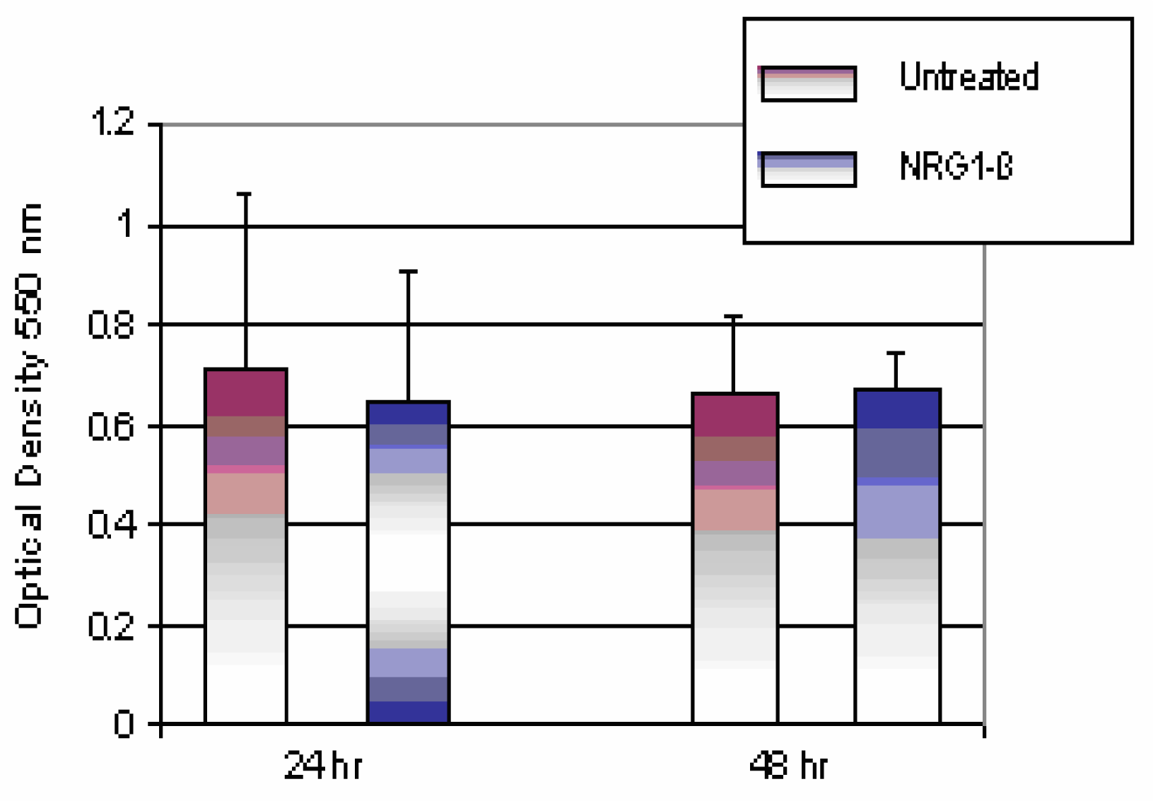
Comparison of Untreated and NRG1-βTtreated Mouse Hepatocytes

The comparison of untreated and NRG1-β-treated mouse hepatocytes in vitro is shown in Figure 1. Neuregulin has the ability to elicit cell proliferation through interaction with members of the ErbB family of receptor tyrosine kinases [5, 6]. Therefore, it was of interest to determine whether NRG1-β treatment of hepatocyte culture in vitro could promote a mitogenic response. Cells were treated with 0.01 nM NRG1-β or left untreated (0) for 24- and 48 h periods according to the methodology section. The cells were assayed for viability by using MTT incorporation. The mean absorbance was recorded as optical density at 550 nm for untreated and NRG1-β treated cells. The optical densities for 24 h untreated (0) cells and 0.01 nM NRG1-β were recorded at 0.71 and 0.64, respectively. The 48 h optical density was recorded at 0.66 and 0.67 respectively for untreated (0) cells and 0.01 nM NRG1-β. Data obtained from these experiments demonstrated that differences in the mean absorbance of untreated and NRG1-β-treated cells were not statistically significant (p>0.05). Our findings suggest that AML 12 mouse hepatocytes do not significantly proliferate in response to NRG1-β treatment in serum-free medium.

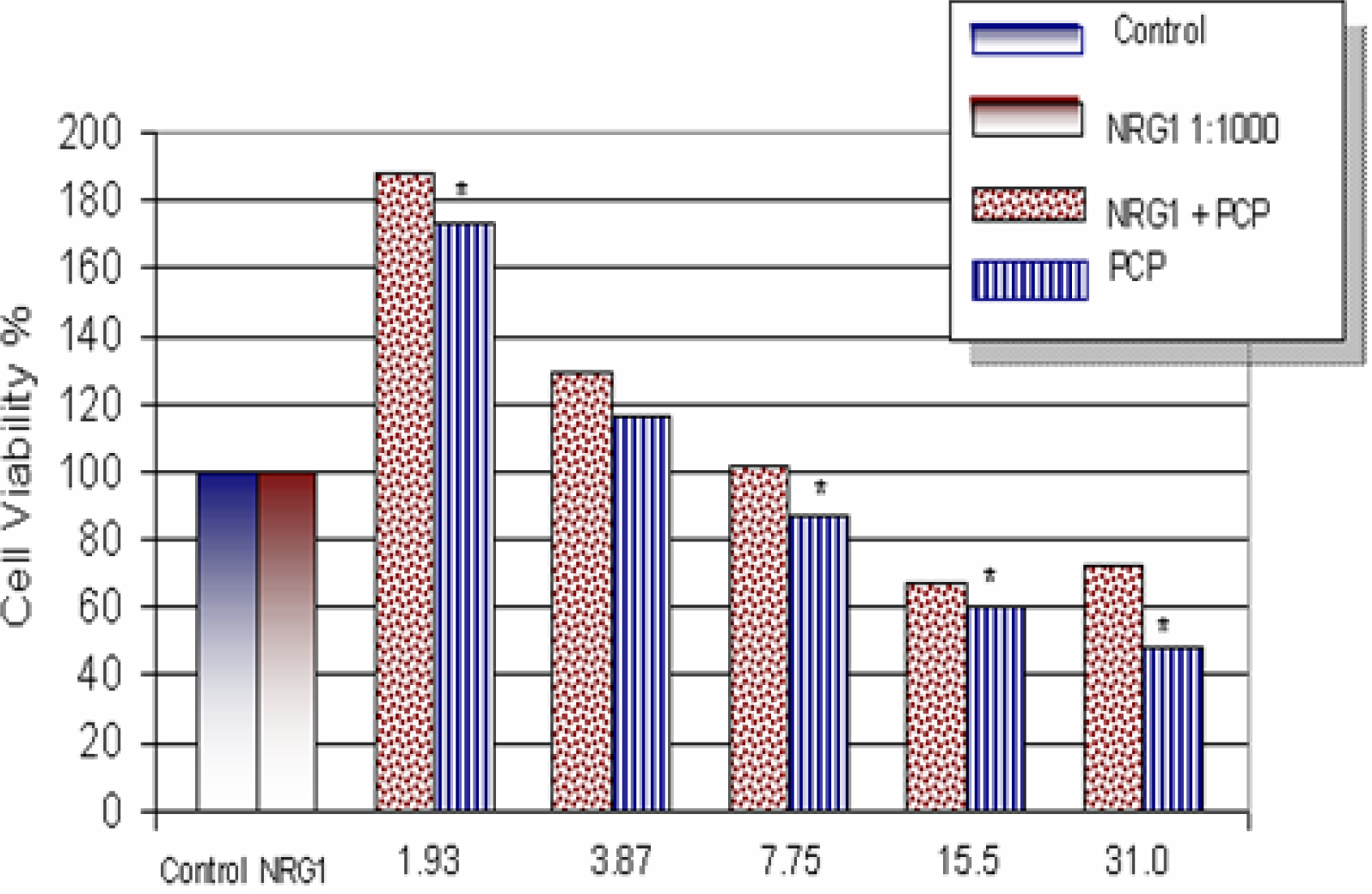
Effects of NRG1-β on PCP-Treated Mouse Hepatocytes

The effects of NRG1-β on PCP toxicity to AML 12 mouse hepatocytes are shown in Figure 2. Within the dose range of 0-31.0 µg/mL, a biphasic-response relationship was observed in both NRG1-β+PCP- and PCP-treated cells. The percentages for cell viability were recorded as 100.0 ± 0.0%, 188.0 ± 0.3%, 129.0 ± 0.3%, 102.0 ± 0.3%, 67.0 ± 0.2%, and 72.0 ± 0.1% at 0, 1.93, 3.87, 7.75, 15.5, and 31.0 µg/mL respectively for NRG1-β+PCP. Cell viability percentages for PCP treatments were recorded as 100 ± 0.0%, 173.0 ± 0.4%, 116.0 ± 0.4%, 87.0 ± 0.2%, 60.0 ± 0.2%, and 48.0 ± 0.1% at 0, 1.93, 3.87, 7.75, 15.5, and 31.0 μg/mL, respectively. The highest viability was achieved at 1.93 µg/mL. Cell viability percentages indicated that NRG1-β+PCP-treated cells had not been significantly impaired (except for the highest PCP concentration), while PCP-treated cells were appreciably reduced. Data obtained from this experiment strongly suggest that NRG1-β has the ability to rescue cell survival after PCP treatment by suppressing the toxic effects of PCP.

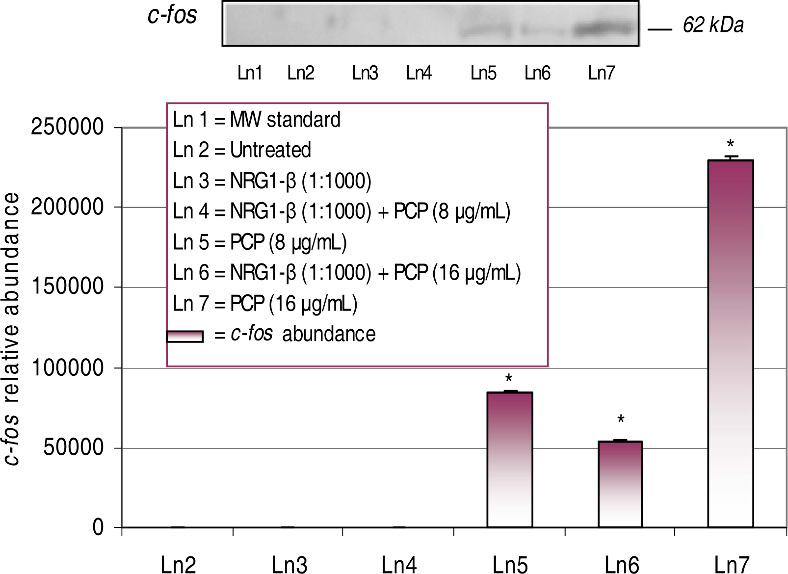
Western Blot and Densitometric Analyses for c-fos Expression

In the studies herein, mouse hepatocytes were acutely exposed to PCP and NRG1-β+PCP treatments (0, 8, 16 μg/mL) and compared to the untreated (0) and 0.01 nM NRG1-β to determine the magnitude of changes in specific protein expressions. In each experiment, basal levels of specific protein expression were not detected in untreated and NRG1-β–treated cultures. Western-blot and densitometric analyses were performed according to the methodology section. The 62-kDa c-fos protooncogene is recognized as an immediate early gene and has been identified as a transcription factor that responds to DNA-damage [22]. We have previously reported that PCP has the ability to markedly induce a dose-dependent upregulation of the c-fos gene protein in HepG2 cells and primary catfish hepatocytes [18, 19, 23].
Western-blot and densitometric analyses of c-fos expression in PCP- and NRG1-β + PCP-treated AML 12 mouse hepatocytes for 48 h are shown in Figure 3 upon 48h of exposure, a dose-dependent upregulation of the c-fos protein was observed in concomitant treatments of PCP and NRG1-β+PCP. The magnitude of c-fos expression was highly correlated with increased levels of PCP toxicity. For example, densitometric analysis showed a significant (p<0.05) increase of c-fos relative abundance of 83,714 at 8 µg PCP/mL, 54,115 at 16 µg NRG1-β+PCP/mL, and 229,374 at 16 µg PCP/mL. We did not detect the c-fos protein at 8 μg NRG1-β+PCP/mL; suggesting that NRG1-β attenuates PCP-stimulated c-fos expression at low levels of PCP-toxicity.

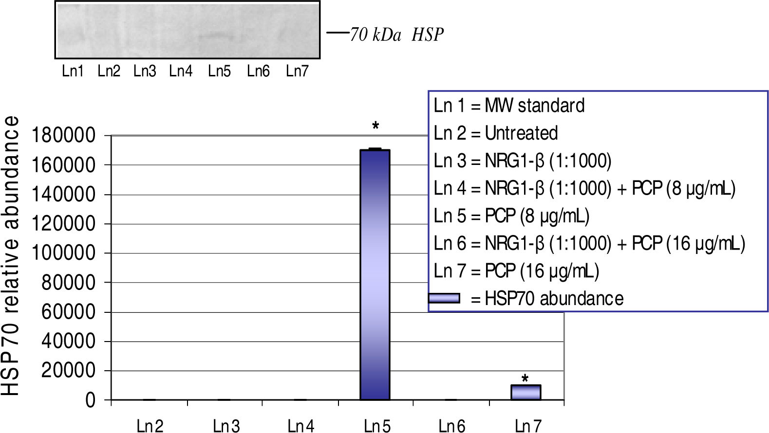
Western Blot and Densitometric Analyses for HSP70 Expression

HSP70, a member of the heat shock protein family, is a highly conservative molecular chaperone with strategic functions that respond to conditions of environmental stress, including tissue damage, inflammation, and mutant proteins associated with genetic abnormalities [24]. We have previously reported that the overexpression of the HSP70 gene protein is a dose-dependent event in HepG2 cells exposed to PCP [18, 23].
The expression and relative abundance of the 70-kDa heat shock protein in AML 12 mouse hepatocytes exposed to PCP and NRG1-β+PCP treatments for 48 h are shown in Figure 4. A dose-dependent upregulation of the HSP70 gene protein was demonstrated in both treatments of PCP and NRG1-β+PCP at 48 h. PCP-toxicity induced the upregulation of HSP70 at 8 μg PCP/mL with a relative abundance of 170,584. However, a drastic decrease (p<0.05) or down-regulation of HSP70 expression was demonstrated at 16 μg PCP/mL with a relative abundance of 10,000. NRG1-β completely suppressed the expression of HSP70 at 8 µg NRG1-β+PCP/mL and 16 µg NRG1-β+PCP/mL. These results suggest that NRG1-β has the ability to rescue cells from the consequences of proteolytic activity induced by PCP-toxicity.

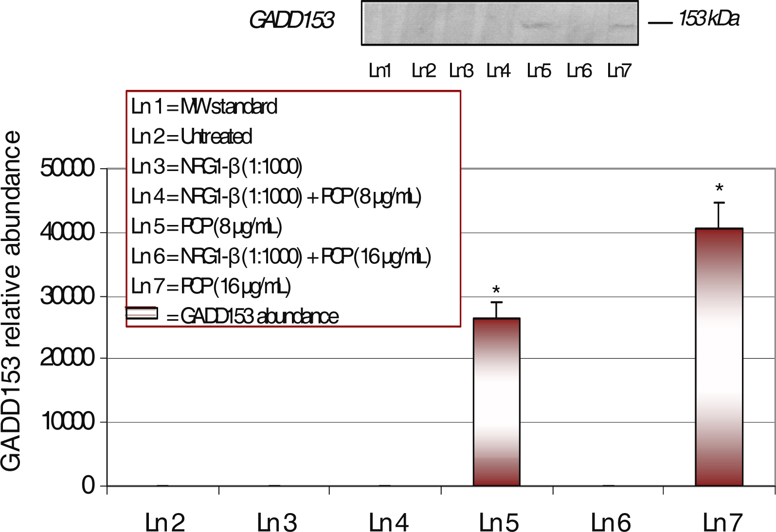
Western-Blot and Densitometric Analyses for GADD153 Expression

The GADD153 gene protein is robustly induced by genotoxic stress and is coordinately regulated with the endoplasmic reticulum (ER) [25]. The expression and relative abundance of the 153-kDa GADD in AML 12 mouse hepatocytes exposed to PCP and NRG1-β+PCP treatments for 48 h are shown in Figure 5.
We have previously reported that PCP can induce a dose-dependent expression of the GADD153 gene protein in HepG2 cells [18]. In this experiment, stimulated cells resulted in a dose-dependent (p<0.05) upregulation of the GADD153 protein with relative abundances of 26,411 and 40,626 respectively for 8 μg PCP/mL and 16 μg PCP/mL. Conversely, the expression of GADD153 was completely repressed at 8 µg NRG1-β+PCP/mL and 16 µg NRG1-β+PCP/mL. Data obtained from this experiment indicated that NRG1-β has the ability to attenuate growth arrest and DNA-damage activity at low and high levels of PCP-toxicity.

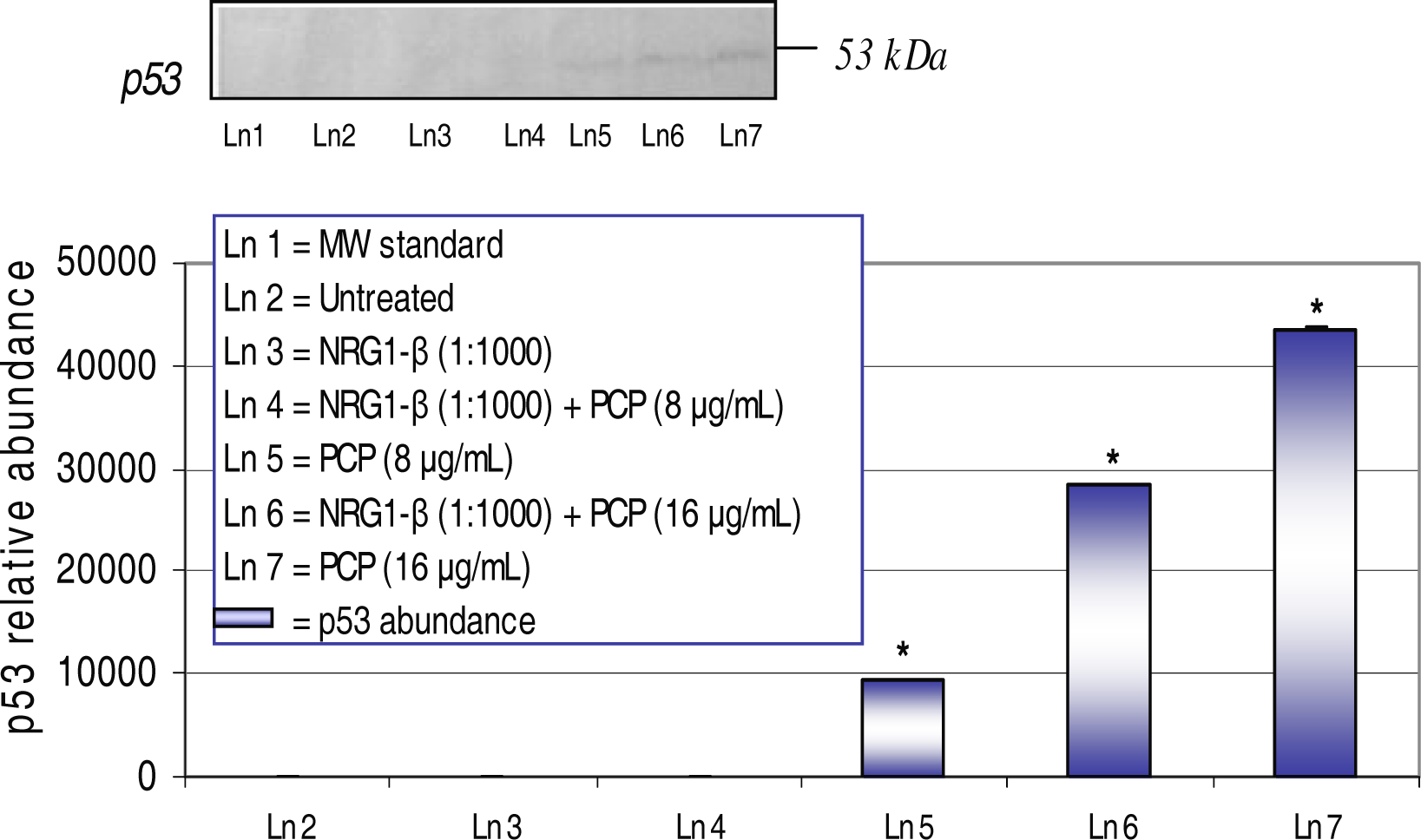
Western-Blot and Densitometric Analyses for p53 Expression

The 53-kDa p53 gene protein, also known as the tumor suppressor gene, senses DNA damage and responds by arresting the cell cycle [26]. Western-blot and densitometric analyses of the p53 gene protein in AML 12 mouse hepatocytes exposed to PCP and NRG1-β + PCP for 48 h are shown in Figure 6. We have previously reported that PCP has the ability to transcriptionally activate the p53 tumor suppressor gene in HepG2 cells [18, 23]. NRG1-β has the ability to repress the p53 protein at lower levels of PCP-toxicity. We did not detect the p53 expression at 8 µg NRG1-β+PCP/mL. However, we observed a dose-dependent increase (p<0.05) upregulation of the p53 protein with relative abundances of 9,502, 28,339 and 43,458 respectively for 8 µg PCP/mL, 16 µg NRG1-β+PCP/mL, and 16 µg PCP/mL; demonstrating the incompetence of NRG1-β to suppress DNA damage at higher levels of PCP toxicity.

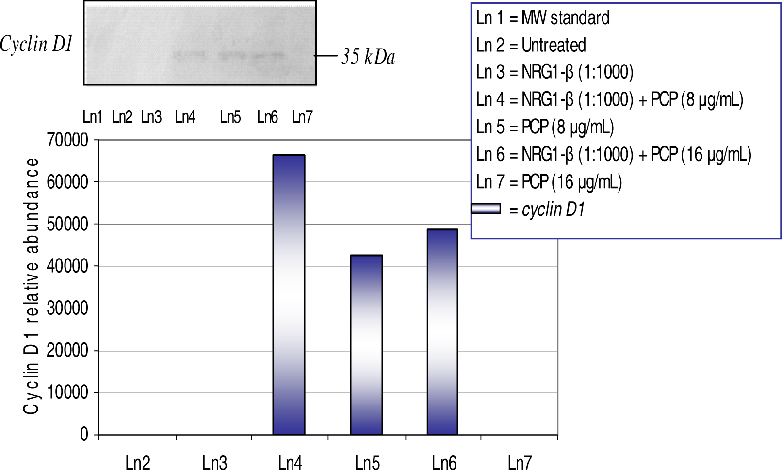
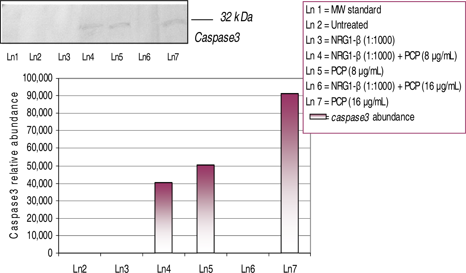
Western-Blot and Densitometric Analyses for Cyclin D1 and Caspase-3 Expression

The overexpression of cyclin D1 in hepatocytes is indicative of G1/S transition and mitogenic response [27]. Expression and relative abundance of the 35–kDa cyclin D1 in AML 12 mouse hepatocytes exposed to PCP and NRG1-β + PCP treatments for 48 h are shown in Figure 7. We have previously reported that PCP has the ability to elicit a mitogenic response in HepG2 cells, primary catfish hepatocytes, and AML 12 mouse hepatocytes [19, 20, 23]. In this experiment, we observed a dose-dependent overexpression of cyclin D1 with relative abundances of 66,371 at 8 μg NRG1-β+PCP/mL, 42,764 at 8 µg PCP/mL, and 48,757 at 16 µg NRG1-β+PCP/mL. This finding supports the fact that NRG1-β has the ability to protect hepatocytes in the presence of PCP.
The upregulation of caspase-3 expression is strongly associated with the readiness of cells to undergo apoptosis [28]. The expression and relative abundance of the 32-kDa caspase-3 protein in AML 12 mouse hepatocytes exposed to PCP and NRG1-β + PCP treatments for 48 h are shown in Figure 8. In this experiment we observed a dose- dependent activation of the caspase-3 protein with relative abundances of 40, 223 at 8 µg NRG1-β+PCP/mL, 50,160 at 8 µg PCP/mL, and 91,239 at 16 µg PCP/mL. In the presence of NRG1-β, the caspase-3 protein was repressed or down-regulated at 16 µg NRG1-β+PCP/mL; demonstrating a protective effect of NRG1-β at higher levels of PCP exposure. Here, we clearly demonstrate the ability of NRG1- β to block programmed cell death in PCP-treated mouse hepatocytes.

Discussion

Neuregulin 1-βeta-Induced Protective Effects

The present study was designed to investigate the protective effects of NRG1- β on PCP-induced cytotoxicity in AML 12 mouse hepatocytes. To our knowledge, the effects of NRG1-β treatment on hepatocyte survival have not been previously reported. We have previously reported that PCP is acutely toxic and causes cell injury to AML 12 mouse hepatocytes [20]. In that study, the 48 h-LC50 was computed to be 16.0 ± 2.0 μg PCP/mL. In the present study, we observed a similar biphasic response pattern with respect to cell viability. Our in vitro cell viability studies indicate that NRG1-β has a direct cytoprotective effect on hepatocytes against PCP-toxicity.
The members of the NRG family are produced by either neuronal or mesenchymal cells, and mediate their effects by binding to and signaling by the ErbB family of receptors [2, 9]. However, we provide clear evidence that NRG1-β was found to display a protective effect on AML 12 mouse hepatocytes caused by PCP toxicity (Figure 2). All NRG proteins contain an extracellular EGF-like domain, which is essential for bioactivity [29]. In a previous study, NRG1-β treatment of neonatal rat ventricular myocytes was shown to inhibit daunorubicin-induced apoptosis and the activation of caspase-3 [30]. A similar investigation demonstrated the survival effects of NRG1-β on norepinephrine cytotoxicity in adult rat ventricular myocytes by suppressing beta-adrenergic receptor-stimulated apoptosis [31]. In the present study, we have demonstrated that NRG1-β has to ability to inhibit PCP-induced apoptosis by completely suppressing the activation of caspase-3 at higher levels of PCP toxicity (Figure 8).
NRGs and their receptors can influence a network of survival signaling pathways, which is likely to vary in many cell types. The phosphoinositide 3-kinase (PIK3), which is specifically recruited by ErbB-3 and the ErbB-1T3 chimeric receptor, is a well-known regulator of cell growth and survival [32]. Abundant evidence has shown that the protective effects of NRGs are mediated by the PI3K signaling pathway [33, 34, 35]. In a very recent study, NRG induced a significant protective effect from β-amyloid 25–35 peptide-induced cell death [33]. The same study revealed that NRG treatment produced elevation in the levels of the antiapoptotic protein BclxL. The NRG-mediated BclxL elevation was regulated by protein kinase C (PKC). Results from that study suggested that NRG might affect cell viability by using two signaling pathways: activation of PI3K/PKB/AkT pathway and activation of PKC, which resulted in increasing levels of the antiapoptotic protein BclxL [33]. More specifically, NRG1-β has been shown to have a prosurvival effect on cardiac myocytes via the PI3K/Akt pathway [34]. The NRG-β isoform is a potent Schwann cell survival factor that binds to and activates a heterodimeric ErbB2/ErbB3 receptor complex [35]. Moreover, corroboration that NRG is mediated by the PIK3 pathway was demonstrated when NRG rapidly signaled the phosphorylation of mitogen-activated-protein kinase (MAPK) and the serine/threonine kinase Akt in serum-starved Schwann cells [35]. The same study used PIK3 inhibitors that blocked the NRG-mediated rescue of Schwann cells, as well as Akt, MAPK, and Bad; demonstrating the involvement of the PIK3 pathway [35].
Another possible mechanism of NRG1-β against PCP-toxicity in mouse hepatocytes is the involvement of the MAPK pathway. The MAPK pathway is thought to be directly responsible for regulating cell proliferation, differentiation, and survival. A number of investigations have implicated the MAPK pathway in NRG-β mediated survival [35, 36, 37]. A recent study demonstrated that NRG1-induced activation of ErbB4 stimulates the MAPK, PI3K, and cyclin-dependent kinase-5 (cdk5) pathways in cultured rat cerebellar granule neurons [37]. MAPK is a known downstream effector of the ErbB receptors. Therefore, in the present study, it is also possible that NRG1-β may exert its protective activity in PCP-treated mouse hepatocytes via the MAPK pathway.
Supporting evidence suggests that ErbB3 requires the association of other ErbB receptors or the EGFr to form an active signaling complex in hepatocytes [38]. Although NRGs are not primary mitogens in rat liver, they could regulate differentiation during development, maintenance of differentiated functions during regeneration, or metabolism in response to nutritional status [38]. A previous study demonstrated that a recombinant peptide corresponding to the EGF domain of the ß-1 isoform of heregulin (NRG1-β) bound to rat hepatocytes via the ErbB3 receptor, induced receptor phosphorylation, and stimulated DNA synthesis [38]. A similar investigation has shown that during rat liver development and regeneration, diverse ErbB receptor proteins are expressed [39]. In the present study, cell survival and cytoprotective effects of NRG1-β were assessed by the MTT- cell respiration assay. Cell viability data indicated that NRG1-β possesses a potent protective effect against PCP-induced cytotoxicity in AML 12 mouse hepatocytes (Figure 2). We propose that NRG1-β protective effects in PCP-treated hepatocytes are orchestrated by binding to ErbB receptors, and in turn the PI3K or MAPK pathway is solicited.

NRG1-β Effects on PCP-Induced c-fos Expression

Gene expression is controlled and regulated by many transcription factors in order for the cell to adjust to environmental or genetic modifications. We present evidence that NRG1-β has the ability to attenuate stress-related events as a consequence of PCP-toxicity. Specifically, we show that PCP induces stress-related gene expression and death of AML12 mouse hepatocytes, where NRG1-β protected them from PCP-induced death. To assess the cellular injury response in AML 12 mouse hepatocytes, we examined the effect of NRG1-β on the expression of stress-related gene proteins as a consequence of PCP-toxicity. In this study, we report that an appreciable dose-dependent expression of the c-fos protein was observed after 48 h of exposure to concomitant treatments of PCP and NRG1-β+PCP (Figure 3, Table 1).
The transcriptional activation of immediate early transcription factors such as c-fos is thought to be essential for mitogen-induced progression through the cell [40]. c-fos is a constituent of the immediate early transcription factor activator protein 1 (AP-1) heterodimeric complex and has been implicated as an positive modulator of G1-to-S-phase progression and cell proliferation [40, 41, 42]. The induction of the c-fos gene involves both transcriptional and post-transcriptional machinery [43]. Once stimulated, c-fos conjoins with c-jun, a transcription factor of the Jun family, and forms the heterodimeric complex, AP-1 [43]. Moreover, oxidative stress and DNA damage can stimulate c-fos expression and thus increase AP-1 transcription factor activity [44, 45]. The c-fos protooncogene plays a vital role in mitogenesis by inducing the expression of genes necessary for the activation of G1 cyclins [46]. For example, cyclin D1 mRNA is increased by the upregulation of c-fos [47] and mitosis-and mitogen-stimulated cyclin D1 transcription are repressed in cells deficient for c-fos and FosB [46]. It is well documented that mitogen-stimulated cyclin D1 requires the PIK3 activity [48, 49] and upon activation, PI3K stimulates c-fos transcription [50]. We have previously reported that the potential toxicity of PCP transcriptionally activates the c-fos gene protein in HepG2 cells and in primary catfish hepatocytes [18, 19, 23]. In the present study, we observed a dose-dependent upregulation of the c-fos protein in PCP-treated mouse hepatocytes (Figure 3, Table 1). This finding is consistent with our previous results and demonstrates similar PCP mechanistic activity across cell lines. We also observed a NRG1-β down regulation or repression of c-fos expression; indicating that NRG1-β has the ability to attenuate the activation of c-fos expression in PCP-treated AML mouse hepatocytes.

NRG1-β Effect on PCP-Induced HSP70 Expression

The 70-kDa HSP gene, a member of the heat shock protein molecular chaperone family, is involved in protein folding, translocation, and refolding of intermediates and proteases, while ensuring the efficient degradation of damaged and short-lived proteins [51]. Under stressful conditions, the accumulation of unfolded proteins in the ER leads to the induction of transcriptionally activated genes that encode molecular chaperones and folding enzymes [52]. Upon a variety of stress stimuli, signals are transduced from the ER to the cytoplasm and the nucleus to eventually result in adaptation for survival or induction of apoptosis [53]. HSP70 is documented as a general anti-apoptosis protein where it protects cells from the consequences of proteolysis by caspase-3-like proteases [54]. Moreover, the HSP70 gene protein is highly associated with the inflammatory response in lung epithelium and myocardial damage [55, 56]. We have previously shown that PCP can potently activate the HSP70 protein in HepG2 cells [18, 23]. In the present study, a dose-dependent increased expression of HSP70 was induced as a result of PCP-toxicity (Figure 4, Table 1). These results may be directly linked to the phenomenon in which the HSP70 gene enhances a cell signaling cascade that initiate protein repair. In the presence of NRG1-β, we observed a down regulation or inhibition of HSP70 expression in PCP-treated cells at 48 h of exposure. Our results are supported by a recent study that demonstrated the ability of NRG1-β to reverse inflammation and oxidative stress-related genes in focal ischemia of the rat brain and in a rat neuroblastoma cell line [13]. Here, we report that NRG1-β has the ability to attenuate proteolytic activity as a consequence of PCP-toxicity.

NRG1-β Effect on PCP-Induced GADD153 Expression

The GADD153 gene protein is a CCAAT/enhancer-binding protein (C/EBP)-related gene whose expression is induced in response to growth arrest and DNA damage [57]. The mechanism responsible for the activation of GADD153 expression after DNA damage remains unclear. GADD153 is induced by DNA- and cellular damage in a dose-dependent manner [58, 59]. The magnitude of GADD153 expression is proportional to the extent of cellular injury with maximal GADD153 promoter activity occurring under circumstances of severe toxicity to the cell [58]. Several investigations have reported that the induction of GADD153 is strongly correlated with the onset of apoptosis [60, 61]. Moreover, it has been demonstrated that elevated GADD153 expression depletes cells of essential thiols and down-regulates Bcl2 expression by inhibiting bcl2 transcription; thus sensitizing cells for death [58]. GADD153 is a potent mediator of the p53 gene protein in response to a variety of DNA damaging agents, inducing directly or indirectly G1 arrest and/or apoptosis [62]. We have previously reported that PCP-toxicity can induce a dose-dependent GADD153 expression in HepG2 cells [18]. In the present study, we also observed a dose-dependent elevated expression of the 153-kDa GADD protein in PCP-treated hepatocytes at 48 h of exposure (Figure 5, Table 1). Consistent with our previous findings, NRG1-β down-regulated or completely repressed the GADD153 expression in PCP-treated hepatocytes. In this study, the GADD153 expression is consistent with a previous study that demonstrated the ability of NRG1-β to reverse stress-related gene expression [13].

NRG1-β Effect on PCP-Induced on p53 Expression

The p53 gene is a tumor suppressor protein that plays a regulatory role in cell cycle control and apoptosis. p53 is a checkpoint molecule in G1 arrest caused by DNA damage and is closely dependent on transcriptional activation of the CKI p21 target gene [63]. Three major events associated with the p53 tumor suppressor gene are growth arrest, DNA repair, and apoptosis. Moreover, the ability of p53 to function as a sequence-specific DNA-binding protein appears to be essential to the function of p53 as a tumor suppressor [64, 65]. It has been reported that increased p53 expression is directly linked to missense mutations, which result in loss of its transcriptional activator function [66]. Our laboratory has previously reported that PCP has the ability to induce a dose-dependent expression of the 53-kDa p53 protein in HepG2 cells [18, 23]. In the present study, NRG1-β attenuated the expression of p53 at lower levels of PCP; however, PCP-treated hepatocytes demonstrated an increase in p53 expression (Figure 6, Table 1). In support of these results, it has been documented that in response to DNA damage, cells facilitate a rapid increase in wild-type p53 levels and a temporarily G1 arrest; allowing time for DNA to be repaired before being copied [67]. When optimal repair after DNA damage is irreparable, p53 initiates the signal to promote apoptosis [68].

NRG1-β Effect on PCP-Induced cyclin D1

Cell cycle transition from G1 to S phase is tightly regulated by distinct cyclin-dependent kinases (cdks) which are instrumental in cell cycle progression [69]. Cyclin D/Cdk4 complexes phosphorylate the retinoblastoma gene product early in the G1 phase of the cell cycle [69]. The 35-kDa cyclin D1 is overexpressed in several types of carcinomas and, therefore, suggested to play an important role in tumorigenesis and tumor progression including hepatocellular carcinoma [70, 71, 72]. When cyclin D1 is upregulated due to gene amplification, gene rearrangement, protein stabilization or other mechanisms, cyclin D1 acts as an oncogene by intensifying cell transformation, either alone [73] or in combination with activated ras [74], thereby shortening the G1 phase of the cell cycle. Interestingly, the induction of apoptosis is closely associated with an increase in cyclin D1-dependent kinase activity [75, 76, 77]. Notably, alterations in cyclin D1/Cdk4 caused by inhibition of ErbB2 support a critically role for cyclin D1 in ErbB2-mediated cell cycle progression [78]. Primary hepatocytes in culture readily proliferate in response to mitogens such as EGF [79]. Previous studies have shown that cyclin D1 is up-regulated during hepatocyte proliferation in culture [27, 80, 81]. We have previously demonstrated that PCP is capable of provoking a mitogenic response in HepG2 cells, primary catfish hepatocytes, and AML 12 mouse hepatocytes [19, 20, 23]. Results from the present study showed a dose-dependent activation of the 35-kDa cyclin D1 protein in PCP-treated cell in the presence of NRG1-β (Figure 7). However, in the absence of NRG1-β, PCP-treated cells demonstrated a down-regulated or repressed expression of the cyclin D1 protein; probably due to cell death at high levels of PCP- toxicity. These results are consistent with previous data in this study that demonstrate the protective effects of NRG1-β against PCP-toxicity.

NRG1-β Effects on PCP-induced Caspase-3 Expression

Caspases are the major enforcers of cell death, serving as molecular executioners to destroy many proteins required for maintenance of cellular homeostasis [82]. Caspase-3 is a major modulator of apoptotic activity. Apoptotic inhibitors, such as Bcl-2 and NF-kappa B, play a crucial role in the mechanism of anti-apoptosis of tumors [83, 84]. Caspase-3 is highly associated with apoptosis and is only cleaved and activated once the process of apoptosis is irreversible [85, 86]. Interestingly, it has been documented that PKC activity precedes the activation of caspase-3 [87]. A recent study demonstrated that the overexpression of PKC resulted in an increase of apoptosis, whereas its inhibition blocked caspase-3 activity and decreased apoptosis [87]. We have previously demonstrated that PCP causes cell injury and is cytotoxic to HepG2, primary catfish hepatocytes, and AML 12 mouse hepatocytes [18, 19, 20, 23]. In this study, we clearly demonstrate the ability of PCP to induce apoptosis in AML 12 mouse hepatocytes. This event was demonstrated by the upregulation of the 32-kDa caspase-3 protein in PCP-treated hepatocytes (Figure 8). Moreover, the caspase-3 protein was down-regulated or repressed in the presence of NRG1-β at higher levels of PCP. These results demonstrate the ability NRG1-β can exert an anti-apoptotic effect in PCP–treated hepatocytes. The mechanism by which NRG1-β attenuates caspase-3 expression is not clear. Future studies from our laboratory will investigate whether NRG1-β regulates pro-apoptotic gene expression by interfering with various transcriptional signaling pathways.

Conclusions

We have demonstrated that NRG1-β plays a cytoprotective role in AML 12 mouse hepatocytes exposed to PCP. NRG1-β was able to protect AML 12 mouse hepatocytes from cell injury by suppressing the toxic effects of PCP. NRG1-β has the ability to attenuate stress-related gene expression in PCP-treated AML 12 mouse hepatocytes. Western-blot analysis strongly indicated that PCP has the ability to cause oxidative stress and inflammatory reaction (c-fos), growth arrest and DNA damage (GADD153), proteotoxic effects (HSP70), and cell cycle arrest as consequence of DNA damage (p53). A mitogenic response was demonstrated by the upregulation of the 35-kDa cyclin D1 protein in PCP-treated hepatocytes. PCP-induced apoptosis was demonstrated by the overexpression of the 32-kDa caspase-3 protein. PCP-induced toxicity was attenuated or reversed in the presence of NRG1-β. To our knowledge, the anti-inflammatory activity of NRG1-β, is a novel finding and could represent efficient treatment for hepatic inflammatory disorders. Future studies from our laboratory will investigate the regulatory activity of NRG1- β on pro-inflammatory gene expression.
Figure 1:. Comparison of untreated and NRG1-β-treated AML 12 mouse hepatocytes. NRG1-β-treated (10 nM NRG1-β; 1:1000) hepatocytes were compared to untreated (0) cells for 24- and 48 h. Hepatocytes were maintained in DMEM medium with 10% FBS supplement. On day of exposure, FBS-medium was replaced with serum-free medium. The MTT-assay was used to determine absorbance at 550 nm after 24- and 48 h exposure periods. Absorbance readings are expressed as optical density. Each bar represents the mean ± S.D (n=3 independent experiments; p>0.05).
Ijerph 03 00011f1
Figure 2:. Effect of NRG1-β on PCP toxicity in AML 12 Mouse Hepatocytes. Cells were treated for 48hrs with PCP in the presence or absence of NRG1-β (0.01 nM). The number of metabolically active cells was determined by the MTT incorporation. The data are expressed as percentages of cell viability. Each point represents a mean value and standard deviation of three independent experiments (n = 3 independent experiments; 8 replications per treatment). *Significantly different (p < 0.05) from NRG1-β-treatment.
Ijerph 03 00011f2
Figure 3:. Expression and relative abundance of the 62 kDa c-fos in AML 12 mouse hepatocytes exposed to PCP and NRG1-β + PCP for 48 h. AML 12 mouse hepatocytes were treated with 8 µg/mL and 16 µg/mL concomitant treatments of PCP and NRG1-β + PCP. c-fos protein identification was assessed following exposure incubation period of 48 h. Inset shows a representative Wester n blot analysis. Each point represents a mean value and standard deviation of three experiments. *Significantly different (p < 0.05) from untreated (0 µg/mL PCP) and NRG1-β (1:1000) treated cells.
Ijerph 03 00011f3
Figure 4:. Expression and relative abundance of the 70 kDa heat shock (HSP70) in AML 12 mouse hepatocytes exposed to PCP and NRG1-β + PCP for 48 h. AML 12 mouse hepatocytes were treated with 8 μg/mL and 16 μg/mL concomitant treatments of PCP and NRG1-β+PCP. HSP70 protein identification was assessed following exposure incubation period of 48 h. Inset shows a representative Western blot analysis. Each point represents a mean value and standard deviation of three experiments. *Significantly different (p<0.05) from untreated (0 µg/mL PCP) and NRG1-β (1:1000) treated cells.
Ijerph 03 00011f4
Figure 5:. Expression and relative abundance of the 153 kDa GADD in AML 12 mouse hepatocytes exposed to PCP and NRG1-β + PCP for 48 h. AML 12 mouse hepatocytes were treated with 8 μg/mL and 16 μg/mL concomitant treatments of PCP and NRG1-β + PCP. GADD153 protein identification was assessed following exposure incubation period of 48 h. Inset shows representative Wester n blot analysis. Each point represents a mean value and standard deviation of three experiments. *Significantly different (p ≤ 0.05) from untreated (0 µg/mL PCP) and NRG1-β (1:1000) treated cells.
Ijerph 03 00011f5
Figure 6:. Expression and relative abundance of p53 in AML 12 mouse hepatocytes exposed to PCP and NRG1-β+ PCP for 48 h. p53 protein identification was assessed following exposure incubation period of 48 h. Inset shows representative Western blot analysis. Each point represents a mean value and standard deviation of three independent experiments. *Significantly different (p < 0.05) from untreated (0 μg/mL) and NRG1-β (1:1000) treated cells.
Ijerph 03 00011f6
Figure 7:. Expression and relative abundance of cyclin D1 in AML 12 mouse hepatocytes exposed to PCP and NRG1-β + PCP for 48h. Cyclin D1 protein identification was assessed following exposure incubation period of 48 h. Inset shows a representative Western-blot analysis. The following values were compared to untreated (0 µg/mL) and NRG1-β (1:1000) treated cells.
Ijerph 03 00011f7
Figure 8:. Expression and relative abundance of caspase-3 in AML 12 mouse hepatocytes exposed to PCP and NRG1-β + PCP for 48h. Caspase-3 protein identification was assessed following exposure incubation period of 48 h. Inset shows a representative Western-blot analysis. The following values were compared to untreated (0 µg/mL) and NRG1-β (1:1000) treated cells.
Ijerph 03 00011f8
TABLE 1:. Relative abundance of significantly up-regulated proteins in AML 12 mouse hepatocytes exposed for 48 hrs to PCP in the absence or presence of NRG1-β (0.01 nM).
Gene ProteinBiological Function / ResponseNRG1+PCP 8μg/mLPCP 8μg/mLNRG1+PCP 16μg/mLPCP 16μg/mL
c-fosAP-1 component Signaling-immediate early gene-83,71454,115229,374
DNA-damage Molecular chaperone
HSP70Oxidative stress Protein alterations-170,584-10,000
Cell cycle regulator
GADD153DNA repair-26,441-40,626
DNA-damage
p53Cell cycle- regulator Apoptosis-9,50228,38943,458
DNA-damage

References

  1. Crovello, CS; Lai, C; Cantley, LC; Carraway, KL, III. Differential signaling by the epidermal growth factor-like growth factors neuregulin-1 and neuregulin -2. J Biol Chem 1998, 273, 26954–26961. [Google Scholar]
  2. Burden, S; Yarden, Y. Neuregulins and their receptors: a versatile signaling module in organogenesis and oncogenesis. Neuron 1997, 18, 847–855. [Google Scholar]
  3. Nakano, N; Higashiyama, S; Ohmoto, H; Ishiguro, H; Taniguchi, N; Wada, Y. The N-terminal region of NTAK/neuregulin-2 isoforms has an inhibitory activity on angiogenesis. J Biol Chem 2004, 279(12), 11465–11470. [Google Scholar]
  4. Parkinson, DB; Langner, K; Namini, SS; Jessen, KR; Mirksy, R. β-Neuregulin and autocrine mediated survival of Schwann cells requires activity of Ets family transcription factors. Molecular and Cellular Neuroscience 2002, 20, 154–167. [Google Scholar]
  5. Murphy, S; Krainock, R; Tham, M. Neuregulin signaling via ErbB receptor assemblies in the nervous system. Molecular Neurobiology 2002, 25, 67–78. [Google Scholar]
  6. Adlkofer, K; Lai, C. Role of neuregulins in glial cell development. Glia 2000, 15, 104–111. [Google Scholar]
  7. Marchionni, MA; Cannella, B; Hoban, C; Gao, YL; Garcia-Arnas, R; Lawson, D; Happel, E; Noel, F; Tofilon, P; Gwynne, D; Raine, CS. Neuregulin in neuron/glial interactions in the central nervous system: GGF2 diminishes auto-immune demyelination, promotes oligodendrocytes progenitor expansion, and enhances remyelination. Adv. Exp. Med. Biol 1999, 468, 283–295. [Google Scholar]
  8. Fischbach, GD; Rosen, KM. ARIA: a neuromuscular junction neuregulin. Annu Rev Neurosci 1997, 20, 429–458. [Google Scholar]
  9. Carraway, KL, III; Burden, SJ. Neuregulins and their receptors. Curr Opin Neurobiol 1995, 5, 606–612. [Google Scholar]
  10. Carraway, KL, III; Cantley, LC. A neu acquaintance for erbB3 and erbB4: a role for receptor heterodimerization in growth signaling. Cell 1994, 78, 5–8. [Google Scholar]
  11. Fallon, KB; Havlioglu, N; Hamilton, LH; Cheng, TP; Carroll, SL. Constitutive activation of the neuregulin-1/erbB signaling path way promotes proliferation of a human peripheral neuroepithelioma cell line. J Neurooncol 2004, 66, 273–284. [Google Scholar]
  12. Xu, Z; Jiang, J; Ford, G; Ford, BD. Neuregulin-1 is neuroprotective and attenuates inflammatory responses induced by ischemic stroke. Biochem Biophys Res Commun 2004, 322, 440–446. [Google Scholar]
  13. Xu, Z; Ford, GD; Croslan, DR; Jiang, J; Gates, A; Allen, R; Ford, BD. Neuroprotection by neuregulin-1 following focal stroke is associated with the Attenuation of ischemia-induced pro-inflammatory and stress gene expression. Neurobiology of Disease 2005, 19, 461–470. [Google Scholar]
  14. Fukazawa, R; Miller, TA; Kuramochi, Y; Frantz, S; Kim, YD; Marchionni, MA; Kelly, RA; Sawyer, DB. Neuregulin-1 protects ventricular myocytes from anthracycline-induced apoptosis via erbB4-dependent activation of PI3-kinase/Akt. J Mol Cell Cardio 2003, 35(12), 1473–1479. [Google Scholar]
  15. Negro, A; Brar, K; Lee, KF. Essential roles of Her2/erbB2 in cardiac development and function. Recent Prog Horm Res 2004, 59, 1–12. [Google Scholar]
  16. Sawyer, DB; Zuppinger, C; Miller, TA; Eppenberger, HM; Suter, TM. Modulation of anthracycline-induced myofibrillar disarray in rat ventricular myocytes by neuregulin-1 beta and anti-erbB2: potential mechanism fortrastuzumab-induced cardiotoxicity. Circulation 2002, 105(13), 1551–1554. [Google Scholar]
  17. Agency for Toxic Substances and Disease Registry (ATSDR), Toxicological Profile for Pentachlorophenol; U.S. Public Health Service, U.S. Department of Health and Human Services: Atlanta, GA, 1992.
  18. Dorsey, WC; Tchounwou, PB; Ishaque, AB; Shen, E. Transcriptional activation of stress genes and cytotoxicity in human liver carcinoma (HepG2) exposed to pentachlorophenol. International J Molecular Sci 2002, 3, 989–1004. [Google Scholar]
  19. Dorsey, WC; Tchounwou, PB. Pentachlorophenol-induced cytotoxic, mitogenic, and endocrine-disrupting activities in channel catfish, Ictalurus punctatus. International J Environ Research and Public Health 2004, 1(2), 74–83. [Google Scholar]
  20. Dorsey, WC; Tchounwou, PB; Sutton, D. Mitogenic and cytotoxic effects of pentachlorophenol to AML 12 mouse hepatocytes. International J Environ Research and Public Health 2004, 1(2), 100–105. [Google Scholar]
  21. Mosmann, T. Rapid colorimetric assay for cellular growth and survival: applications to proliferation and cytotoxicity assays. J Immunol Methods 1983, 65, 55–63. [Google Scholar]
  22. Holbrook, NJ; Fornace, AJ. Response to adversity: molecular control of gene activation following genotoxic stress. New Biol 1991, 3, 825–833. [Google Scholar]
  23. Dorsey, WC; Tchounwou, PB. CYP1A1, HSP70, p53, and c-Fos expression in human liver carcinoma cells (HepG2) exposed to pentachlorophenol. ISA-Biomedical Sciences Instrumentation 2003, 39, 389–396. [Google Scholar]
  24. Jolly, C; Morimoto, RI. Role of the heat shock response and molecular chaperones in oncogenesis and cell death. Journal of the National Cancer Institute 2000, 92(19), 1564–1572. [Google Scholar]
  25. Zinszer, H; Kuroda, M; Wang, X; Batchvarova, N; Lightfoot, RT; Remotti, H; Stevens, JL; Ron, D. CHOP is implicated in programmed cell death in response to impaired function of the endoplasmic reticulum. Genes & Development 1998, 12(7), 982–995. [Google Scholar]
  26. Quelle, FW; Wang, J; Feng, J; Wang, D; Cleveland, JL; Ihle, JN; Zambetti, GP. Cytokine rescue of p53-dependent apoptosis and cell cycle arrest is mediated by distinct Jakkinase signaling pathways. Genes & Development 1998, 12(8), 1099–1107. [Google Scholar]
  27. Albrecht, JH; Hansen, LK. Cyclin D1 promotes mitogenic-independent cell cycle progression in hepatocytes. Cell Growth & Differentiation 1999, 10, 397–404. [Google Scholar]
  28. Tormanen-Napankangas, U; Soini, Y; Kahlos, K; Kinnula, V; Paakko, P. Expression of Caspases-3, -6 and -8 and their relation to apoptosis in non-small cell lung carcinoma. Int J Cancer 2001, 93(2), 192–98. [Google Scholar]
  29. Adlkofer, K; Lai, C. Role of neuregulins in glial cell development. Glia 2000, 29, 104–111. [Google Scholar]
  30. Fukazawa, R; Miller, TA; Kuramochi, Y; Frantz, S; Kim, YD; Marchionni, MA; Kelly, RA; Sawyer, DB. Neuregulin-1 protects ventricular myocytes from anthracyline-induced apoptosis via erbB4-dependent activation of PI3-Akt. J Mol Cell Cardiol 2003, 35(12), 1473–1479. [Google Scholar]
  31. Kuramochi, Y; Lim, CC; Guo, X; Colucci, WS; Liao, R; Sawyer, DB. Myocyte contractile activity modulates norepinephrine cytotoxicity and survival effects of neuregulin-1 beta. Am J Physiol Cell Physiol 2004, 286(2), C222–229. [Google Scholar]
  32. Carpenter, CL; Cantley, LC. Phosphoinositide 3-kinase and the regulation of cell growth. Biochem Biophys Acta 1996, 1288, M11–M16. [Google Scholar]
  33. Di Segni, A; Shaharabani, E; Stein, R; Pinkas-Kramarski, R. Neuregulins rescue PC12-ErbB-4 cells from cell death induced by ß-amyloid 34.
  34. Parkinson, DB; Langner, K; Namini, SS; Jessen, KR; Mirsky, R. beta- Neuregulin and autocrine mediated survival of Schwann cells requires activity of Ets family transcription factors. Mol Cell Neurosci 2002, 20(1), 154–167. [Google Scholar]
  35. Xie, F; Raetzman, LT; Siegal, RE. Neuregulin induces GABAA receptor β2 subunit expression in cultured rat cerebellar granule neurons by activating multiple signal pathways. Journal of Neurochemistry 2004, 90, 1521–1529. [Google Scholar]
  36. Carver, RS; Sliwkowski, MX; Sitaric, S; Russell, WE. Insulin regulates heregulin binding and ErbB3 expression in rat hepatocytes. J Biol Chem 1996, 271(23), 13491–13496. [Google Scholar]
  37. Carver, RS; Stevenson, MC; Scheving, LA; Russell, WE. Diverse expression of ErbB receptor proteins during rat liver development and regeneration. Gastroenterology 2002, 123(6), 2017–2027. [Google Scholar]
  38. Schreiber, M; Kolbus, A; Piu, F; Szabowski, A; Mohle-Steinlein, U; Tian, J; Karin, M; Angel, P; Wagner, EF. Control of cell cycle progression by c-jun is p53 dependent. Genes & Development 1999, 13, 617–619. [Google Scholar]
  39. Deschamps, J; Meijlink, F; Verma, IM. Identification of a transcriptional enhancer element upstream from the proto-oncogene fos. Science 1985, 230, 1174–1177. [Google Scholar]
  40. Kovary, K; Bravo, R. The jun and fos protein families are both required for cell cycle progression in fibroblasts. Mol Cell Biol 1991, 11, 4466–4472. [Google Scholar]
  41. Takeuchi, K; Shibamoto, S; Nagamine, K; Shigemori, I; Omura, S; Kitamura, N; Ito, F. Signaling pathways leading to transcription and translation cooperatively regulate the transient increase in expression of c-fos protein. J Biol Chem 2001, 276(280), 26077–26083. [Google Scholar]
  42. Schenk, H. Distinct effects of thioredoxin and antioxidants on the activation of transcription factors NF-*B and AP-1. Proc Natl Acad Sci USA 1994, 91, 1672–1676. [Google Scholar]
  43. Jackson, SP; Jeggo, PA. DNA double-strand break repair and V(D)J recombination: Involvement of DNA-PK. Trends Biochem Sci 1995, 20, 412–415. [Google Scholar]
  44. Brown, JR; Nigh, E; Lee, RJ; Ye, H; Thompson, MA; Saudou, F; Pestell, RG; Greenburg, ME. fos family members induce cell cycle entry by activating cyclin D1. Mol Cell Biol 1998, 18, 5609–5619. [Google Scholar]
  45. Miao, GG; Curran, T. Cell transformation by c-fos requires an extended period of expression and is independent of the cell cycle. Mol Cell Biol 1994, 14, 4295–4310. [Google Scholar]
  46. Yamauchi, K; Holt, K; Pessin, JE. Phosphatidylinositol 3-kinase functions upstream of Ras and Raf in mediating insulin stimulation of c-fos transcription. J Biol Chem 1993, 268, 14597–14600. [Google Scholar]
  47. Jhun, BH; Rose, DW; Seely, BL; Rameh, L; Cantley, L; Saltiel, AR; Olefsky, JM. Microinjection of the SH2 domain of the 85-kilodalton subunit phosphatidylinositol 3-kinase inhibits insulin induced DNA synthesis and c-fos expression. Mol Cell Biol 1994, 14, 7466–7475. [Google Scholar]
  48. Wang, Y; Falasca, M; Schlessinger, J; Malstrom, S; Tsichlis, P; Settlemen, J; Hu, W; Lim, B; Prywes, R. Activation of the c-fos serum response element by phosphatidyl inositol 3-kinase and rho pathways in HeLa cells. Cell Growth Differ 1998, 9, 513–522. [Google Scholar]
  49. Jolly, C; Morimoto, RI. Role of the heat shock response and molecular chaperones in oncogenesis and cell death. J. Nation. Cancer Inst 2000, 92, 1564–1572. [Google Scholar]
  50. Kozutsumi, Y; Segal, M; Normington, K; Gething, MJ; Sambrook, J. The presence of malfolded proteins in the endoplasmic reticulum signals the induction of glucose-regulated proteins. Nature 1988, 332, 462–464. [Google Scholar]
  51. Kaufman, RJ. Stress signaling from the lumen of the endoplasmic reticulum: coordination of gene transcriptional and translational controls. Genes & Development 1999, 13, 1211–1233. [Google Scholar]
  52. Jaattela, M; Wissing, D; Kokholm, K; Kallunki, T; Egeblad, M. Hsp70 exerts its anti-apoptotic function downstream of Caspase-3-like proteases. EMBO J 1998, 17(21), 6124–6134. [Google Scholar]
  53. Ramage, L; Guy, K. Expression of C-reactive protein and heat-shock protein-70 in the lung epithelial cell line A549, in response to PM10 exposure. Inhal Toxicol 2004, 16(6–7), 447–452. [Google Scholar]
  54. Dybdahl, B; Slordahl, SA; Waage, A; Kierulf, P; Espevik, T; Sundan, A. Myocardial ischaemia and the inflammatory response: release of heat shock protein 70 after myocardial infarction. Heart 2005, 91, 299–304. [Google Scholar]
  55. Bartlett, JD; Luethy, JD; Carlson, SG; Sollott, SJ; Holbrook, NJ. Calcium ionophore A23187 induces expression of the growth arrest and DNA damage inducible CCAAT/enhancer-binding protein (C/EBP)-related gene, gadd153. Ca2+ increases transcriptional activity and mRNA stability. J Biol Chem 1992, 267(28), 20465–20470. [Google Scholar]
  56. Los, G; Benbatoul, K; Gately, DP; Barton, R; Christen, R; Robbins, KT; Vicario, D; Kirmani, S; Orloff, LA; Weisman, R; Howell, SB. Quantitation of the change in GADD153 messenger RNA Level as a molecular marker of tumor response in head and neck cancer. Clinical Cancer Research 1999, 5, 1610–1618. [Google Scholar]
  57. Gately, DP; Jones, JA; Christen, R; Barton, R; Los, G; Howell, SB. Induction of growth arrest and DNA damage inducible gene gadd153 by cisplatin in vitro and in vivo. Br J Cancer 1994, 70, 1102–1106. [Google Scholar]
  58. Eymim, B; Dubrez, L; Allouche, M; Solary, E. Increased gadd153 messenger RNA level is associated with apoptosis in human leukemic cells treated with etoposide. Cancer Res 1997, 57, 686–695. [Google Scholar]
  59. Friedman, AD. GADD153/CHOP, a DNA damage-inducible protein, reduced CAAT/enhancer binding protein activities and increased apoptosis in 32D c13 myeloid cells. Cancer Res 1996, 56, 3250–3256. [Google Scholar]
  60. Jackman, J; Alamo, I, Jr; Fornace, AJ, Jr. Genotoxic stress confers preferential and coordinate messenger RNA stability on the five gadd genes. Cancer Res 1994, 54, 5656–5662. [Google Scholar]
  61. Levine, AJ. p53, the cellular gatekeeper for growth and division. Cell 1997, 88, 323–331. [Google Scholar]
  62. Bargonetti, J; Friedman, PN; Kern, SE; Vogelstein, B; Prives, C. Wild-type but not mutant p53 immunopurified proteins bind to sequences adjacent to the SV40 origin of replication. Cell 1991, 65(6), 1083–91. [Google Scholar]
  63. Kern, SE; Kinzler, KW; Bruskin, A; Jarosz, D; Friedman, P; Prives, C; Vogelstein, B. Identification of p53 as a sequence-specific DNA-binding protein. Science 1991, 252(5013), 1708–1711. [Google Scholar]
  64. Midgley, CA; Lane, D. p53 protein stability in tumour cells is not determined by mutation but is dependent on Mdm2 binding. Oncogene 1997, 15, 1179–1189. [Google Scholar]
  65. Kastan, MB; Onyekwere, O; Sidransky, D; Vogelstein, B; Craig, RW. Participation of p53 protein in cellular response to DNA damage. Cancer Res 1991, 51, 6304–6311. [Google Scholar]
  66. Lane, DP. p53, guardian of the genome. Nature 1992, 258, 15–16. [Google Scholar]
  67. Muraoka, RS; Lenferink, AEG; Law, B; Hamilton, E; Brantley, DM; Roebuck, LR; Arteaga, CL. ErbB2/neu-induced, cyclin D1-dependent transformation is accelerated in p27-haploinsufficient mammary epithelial cells but impaired in p27-null cells. Molecular and Cellular Biology 2002, 22(7), 2204–2219. [Google Scholar]
  68. Gillett, C; Smith, P; Gregory, W; Richards, M; Millis, R; Peters, G; Barnes, D. Cyclin D1 and prognosis in human breast cancer. Int J Cancer 1996, 69, 92–99. [Google Scholar]
  69. Hall, M; Peters, G. Genetic alterations of cyclins, cyclin-dependent kinases, and cdk inhibitors in human cancer. Adv Cancer Res 1996, 68, 67–108. [Google Scholar]
  70. Uto, H; Ido, A; Moriuchi, A; Onaga, Y; Nagata, K; Onaga, M; Tahara, Y; Hori, T; Hirono, S; Hayashi, K; Tsubouchi, H. Transduction of antisense cyclin D1 using two-step gene transfer inhibits the growth of rat hepatoma cells. Cancer Res 2001, 61, 4779–4783. [Google Scholar]
  71. Jiang, W; Kahn, SM; Zhou, P; Zhang, YJ; Cacace, AM; Infante, AS; Doi, S; Santella, RM; Weinstein, IB. Overexpression of cyclin D1 in rat fibroblasts causes abnormalities in growth control, cell cycle progression and gene expression. Oncogene 1993, 8, 3447–3457. [Google Scholar]
  72. Lovec, H; Sewing, A; Lucibello, FC; Müller, R; Möröy, T. Oncogenic activity of cyclin D1 revealed through cooperation with Ha-ras: link between cell cycle con trol and malignant transformation. Oncogene 1994, 9, 323–326. [Google Scholar]
  73. Kranenburg, O; van der Eb, AJ; Zentema, A. Cyclin D1 is an essential mediator of apoptotic neuronal cell death. EMBO J 1996, 15, 46–54. [Google Scholar]
  74. Sofer-Levi, Y; Resnitzky, D. Apoptosis induced by ectopic expression of cyclin D1 but not cyclin E. Oncogene 1996, 13, 2431–2437. [Google Scholar]
  75. Hiyama, H; Reeves, SA. Role for cyclin D1 in UVC-induced and p53-mediated apoptosis. Cell Death and Differentiation 1999, 6, 565–569. [Google Scholar]
  76. Lenferink, AEG; Busse, D; Flanagan, WM; Yakes, FM; Arteaga, CL. ErbB2/ neu kinase modulates cellular p27Kip1 and cyclin D1 through multiple pathways. Cancer Res 2001, 61, 6583–6581. [Google Scholar]
  77. Michalopoulos, GK; De Frances, MC. Liver regeneration. Science 1997, 276, 60–66. [Google Scholar]
  78. Albrecht, JH; Hu, MY; Cerra, FB. Distinct patterns of cyclin D1 regulation in models of liver regeneration and human liver. Biochem. Biophys. Res. Commun 1995, 209, 648–655. [Google Scholar]
  79. Ehrenfried, JA; Ko, TC; Thompson, EA; Evers, BM. Cell cycle-mediated regulation of hepatic regeneration. Surgery 1997, 122, 927–935. [Google Scholar]
  80. Utz, PJ; Anderson, P. Life and death decisions: regulation of apoptosis by proteolysis of signaling molecules. Cell Death Differ 2000, 7(7), 589–602. [Google Scholar]
  81. Ohshima, K; Sugihara, M; Haraoka, S; Suzumiya, J; Kanda, M; Kawasaki, C; Shimazaki, K; Kikuchi, M. Possible immortalization of Hodgkin and Reed-Sternberg cells: telomerase expression, lengthening of telomere, and inhibition of apoptosis by NF-kappa B expression. Leuk Lymphoma 2001, 41, 367–376. [Google Scholar]
  82. Hussein, MR; Haemel, AK; Wood, GS. Apoptosis and melanoma: molecular mechanisms. J Pathol 2003, 199, 275–288. [Google Scholar]
  83. Shirin, H; Pinto, JT; Kawabata, Y; Soh, J; Delohery, T; Moss, SF; Murty, V; Rivlin, RS; Holt, PR; Weinstein, IB. Antiproliferative effects of s-allylmer- captocysteine on colon cancer cells when tested alone or in combination with sulindac sulfide. Cancer Res 2001, 61, 725–731. [Google Scholar]
  84. Hirota, J; Furuichi, T; Mikoshiba, K. Inositol 1,4,5–triphosphate receptor type 1 is a for caspase-3 and is cleaved during apoptosis in a caspase-3-dependent manner. J Biol Chem 1999, 274, 34433–34437. [Google Scholar]
  85. Voss, OH; Kim, S; Wewers, MD; Doseff, AI. Regulation of monocyte apoptosis by the protein kinase Cδ-dependent phosphorylation of caspase-3. J Biol Chem 2005, 17371–17379. [Google Scholar]

