
Cobalt Oxide Nanoparticles: Behavior towards Intact and Impaired Human Skin and Keratinocytes Toxicity

 ,
, 
Abstract
:1. Introduction
2. Materials and Methods
2.1. Chemicals
2.2. Nanoparticles Characterization
2.3. Nanoparticles Dissolution
2.4. Preparation of Skin Membranes
2.5. In Vitro Diffusion System
2.5.1. Experiment 1
2.5.2. Experiment 2
2.5.3. Blanks
2.5.4. Skin Digestion after the Experiment
2.6. Analytical Measurements
2.6.1. Cell Tests
2.6.2. Cell Culture
2.6.3. MTT Assay
2.6.4. AlamarBlue® Assay
2.6.5. Propidium Iodide Uptake
2.7. Cell Fixation for TEM Analysis
2.8. Statistical Analysis
3. Results
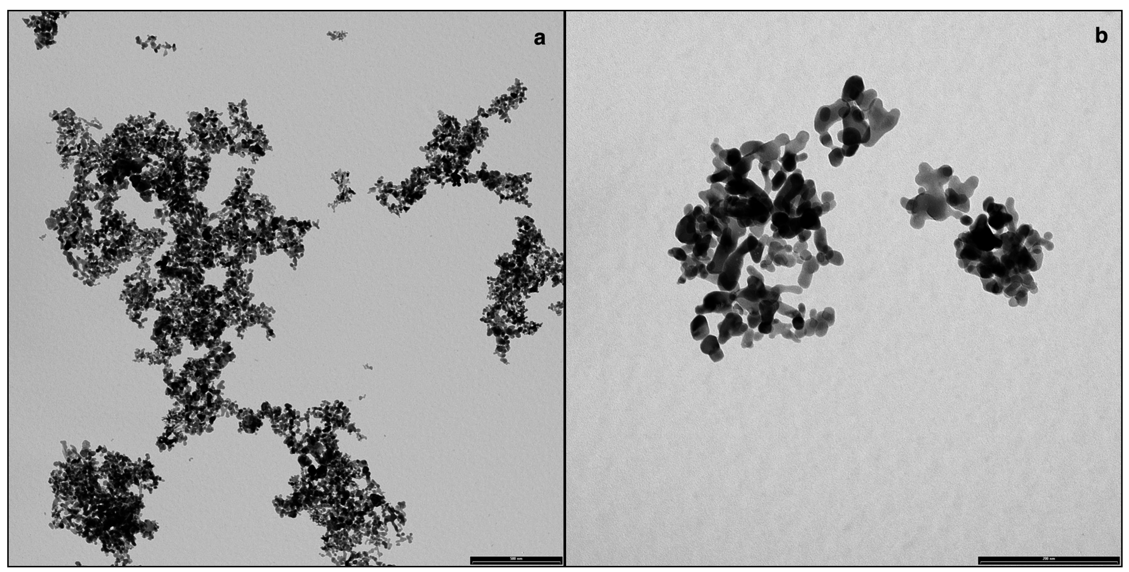
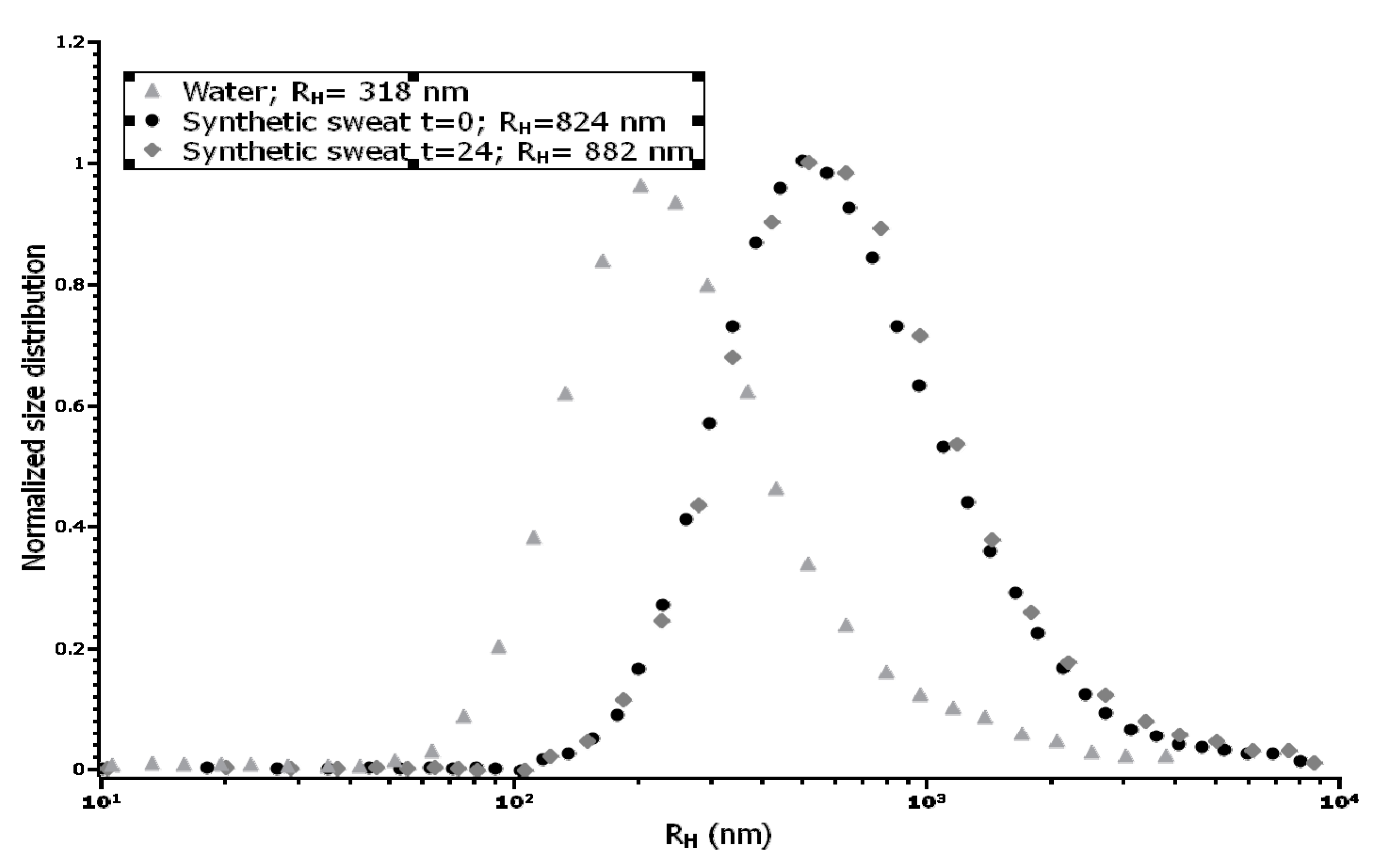
3.1. Nanoparticles Characterization


| Medium Specimen | Water | Synthetic Sweat T = 0 | Synthetic Sweat T = 24 h |
|---|---|---|---|
| Co3O4 | Mean: –19.8 +/− 1.15 mV | Mean: –18.5 +/− 3.5 mV | Mean: –15.9 +/− 4.2 mV |

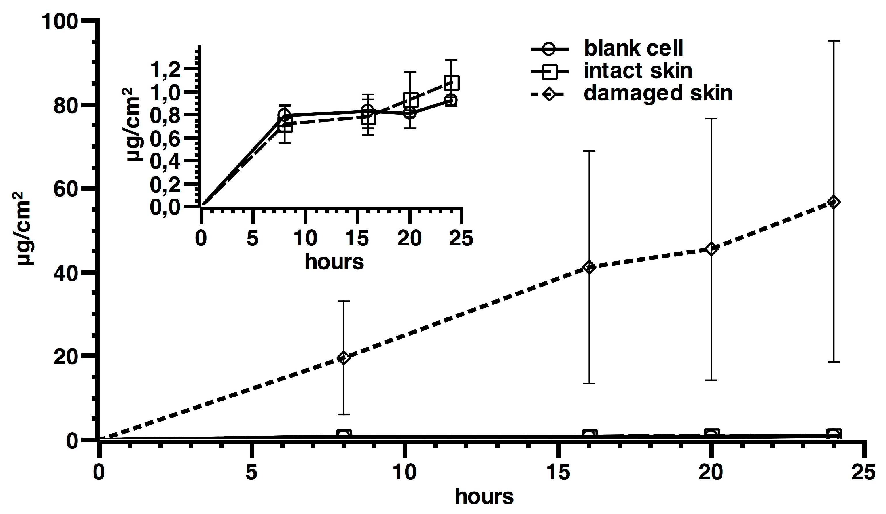
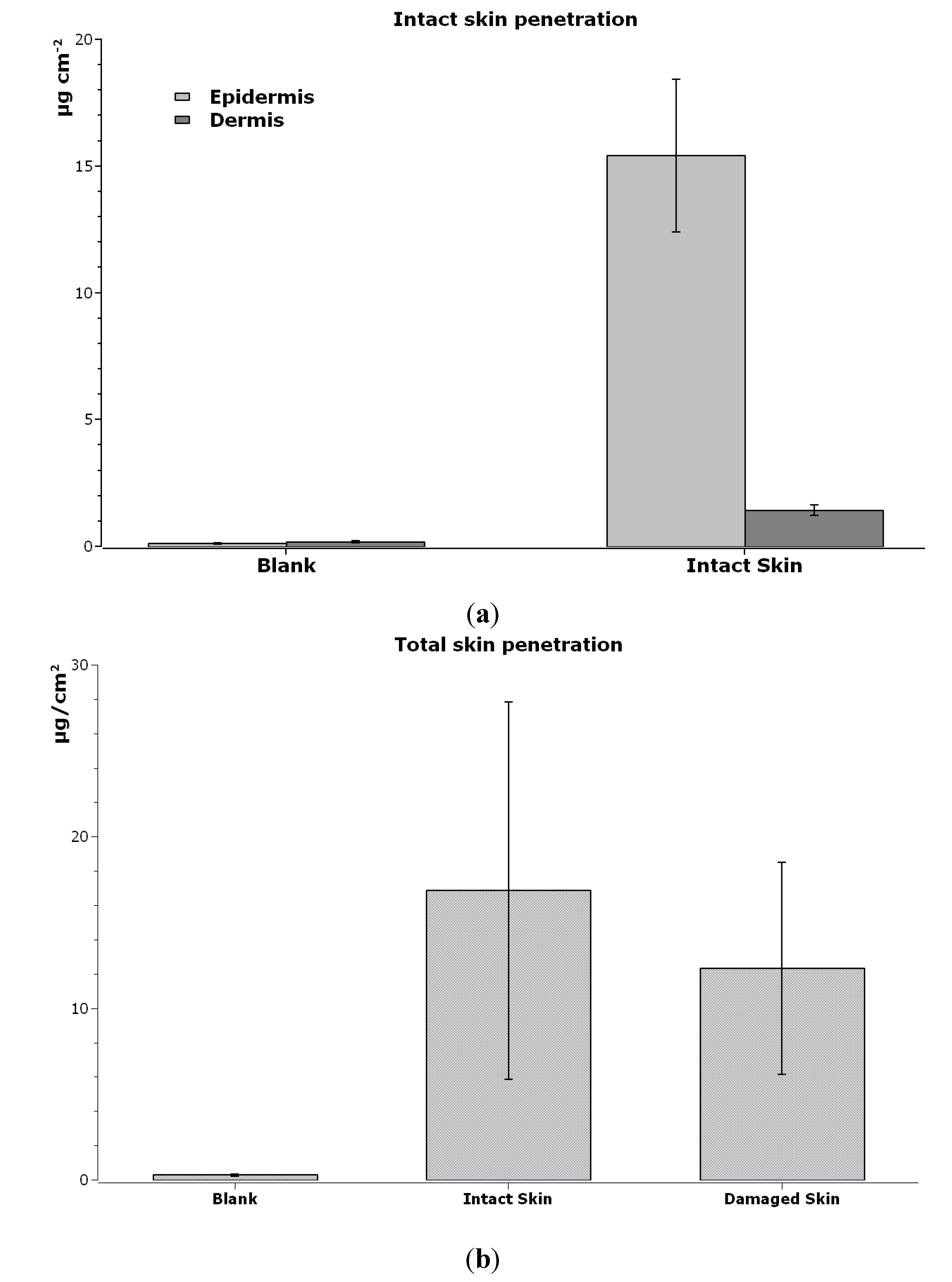
3.2. NPs Skin Permeation


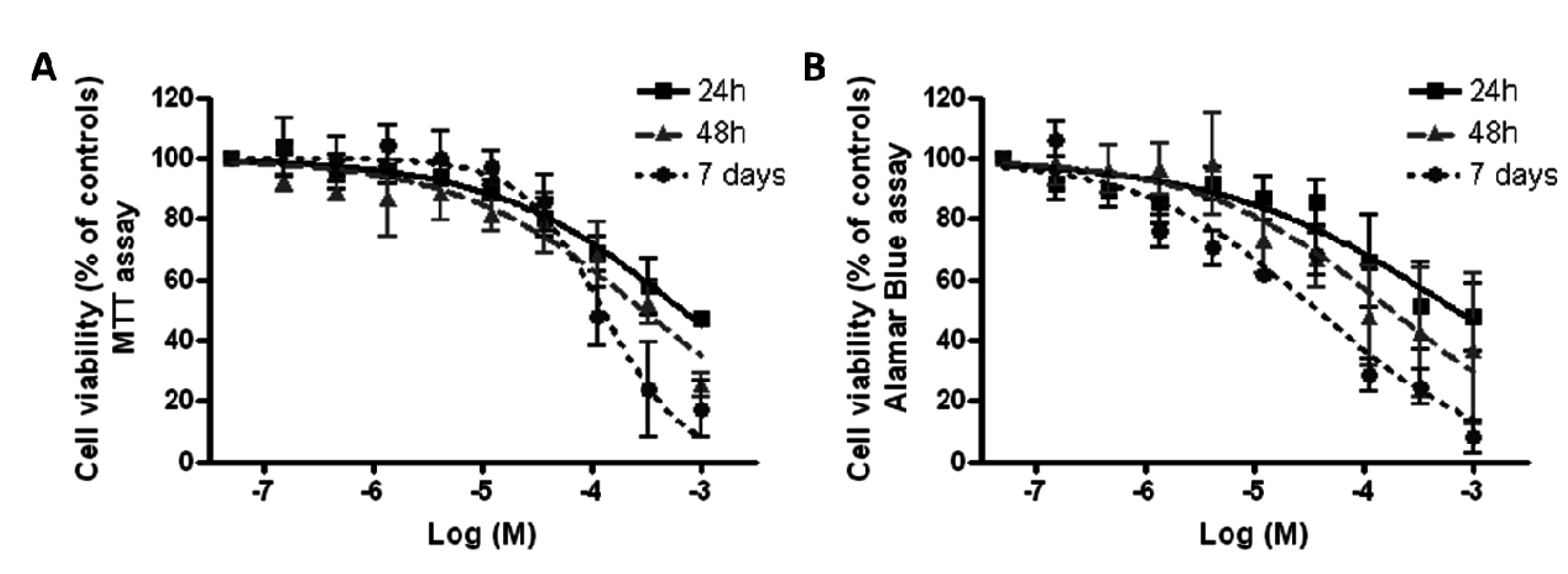
3.3. Effect of Co3O4NPs on Cell Viability
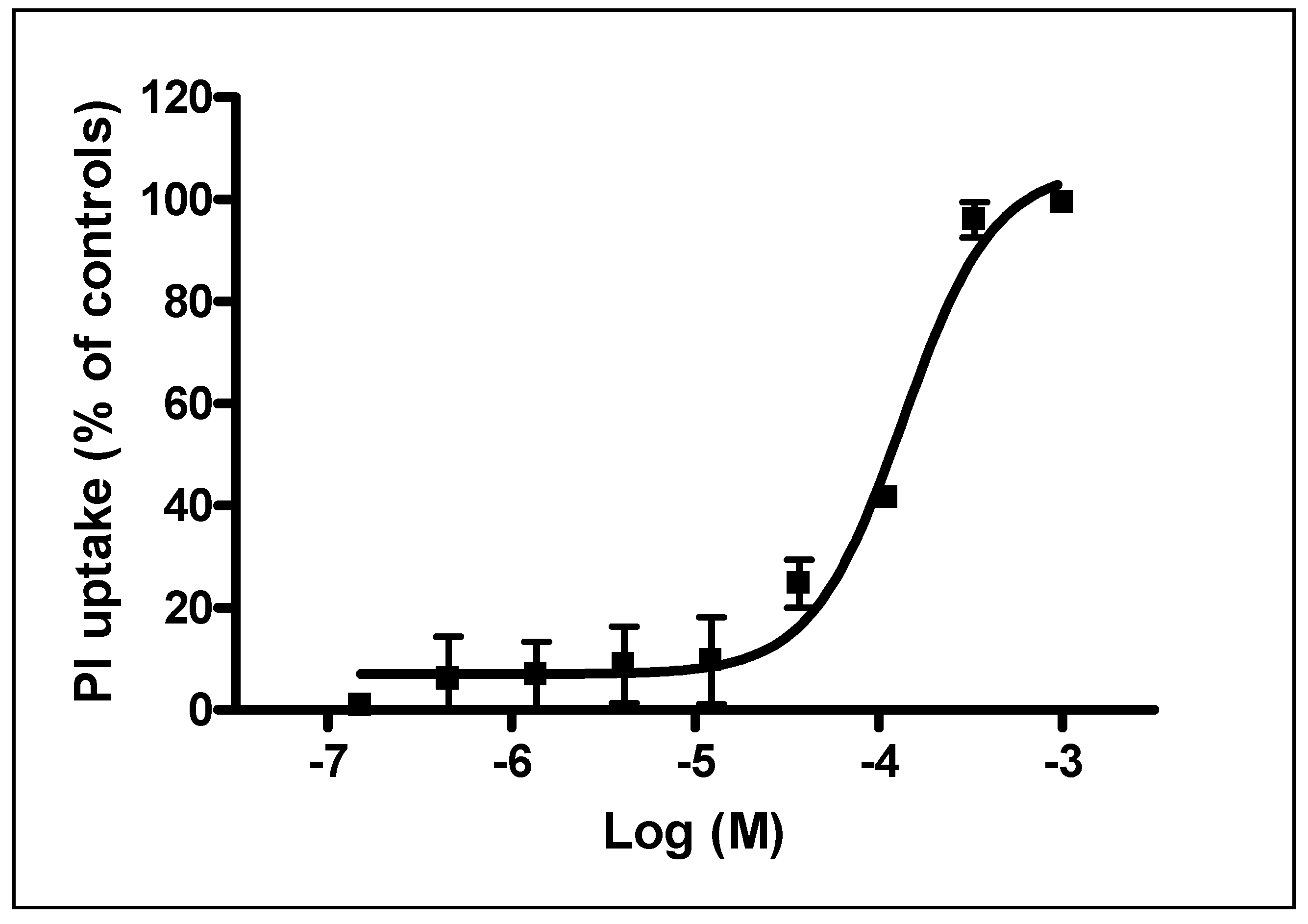
3.4. Effect of Co3O4NPs on Plasma Membrane Damage
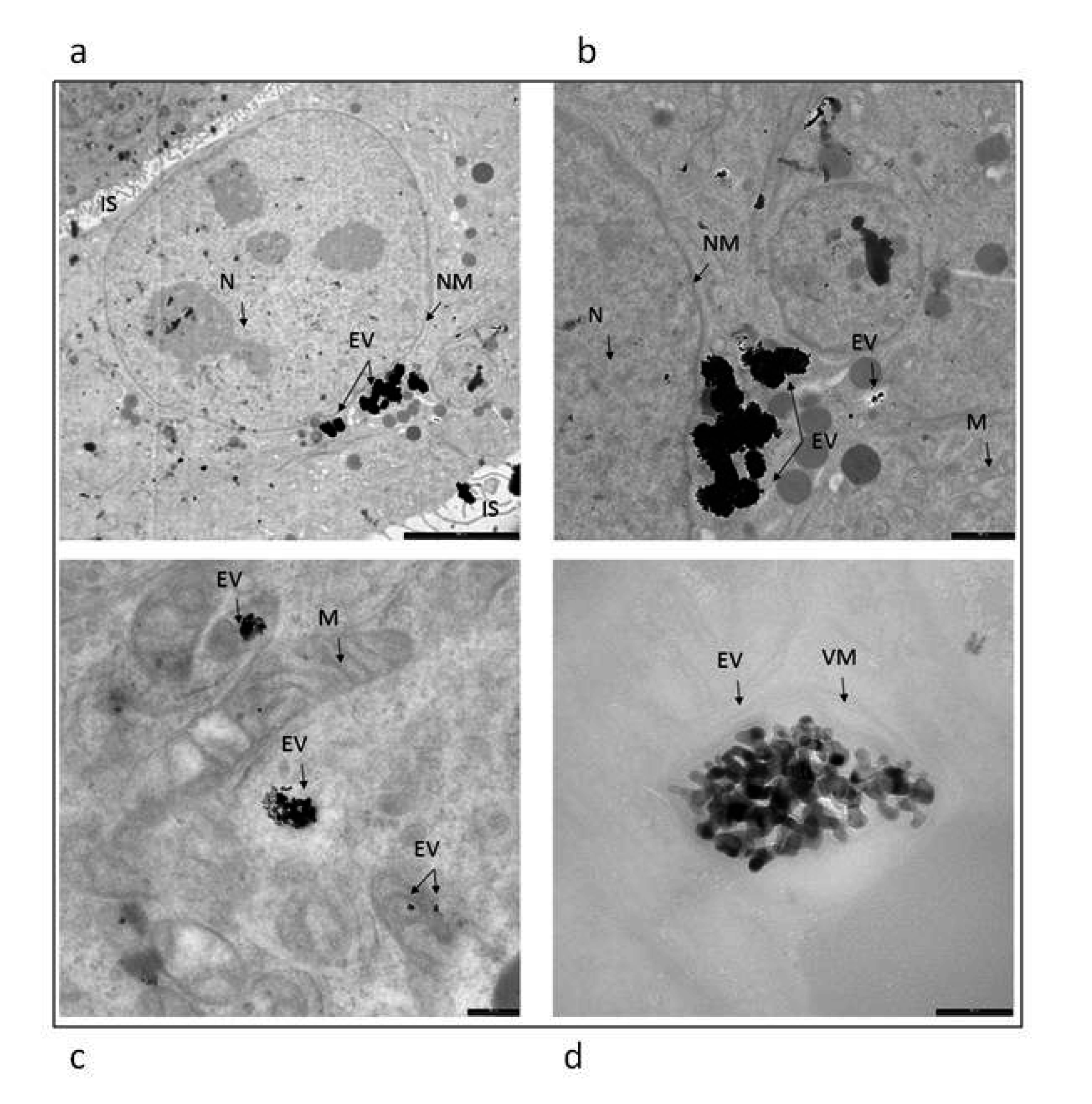
3.5. Evaluation of Cellular Internalization of NPs Using Electron Microscopy Imaging

4. Discussion
| Damaged Skin | Donor Suspension | Co3O4NPs (Peak 17 nm) 606 μg·cm−2 (445 μg·cm−2 as Cobalt) | CoNPs (Peak 80 nm) 1000 μg·cm−2 | Co3O4NPs Standardized Values 1000 μg·cm−2 | |||
| Mean | SD | Mean | SD | Mean | % | ||
| Membrane (μg·cm−2) | 4.78 | 0.90 | 12.0 | 3.8 | 10.75 | 89.6% | |
| Receiving Solution (ng·cm−2) | 47 | 41 | 1870 * | 860 | 106 | 5.6% | |
| Flux (ng·cm−2·h−1) | 1.7 | 2.0 | 76 * | 49.3 | 3.82 | 5.0% | |
5. Conclusions
Acknowledgements
Author Contributions
Conflicts of Interest
References
- Chen, H.C.; Qiu, J.T.; Yang, F.L.; Liu, Y.C.; Chen, M.C.; Tsai, R.Y.; Yang, H.W.; Lin, C.Y.; Lin, C.C.; Wu, T.S.; et al. Magnetic-composite-modified polycrystalline silicon nanowire field-effect transistor for vascular endothelial growth factor detection and cancer diagnosis. Anal. Chem. 2014, 86, 9443–9450. [Google Scholar] [CrossRef] [PubMed]
- Radović, M.; Calatayud, M.P.; Goya, G.F.; Ibarra, M.R.; Antić, B.; Spasojević, V.; Nikolić, N.; Janković, D.; Mirković, M.; Vranješ-Đurić, S. Preparation and in vivo evaluation of multifunctional 90Y-labeled magnetic nanoparticles designed for cancer therapy. J. Biomed. Mater. Res. A. 2015, 103, 126–134. [Google Scholar] [CrossRef] [PubMed]
- Da Silva, E.P.; Sitta, D.L.; Fragal, V.H.; Cellet, T.S.; Mauricio, M.R.; Garcia, F.P.; Nakamura, C.V.; Guilherme, M.R.; Rubira, A.F.; Kunita, M.H. Covalent TiO(2)/pectin microspheres with Fe(3)O(4) nanoparticles for magnetic field-modulated drug delivery. Int. J. Biol. Macromol. 2014, 67, 43–52. [Google Scholar] [CrossRef] [PubMed]
- Shi, R.; Chen, G.; Ma, W.; Zhang, D.; Qiu, G.; Liu, X. Shape-controlled synthesis and characterization of cobalt oxides hollow spheres and octahedra. Dalton Trans. 2012, 41, 5981–5987. [Google Scholar] [CrossRef] [PubMed]
- Wei-Yang, L.; Li-Na, X.; Jun, C. Co3O4. Nanomaterials in Lithium-Ion Batteries and Gas Sensors. Adv. Funct. Mater. 2005, 15, 851–856. [Google Scholar]
- Ren-Jang, W.; Cheng-Hung, H.; Chuin-Tih, Y.; Pi-Guey, S. Nanogold on powdered cobalt oxide for carbon monoxide sensor. Sensor. Actuat. B-Chem. 2003, 96, 596–601. [Google Scholar]
- Rahman, M.M.; Khan, S.B.; Faisal, M.; Rub, M.A.; Al-Youbi, A.O.; Asiri, A.M. Electrochemical determination of olmesartan medoxomil using hydrothermally prepared nanoparticles composed SnO2-Co3O4 nanocubes in tablet dosage forms. Talanta 2012, 15, 924–931. [Google Scholar] [CrossRef] [PubMed]
- Lou, X.W.; Deng, D.; Lee, J.Y.; Feng, J.; Archer, L.A. Self-supported formation of needlelike Co3O4 nanotubes and their application as lithium-ion battery electrodes. Adv. Mater. 2008, 20, 258–262. [Google Scholar] [CrossRef]
- Shu-Lei, C.; Jia-Zhao, W.; Hua-Kun, L.; Shi-Xue, D. Electrochemical deposition of porous Co3O4 nanostructured thin film for lithium-ion battery. J. Power Sources 2008, 182, 359–364. [Google Scholar]
- Makhlouf, S.A. Magnetic properties of Co3O4 nanoparticles. J. Magn. Magn. Mater. 2002, 246, 184–190. [Google Scholar] [CrossRef]
- Ando, M.; Kadono, K.; Kamada, K.; Ohta, K. Third-order nonlinear optical responses of nanoparticulate Co3O4 films. Thin Solid Films 2004, 446, 271–276. [Google Scholar] [CrossRef]
- Karimi, Z.; Karimi, L.; Shokrollahi, H. Nano-magnetic particles used in biomedicine: Core and coating materials. Mater. Sci. Eng. C Mater. 2013, 33, 2465–2475. [Google Scholar] [CrossRef] [PubMed]
- Papis, E.; Rossi, F.; Raspanti, M.; Dalle-Donne, I.; Colombo, G.; Milzani, A.; Bernardini, G.; Gornati, R. Engineered cobalt oxide nanoparticles readily enter cells. Toxicol. Lett. 2009, 189, 253–259. [Google Scholar] [CrossRef] [PubMed]
- Cho, W.S.; Dart, K.; Nowakowska, D.J.; Zheng, X.; Donaldson, K.; Howie, S.E. Adjuvanticity and toxicity of cobalt oxide nanoparticles as an alternative vaccine adjuvant. Nanomedicine 2012, 7, 1495–1505. [Google Scholar] [CrossRef] [PubMed]
- Alarifi, S.; Ali, D.; Suliman Y, A.O.; Ahamed, M.; Siddiqui, M.A.; Al-Khedhairy, A.A. Oxidative stress contributes to cobalt oxide nanoparticles-induced cytotoxicity and DNA damage in human hepatocarcinoma cells. Int. J. Nanomed. 2013, 8, 189–199. [Google Scholar]
- Rui, F.; Bovenzi, M.; Prodi, A.; Belloni Fortina, A.; Romano, I.; Corradin, M.T.; Larese Filon, F. Nickel, chromium and cobalt sensitization in a patch test population in north-eastern Italy (1996–2010). Contact Dermatitis 2013, 68, 23–31. [Google Scholar] [CrossRef] [PubMed]
- Larese Filon, F.; Crosera, M.; Timeus, E.; Adami, G.; Bovenzi, M.; Ponti, J.; Maina, G. Human skin penetration of cobalt nanoparticles through intact and damaged skin. Toxicol. In Vitro. 2013, 27, 121–127. [Google Scholar] [CrossRef] [PubMed]
- Williams, F.M.; Cage, S.; Carmichael, P.; Corish, J.; Dick, I.; Fitzpatrick, D.; Golden, D.; Jakasa, I.; Kenyon, S.; Kezic, S.; et al. Evaluations and predictions of dermal absorption of toxic chemicals. In Proceedings of Occupational and Environmental Exposures of Skin to Chemicals, Stockholm, Švedska, 12–15 June 2005.
- Larese Filon, F.; D’Agostin, F.; Crosera, M.; Adami, G.; Renzi, N.; Bovenzi, M.; Maina, G. Human skin penetration of silver nanoparticles through intact and damaged skin. Toxicology 2009, 255, 33–37. [Google Scholar] [CrossRef] [PubMed]
- Larese Filon, F.; Crosera, M.; Adami, G.; Bovenzi, M.; Rossi, F.; Maina, G. Human skin penetration of gold nanoparticles through intact and damaged skin. Nanotoxicology 2011, 5, 493–501. [Google Scholar] [CrossRef] [PubMed]
- Bronaugh, R.; Steward, R. Methods for in vitro percutaneous absorption studies V: Permeation through damaged skin. J. Pharm Sci. 1985, 15, 1062–1066. [Google Scholar] [CrossRef]
- Fasano, W.; Manning, L.; Green, J. Rapid assessment of rat and human epidermal membranes for in vitro dermal regulatory testing: Correlation of electrical resistance with tritiated water permeability. Toxicol. In Vitro 2002, 16, 731–740. [Google Scholar] [CrossRef]
- Davies, D.J.; Ward, R.J.; Heylings, J.R. Multi-species assessment of electrical resistance as a skin integrity marker for in vivo percutaneous absorption studies. Toxicol. In Vitro 2004, 18, 351–358. [Google Scholar] [CrossRef] [PubMed]
- Franz, T.J. On the relevance of in vitro data. J. Invest. Dermatol. 1975, 93, 633–640. [Google Scholar]
- Boukamp, P.; Petrussevska, R.T.; Breitkreutz, D.; Hornung, J.; Markham, A.; Fusenig, N.E. Normal keratinization in a spontaneously immortalized aneuploid human keratinocyte cell line. J. Cell Biol. 1988, 106, 761–771. [Google Scholar] [CrossRef] [PubMed]
- Mosmann, T. Rapid colorimetric assay for cellular growth and survival: Application to proliferation and cytotoxicity assays. J. Immunol. Methods 1983, 65, 55–63. [Google Scholar] [CrossRef]
- Pelin, M.; Sosa, S.; Della Loggia, R.; Poli, M.; Tubaro, A.; Decorti, G.; Florio, C. The cytotoxic effect of palytoxin on Caco-2 cells hinders their use for in vitro absorption studies. Food Chem. Toxicol. 2012, 50, 206–211. [Google Scholar] [CrossRef] [PubMed]
- Pelin, M.; Sosa, S.; Pacor, S.; Tubaro, A.; Florio, C. The marine toxin palytoxin induces necrotic death in HaCaT cells through a rapid mitochondrial damage. Toxicol. Lett. 2014, 229, 440–450. [Google Scholar] [CrossRef] [PubMed]
- Alinovi, R.; Goldoni, M.; Pinelli, S.; Campanini, M.; Aliatis, I.; Bersani, D.; Lottici, P.P.; Iavicoli, S.; Petyx, M.; Mozzoni, P.; Mutti, A. Oxidative and pro-inflammatory effects of cobalt and titanium oxide nanoparticles on aortic and venous endothelial cells. Toxicol. In Vitro. 2015, 29, 426–437. [Google Scholar] [CrossRef] [PubMed]
- Chu, M.; Wu, Q.; Wang, J.; Hou, S.; Miao, Y.; Peng, J.; Sun, Y. In vitro and in vivo transdermal delivery capacity of quantum dots through mouse skin. Nanotechnology 2007, 18. [Google Scholar] [CrossRef]
- Crosera, M.; Bovenzi, M.; Maina, G.; Adami, G.; Zanette, C.; Florio, C.; Filon Larese, F. Nanoparticle dermal absorption and toxicity: A review of the literature. Int. Arch. Occup. Environ. Health 2009, 82, 1043–1055. [Google Scholar] [CrossRef] [PubMed]
- Ortega, R.; Bresson, C.; Darolles, C.; Gautier, C.; Roudeau, S.; Perrin, L.; Janin, M.; Floriani, M.; Aloin, V.; Carmona, A.; Malard, V. Low-solubility particles and a Trojan-horse type mechanism of toxicity: The case of cobalt oxide on human lung cells. Part. Fibre Toxicol. 2014, 11. [Google Scholar] [CrossRef] [PubMed]
- Barceloux, D.G.; Barceloux, D. Cobalt. J. Toxicol-Clin. Toxic. 1999, 37, 201–206. [Google Scholar] [CrossRef]
- Collier, C.G.; Pearce, M.J.; Hodgson, A.; Ball, A. Factors affecting the in vitro dissolution of cobalt oxide. Environ. Health Persp. 1992, 97, 109–113. [Google Scholar] [CrossRef]
- Chattopadhyay, S.; Dash, S.K.; Tripathy, S.; Das, B.; Mandal, D.; Pramanik, P.; Roy, S. Toxicity of cobalt oxide nanoparticles to normal cells; an in vitro and in vivo study. Chem-Biol. Interact. 2015, 226, 58–71. [Google Scholar] [CrossRef] [PubMed]
- Limbach, L.K.; Wick, P.; Manser, P.; Grass, R.N.; Bruinink, A.; Stark, W.J. Exposure of engineered nanoparticles to human lung epithelial cells: Influence of chemical composition and catalytic activity on oxidative stress. Environ. Sci. Technol. 2007, 41, 4158–4163. [Google Scholar] [CrossRef] [PubMed]
- Lundborg, M.; Falk, R.; Johansson, A.; Kreyling, W.; Camner, P. Phagolysosomal pH and dissolution of cobalt oxide particles by alveolar macrophages. Environ. Health Persp. 1992, 97, 153–157. [Google Scholar] [CrossRef]
- Spigoni, V.; Cito, M.; Alinovi, R.; Pinelli, S.; Passeri, G.; Zavaroni, I.; Goldoni, M.; Campanini, M.; Aliatis, I.; Mutti, A.; Bonadonna, R.C.; Dei Cas, A. Effects of TiO2 and Co3O4 Nanoparticles on Circulating Angiogenic Cells. PLoS ONE 2015, 10. [Google Scholar] [CrossRef] [PubMed]
- Sabbioni, E.; Fortaner, S.; Farina, M.; Del Torchio, R.; Petrarca, C.; Bernardini, G.; Mariani-Costantini, R.; Perconti, S.; Di Giampaolo, L.; Gornati, R.; Di Gioacchino, M. Interaction with culture medium components, cellular uptake and intracellular distribution of cobalt nanoparticles, microparticles and ions in Balb/3 T3 mouse fibroblasto. Nanotoxicology 2014, 8, 88–99. [Google Scholar] [PubMed]
- Bauer, A.; Schmitt, J.; Bennett, C.; Coenraads, P.J.; Elsner, P.; English, J.; Williams, H.C. Interventions for preventing occupational irritant hand dermatitis. Cochrane DB. Syst. Rev. 2010, 16. [Google Scholar] [CrossRef] [Green Version]
- Gibbs, S. In vitro irritation models and immune reactions. Skin Pharmacol. Phys. 2009, 22, 103–113. [Google Scholar] [CrossRef] [PubMed]
- Rampersad, S.N. Multiple applications of Alamar Blue as an indicator of metabolic function and cellular health in cell viability bioassays. Sensors 2012, 12, 12347–12360. [Google Scholar] [CrossRef] [PubMed]
- Gonzalez, R.J.; Tarloff, J.B. Evaluation of hepatic subcellular fractions for Alamar blue and MTT reductase activity. Toxicol. In Vitro. 2001, 15, 257–259. [Google Scholar] [CrossRef]
© 2015 by the authors; licensee MDPI, Basel, Switzerland. This article is an open access article distributed under the terms and conditions of the Creative Commons Attribution license (http://creativecommons.org/licenses/by/4.0/).
Share and Cite
Mauro, M.; Crosera, M.; Pelin, M.; Florio, C.; Bellomo, F.; Adami, G.; Apostoli, P.; De Palma, G.; Bovenzi, M.; Campanini, M.; et al. Cobalt Oxide Nanoparticles: Behavior towards Intact and Impaired Human Skin and Keratinocytes Toxicity. Int. J. Environ. Res. Public Health 2015, 12, 8263-8280. https://doi.org/10.3390/ijerph120708263
Mauro M, Crosera M, Pelin M, Florio C, Bellomo F, Adami G, Apostoli P, De Palma G, Bovenzi M, Campanini M, et al. Cobalt Oxide Nanoparticles: Behavior towards Intact and Impaired Human Skin and Keratinocytes Toxicity. International Journal of Environmental Research and Public Health. 2015; 12(7):8263-8280. https://doi.org/10.3390/ijerph120708263
Chicago/Turabian StyleMauro, Marcella, Matteo Crosera, Marco Pelin, Chiara Florio, Francesca Bellomo, Gianpiero Adami, Piero Apostoli, Giuseppe De Palma, Massimo Bovenzi, Marco Campanini, and et al. 2015. "Cobalt Oxide Nanoparticles: Behavior towards Intact and Impaired Human Skin and Keratinocytes Toxicity" International Journal of Environmental Research and Public Health 12, no. 7: 8263-8280. https://doi.org/10.3390/ijerph120708263