Article

First Evidence of Palytoxin and 42-Hydroxy-palytoxin in the Marine Cyanobacterium Trichodesmium

1
Toulouse University, UPS, UMR152 UPS-IRD (PHARMA-DEV), 118, route de Narbonne, F-31062 Toulouse cedex 9, France
2
Research Institute for the Development (IRD), UMR152, 98848 Noumea, New Caledonia
3
Laboratory of Phycotoxins, IFREMER, Rue de l’Ile d’Yeu, BP21105, F-44311 Nantes cedex 3, France
4
Laboratory of toxic micro-algae (LMT), Louis Malarde Institute (ILM), BP30, 98713 Papeete, Tahiti, French Polynesia
5
Biological Science Center, Boston University, 5 Cummington Street, Boston, MA 02215, USA
6
Research Institute for the Development (IRD), UMR152, 98713 Papeete, Tahiti, French Polynesia
*
Author to whom correspondence should be addressed.
Mar. Drugs 2011, 9(4), 543-560; https://doi.org/10.3390/md9040543
Submission received: 15 February 2011 / Revised: 23 March 2011 / Accepted: 25 March 2011 / Published: 31 March 2011

Abstract

:
Marine pelagic diazotrophic cyanobacteria of the genus Trichodesmium (Oscillatoriales) are widespread throughout the tropics and subtropics, and are particularly common in the waters of New Caledonia. Blooms of Trichodesmium are suspected to be a potential source of toxins in the ciguatera food chain and were previously reported to contain several types of paralyzing toxins. The toxicity of water-soluble extracts of Trichodesmium spp. were analyzed by mouse bioassay and Neuroblastoma assay and their toxic compounds characterized using liquid chromatography coupled with tandem mass spectrometry techniques. Here, we report the first identification of palytoxin and one of its derivatives, 42-hydroxy-palytoxin, in field samples of Trichodesmium collected in the New Caledonian lagoon. The possible role played by Trichodesmium blooms in the development of clupeotoxism, this human intoxication following the ingestion of plankton-eating fish and classically associated with Ostreopsis blooms, is also discussed.

Graphical Abstract

1. Introduction

Trichodesmium spp. are marine pelagic cyanobacteria belonging to the order Oscillatoriales. These filamentous, non-heterocystous cyanobacteria are known for their ability to fix atmospheric dinitrogen [13]. They are characterized by trichomes (linear arrangements of about 100–200 cells) that form colonies and occur in extensive floating blooms also called “sea sawdust” by the sailors. Trichodesmium blooms are widely distributed in oligotrophic regions of the oceans throughout the tropics and subtropics [1,4,5].
Despite a number of surveys dedicated to the ecological aspects of Trichodesmium spp. and their importance for the coral reef ecosystems [1,3,6,7], their toxicity remain sparsely documented [3,8]. The stochastic nature of the blooms (and the difficulties inherent in establishing laboratory cultures) has greatly hampered toxicological studies [9,10]. In 1991, Hawser [11] reported the death of oysters following Trichodesmium blooms. The toxicity of these cyanobacteria was tested on various species of zooplankton and mortality of certain crustaceans (brine shrimp and two species of copepods) was demonstrated. However, grazers (Macrosetella gracilis and Miracia efferata) that are known to feed on Trichodesmium were not affected. No information was provided on the nature of the toxins involved [1113]. Based on chemical analysis studies, Hahn and Capra [14] were the first to hypothesize that Trichodesmium erythraeum could be a potential source of toxin in ciguatera, a typical foodborne intoxication in the tropics due to the ingestion of fish contaminated with ciguatoxins (CTXs) [1517]. The compounds extracted from T. erythraeum and from samples of molluscs, collected during, and shortly after, these Trichodesmium blooms, were positive for CTXs-like toxins [14]. In 1993, Endean et al. [18] demonstrated that the toxin profiles of the lipid- and water-soluble extracts from T. erythraeum were similar to those of corresponding fractions extracted from the flesh extracts of the pelagic carnivore Scomberomorus commerson, a fish often implicated in ciguatera. Chromatographic elutions of water-soluble and lipid-soluble fractions from both Trichodesmium and Scomberomorus samples further showed the presence of an alkaloid, in addition to a peptide and CTXs-like compounds. These observations, as well as recent studies by Kerbrat et al. [19], conducted mainly on T. erythraeum blooms in the New Caledonia lagoon, tend to indicate that Trichodesmium spp. could be the source of some of the toxins carried by ciguateric fish, and may contribute to the ciguatera syndrome. Recently, Ramos et al. [20] detected the presence of microcystin-LR in T. erythraeum by chromatographic analysis. Moreover, Proença et al. [21] analyzed the contents of analogues of microcystin, cylindrospermopsin and saxitoxin in Trichodesmium blooms off the Brazilian coasts. Saxitoxin analogues and microcystins were present at low concentrations in all samples, but the authors concluded that these toxins do not represent a potential harm to human health by primary contact. The only reported harmful effect of Trichodesmium to humans refers to the “Tamandare fever” on the coast of Tamandare, Brazil [22]. The possible involvement of marine benthic cyanobacteria in ciguatera outbreaks in New Caledonia has recently been documented by Laurent et al. [23]. The incriminated cyanobacterium Hydrocoleum lyngbyaceum was found phylogenetically very close to the species of Trichodesmium [24,25]. The presence of homoanatoxin-a, a derivative of anatoxin-a, in mats of H. lyngbyaceum, as well as in giant clams (Tridacna spp.) collected in the surroundings of contaminated area, has been recently reported by Méjean et al. [26]. Both these neurotoxins are well known in freshwater cyanobacteria involved in dog poisonings in France, New Zealand and Scotland [27,28].
Originally, palytoxin (PLTX) (Figure 1) and 42-OH-palytoxin (42-OH-PLTX) were isolated from the zoanthid anemone Palythoa sp. [29,30]. PLTX and analogues like ovatoxins, ostreocins, ostreotoxins, mascarenotoxins constitute the family of PLTXs [31]. PLTXs were evidenced in marine organisms ranging from dinoflagellates (Ostreopsis) to fishes [3235] but, to our knowledge, never in cyanobacteria. PLTX is one of the largest nonpolymeric natural molecules with a molecular weight of 2680 Da and one of the most potent non-protein compounds known to date, exhibiting high toxicity in mammals with intravenous LD50 ranging between 10 and 100 ng/kg [3133]. One of the main actions of PLTX is to bind to the Na, K-ATPase, converting the pump into an ion channel and causing a K+ efflux, an Na+ influx and membrane depolarization [32]. The osmotic imbalance that results from this flux of ions can be compared to CTX mechanisms and could explain why PLTX has often been implicated in ciguatera [36,37].
PLTX is also likely to play a role in clupeotoxism, a marine poisoning resulting from the ingestion of plankton-eating fish such as herrings and sardines (Clupeidae), anchovies (Engaulidae) or mullets (Mugillidae) in tropical regions [37,39]. This has been postulated after the detection of PLTX and analogues in the remains of fish instigating serious human intoxications [36,40,41]. Symptoms appear abruptly: metallic taste, digestive disorders, generalized paralysis, tachycardia, convulsions and acute respiratory distress. Their variety and intensity depend upon the route of exposure which occurred through the consumption of PLTX-contaminated organisms and through dermal, ocular and inhalation routes [42]. Although rare, this poisoning can be fatal [37]. Clupeotoxism is classically associated with blooms of the benthic dinoflagellate Ostreopsis, most notably with the species O. siamensis and O. mascarenensis known as sources of PLTX [43,44] whereas two other species, O. lenticularis and O. ovata, are potentially progenitors of PLTX analogues: ostreotoxins and ovatoxin-a, respectively [45].
Detection and quantification of PLTX in biological samples can be conducted by biological and analytical means, but there is currently no officially approved method [38,46]. Here, we used two biological methods (mouse bioassay and Neuroblastoma cell-based assay) and one analytical method (LC-MS/MS) to detect this toxin in marine cyanobacteria.
The present contribution provides the first evidence of the production of PLTX and one of its analogues, 42-OH-PLTX by Trichodesmium in tropical and subtropical waters. The potential role played by Trichodesmium blooms in clupeotoxicity, via the ingestion of the trichomes of this pelagic cyanobacterium by plankton-eating fish is also discussed.

2. Materials and Methods

2.1. Materials

All reagents and chemicals were obtained from Sigma-Aldrich (St. Louis, MO, USA) unless otherwise stated. Solvents used for extraction and purification were of analytical grade and were purchased from Prolabo (Paris, France). For chromatographic techniques, methanol and acetonitrile were HPLC grade, obtained from J.T. Baker (Deventer, The Netherlands). Water was deionised and purified to 18.2 MΩ quality through a MilliQ water purification system (Purelab Elga, UK). Standard solution of PLTX was purchased from Wako chemicals GmbH (Neuss, Germany). In addition to PLTX, this solution contains traces of ovatoxin-a and 42-OH-PLTX.

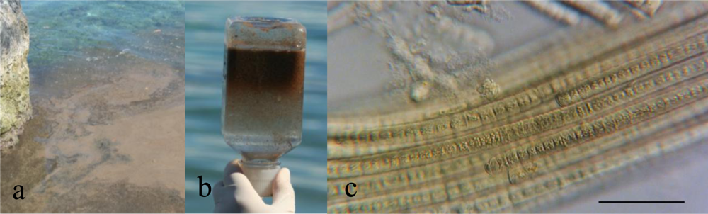
2.2. Sampling Sites and Collection of Cyanobacteria

Trichodesmium sampling took place in the southern lagoon of New Caledonia, primarily during the southern summer (September to March), as soon as the blooms were reported (Table 1). Massive blooms were subject to drifts by wind and concentrated especially in bays (Figure 2a). Samples were collected on the surface and sub-surface (0–1 m). Two sampling techniques were used, depending on trichome concentrations: manually with a 35 μm phytoplankton net or with a gentle suction using a vacuum pump. All samples were handled delicately to avoid cell lysis leading to toxin release. The buoyancy of cyanobacteria conferred by intracellular gas vesicles separates them from debris and other organisms (Figure 2b). The trichomes were separated from remaining macroalgae, phanerogams and debris and further concentrated by reversing the sampling bottles. Concentrated samples were frozen and kept freeze-dried until extracted and tested for their toxicity. Subsamples from each batch were fixed in 5% formaldehyde solution in Millipore®-filtered sea-water (0.45 μm) for morphological identification purposes.

2.3. Taxonomic Identification of Cyanobacterial Samples

Samples collected from various occurrences of Trichodesmium blooms and different locations are presented in Table 1. T. erythraeum is known to be prevalent in the New Caledonian lagoon [7]. T. erythraeum forms typically spindle-shaped colonies composed of trichomes oriented in parallel (Figure 2c) [47]. Cells are shorter (5.4–11 μm) than wide (7–11 μm). The end cells are often capped by a calyptra. Although associated with a variety of organisms, including hydrozoans, copepods, diatoms, dinoflagellates, fungi and other protists and bacteria, Trichodesmium species are usually the major component of the blooms.

2.4. Extraction

Freeze dried pellets of cyanobacterial samples were extracted using solvent partition. Briefly, pellets (≈100 g) were extracted three times with methanol (1 L) with each time one hour of ultra-sonication and by agitation overnight. This extract was subsequently filtered and dried under vacuum, and the residue was partitioned between 60% aqueous methanol (500 mL) and diethyl ether (250 mL). The water-soluble fraction was saved and dried under vacuum. Data concerning the respective extraction yields are summarized in Table 1.

2.5. Mouse Bioassay

The mouse bioassays (MBA) were based on careful observation of the symptoms displayed by the animals following injection of toxic extracts. MBA were performed on aqueous methanolic extracts using 20 g ± 2 g mice (OF1, Iffa-Credo, L’Arbresle, France) of either sex. All tested animals were treated under conditions, which meet the ethical standards defined by the European Community Council Directive of November 24, 1986 (86/609/EEC). The animals were allowed food and water ad libitum.
The dried extracts were dissolved in 300 μL of phosphate buffer saline (PBS, pH 7.2) containing 0.1% Tween 80, prior to administration via intraperitoneal (i.p.) injection. The tested doses varied from 0.5 to 5.0 mg of extract/g of mouse body weight (n ≥ 2; 3 different concentrations depending on the extract). In total, six animals were used per extract. Control animals received 300 μL of vehicle (n = 2). Animal behavioral changes were observed over a period of approximately 48 h.

2.6. Neuroblastoma Cell-Based Assays (CBA)

The Neuroblastoma cell-based assays (CBA) was performed to quantify the cytotoxic effect of water-soluble extract following the method previously described by Ledreux et al. [48] with some modifications described below. The Neuroblastoma cells (Neuro-2a) were obtained from the American Type Culture Collection (ATCC CCL 131). Neuro-2a cells were maintained in RPMI-1640 medium supplemented with 1 mM sodium pyruvate, 2.5 μg/mL amphotericin B, 50 units/mL penicillin G, 50 μg/mL streptomycin sulfate, 1% glutaMAX™-I, and 10% FBS (fetal bovine serum), at 37 °C in a humidified 5% CO2 atmosphere. Briefly, Neuro-2a were harvested with a trypsin-EDTA solution and 50,000 cells in a 5% FBS RPMI-1640 supplemented medium were seeded into each well of a 96-well microtiter plate, and incubated for 24 h at 37 °C.

2.6.1. Evaluation of Cytotoxic Effects of PLTX

The incubation step in a 96-well microtiter plate was followed by a pre-treatment for 2 h with a control solution or ouabain solution at 100, 250 or 500 μM. Different pre-treatment times (0, 1 and 2 h) with ouabain 500 μM were also tested. The final PLTX concentrations ranged from 1.8 × 10⁻¹⁵ to 1.8 × 10⁻⁸ M. At least 3 replicates per dilution were tested and for each microplate, 6 wells were processed as untreated controls and 6 wells as ouabain control. After a 20–22 h incubation period at 37 °C, cell viability was assessed using the quantitative colorimetric 3-(4,5-dimethylthiazol-2-yl)-2,5-diphenyl tetrazolium bromide (MTT) assay, a method previously described by Mossman [49]. Medium was removed, and 60 μL of medium containing 0.83 mg/mL of MTT were added to each well. The plates were incubated for 1 h at 37 °C. Finally, the MTT was discarded and 100 μL dimethyl sulfoxide added to each well to dissolve the formazan. The absorbance was read at 570 nm on a plate reader (Imark microplate reader, BioRad). Compared to the absorbance of cells alone (100% of viability), the results were expressed as the percentage of viability. Data were fitted to a sigmoid curve with variable slope using GraphPad Prism version 4. EC50 values (concentration of toxin or extract that reduces by half the cell survival) were determined for samples that showed a sigmoidal curve. R2 values, not shown here, were found higher than 0.97.

2.6.2. Evaluation of Cytotoxic Effects of the Extracts

The incubation step was followed by a pre-treatment for 2 h with a control solution, or with an ouabain solution at a final concentration of 500 μM. Different dilutions of the toxic extracts were then added to each well, at a final concentration ranging from 178.5 to 4464 μg/mL followed by 20–22 h incubation and analyzed using MTT assay.

2.6.3. Evaluation of Cytotoxic Effects of Extracts Spiked with PLTX

To evaluate the effectiveness and the specificity of the CBA for PLTXs and the matrix effect on Neuroblastoma cells, a non-toxic extract was spiked on pure PLTX. The sample No. 8 (Tricho Lifou C02) was spiked to obtain a final concentration of 1.7 μg PLTX/g of extract corresponding to the concentration estimated in the most toxic extract (No. 1, Tricho 5îles) by chromatographic analyses. Different dilutions of the spiked extract were then added to each well: (i) at a final extract concentration ranging from 178.5 to 4,464 μg/mL, or (ii) at a final PLTX concentration ranging from 6.4 × 10⁻⁹ to 1.6 × 10⁻⁷ M with pre-treatment for 2 h with ouabain (500 μM), followed by MTT assay.

2.7. LC-MS/MS Analysis

Liquid Chromatography coupled with tandem mass spectrometry (LC-MS/MS) analyses was performed with aqueous methanol 80% extracts. An aliquot (300 μL) was filtered through a 0.2 μm Whatman® Vectaspin filter. Five microliters of the filtrate were injected for analyses by LC-MS/MS.
PLTXs analysis were carried out using an LC system (HP 1200, Agilent) coupled to a hybrid triple quadrupole/ion trap mass spectrometer (API-4000Qtrap, PE/SCIEX) equipped with a turbo spray® interface, according to modified Ciminiello method [45]. A C18 Gemini column (5 μm, 150 mm × 2.0 mm, Phenomenex) was employed at 20 °C and eluted at 200 μL/min. Eluent A was water and eluent B was 95% acetonitrile/water, both eluents containing 2 mM ammonium formate and 50 mM formic acid. The gradient of B was raised from 20 to 100% over 10 min and held for a further 4 min before returning to initial conditions. The instrument control, data processing and analysis were conducted using Analyst software.
Mass spectrometry detection was performed in positive mode and optimized from a PLTX standard solution using Selected Reaction Monitoring (SRM) (Table 2). The SRM experiments were established by using the following source settings: curtain gas set at 30, ion spray at 5500 V, a turbogas temperature of 450 °C, gas 1 and 2 set at 50 (arbitrary units) and an entrance potential of 10 V. The collision energy was applied at 45 eV for doubly charged ions [M + 2H]2+, [M + 2H − H2O]2+ and at 33 eV for triply charged ions [M + 3H]3+ to give the characteristic product ion at 327.

3. Results

3.1. MBA Toxicity Data

The five water-soluble extracts of Trichodesmium injected (No. 1, 4, 5, 6 and 8) were found toxic. Symptoms in mice included reduced activity and responsiveness, frequent convulsive spasms, respiratory difficulty and partial paralysis, which quickly advanced to total paralysis. No salivation or lacrimation was observed. The i.p. injection of 2.5 mg of water-soluble extracts/g of mouse body weight (corresponding to ca. 12 mg of freeze-dried pellets of cyanobacteria/g of mouse body weight) killed all mice within 5 min. Below this concentration, a complete recovery of mice was observed accompanied with a transient comatose phase lasting from 40 min to 2 h. The extracts No. 2, 3, and 7 were not injected in mice.

3.2. CBA Cytotoxicity Data

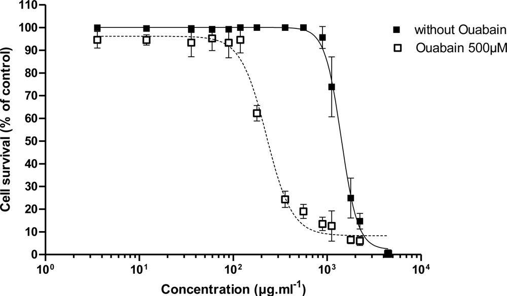
3.2.1. Effect of PLTX

EC50 values for the dose-response curves obtained for the direct effect of PLTX or after pre-incubating the Neuro-2a cells with 100, 250 or 500 μM ouabain (O) and for different pre-incubation times (0, 1 or 2 h) are presented in Table 3. The dose-response curves with different ouabain concentration are presented in Figure 3 while the dose-response curves with different pre-incubation time are presented in Figure 4. Following a pretreatment to ouabain (2 h), Neuro-2a cells were sensitized in a positive dose-dependent manner to the action of PLTX (Figure 3). The action of ouabain on the sensitivity of Neuro-2a to PLTX did not depend on its administration time (Figure 5). In all cases, ouabain sensitized the Neuro-2a cells.

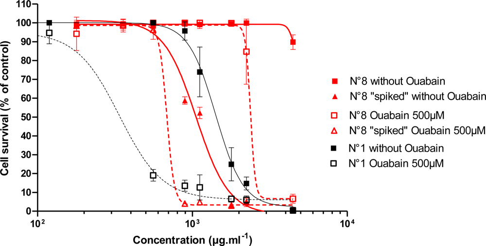
3.2.2. Effect of Trichodesmium Extracts

EC50 values for the dose-response curves obtained for the effect of Trichodesmium extracts alone or after pre-incubating the Neuro-2a cells for 2 h with 500 μM ouabain were presented in Table 2. The effect of Trichodesmium extract No. 1 from “5îles” on the viability of Neuro-2a cells, without or with pre-incubation with 500 μM ouabain for 2 h before adding extract is illustrated in Figure 5. As in the experiments with PLTX, the sensitivity of the Neuro-2a cells to the Trichodesmium extract No. 1 has increased with pre-incubation with 500 μM ouabain for 2 h (Figure 5).

3.2.3. Effect of Trichodesmium Non-Toxic Extracts Spiked with PLTX

When the non-cytotoxic extract (Tricho Lifou C02, No. 8) was spiked with a PLTX dose equivalent at 1.7 μg of PLTX/g of extract (PLTX content of the most toxic Trichodesmium extract No. 1, Tricho “5îles”), this atoxic extract became toxic with an EC50 of 1058 μg/mL as opposed to 1337 μg/mL for extract No. 1. With 2 h of pre-incubation, EC50 decreased from 2054 to 683 μg/mL as opposed to 114 μg/mL for non-spiked extract No. 1 (Figure 6).
Without pre-treatment with ouabain, the cytotoxicity of the extract No. 8 spiked with PLTX is slightly stronger than that of the natural toxic extract No. 1. With pre-incubation with ouabain, we observed the inverse phenomenon: a stronger cytotoxic potential of the non-spiked extract than that of the spiked extract.

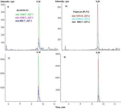
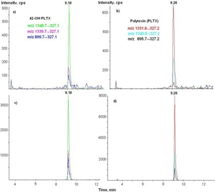
3.3. LC-MS/MS Analysis

Several toxins were submitted to LC-MS/MS analysis: PLTX, 42-OH-PLTX, ovatoxin-A (analogue of PLTX isolated from Ostreopsis cf. ovata), ostreocin-D (analogue of PLTX isolated from O. siamensis), mascarenotoxins A and B (analogue of PLTX isolated from O. mascarensis). Eight water extracts were tested: among all the toxins screened, only PLTX and 42-OH PLTX were detected in four samples of Trichodesmium (No. 1, 4, 5, 6) (Figure 7, Table 4). No toxins were detected in the samples No. 2, 3, 7, 8 (Limit of Detection = 0.01 μg of PLTX/g of extract). In the extract containing PLTX and 42-OH-PLTX, the concentration of these combined toxins varies from 1.08 to 1.70 μg/g of extract (0.28 to 1.10 μg/g eqv. of dried cyanobacteria) (Table 4).

4. Discussion

The first evidence of the production of PLTX and one of its analogues, 42-hydroxy-palytoxin (42-OH-PLTX), by Trichodesmium cyanobacteria in New Caledonian waters is demonstrated by CBA and LC-MS/MS.
In our experiments, Neuro-2a cells seemed to be more sensitive to PLTX standards and to both cytotoxic and atoxic extracts after a pre-treatment with ouabain, whereas according to Ledreux et al. [48], the presence of ouabain, in counteracting the effects of PLTX on Na+/K+ ATPase, should inhibit partially the cytotoxicity of PLTX or PLTX-contaminated extracts. This PLTX dose-dependent decrease in viability was also specifically inhibited by ouabain in the case of BE (2)-M17 Neuroblastoma cells [50]. Ouabain also showed inhibition of the lysis of sheep erythrocytes by PLTX [51].
Studying the suitability of the Neuro-2a cell line for the detection of PLTX and analogs, Ledreux et al. [48] found an EC50 value of 42.9 pM for a direct cytolitic effect of PLTX, and an EC50 value of 290.7 pM for a specific effect of PLTX when ouabain was used as a competitor and pre-added 2 h before, at the concentration of 500 μM. For our part, we found an EC50 of 170 ± 60 pM for direct effect of PLTX and 6.0 ± 2.2 pM for the specific effect, respectively, in the same conditions, i.e., ouabain concentration and pre-incubation time (Table 3). The CBA method was largely based on Ledreux’s method, with the exception that after 24 h incubation time, the medium was not removed and replaced by MWS (Medium without Serum) for economic reasons, and that the final working volume was 112 μL. We also tried Ledreux’s method entirely (data not shown), and we made the same observations as previously found, that Neuro-2a with 2 h ouabain 500 μM pre-treatment were more sensitive to PLTX than cells without pre-treatment.
To explore the observed difference in the specific effect of PLTX after pre-incubation of Neuro-2a cells with ouabain, compared to literature, different sets of experiments were performed with respect to ouabain concentration and pre-incubation time (Table 3). As a result, the highest sensitivity of the Neuro-2a cells was observed when ouabain was added prior to PLTX. This sensitivity was dose-dependent (from 100 to 500 μM ouabain) (Figure 3) and was not dependent of pre-incubation time (Figure 4).
Cañete and Diogene [52] obtained in 24 h growth and 24 h exposure conditions, a dose-response curve with an EC50 of 100 pM without ouabain/veratridine (O/V) and 40 pM with O/V added simultaneously with the toxins. Ouabain and veratridine treatment affects the ionic cell equilibrium; therefore, cells treated with O/V were more sensitive to PLTX than the untreated cells. The synergistic effect of PLTX and ouabain was observed (EC50 = 6.3 ± 4.7 pM) when these compounds were both added simultaneously to the Neuro-2a cells [48]. Our results were very similar with an EC50 of 5.08 pM or 6.37 with a post-treatment of 500 μM ouabain, respectively, showing that our ouabain sample is active (Table 3).
The mouse bioassay revealed a global toxicity of the five tested extracts with paralyzing effect. No difference was noted in the activity and toxic potency of T. erythraeum blooms collected from different locations.
However, the bioassay with Neuroblastoma cells seems to show the potential cytotoxicity of extracts even if the results do not fully correlate with the presence or the absence of PLTX detected by LC-MS/MS. Only one of the eight extracts, the bloom collected in Nouméa (No. 7), was found cytotoxic while PLTX was not detected by LC-MS/MS. It is possible that the toxicity in mice and the cytotoxicity observed on Neuro-2a cells is due to other paralyzing toxins or cytotoxins. However, all these samples were analyzed by LC-MS/MS for their content in cyanotoxins (anatoxin-a, homoanatoxin-a, cylindrospermopsin, nodularin-R, microcystins), paralyzing shellfish toxins (STX, NEO-STX, GTX1 to GTX6, C1 to C4, dcSTX, dcNEO, dcGTX1 toDCGTX4), lipophilic toxins (okadaic acid, dinophysistoxins, pectenotoxins, azaspiracids, yessotoxins) and fast-acting toxins (spirolides and gymnodimines) but none of these was detected. In addition, cyanobacteria, proliferating in marine environments, are an important source of structurally diverse bioactive secondary metabolites. Some of these compounds show a strong cytotoxicity (lyngbyatoxins, lyngbyabellins, aplysiatoxins, dolastatins, curacin, aurilide, antillatoxin, kalkitoxin, jamaïcamide) which could interfere if these compounds were present.
According to the results of LC-MS/MS analysis, four extracts from eight Trichodesmium blooms appeared to contain PLTX and its congener, 42-OH-PLTX. This presence of PLTX depends neither on the location nor the collection season. The four non-toxic extracts may come from blooms composed either of non-toxigenic strains, or from blooms that were harvested at a physiological level close to senescence. Indeed, these non-toxic extracts came from blooms of weak orange color with white streaks, which may have already lost some of their pigments; the lysis of the cells may have already begun, causing the release of toxins in the environment.
The concentrations of PLTX in the toxic samples, detected by CBA or by LC-MS/MS, are relatively low, the highest being 1.70 μg/g of aqueous extract or 1.1 μg equivalent of total PLTX/g of freeze-dried material (Table 4). The maximum levels of PLTX in shellfish are not regulated, but a health value was proposed by the European Food Safety Authority (EFSA) in 2009 as 0.03 μg/g of flesh [53]. Given this level, Trichodesmium blooms could lead to a risk for human health by bathing but also by consumption of shellfish or fish, which, in addition, could absorb and bioaccumulate these toxins.
Trichodesmium colonies constitute a living habitat for a variety of small marine organisms [54,55]. They are also consumed by certain invertebrate species showing tolerance to the concentrated toxins, like the pelagic copepods, Macrosetella gracilis and Miracia efferata [8,13,54]. Accordingly, the toxins produced by these cyanobacteria have the potential to enter the food chain. Mullets (Mugilidae), for instance, are known to graze on Trichodesmium as reported by local fishermen, which also reported cases of ciguatera-like intoxications following the ingestion of this fish. During the course of this study, we could personally observe schools of mullets grazing on the large bloom of Trichodesmium harvested in Lifou in November 2008 and 2009. In addition, Endean et al. [18] demonstrated that the toxins produced by T. erythraeum were indistinguishable from those present in the flesh of the narrow-barred Spanish mackerel Scomberomorus commerson, frequently implicated in Ciguatera Fish Poisoning (CFP) outbreaks. Wachi et al. [51] using a hemolysis neutralization assay with both ouabain and an anti-PLTX antibody showed that a moderate percentage of gut extracts from herbivorous reef fish, and flesh extracts from carnivorous species did exhibit PLTX-like-activities. Then, combining mouse bioassay and hemolysis neutralization assay, Wachi and Hokama [56] concluded that several toxins, PLTX-like compounds and probably CTX-like compounds, appear to be present in herbivorous and carnivorous Hawaiian reef fishes. The presence of PLTX in flesh of some carnivorous species is surprising given the water soluble nature of PLTX [37].
Recently, our studies supported the hypothesis of the presence of CTX-like compounds in Trichodesmium blooms [19]. The combination of these toxins with paralyzing toxins such as PLTX, may lead to poisoning described by inhabitants of New Caledonia after consumption of mullets. As was the case for saxitoxins, CTX-like compounds and PLTX are two types of toxins that were thought to be of marine dinoflagellate origin, but which may also be produced by a prokaryote.
In conclusion, Trichodesmium blooms are an esthetically unpleasant nuisance, which could become a health hazard to swimmers, fishermen and researchers repeatedly exposed during collection. Moreover, by bioaccumulation of PLTX and congeners, they may cause a danger to consumers of planktivorous fish that have ingested trichomes of cyanobacteria. To confirm this hypothesis and the involvement of Trichodesmium in the clupeotoxism, further studies are required, including analyses of PLTX content of planktivorous fish in contact with a bloom.

Acknowledgments

We are grateful to Robert Le Borgne for the expertise and the morphological identification of the collected samples. We wish to thank Martine Rodier, Claire Goiran and Fanny Jeffroy for their contribution in this survey. This work was supported by the ANRT and Vale NC (CIFRE graduate fellowship to A.S.K.) and was funded by ANR CES2008. International collaboration was supported by Hanse Institute for Advanced Studies, Delmenhorst, Germany (SG).

References

  1. Capone, DG; Zehr, JP; Paerl, HW; Bergman, B; Carpenter, EJ. Trichodesmium, a globally significant marine cyanobacterium. Science 1997, 276, 1221–1229. [Google Scholar]
  2. Karl, D; Michaels, A; Bergman, B; Capone, D; Carpenter, E; Letelier, R; Lipschultz, F; Paerl, H; Sigman, D; Stal, L. Dinitrogen fixation in the world’s oceans. Biogeochemistry 2002, 57–58, 47–98. [Google Scholar]
  3. Laroche, J; Breitbarth, E. Importance of the diazotrophs as a source of new nitrogen in the ocean. J Sea Res 2005, 53, 67–91. [Google Scholar]
  4. Carpenter, EJ; Subramaniam, A; Capone, DG. Biomass and primary productivity of the cyanobacterium Trichodesmium spp. in the tropical N Atlantic ocean. Deep Sea Res Part I: Oceanogr Res Pap 2004, 51, 173–203. [Google Scholar]
  5. Wilson, C; Qiu, X. Global distribution of summer chlorophyll blooms in the oligotrophic gyres. Prog Oceanogr 2008, 78, 107–134. [Google Scholar]
  6. Rodier, M; Le Borgne, R. Population dynamics and environmental conditions affecting Trichodesmium spp. (filamentous cyanobacteria) blooms in the south-west lagoon of New Caledonia. J Exp Mar Biol Ecol 2008, 358, 20–32. [Google Scholar]
  7. Rodier, M; Le Borgne, R. Population and trophic dynamics of Trichodesmium thiebautii in the SE lagoon of New Caledonia. Comparison with T. erythraeum in the SW lagoon. Mar Pollut Bull 2010, 61, 349–359. [Google Scholar]
  8. Sellner, KG. Physiology, ecology, and toxic properties of marine cyanobacteria blooms. Limnol Oceanogr 1997, 42, 1089–1104. [Google Scholar]
  9. Chen, YB; Zehr, JP; Mellon, M. Growth and nitrogen fixation of the diazotrophic filamentous nonheterocystous cyanobacterium Trichodesmium sp. IMS 101 in defined media: evidence for a circadian rhythm. J Phycol 1996, 32, 916–923. [Google Scholar]
  10. Bell, PRF; Uwins, PJR; Elmetri, I; Phillips, JA; Fu, FX; Yago, AJE. Laboratory culture studies of Trichodesmium isolated from the Great Barrier Reef Lagoon, Australia. Hydrobiologia 2005, 532, 9–21. [Google Scholar]
  11. Hawser, SP; Codd, GA; Capone, DG; Carpenter, EJ. A neurotoxic factor associated with the bloom-forming cyanobacterium Trichodesmium. Toxicon 1991, 29, 227–278. [Google Scholar]
  12. Hawser, SP; O’Neil, J; Roman, MR; Codd, GA. Toxicity of blooms of the cyanobacterium Trichodesmium to zooplankton. J Appl Phycol 1992, 4, 79–86. [Google Scholar]
  13. O’Neil, JM; Roman, MR. Grazers and associated organisms of Trichodesmium. In Marine Pelagic Cyanobacteria: Trichodesmium and Other Diazotrophs; Carpenter, EJ, Capone, DG, Eds.; Kluwer Academic Publishers: Dordrecht, The Netherlands, 1992; pp. 61–73. [Google Scholar]
  14. Hahn, ST; Capra, M. The cyanobacterium Oscillatoria erythraea—a potential source of toxin in the ciguatera food-chain. Food Addit Contam 1992, 9, 351–355. [Google Scholar]
  15. Lehane, L; Lewis, RJ. Ciguatera: recent advances but the risk remains. Int J Food Microbiol 2000, 61, 91–125. [Google Scholar]
  16. Friedman, MA; Fleming, LE; Fernandez, M; Bienfang, P; Schrank, K; Dickey, R; Bottein, MY; Backer, L; Ayyar, R; Weisman, R; Watkins, S; Granade, R; Reich, A. Ciguatera Fish Poisoning: Treatment, Prevention and Management. Mar Drugs 2008, 6, 456–479. [Google Scholar]
  17. Dickey, RW; Plakas, SM. Ciguatera: A public health perspective. Toxicon 2010, 56, 123–136. [Google Scholar]
  18. Endean, R; Monks, SA; Griffith, JK; Llewellyn, LE. Apparent relationships between toxins elaborated by the cyanobacterium Trichodesmium erythraeum and those present in the flesh of the narrow-barred Spanish mackerel Scomberomorus commersoni. Toxicon 1993, 31, 1155–1165. [Google Scholar]
  19. Kerbrat, AS; Darius, HT; Pauillac, S; Chinain, M; Laurent, D. Detection of ciguatoxin-like and paralysing toxins in Trichodesmium spp. from New Caledonia lagoon. Mar Pollut Bull 2010, 61, 360–366. [Google Scholar]
  20. Ramos, AG; Geoffrey, AM; Codd, A; Soler, E; Coca, J; Redondo, A; Morrison, LF; Metcalf, JS; Ojeda, A; Suárez, S; Petit, M. Bloom of the marine diazotrophic cyanobacterium Trichodesmium erythraeum in the Northwest African Upwelling. Mar Ecol Prog Ser 2005, 30, 303–305. [Google Scholar]
  21. Proença, LAO; Tamanaha, MS. Screening the toxicity and toxin content of blooms of the cyanobacterium Trichodesmium erythraeum (Ehrenberg) in northeast Brasil. J Venom Anim Toxins Incl Trop Dis 2009, 15, 204–215. [Google Scholar]
  22. Sato, S; Paranagua, M; Eskinazi, E. On the mechanism of red tide of Trichodesmium in Recife northeastern Brazil, with some considerations of the relation to the human disease, “Tamandare fever”. Trab Inst Oceanogr Univ Recife 1963, 5–6, 7–49. [Google Scholar]
  23. Laurent, D; Kerbrat, AS; Darius, HT; Girard, E; Golubic, S; Benoit, E; Sauviat, MP; Chinain, M; Molgo, J; Pauillac, S. Are cyanobacteria involved in Ciguatera Fish Poisoning-like outbreaks in New Caledonia. Harmful Algae 2008, 7, 827–838. [Google Scholar]
  24. Abed, RMM; Palinska, KA; Camoin, G; Golubic, S. Common evolutionary origin of planktonic and benthic nitrogen-fixing oscillatoriacean cyanobacteria from tropical oceans. FEMS Microbiol Lett 2006, 260, 171–177. [Google Scholar]
  25. Charpy, L; Palinska, K; Casareto, B; Langlade, M; Suzuki, Y; Abed, R; Golubic, S. Dinitrogen-Fixing Cyanobacteria in Microbial Mats of Two Shallow Coral Reef Ecosystems. Microb Ecol 2010, 59, 174–186. [Google Scholar]
  26. Méjean, A; Peyraud-Thomas, C; Kerbrat, AS; Golubic, S; Pauillac, S; Chinain, M; Laurent, D. First identification of the neurotoxin homoanatoxin-a from mats of Hydrocoleum lyngbyaceum (marine cyanobacterium) possibly linked to giant clam poisoning in New Caledonia. Toxicon 2010, 56, 829–835. [Google Scholar]
  27. Edwards, C; Beattie, KA; Scrimgeour, CM; Codd, GA. Identification of anatoxin-a in benthic cyanobacteria (blue-green algae) and in associated dog poisonings at Loch Insh, Scotland. Toxicon 1992, 30, 1165–1175. [Google Scholar]
  28. Gugger, M; Lenoir, S; Berger, C; Ledreux, A; Druart, J-C; Humbert, JF; Guette, C; Bernard, C. First report in a river in France of the benthic cyanobacterium Phormidium favosum producing anatoxin-a associated with dog neurotoxicosis. Toxicon 2005, 45, 919–928. [Google Scholar]
  29. Moore, RE; Scheuer, PJ. Palytoxin: A new marine toxin from a coelenterate. Science 1971, 172, 495–498. [Google Scholar]
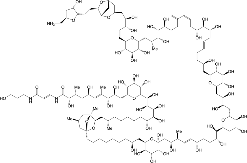
  30. Ciminiello, P; Dell’Aversano, C; Dello Iacovo, E; Fattorusso, E; Forino, M; Grauso, L; Tartaglione, L; Florio, C; Lorenzon, P; De Bortoli, M; Tubaro, A; Poli, M; Bignami, G. Stereostructure and biological activity of 42-hydroxy-palytoxin: A new palytoxin analogue from hawaiian palythoa subspecies. Chem Res Toxicol 2009, 22, 1851–1859. [Google Scholar]
  31. Katikou, P. Palytoxins and analogues: Ecobiology and origin, chemistry, metabolism and chemical analysis. In Phycotoxins: Chemistry and Biochemistry, 2nd ed; Botana, LM, Ed.; Blackwell Publishing: Oxford, UK, 2008; pp. 631–663. [Google Scholar]
  32. Vale, C; Ares, IR. Biochemistry of palytoxins and ostreocins. In Phycotoxins: Chemistry and Biochemistry; Botana, LM, Ed.; Blackwell Publishing: Oxford, UK, 2007; pp. 95–118. [Google Scholar]
  33. Wu, CH. Palytoxin: Membrane mechanisms of action. Toxicon 2009, 54, 1183–1189. [Google Scholar]
  34. Ramos, V; Vasconcelos, V. Palytoxin and analogs: Biological and ecological effects. Mar Drugs 2010, 8, 2021–2037. [Google Scholar]
  35. Aligizaki, K; Katikou, P; Milandri, A; Diogene, J. Occurrence of palytoxin-group toxins in seafood and future strategies to complement the present state of the art. Toxicon 2011, 57, 390–399. [Google Scholar]
  36. Kodama, AM; Hokama, Y; Yasumoto, T; Fukui, M; Manea, SJ; Sutherland, N. Clinical and laboratory findings implicating palytoxin as cause of ciguatera poisoning due to Decapterus macrosoma (mackerel). Toxicon 1989, 27, 1051–1053. [Google Scholar]
  37. Deeds, JR; Schwartz, MD. Human risk associated with palytoxin exposure. Toxicon 2010, 56, 150–162. [Google Scholar]
  38. Riobó, P; Franco, JM. Palytoxins: Biological and chemical determination. Toxicon 2011, 57, 368–375. [Google Scholar]
  39. Randall, JE. Review of clupeotoxism, an often fatal illness from the consumption of clupeoid fishes. Pac Sci 2005, 59, 73–77. [Google Scholar]
  40. Onuma, Y; Satake, M; Ukena, T; Roux, J; Chanteau, S; Rasolofonirina, N. Identification of putative palytoxin as the cause of clupeotoxism. Toxicon 1999, 37, 55–65. [Google Scholar]
  41. Taniyama, S; Arakawa, O; Terada, M; Nishio, S; Takatani, T; Mahmud, Y; Noguchi, T. Ostreopsis sp., a possible origin of palytoxin (PLTX) in parrotfish Scarus ovifrons. Toxicon 2003, 42, 29–33. [Google Scholar]
  42. Tubaro, A; Durando, P; Del Favero, G; Ansaldi, F; Icardi, G; Deeds, JR; Soda, S. Case definitions for human poisonings postulated to palytoxine exposure. Toxicon 2011, 57, 478–495. [Google Scholar]
  43. Rhodes, L; Towers, N; Briggs, L; Munday, R; Adamson, J. Uptake of palytoxin-like compounds by shellfish fed Ostreopsis siamensis (Dinophyceae). N Z J Mar Freshw Res 2002, 36, 631–636. [Google Scholar]
  44. Lenoir, S; Ten-Hage, L; Turquet, J; Quod, JP; Bernard, C; Hennion, MC. First evidence of palytoxin analogues from an Ostreopsis mascarenensis (Dinophyceae) benthic bloom in southwestern Indian Ocean. J Phycol 2004, 40, 1042–1051. [Google Scholar]
  45. Ciminiello, P; Dell’Aversano, C; Fattorusso, E; Forino, M; Magno, GS; Tartaglione, L; Grillo, C; Melchiorre, N. The Genoa 2005 outbreak. Determination of putative Palytoxin in Mediterranean Ostreopsis ovata by a new Liquid Chromatography tandem Mass Spectrometry Method. Anal Chem 2006, 78, 6153–6159. [Google Scholar]
  46. Ciminiello, P; Dell’Aversano, C; Dello Iacovo, E; Fattorusso, E; Forino, M; Tartaglione, L. LC-MS of palytoxin and its analogues: State of the art and future perspectives. Toxicon 2011, 57, 376–389. [Google Scholar]
  47. Janson, S; Siddiqui, PJA; Walsby, AE; Romans, KM; Carpenter, EJ; Bergman, B. Cytomorphological characterization of the planktonic diazotrophic cyanobacteria Trichodesmium spp. from the Indian Ocean and Caribbean and Sargasso seas. J Phycol 1995, 31, 463–477. [Google Scholar]
  48. Ledreux, A; Krys, S; Bernard, C. Suitability of the Neuro-2a cell line for the detection of palytoxin and analogues (neurotoxic phycotoxins). Toxicon 2009, 53, 300–308. [Google Scholar]
  49. Mossman, T. Rapid colorimetric assay for cellular growth and survival: application to proliferation and cytotoxicity assays. J Immunol Methods 1983, 65, 55–63. [Google Scholar]
  50. Espiña, B; Cagide, E; Louzao, MC; Fernandez, MM; Vieytes, MR; Katikou, P; Villar, A; Jaen, D; Maman, L; Botana, L. Specific and dynamic detection of palytoxins by in vitro microplate assay with Neuroblastoma cells. Biosci Rep 2009, 29, 13–23. [Google Scholar]
  51. Wachi, KM; Hokama, Y; Haga, LS; Shiraka, A; Takenaka, WE; Bignami, GS; Levine, L. Evidence for palytoxin as one of the sheep erythrocyte lytic factors in crude extracts of ciguateric and non-ciguateric reef fish tissue. J Nat Toxins 2000, 9, 139–146. [Google Scholar]
  52. Cañete, E; Diogene, J. Comparative study of the use of Neuroblastoma cells (Neuro-2a) and Neuroblastoma x glioma hybrids cells (NG108-15) for the toxic effect quantification of marine toxins. Toxicon 2008, 52, 541–550. [Google Scholar]
  53. European Food Safety Authority (EFSA). Scientific Opinion on marine biotoxins in shellfish—Palytoxin group. EFSA Panel on Contamination in the Food Chain (CONTAM). EFSA J 2009, 7, 1393.
  54. O’Neil, JM. The colonial cyanobacterium Trichodesmium as a physical and nutritional substrate for the harpacticoid copepod Macrosetella gracilis. J Plankton Res 1998, 20, 43–59. [Google Scholar]
  55. Sheridan, C; Steinberg, D; Kling, G. The microbial and metazoan community associated with colonies of Trichodesmium spp.: A quantitative survey. J Plankton Res 2002, 24, 913–922. [Google Scholar]
  56. Wachi, KM; Hokama, Y. Diversity of marine biotoxins in the near-shore ocean area: Presence of a palytoxin-like entity at Barbers Point Harbor, Oahu. J Nat Toxins 2001, 10, 317–333. [Google Scholar]
Figure 1. Structure of palytoxin from Riobó [38].
Marinedrugs 09 00543f1
Figure 2. A bloom of Trichodesmium erythraeum Ehrenberg: (a) Field view of wind-blown concentration of colonies; (b) Concentration of trichomes using their buoyancy properties; (c) Trichodesmium trichomes in bundles oriented in parallel (scale bar = 50 μm).
Marinedrugs 09 00543f2
Figure 3. Effect of PLTX alone on the viability of Neuro-2a cells, and with pre-incubation with 100, 250 or 500 μM ouabain for 2 h before adding PLTX.
Marinedrugs 09 00543f3
Figure 4. Effect of PLTX alone on the viability of Neuro-2a cells, and with pre-incubation of PLTX for 2 h before adding 500 μM ouabain, with 500 μM ouabain administered simultaneously, with pre-incubation of 500 μM ouabain for 1 h or 2 h before adding PLTX.
Marinedrugs 09 00543f4
Figure 5. Effect of Trichodesmium extract No. 1, “5îles”, alone on the viability of Neuro-2a cells and with pre-incubation with 500 μM ouabain for 2 h before adding extract.
Marinedrugs 09 00543f5
Figure 6. Effect of Trichodesmium extract (No. 8) from Lifou on the viability of Neuro-2a cells alone or spiked with an equivalent of 1.7 μg PLTX/g of extract, and with pre-incubation with 500 μM ouabain for 2 h before adding extract. Toxicity of Trichodesmium extract No. 1 from 5îles was compared with the toxicity of Trichodesmium extract No. 8 and with pre-incubation with ouabain.
Marinedrugs 09 00543f6
Figure 7. LC-MS/MS chromatograms of 42-OH-PLTX (a) and PLTX (b) in a sample of Trichodesmium spp. Standards of 42-OH-PLTX (c) and PLTX (d) were purchased from Wako.
Marinedrugs 09 00543f7
Table 1. Trichodesmium collections: date, location, identification and yield extraction.
No.DateReferenceLocationLatitudeLongitudeWater-soluble fraction % of dried material

12007-03-01Tricho 5îles5îles−22.771900166.80099564.3
22007-09-24Tricho BD 2007Baie des citrons−22.297600166.43800451.6
32008-02-08Tricho BD 2008Baie des citrons−22.295700166.43600568.8
42008-02-18Tricho DumbéaPasse de Dumbéa−22.349501166.27499426.2
52008-02-18Tricho RicaudyRécif Ricaudy−22.306900166.46021021.5
62008-11-04Tricho Lifou C01Lifou–Hunëtë−20.767310167.09300652.5
72009-02-01Tricho Nouméa 3Passe de Dumbéa−22.349501166.27499449.7
82009-11-18Tricho Lifou C02Lifou–Hunëtë−20.767310167.09300655.4
Table 2. SRM parameters setting used for PLTXs-like detection.
ToxinsTransitions m/zDeclustering potential (V)Cell exit potential (V)Dwell time (ms)
PLTX1340→3272618250
1332→3272618250
896→327618250
Ovatoxin-a1324→3272618250
1315→3272618250
889→327618250
42-OH-PLTX1348.7→3272618250
1339.7→3272618250
899.7→327618250
Table 3. EC50 values of the dose-response curves obtained for the PLTX with or without ouabain pre-incubation *.
ConditionsWithout OPre PLTXOPre O
O (μM)
Pre-incubation time (h)
0500
2
500
0
500
1
500
2
250
2
100
2
C50 (pM)170 ± 60 (n = 3)6.375.089.416.0 ± 2.2 (n = 3)11.6521.74
*Neuro-2a cells with 100, 250 and 500 μM ouabain (O) with different preincubation times (0, 1 and 2 h). R2 values showed always good fit. Each point represents at least the mean of 3 well values. For some conditions (without O and PreO 500, 2 h), data represent the mean ±SD of 3 separate experiments.
Table 4. Results of Neuro-2a cells cytotoxicity (CBA) and LC-MS/MS analysis *.
No.ReferenceCBALC-MS/MS
preO−preO+PLTX42-OH-PLTXTotal PLTX eqv.
EC50
μg/mL
μg/g extractμg/g extractμg/g eqv. dried material

1Tricho 5îles1337 ± 126113.8 ± 110.80.820.871.701.10
2Tricho BD 2007>LOQ2261<LOD<LOD<LOD<LOD
3Tricho BD 2008>LOQ1138<LOD<LOD<LOD<LOD
4Tricho Dumbéa1324/1066494/1580.570.521.080.28
5Tricho Ricaudy927NA0.890.641.530.33
6Tricho Lifou C0112143970.860.591.450.76
7Tricho Nouméa 3121291<LOD<LOD<LOD<LOD
8Tricho Lifou C02>LOQ2054 ± 277<LOD<LOD<LOD<LOD
*preO−: Without ouabain pre-treatment; preO+: With ouabain pre-incubation; LOQ (Limit of Quantification): 4464 μg/mL for CBA; LOD (Limit of Detection): 0.01 μg/g for LC-MS/MS; NA: Not Available.

