Article

Purification of Antioxidant Peptides by High Resolution Mass Spectrometry from Simulated Gastrointestinal Digestion Hydrolysates of Alaska Pollock (Theragra chalcogramma) Skin Collagen

Yunnan Institute of Food Safety, Kunming University of Science and Technology, No. 727 South Jingming Road, Kunming 650500, Yunnan, China
*
Author to whom correspondence should be addressed.
Mar. Drugs 2016, 14(10), 186; https://doi.org/10.3390/md14100186
Submission received: 14 July 2016 / Revised: 20 September 2016 / Accepted: 26 September 2016 / Published: 17 October 2016
(This article belongs to the Special Issue Marine Proteins and Peptides)

Abstract

:
In this study, the stable collagen hydrolysate was prepared by alcalase hydrolysis and twice simulated gastrointestinal digestion from Alaska pollock skin. The characteristics of hydrolysates and antioxidant activities in vitro, including 2,2′-azino-bis (3-ethylbenzothiazoline-6-sulfonic acid) radical (ABTS•+) scavenging activity, ferric-reducing antioxidant power (FRAP) and hydroxyl radical (OH·) scavenging activity, were determined. After twice simulated gastrointestinal digestion of skin collagen (SGI-2), the degree of hydrolysis (DH) reached 26.17%. The main molecular weight fractions of SGI-2 were 1026.26 and 640.53 Da, accounting for 59.49% and 18.34%, respectively. Amino acid composition analysis showed that SGI-2 had high content of total hydrophobic amino acid (307.98/1000). With the simulated gastrointestinal digestion progressing, the antioxidant activities increased significantly (p < 0.05). SGI-2 was further purified by gel filtration chromatography, ion exchange chromatography and high performance liquid chromatography, and the A1a3c–p fraction with high hydroxyl radical scavenging activity (IC50 = 7.63 μg/mL) was obtained. The molecular weights and amino acid sequences of key peptides of A1a3c–p were analyzed using high resolution mass spectrometry (LC-ESI-LTQ-Orbitrap-MS) combined with de novo software and UniProt of MaxQuant software. Four peptides were identified from A1a3c–p, including YGCC (444.1137 Da) and DSSCSG (554.1642 Da) identified by de novo software and NNAEYYK (900.3978 Da) and PAGNVR (612.3344 Da) identified by UniProt of MaxQuant software. The molecular weights and amino acid sequences of four peptides were in accordance with the features of antioxidant peptides. The results indicated that different peptides were identified by different data analysis software according to spectrometry mass data. Considering the complexity of LC-ESI-LTQ-Orbitrap-MS, it was necessary to use the different methods to identify the key peptides from protein hydrolysates.

1. Introduction

Reactive oxygen species (ROS) and free radicals are very unstable and react rapidly with lipids and proteins in the body, generating damage in the biological system, such as DNA, protein and membrane lipid, if the human body cannot control their formation or eliminate them [1]. Therefore, it is important to inhibit oxidation reactions and the formation of free radicals in the living body. In recent years, some peptides have been found to possess antioxidant activity [2,3]. Collagen is rich in hydrophobic amino acids, and the abundance of these amino acids favors higher affinity to oil and better emulsifying ability. Therefore, collagen is expected to provide natural antioxidant peptides and exert higher antioxidant effects than other proteins. In addition, collagen peptides have good biocompatibility, good penetrability and cause no irritation to the body [4]. Researchers have reported the characteristic and antioxidant activity of different collagen resources, especially from aquatic animals, such as sea jumbo squid skin [5], jellyfish umbrella [2], smooth hound protein [6], sea cucumber [7], pacific hake [8] and tilapia skin [3].
The structure of bioactive peptides may be changed when they are digested, absorbed and transferred in gastrointestinal tract [9]. It is expected that bioactive peptides would not be further digested in the gastrointestinal tract and thereby ensure its stability after digestion. The stable peptides should ideally be isolated and identified in vivo. However, in vivo studies are costly and time-consuming, are rather complicated to perform. As an alternative to in vivo studies, a simple, rapid, and inexpensive in vitro simulated gastric and intestinal digestive method has been established to isolate bioactive peptides. In this method, the prior hydrolysis with the endopeptidases would increase the stability and bioavailability of the bioactive peptide in vivo. In the previous studies, it has been shown that gastrointestinal enzyme digestion results in more potent peptides compared with other enzymatic digestions [10].
Alaska pollock (Theragra chalcogramma) is one of the commercially important fish species in China, but a large number of scraps containing fish skin, head and bones are left in the processing of fillet production. It has been reported that 70% of the dry matter of fish skin is collagen. Some studies reported the characteristics of collagen and bioactivities of collagen peptides from Alaska pollock skin. Yan et al. [11] reported the characterization of collagen from the skin of Alaska pollock. Hou et al. [12] studied the immunomodulatory activity of Alaska pollock hydrolysates and the active peptide was identified. Byun and Kim [13] purified the key peptide from Alaska Pollock skin with angiotensin I converting enzyme inhibitory activity. Jia et al. [14] studied the enzymatic hydrolysates of Alaska Pollock skin with antioxidant activity.
In this study, twice simulated gastrointestinal (GI) digestion was used to prepare hydrolysates from the collagen of Alaska Pollock skin, in order to obtain the stable antioxidant peptides. The antioxidant activity of the hydrolysates was evaluated in vitro. The bioactive fraction from hydrolysates with the highest antioxidant activity was separated by gel filtration chromatography, ion exchange chromatography and high performance liquid chromatography. Furthermore, two methods, including de novo analysis software and UniProt of MaxQuant software, were used to identify the key peptides from protein hydrolysates.

2. Results and Discussion

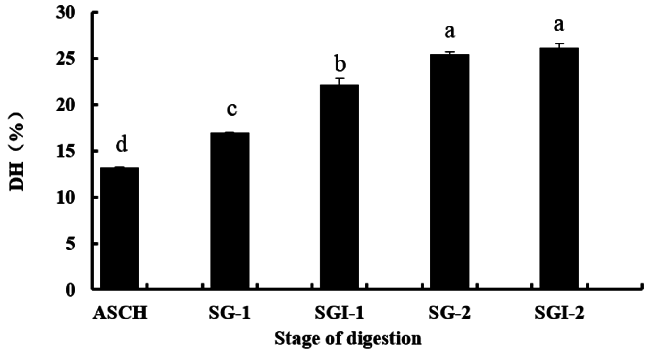
2.1. Analyses, DH, Molecular Weight (MW) and Amino Acid Composition of Hydrolysates

In this study, Alaska Pollock skin collagen was hydrolysed by alcalase to obtain its hydrolysates (ASCH). Two successive simulated GI digestions of ASCH were conducted in order to get the stable collagen hydrolysates. The DHs of collagen hydrolysates were studied at different stages, including the first simulated gastric digestion (SG-1), the first simulated intestinal digestion (SGI-1), the second simulated gastric digestion (SG-2) and the second simulated intestinal digestion (SGI-2). As shown in Figure 1, the DH of ASCH was 13.17%. It increased from 16.92% (SG-1) to 22.65% (SGI-1) at the first simulated GI digestion. It was similar to the study of You et al. which indicated that more peptide bonds were broken using pancreatin digestion than using pepsin digestion [15]. In the second simulated GI digestion, the increase of DH was significant (p < 0.05) at the stage of SG-2 (25.47%), but it was not significant between the SG-2 and SGI-2 stage (26.17%) (p > 0.05). It might be because the DH became basically stable as the second simulated intestinal digestion was processed.
Furthermore, the molecular weight distributions of the different stages of digestion from skin collagen of Alaska pollock were shown in Table 1. The main molecular weight fractions of ASCH, SGI-1 and SGI-2 were 3198.76 (66.82%), 1552.34 (74.66%) and 1026.26 Da (59.49%), respectively. With the increasing of DH, the molecular weight of the hydrolysates significantly decreased. As shown in Table 1, the MW distribution of SGI-2 was 1026.26, 640.53, 284.97 and 96.58 Da. Based on the peak area, they accounted for 59.49, 18.34, 16.60 and 4.56%, respectively.
The amino acid compositions of the different stages of digestion were showed in Table 2. The compositions of three stages were similar and they were rich in Gly, Glu, Pro, Asp, and Arg. The result was similar to that of other fish skin hydrolysates [4,5] and in accordance with the characteristics of collagen hydrolysates. In this study, the total hydrophobic amino acid (THAA) contents of three hydrolysates were high, showing 315.11, 302.22 and 307.98 per 1000 residues, respectively. High content of hydrophobic amino acids could increase the solubility in lipids and therefore enhance the antioxidative activity of hydrolysates [4].

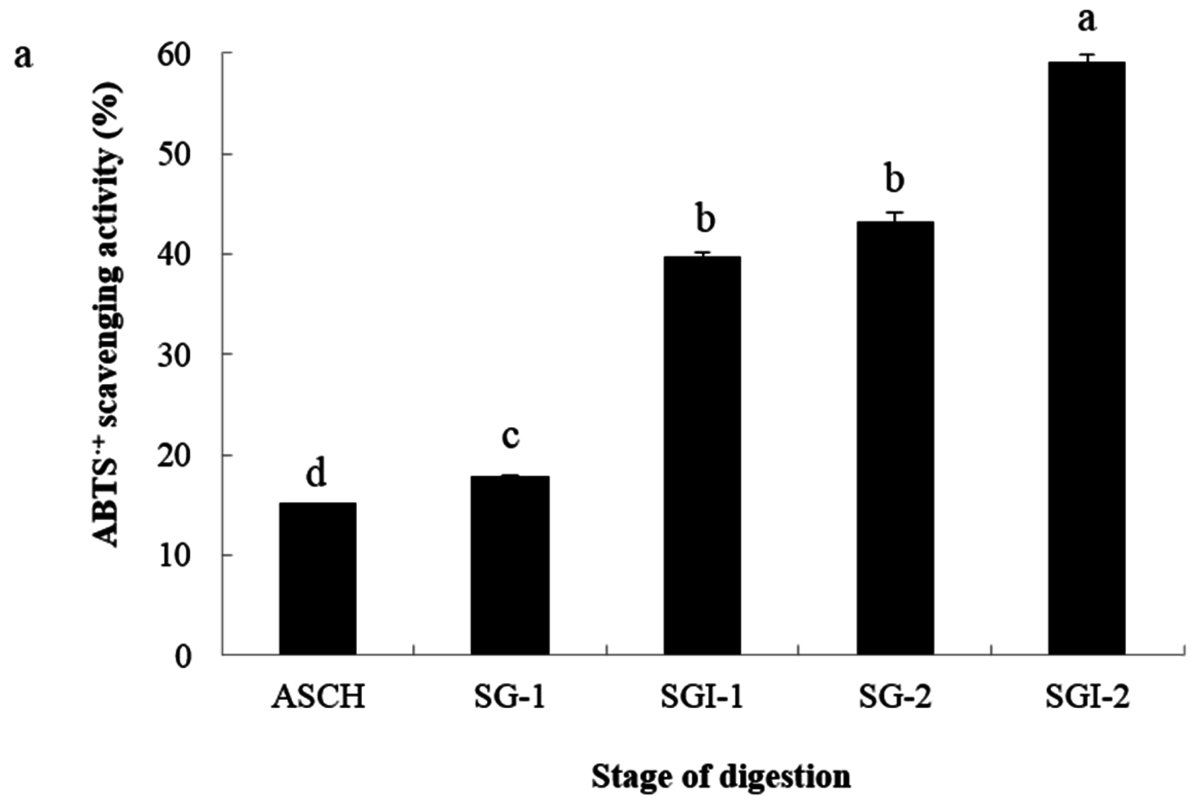
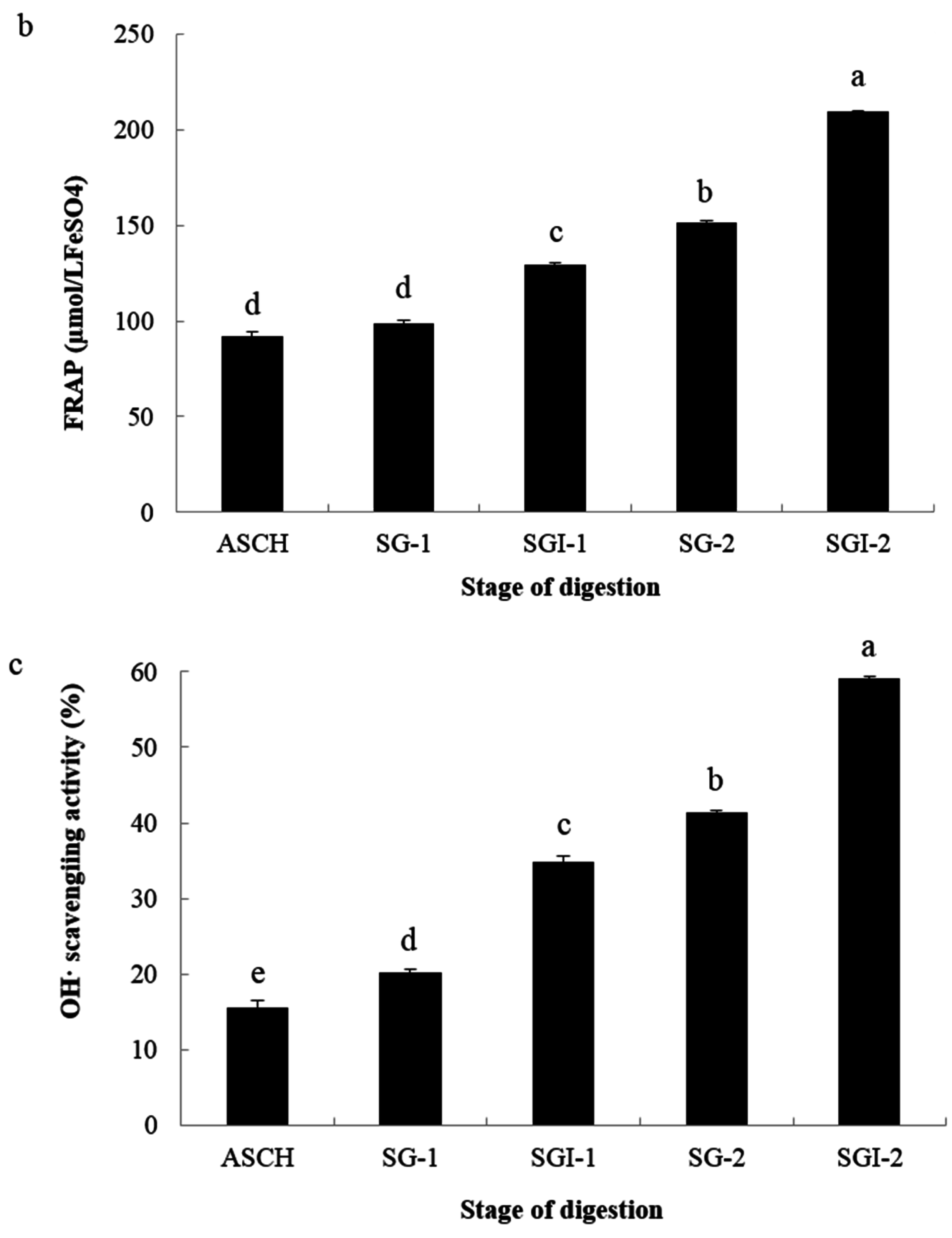
2.2. Analyses of Antioxidant Activities

Since there is no single antioxidant standard method to test for antioxidant capacity, it is recommended to use different methods for investigating the different mechanisms of antioxidant capacity [16]. In order to evaluate the antioxidant activity of collagen hydrolysates, the pHs was adjusted to about 7.0 in this study. As shown in Figure 2, the ABTS•+ scavenging activity, FRAP and OH· scavenging activity were measured.
The ABTS assay is often used to evaluate the ability of antioxidants to scavenge free radicals. After simulated GI digestion, the ABTS•+ scavenging activity obviously increased (Figure 2a). The ABTS•+ scavenging activity was 39.79% in the SGI-1 stage and 59.17% in the SGI-2 stage at the concentrations of 0.5 mg/mL. The FRAP assay is based on the ability of antioxidants to reduce Fe3+ to Fe2+ in the presence of 1,3,5-tri(2-pyridyl)-2,4,6-triazine (TPTZ). As shown in Figure 2b, with the simulated gastrointestinal digestion progressing, FRAP of the hydrolysates increased significantly (p < 0.05). At the dose of 10 mg/mL, the activities of SGI-1 and SGI-2 were 129.42 and 209.27 μmol/L FeSO4, respectively. Hydroxyl radical is the most reactive radical, which has been demonstrated to be a highly damaging species in free radical pathology, attacking almost every molecule in living cells. As shown in Figure 2c, the simulated GI digestion significantly increased the OH· scavenging activity (p < 0.05). Indeed, the OH· scavenging activity of ASCH was 15.53% at the concentration of 2 mg/mL, and the activities of SGI-1 and SGI-2 were 34.77 and 59.11%, respectively. The increase of scavenging activity of the pancreatin digestion process was higher than the pepsin digestion.

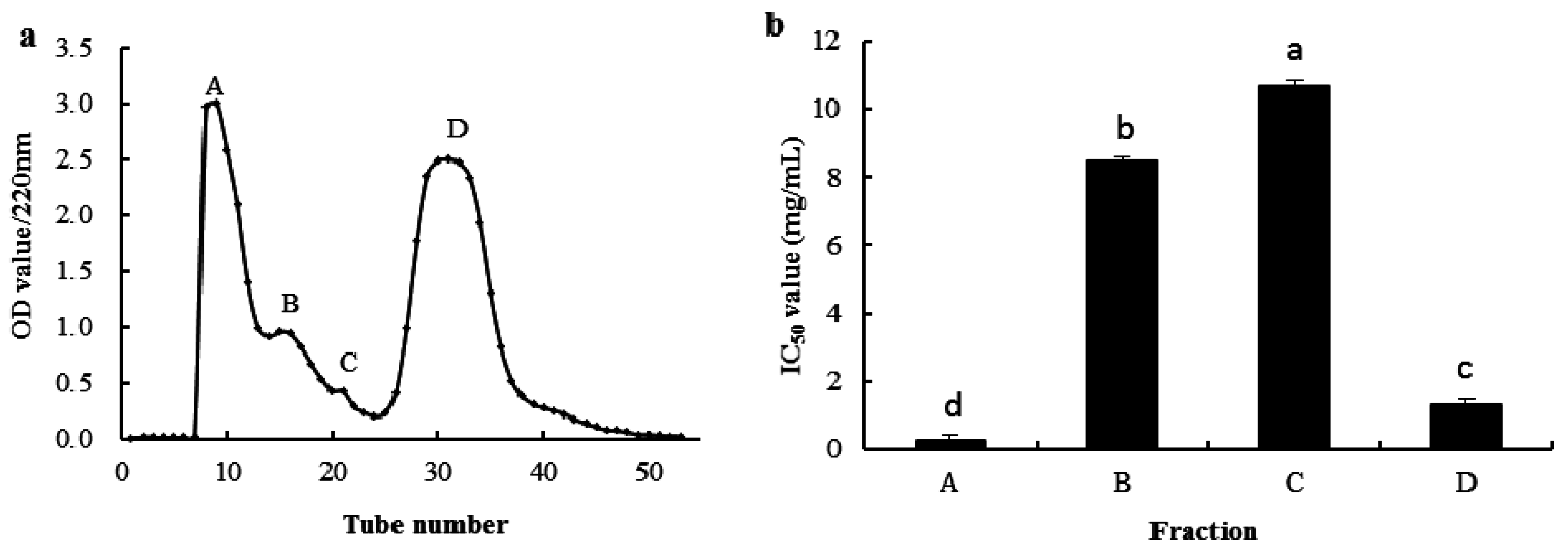
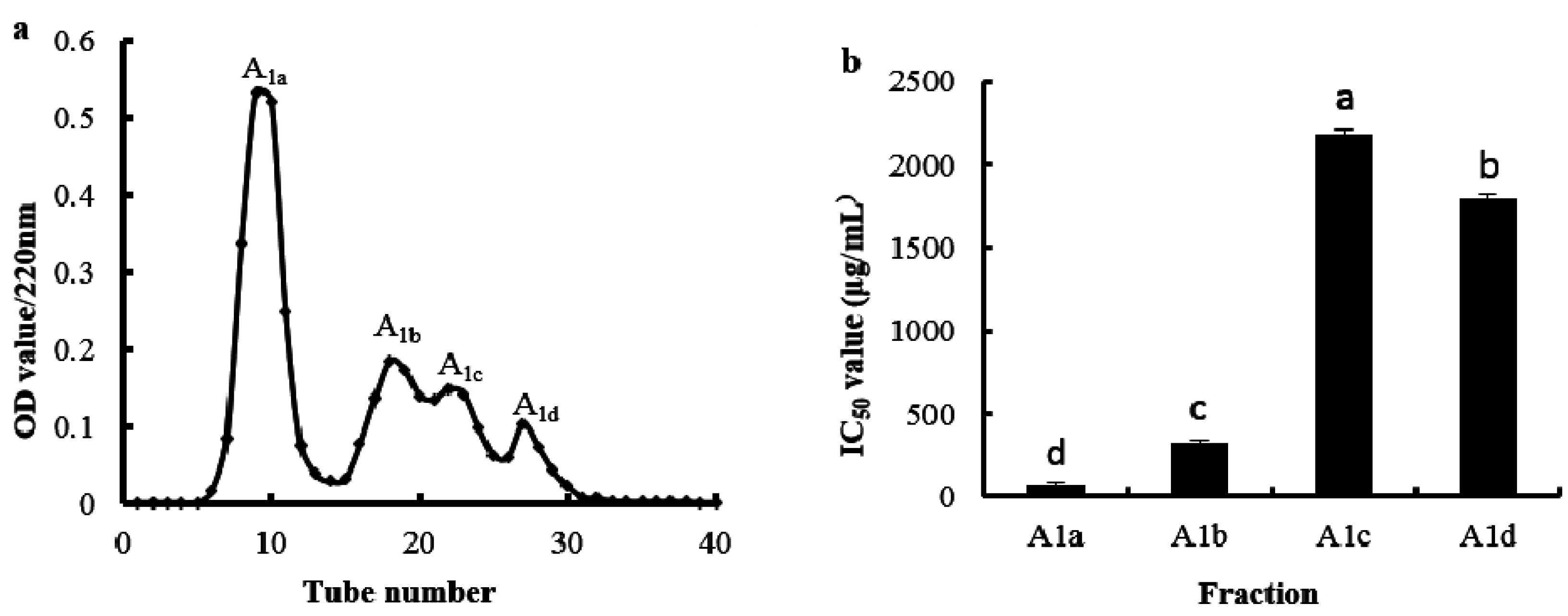
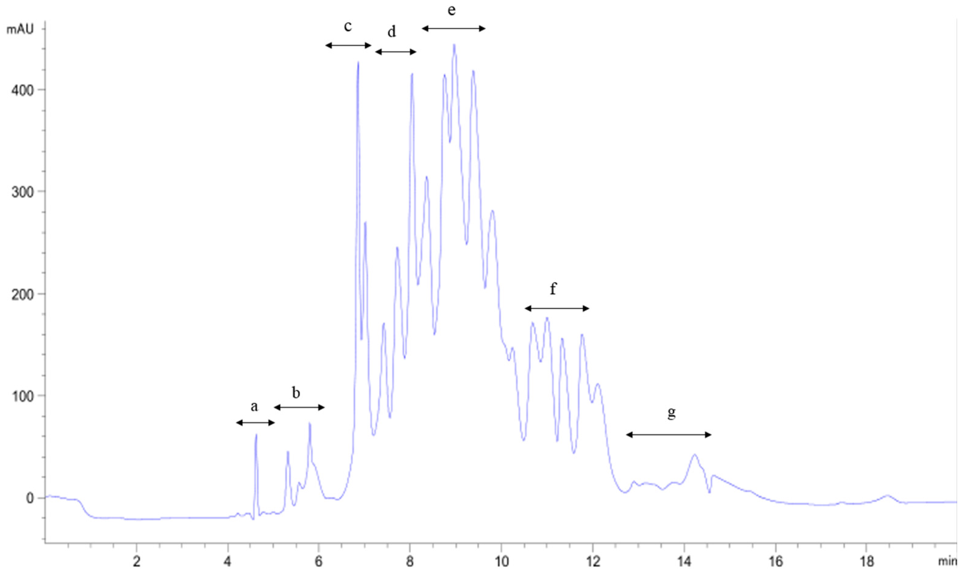
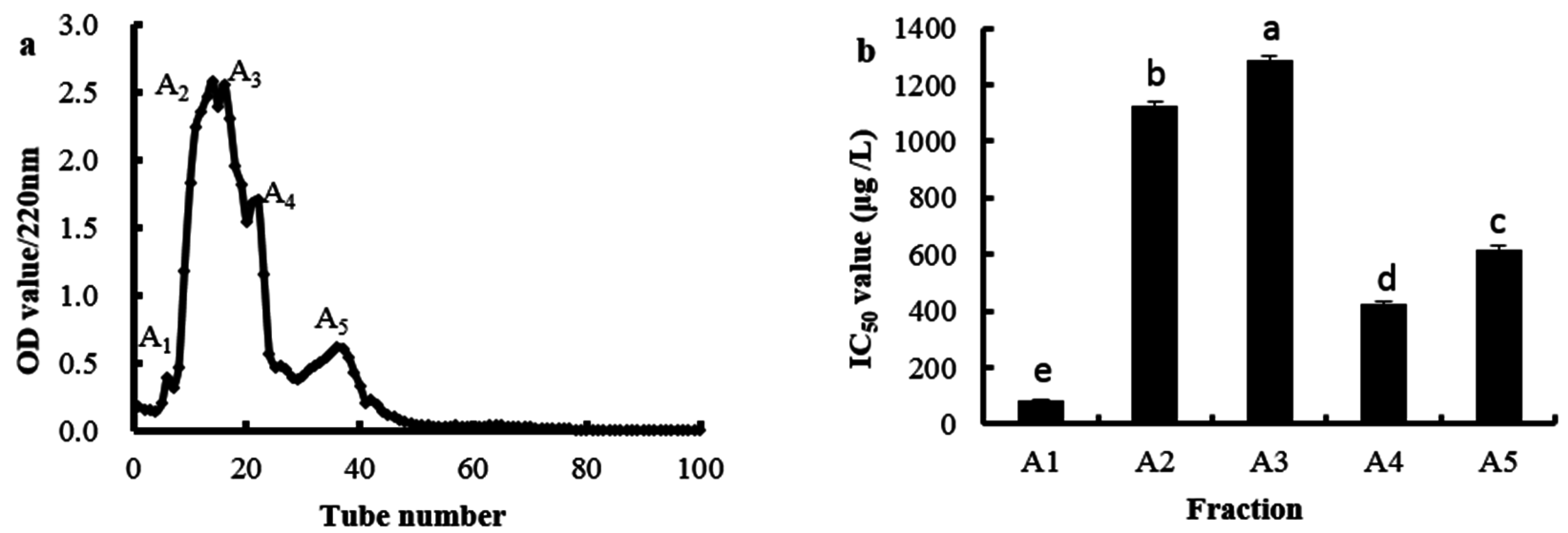
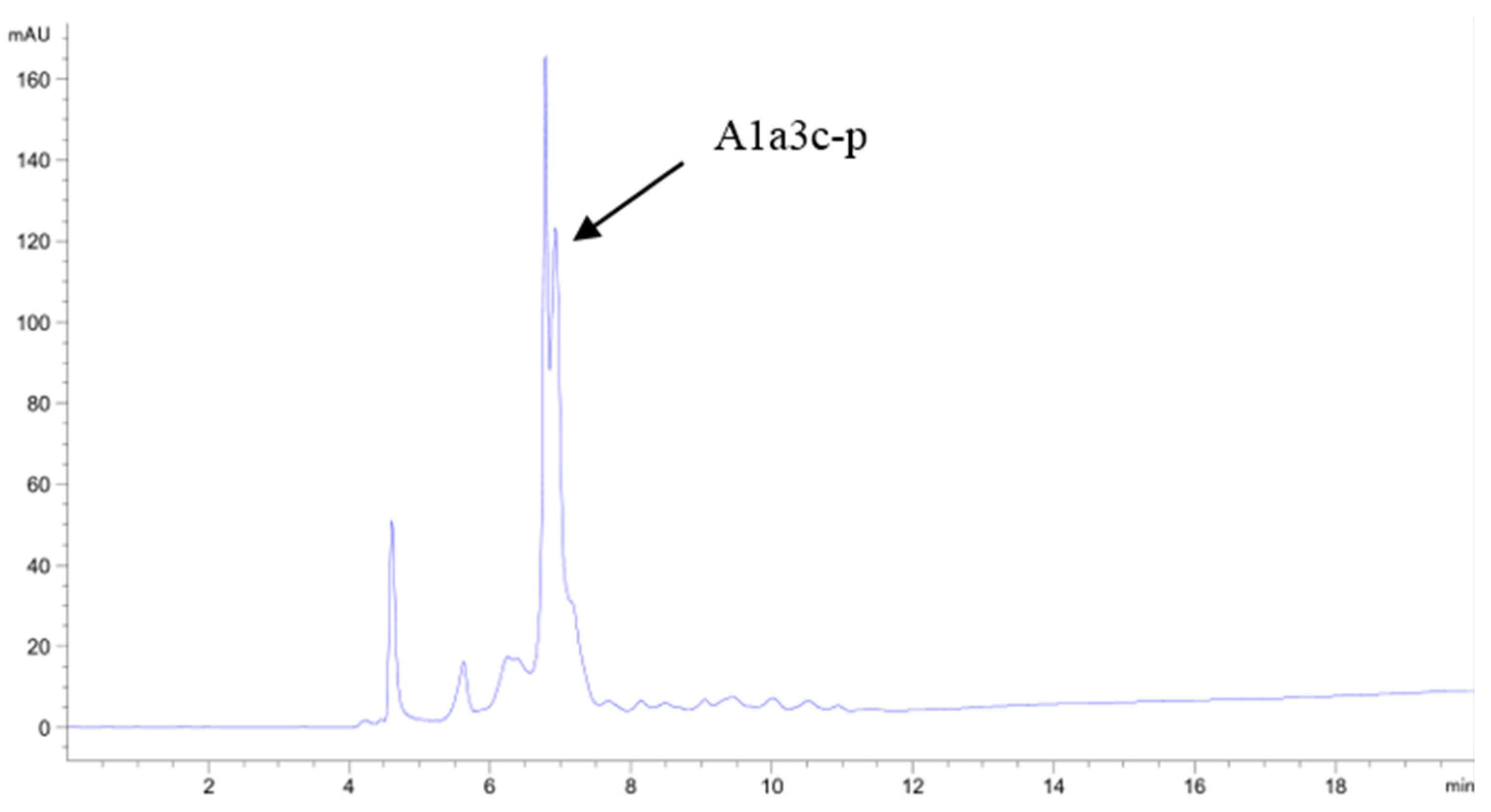
2.3. Purification of the Antioxidant Peptides from SGI-2

The removal of OH· is probably one of the most effective defenses of a living body against various diseases [17]. Based on this reason, the OH· scavenging activity was selected as the indicator of purification of antioxidant peptides in the study.
The SGI-2 solution was purified by a Sephadex G-25 gel filtration column, and four fractions were obtained, noting A–D respectively (Figure 3a). Four fractions were collected, concentrated and the OH· scavenging activities were determined. Results showed that fraction A had the highest OH· scavenging activity among the four fractions, with the IC50 value being 0.26 mg/mL (Figure 3b).
Ion-exchange chromatography was a method of separation according to the substance with a different acid-base property and polarity. SP Sephadex C-25 was one of the strong cation exchangers with a main functional group of sulfopropyl and it was widely used in separating bioactive peptides [18]. The fraction A collected from Sephadex G-25 was further separated by the SP Sephadex C-25 column and five fractions were obtained, noting A1, A2, A3, A4 and A5, respectively (Figure 4a). The OH· scavenging activities of these five fractions were shown in Figure 4b; fraction A1 had the highest OH· scavenging activity with the IC50 value being 81.15 μg/mL. Thus, the fraction A1 was selected for next separation.
Sephadex G-15 was used to remove NaCl from the eluate of SP Sephadex C-25 in the fraction A1. As shown in Figure 5, four fractions (A1a, A1b, A1c, and A1d) were obtained and their OH· scavenging activities were measured. The OH· scavenging activity of the fraction A1a was the highest compared with the other three fractions, and the IC50 value was 73.52 μg/mL.
The fraction A1a was further isolated by a Shim-pack GIS C18 preparative column with a liner gradient of acetonitrile containing 0.1% trifluoroacetic acid (TFA) from 5% to 30% in 30 min. The elution profile was shown in Figure 6. A total of 12 peaks were obtained and named as A1a1–A1a12, respectively. Each peak was collected, concentrated and it’s OH· scavenging activities were measured. The result showed that A1a3 had the highest antioxidant activity and the IC50 value of A1a3 was 31.72 μg/mL.
The fraction A1a3 was isolated by HPLC on the semi-preparative C18 column using a liner gradient of acetonitrile containing 0.1% TFA from 5% to 25% in 30 min. Seven fractions were collected and designated as A1a3a–A1a3g in turn respectively (Figure 7). After the OH· scavenging activities of seven fractions were determined, we found that A1a3c had the highest OH· scavenging activity, and the IC50 value of A1a3c was 14.36 μg/mL. The most active A1a3c faction was isolated again by the semi-preparative C18 column using a different liner gradient of acetonitrile containing 0.1% TFA from 5% to 20% in 30 min. The main peak A1a3c–p with high antioxidant activity was collected and concentrated (Figure 8). The IC50 value of OH· scavenging activity of the A1a3c–p fraction was 7.63 μg/mL.

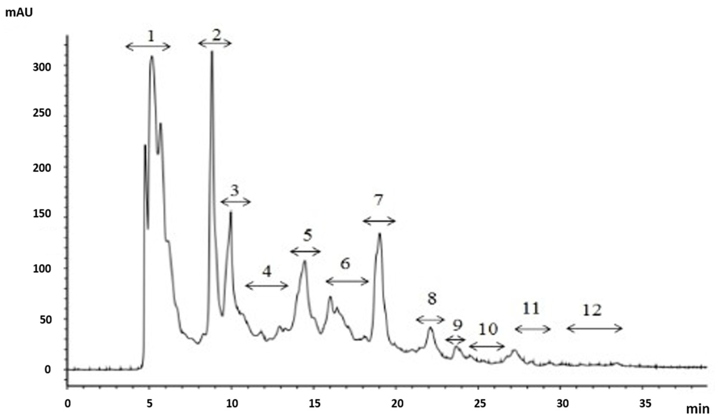
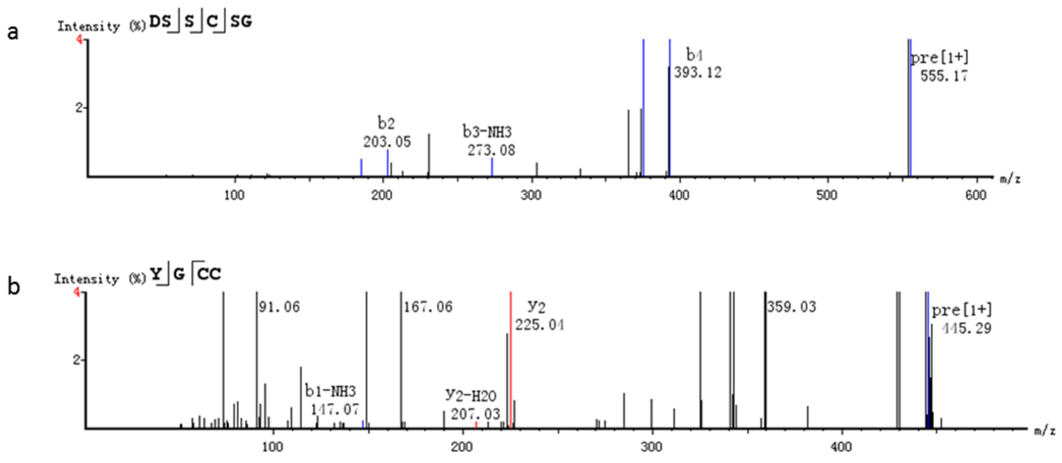
2.4. Identification of Purified Peptide

The antioxidant activity of peptides is connected with their molecular weights, amino acid compositions, amino acid sequences and so on [19]. In this study, the fraction A1a3c–p was analyzed by high resolution mass spectrometry combined with two methods, including de novo software and MaxQuant software. As shown in Figure 9, two peptides were obtained by de novo software. The peptide sequences were Try–Gly–Cys–Cys (YGCC) and Asp–Ser–Ser–Cys–Ser–Gly (DSSCSG), and their molecular weight was 444.1137 and 554.1642 Da, respectively. The data was scanned in the “fish collagen” database by UniProt of MaxQuant software. Two peptides, Asn-Asn-Ala-Gln-Tyr-Tyr-Lys (NNAEYYK) and Pro-Ala-Gly-Asn-Val-Arg (PAGNVR), were identified, and their molecular weight was 900.3978 and 612.3344 Da, respectively. The amino acids at the C-terminus of two peptides were K and R, which conformed to the fracture characters of simulated gastric and intestinal digestions.
Generally, there is no direct relationship between antioxidant activity and molecular weight. However, the previous study indicated that the peptides with smaller molecular weights have stronger antioxidant activities, more resistant to the gastrointestinal digestion and easier to cross the intestinal barrier to exert biological activities than larger peptides [20]. Antioxidative peptides usually contain 2–20 amino acids with molecular weights below 3000 Da [5]. In this study, the amino acid numbers of four peptides identified from Alaska pollock skin collagen were 4, 6, 7, and 6 and the molecular weight was lower 1000 Da. The amino acid number and molecular weight was in accordance with the feature of antioxidant peptide. Our study was similar to the peptides purified from walnut [21] and loach protein hydrolysates [15].
Moreover, compositions and the specific position of amino acids in the peptide may play an important role in its antioxidant activities. High content of hydrophobic amino acids, especially at the N- or C-terminus of peptides, could enhance the activities of antioxidative peptides by interacting with lipid molecules and donating protons into radicals to scavenge radicals [22]. Moreover, polar/charged amino acids such as Arg at the C-terminus position also contribute to the antioxidant activity [23]. Our results were similar to these previous reports, and hydrophobic amino acids or arginine existed in the terminus of four peptides.
Some studies have reported that peptide sequences containing Tyr show strong antioxidant activity, especially when the presence of Tyr was at terminals of the peptide sequence. The antioxidant activity of Tyr may be explained by the special capability of phenolic groups to serve as hydrogen donors, which is one mechanism of inhibiting the radical-mediated peroxidizing chain reaction [24]. YGCC obtained from this study had a Tyrat N-terminus, and this might be one of the reasons why YGCC showed higher radical scavenging activity. In addition, NNAEYYK had two Tyr, which could effectively increase its antioxidant activity. Previous studies show that Cys is hydrophobic in nature and can interact directly with free radicals by donating the sulfur hydrogen, so the presence of Cys is one of the reasons for the good antioxidant activity of the isolated peptide [25]. Li et al. considered that Cys residue at the C-terminus or next to the C-terminus plays an important role in antioxidative activities [26]. It was similar to our study and YGCC and DSSCSG contained Cys, YGCC in particular had two Cys at the C-terminus, which might improve its antioxidant activity. Acidic amino acids, such as Asn and Gln, play important roles in the chelation of metal ions by their side chains, which may inhibit the formation of the hydroxyl radical [27]. Rajapakse et al. reported that the presence of Asp seemed to play a vital role, irrespective of its position, as observed in several antioxidative peptide sequences [28]. It was similar to our results. DSSCSG and NNAEYYK had Asp and Asn at the N-terminus, respectively.
The fraction A1a3c–p was analyzed using high resolution mass spectrometry, and the key peptides obtained by de novo and MaxQuant software were different. Therefore, to adequately identify the key peptides from protein hydrolysates fractions, it was necessary to use different methods to analyze mass data. A further study about the quantitative analysis of key peptides will be carried out.

3. Materials and Methods

3.1. Materials

Collagen of Alaska pollock skin was prepared by the previous methods. Alcalase was purchase from Genencor International Co. (Wuxi, China); Pepsin, pancreatin, 2,2′-azino-bis (3-ethylbenzothiazoline-6-sulfonic acid) (ABTS) and 2,4,6-Tris(2-pyridyl)-S-triazine (TPTZ) were purchased from Sigma Chemical Co., (St. Louis, MO, USA). Sephadex G-25, Sephadex G-15 and SP Sephadex C-25 were purchased from GE Healthcare (Fairfield, CT, USA). Acetonitrile (HPLC grade) was purchased from Merck KGaA (Darmstadt, Germany). All other reagents used in this study were analytical grade.

3.2. Preparation of Skin Collagen Hydrolysates of Alaska Pollock

The Alaska pollock skin collagen was mixed with distilled water at a concentration of 1% (w/v). The mixture was adjusted to pH 9.0 by 1 M NaOH solution and then hydrolyzed using Alcalase (E/S: 5/100, w/w) at 55 °C for 2 h. Alcalase were inactivated in boiling water for 10 min and centrifuged at 5000 rpm for 20 min. The supernatants (ASCH) were collected and then lyophilized. ASCH was hydrolyzed by the simulated GI digestion [15,29]. A total of 2 g ASCH was dissolved in 150 mL distilled water and adjusted to pH 2.5 with 6 M HCl. Then, pepsin was added at a ratio of enzyme to substrate of 1:35 (w/w). After the mixture was incubated at 37 °C for 1 h with shaking (SG-1), sodium cholate (0.2 mM) and SG-1 (1:1, v/v) were mixed. The pH was adjusted to 7.5 using 2 M NaOH. Then, pancreatin was added at a ratio of enzyme to substrate of 1:25 (w/w). The mixture was incubated at 37 °C for 2 h with shaking and then inactivated in boiling water for 15 min and centrifuged at 5000 rpm for 20 min. The supernatants (SGI-1) were desalinized and then lyophilized. The second simulated gastric and intestinal digestions were conducted using the same method with the above processes and SG-2 and SGI-2 were obtained.

3.3. Determination of Characteristics of Hydrolysates

3.3.1. Determination of the Degree of Hydrolysis (DH)

The contents of free amino (–NH2) and protein (N) were evaluated according to the ninhydrin colorimetric method and kjeldah method, respectively. DH was calculated as follows [30]:
D H = h ( m m o l / g ) h tot ( m m o l / g ) × 100 % = [ M 1 ( μ m o l / m L ) N ( m g / m L ) M 0 ( m m o l / g ) ] ÷ h tot ( m m o l / g ) × 100 %
where h is the number of broken peptide bonds per gram protein; htot is the total number of peptide bonds per gram original protein (the htot of collagen was 8.41 mmol per gram protein); M1 is the content of –NH2 in hydrolysate; M0 is the content of –NH2 in original protein; N is the content of protein in hydrolysate.

3.3.2. Molecular Weight (MW) Distribution

The molecular weight distribution of the different hydrolysates was measured using a high-performance liquid chromatography (HPLC) system (1260 series, Agilent Scientific, Santa Clara, CA, USA) with a TSK gel 3000 PWXL column (30 mm i.d. × 7.8 mm, Tosoh, Tokyo, Japan) [17]. The mobile phases were acetonitrile-water (1:1, v/v) in the presence of 0.1% (v/v) trifluoroacetic acid, and the flow rate was 0.6 mL/min. The process was monitored at 220 nm at 30 °C. A calibration curve of molecular weight was prepared according to the following standards: cytochrome C (12,500 Da), insulin (5734 Da), vitamin B12 (1355 Da), hippuryl-histydilleucine (429.5 Da), and glutathione (309.5 Da). The logarithm of molecular weight (MW) and the retention time (tR) were in a linear relationship and the formula was calculated as lg MW = −0.284tR + 7.310 (R2 = 0.9922, p < 0.01).

3.3.3. Amino Acid Composition

The different hydrolysates were hydrolyzed under reduced pressure with 6 mol/L HCl at 110 °C for 22 h and the amino acid compositions were analyzed on a Hitachi amino acid analyzer 835-50 (Hitachi, Tokyo, Japan).

3.4. Antioxidative Activity Assay

3.4.1. ABTS•+ Scavenging Activity Assay

ABTS•+ scavenging activities were determined as described by previous method with a slight modification [31]. A total of 5 mL of 7 mM ABTS and 88 μL of 40 mM potassium persulfate was mixed to prepare ABTS•+ stock solution. The mixture was left in the dark at room temperature for 12 h. The ABTS•+ stock solution was diluted with PBS (2 mM, pH 7.4) to an absorbance of 0.70 ± 0.02 at 734 nm. Then, 0.5 mL of samples were mixed with 4 mL ABTS•+ stock solution. The mixture was shaken for 10 s and left in the 30 °C water bath for 6 min. The absorbance was measured at 734 nm. The capability of ABTS•+ scavenging was calculated according to the following equation.
Radical   scavenging   activity   ( % ) = A c ( A s A c s ) A c × 100
where Ac was 0.5 mL ethanol + 4.0 mL ABTS•+ solution; As was 0.5 mL sample + 4.0 mL ABTS•+ solution; Acs was 0.5 mL sample + 4.0 mL ethanol.

3.4.2. FRAP Assay

FRAP was determined according to the method of Alemania et al. [32] with a slight modification. A total of 300 mM acetic acid buffer solution (pH 3.6) was mixed with 10 mM TPTZ and 20 mM FeCl3∙6H2O according to the rate of 10:1:1. Then the mixture was left in a 37 °C water bath to prepare the FRAP solution. The mixture of 150 μL samples and 4.5 mL FRAP solution was reacted at 37 °C for 10 min and then determined the absorbance at 593 nm. A total of 150 μL of distilled water was used instead of samples solution as a control. The absorbance of different concentrations of FeSO4 solution (0–500 μmol/mL) were determined at 593 nm. The FRAP of samples were expressed as equal to μmol/mL FeSO4.

3.4.3. OH· Scavenging Activity Assay

Hydroxyl radical (OH·) scavenging activity was determined by the previous method with slight modification [33]. Briefly, after 1 mL of samples mixed with 0.3 mL of FeSO4 (8 mM), 1 mL of salicylic acid (3 mM) and 0.25 mL of H2O2 (20 mM), the mixture was incubated at 37 °C for 30 min. The reaction mixture was cooled by flowing water to room temperature. Then, 0.45 mL distilled water was added into the mixture to make the end volume 3.0 mL. The mixture was centrifuged at 3000 rpm for 10 min. The absorbance of supernatant was measured at 510 nm, and 1 mL of the solvent solution was used instead of the sample solution as a control. The capability of scavenging the hydroxyl radical was calculated according to following equation:
Radical   scavenging   activity   ( % ) = A 0 ( A 1 A 2 ) A 0 × 100
where A0 was the absorbance of the control without a sample, A1 was the absorbance with a sample, and A2 was the absorbance of the reagent blank. The IC50 value was defined as an effective concentration that is required to scavenge 50% of radical activity.

3.5. Purification of Antioxidant Peptides

The SGI-2 was dissolved in distilled water and preliminarily separated by a Sephadex G-25 gel filtration column (Φ 2.6 cm × 30 cm). The SGI-2 was eluted at a flow rate of 0.5 mL/min and collected every 6 min. Then, the eluted solution was monitored at 220 nm. The peptide fraction showing the highest OH· scavenging activity was collected and concentrated.
The peptide fraction with the highest OH· scavenging activity was loaded onto a SP Sephadex C-25 of cationic exchange column (Φ 1.6 cm × 80 cm), which was previously equilibrated with a 0.02 M sodium acetate buffer (pH 4.0). The peptide fraction was eluted with a linear gradient of NaCl concentration from 0 to 1.0 M in the same buffer at a flow rate of 0.8 mL/min and monitored at 220 nm. The elution solution was collected at 6 min intervals and then concentrated. The OH· scavenging activity of isolated fractions was determined.
The peptide fraction with the highest OH· scavenging activity obtained from SP Sephadex C-25 was dissolved and further separated and desalinated by the Sephadex G-15 gel filtration column (Φ 2.6 cm × 30 cm). The peptide fraction was eluted at a flow rate of 0.5 mL/min and collected every 6 min. The solution was monitored at 220 nm. The OH· scavenging activity of isolated fractions was determined.
The highest active faction after Sephadex G-15 was further purified by preparative high performance liquid on a Shim-pack GIS C18 preparative column (Φ 20 mm × 250 mm, Shimadzu, Kyoto, Japan). The mobile phase A was water, and mobile phase B was acetonitrile containing 0.1% TFA. The column was eluted by a linear gradient of 5% B to 30% B in 30 min. The flow rate was 10.0 mL/min, and detection wavelength was 220 nm. The above steps were repeated several times until the different eluted fractions were able to measure the OH· scavenging activity and purify further. The same fractions were pooled and concentrated to remove acetonitrile and TFA.
The fraction with the highest OH· scavenging activity was passed through a Zorbax semi-preparative SB-C18 column (Φ 9.4 mm × 250 mm, Agilent Scientific, Santa Clara, CA, USA) by Agilent HPLC 1260 system (Agilent Scientific, Santa Clara, CA, USA). The fraction was eluted using a linear gradient of 5% to 25% acetonitrile containing 0.1% TFA (0 to 30 min) at a flow rate of 2.0 mL/min [3]. The column temperature was controlled at 35 °C and the detection wavelength was 220 nm. The fraction showing the high antioxidant activities was concentrated to remove acetonitrile and TFA and lyophilized.

3.6. Analysis and Identification of Purified Peptide

3.6.1. Assay of High Resolution Mass Spectrometry (LC-ESI-LTQ-Orbitrap-MS)

Purified peptides were eluted from Q Exactive Focus (Thermo Fisher, Tewksbury, MA, USA) with a Hypersil Gold C18 chromatographic column (1.9 μm, Φ 2.1 mm × 100 mm) at a flow rate of 0.2 mL/min. The mobile phase A was acetonitrile containing 0.1% formic acid, and mobile phase B was water with 0.1% formic acid. The column was equilibrated for 1 min at 5% A and eluted as the following flow gradient: 1–2.5 min, 5.0%–10.0% A; 2.5–12.5 min, 10.0%–25.0% A; 12.5–20 min, 25.0%–52.5% A; 20–22 min, 52.5%–95.0% A; 22–24 min, 95.0%–5.0% A; 24–30 min, 5.0% A. The mass spectrogram was scanned in the positive ion mode. The instrument was set up as follows: scanning mode, Full MS-ddMS2; resolution, Full MS 35000, ddMS2 17500; scan range: 120~1800 m/z; stepped CE: 10 eV, 20 eV, 30 eV; AGC target: 1 × 10⁵.

3.6.2. Identification of the Key Peptides

The molecular weights and amino acid sequences of purified peptides were identified by two software methods: (1) De novo analysis software. The peptide was automatically selected for fragmentation. The molecular weight and amino acid sequence of the MS date was processed using de novo software. Peptide identifications were accepted if they could be established at greater than 85% probability; (2) UniProt of MaxQuant software. Peptides identification was achieved by comparing mass data against the UniProt data using MaxQuant Server (version 1.5.3.28) [34]. The “fishcollagen” database was downloaded from http://www.uniprot.org/. The parameters of database searches were as follows: variable oxidation of methionins, and tolerance of the ions at 5 ppm for parents and 0.5 Da for fragments [35]. No enzyme or static modification was set for database searching. No missed cleavage was allowed.

3.7. Statistical Analysis

All results obtained were expressed as means ± standard deviation and analyzed by the SPSS 19.0 statistical software (Armonk, NY, USA). Data were analyzed using one-way analysis of variance (ANOVA). p < 0.05 indicated statistical significance.

4. Conclusions

In this study, the stable collagen hydrolysate of Alaska pollock skin was prepared by successive simulated gastrointestinal digestion. The DHs, molecular weight distributions, amino acid compositions and antioxidant activities in vitro were evaluated. With the simulated gastrointestinal digestions, The DHs and antioxidant activities increased obviously. An antioxidant fraction (A1a3c–p) was purified by gel filtration chromatography, ion exchange chromatography and high performance liquid chromatography, and the IC50 value of hydroxyl radical scavenging activity was 7.63 μg/mL. Furthermore, four key peptides of A1a3c–p, including YGCC, DSSCSG, NNAEYYK and PAGNVR, were analyzed by high resolution mass spectrometry combined with de novo software and UniProt of MaxQuant software. This paper could provide some help for the application of fish skin collagen and the identification of key peptides from protein hydrolysates.

Acknowledgments

This work was financially supported by National Grants of China (31360381).

Author Contributions

The authors contributions were as follows: Liping Sun, Weidan Chuang: performing of the experiments, analysis and interpretation of data, drafting of manuscript; Qingyu Ma: collection of reagents/materials/analysis tools; Yongliang Zhuang: study concept and design, providing guidance on revising the manuscript.

Conflicts of Interest

The authors declare that there are no conflict of interest.

References

  1. Wattanasiritham, L.; Theerakulkait, C.; Wickramasekara, S.; Maier, C.; Stevens, J.F. Isolation and identification of antioxidant peptides from enzymatically hydrolyzed rice bran protein. Food Chem. 2016, 192, 156–162. [Google Scholar] [CrossRef] [PubMed]
  2. Zhuang, Y.; Sun, L.; Zhao, X.; Wang, J.; Hou, H.; Li, B. Antioxidant and melanogenesis inhibitory activities of collagen peptide from jellyfish (Rhopilemaesculentum). J. Sci. Food Agric. 2009, 89, 1722–1727. [Google Scholar] [CrossRef]
  3. Zhang, Y.F.; Duan, X.; Zhuang, Y.L. Purification and characterization of novel antioxidant peptides from enzymatic hydrolysates of tilapia (Oreochromis niloticus) skin gelatin. Peptides 2012, 38, 13–22. [Google Scholar] [CrossRef] [PubMed]
  4. Zhuang, Y.L.; Sun, L.P.; Zhao, X.; Hou, H.; Li, B.F. Investigation of gelatin polypeptides of jellyfish (Rhopilema esculentum) for their antioxidant activity in vitro. Food Technol. Biotechnol. 2010, 48, 222–228. [Google Scholar]
  5. Mendis, E.; Rajapakse, N.; Byun, H.G.; Kim, S.K. Investigation of jumbo squid (Dosidicus gigas) skin gelatin peptides for their in vitro antioxidant effects. Life Sci. 2005, 17, 2166–2178. [Google Scholar] [CrossRef] [PubMed]
  6. Bougatef, A.; Hajji, M.; Balti, R.; Lassoued, I.; Triki-Ellouz, Y.; Nasri, M. Antioxidant and free radical-scavenging activities of smooth hound (Mustelus mustelus) muscle protein hydrolysates obtained by gastrointestinal proteases. Food Chem. 2009, 114, 1198–1205. [Google Scholar] [CrossRef]
  7. Zhou, X.; Wang, C.; Jiang, A. Antioxiant peptides isolated from sea cucumber Stichopus Japonicus. Eur. Food Res. Technol. 2012, 234, 441–447. [Google Scholar] [CrossRef]
  8. Cheung, I.W.Y.; Cheung, L.K.Y.; Tan, N.Y.; Li-Chan, E.C.Y. The role of molecular size in antioxidant activity of peptide fractions from Pacific hake (Merluccius productus) hydrolysates. Food Chem. 2012, 134, 1297–1306. [Google Scholar] [CrossRef] [PubMed]
  9. Minekus, M.; Alminger, M.; Alvito, P.; Balance, S.; Bohn, T.; Bourlieu, C.; Carrière, F.; Boutrou, R.; Corredig, M.; Dupont, D.; et al. A standardised static in vitro digestion method suitable for food—An international consensus. Food Funct. 2014, 5, 1113–1124. [Google Scholar] [CrossRef] [PubMed]
  10. Samaranayaka, A.G.; Kitts, D.D.; Li-Chan, E.C. Antioxidative and angiotensin-I-converting enzyme inhibitory potential of a Pacific Hake (Merluccius productus) fish protein hydrolysate subjected to simulated gastrointestinal digestion and Caco-2 cell permeation. J. Agric. Food Chem. 2010, 58, 1535–1542. [Google Scholar] [CrossRef] [PubMed]
  11. Yan, M.; Li, B.; Zhao, X.; Yi, J. Physicochemical properties of gelatin gels from walleye pollock (Theragra chalcogramma) skin cross-linked by gallic acid and rutin. Food Hydrocol. 2011, 25, 907–914. [Google Scholar] [CrossRef]
  12. Hou, H.; Fan, Y.; Wang, S.; Si, L.; Li, B. Immunomodulatory activity of Alaska Pollock hydrolysates obtained by glutamic acid biosensor-Artificial neural network and the identification of its active central fragmen. J. Funct. Foods 2016, 24, 37–47. [Google Scholar] [CrossRef]
  13. Byun, H.G.; Kim, S.K. Purification and characterization of angiotensin I converting enzyme (ACE) inhibitory peptides from Alaska pollock (Theragra chalcogramma) skin. Process Biochem. 2001, 36, 1155–1162. [Google Scholar] [CrossRef]
  14. Jia, J.; Zhou, Y.; Lu, J.; Chen, A.; Li, Y.; Zheng, G. Enzymatic hydrolysis of Alaska Pollock (Theragra chalcogramma) skin and antioxidant activity of the resulting hydrolysate. J. Sci. Food Agric. 2010, 90, 635–640. [Google Scholar] [PubMed]
  15. You, L.; Zhao, M.; Regenstein, J.M. Changes in the antioxidant activity of loach (Misgurnus anguillicaudatus) protein hydrolysates during a simulated gastrointestinal digestion. Food Chem. 2010, 120, 810–816. [Google Scholar] [CrossRef]
  16. Sun, L.; Bai, X.; Zhuang, Y. Effect of different cooking methods on total phenolic contents and antioxidant activities of four Boletus mushrooms. J. Food Sci. Technol. 2014, 51, 3362–3368. [Google Scholar] [CrossRef] [PubMed]
  17. Zhuang, Y.; Zhao, X.; Li, B. Optimization of antioxidant activity by response surface methodology in hydrolysates of jellyfish (Rhopilema esculentum) umbrella collagen. J. Zhejiang Univ. Sci. B 2009, 10, 572–579. [Google Scholar] [CrossRef] [PubMed]
  18. Hou, H.; Fan, Y.; Li, B. Purification and identification of immunomodulating peptides from enzymatic hydrolysates of Alaska pollock frame. Food Chem. 2012, 134, 821–828. [Google Scholar] [CrossRef] [PubMed]
  19. Song, R.; Wei, R.B.; Ruan, G.Q. Isolation and identification of antioxidative peptides from peptic hydrolysates of half-fin anchovy (Setipinna taty). LWT Food Sci. Technol. 2015, 60, 221–229. [Google Scholar] [CrossRef]
  20. Nimalaratne, C.; Bandara, N.; Wu, J. Purification and characterization of antioxidant peptides from enzymatically hydrolyzed chicken egg white. Food Chem. 2015, 188, 467–472. [Google Scholar] [CrossRef] [PubMed]
  21. Chen, N.; Yang, H.; Sun, Y. Purification and identification of antioxidant peptides from walnut (Juglans regia L.) protein hydrolysates. Peptides 2012, 38, 344–349. [Google Scholar] [CrossRef] [PubMed]
  22. Hong, J.; Chen, T.T.; Hu, P. Purification and characterization of an antioxidant peptide (GSQ) from Chinese leek (Allium tuberosum Rottler) seeds. J. Funct. Foods 2014, 10, 144–153. [Google Scholar] [CrossRef]
  23. Li, Y.W.; Li, B. Characterization of structure-antioxidant activity relationship of peptides in free radical systems using QSAR models: Key sequence positions and their amino acid properties. J. Theor. Biol. 2013, 318, 29–43. [Google Scholar] [CrossRef] [PubMed]
  24. Fan, J.; He, J.; Zhuang, Y.; Sun, L. Purification and Identification of Antioxidant Peptides from Enzymatic Hydrolysates of Tilapia (Oreochromis niloticus) Frame Protein. Molecules 2012, 17, 12836–12850. [Google Scholar] [CrossRef] [PubMed]
  25. Kumar, N.S.S.; Nazeer, R.A.; Jaiganesh, R. Purification and biochemical characterization of antioxidant peptide from horse mackerel (Magalaspis cordyla) viscera protein. Peptides 2011, 32, 1496–1501. [Google Scholar] [CrossRef] [PubMed]
  26. Li, Y.W.; Li, B.; He, J. Structure–activity relationship study of antioxidative peptides by QSAR modeling: The amino acid next to C-terminus affects the activity. J. Pept. Sci. 2011, 17, 454–462. [Google Scholar] [CrossRef] [PubMed]
  27. Zhang, J.H.; Zhang, H.; Wang, L.; Guo, X.N.; Wang, X.G.; Yao, H.Y. Isolation and identification of antioxidative peptides from rice endosperm protein enzymatic hydrolysate by consecutive chromatography and MALDI-TOF/TOF MS/MS. Food Chem. 2010, 119, 226–234. [Google Scholar] [CrossRef]
  28. Rajapakse, N.; Mendis, E.; Byun, H.G.; Kim, S.K. Purification and in vitro antioxidative effects of giant squid muscle peptides on free radical-mediated oxidative systems. J. Nutr. Biochem. 2005, 16, 562–569. [Google Scholar] [CrossRef] [PubMed]
  29. Liang, Q.; Wang, L.; He, Y.; Wang, Z.; Xu, J.; Ma, H. Hydrolysis kinetics and antioxidant activity of collagen under simulated gastrointestinal digestion. J. Funct. Food 2014, 11, 493–499. [Google Scholar] [CrossRef]
  30. Hou, H.; Li, B.; Zhao, X.; Zhang, Z.; Li, P. Optimization of enzymatic hydrolysis of Alaska pollock frame for preparing protein hydrolysates with low-bitterness. LWT Food Sci. Technol. 2011, 44, 421–428. [Google Scholar] [CrossRef]
  31. Ma, Y.; Xiong, Y.L.; Zhai, J. Fractionation and evaluation of radical scavenging peptides from in vitro digests of buckwheat protein. Food Chem. 2010, 118, 582–588. [Google Scholar] [CrossRef] [PubMed]
  32. Aleman, A.; Gimenez, B.; Perez-Santin, E. Contribution of Leu and Hyp residues to antioxidant and ACE-inhibitory activities of peptide sequences isolated from squid gelatin hydrolysate. Food Chem. 2011, 125, 334–341. [Google Scholar] [CrossRef] [Green Version]
  33. Guo, Z.; Liu, H.X.; Ji, X. Hydroxyl radicals scavenging activity of N-substituted chitosan and quaternized chitosan. Bioorg. Med. Chem. Lett. 2006, 16, 6348–6350. [Google Scholar] [CrossRef] [PubMed]
  34. MaxQuant Server (version 1.5.3.28). Available online: http://www.coxdocs.org/doku.php?id=maxquant:common:download_and_installation (accessed on 12 October 2016).
  35. Orsini Delgado, M.C.; Nardo, A.; Pavlovic, M.; Rogniaux, H.; Añón, M.; Tironi, V.A. Identification and characterization of antioxidant peptides obtained by gastrointestinal digestion of amaranth proteins. Food Chem. 2016, 197, 1160–1167. [Google Scholar] [CrossRef] [PubMed]
Figure 1. The hydrolysis degree changes of different stages of digestion from skin collagen of Alaska pollock ASCH: alcalase hydrolysates; SG-1: the first simulated gastric digestion; SGI-1: the first simulated intestinal digestion; SG-2: the second simulated gastric digestion; SGI-2: the second simulated intestinal digestion. Different letters indicated significant differences (p < 0.05).
Marinedrugs 14 00186 g001
Figure 2. The antioxidant acitivties of different stages of digestion of skin collagen of Alaska pollock. (a): ABTS•+ scavenging activity (at 0.5 mg/mL); (b): FRAP (at 10 mg/mL); (c): OH· scavenging activity (at 2 mg/mL). Different letters indicated significant differences (p < 0.05).
Marinedrugs 14 00186 g002aMarinedrugs 14 00186 g002b
Figure 3. Sephadex G-25 gel chromatography (a) and the IC50 value (mg/mL) of each fraction was measured by OH· scavenging activities (b). Different letters indicate significant differences (p < 0.05).
Marinedrugs 14 00186 g003
Figure 4. Elution profile of fraction A separated by SP Sephadex C-25 chromatography (a) and the IC50 value (μg/mL) of the OH· scavenging activities of each fraction (b). Different letters indicate significant differences (p < 0.05).
Marinedrugs 14 00186 g004
Figure 5. Elution profile of fraction A1 separated by Sephadex G-15 chromatography (a) and the IC50 value (μg/mL) of the OH· scavenging activities of each fraction (b). Different letters indicate significant differences (p < 0.05).
Marinedrugs 14 00186 g005
Figure 6. Chromatography of A1a separated by a Shim-pack GIS C18. Liner gradient was 5%–30% acetonitrile containing 0.1% TFA from 0 to 30 min.
Marinedrugs 14 00186 g006
Figure 7. Chromatography of A1a3 separated by C18 semi-preparing HPLC. The liner gradient was 5%–25% acetonitrile containing 0.1% TFA from 0 to 30 min.
Marinedrugs 14 00186 g007
Figure 8. Chromatography of A1a3c separated by C18 semi-preparing HPLC. The liner gradient was 5%–20% acetonitrile containing 0.1% TFA from 0 to 30 min.
Marinedrugs 14 00186 g008
Figure 9. MS/MS spectrum analysis of the active peptides. (a): DSSCSG; (b): YGCC.
Marinedrugs 14 00186 g009
Table 1. Molecular weight distributions of three stages of hydrolysates.
HydrolysatesASCHSGI-1SGI-2
NumMW (Da)Content (%) *MW (Da)Content (%)MW (Da)Content (%)
13198.7666.821552.3474.661026.2659.49
22245.4916.45976.8318.04640.5318.34
3647.398.09505.595.08284.9716.60
4199.638.08180.092.1596.584.56
* the percentage of the peak area.
Table 2. Amino acid compositions of three stages of hydrolysates (No. of residues per 1000 residues).
Amino AcidsASCHSGI-1SGI-2
Asp54.1956.3756.81
Thr25.5626.4327.37
Ser64.7864.8162.17
Glu75.2476.5376.38
Gly314.42325.46319.66
Ala109.31107.95105.06
Cys31.8019.1723.18
Val17.9019.4420.56
Met15.0114.5314.39
Ile11.8212.4913.42
Leu22.2522.7123.58
Tyr3.403.774.90
Phe13.0412.8113.89
Lys27.9528.7030.59
NH357.5154.2969.71
His8.838.879.17
Arg53.0152.5535.25
Pro93.9993.1393.90
THAA*315.11302.22307.98
Total100010001000
* THAA: total hydrophobic amino acid.

