Article

Phylogenetic Diversity and Biological Activity of Actinobacteria Isolated from the Chukchi Shelf Marine Sediments in the Arctic Ocean

1
SOA Key Laboratory for Polar Science, Polar Research Institute of China, Shanghai 200136, China
2
Department of Marine Biology, Ocean University of China, Qingdao 266003, China
*
Author to whom correspondence should be addressed.
Mar. Drugs 2014, 12(3), 1281-1297; https://doi.org/10.3390/md12031281
Submission received: 4 November 2013 / Revised: 22 November 2013 / Accepted: 31 December 2013 / Published: 6 March 2014

Abstract

:
Marine environments are a rich source of Actinobacteria and have the potential to produce a wide variety of biologically active secondary metabolites. In this study, we used four selective isolation media to culture Actinobacteria from the sediments collected from the Chukchi Shelf in the Arctic Ocean. A total of 73 actinobacterial strains were isolated. Based on repetitive DNA fingerprinting analysis, we selected 30 representatives for partial characterization according to their phylogenetic diversity, antimicrobial activities and secondary-metabolite biosynthesis genes. Results from the 16S rRNA gene sequence analysis indicated that the 30 strains could be sorted into 18 phylotypes belonging to 14 different genera: Agrococcus, Arsenicicoccus, Arthrobacter, Brevibacterium, Citricoccus, Janibacter, Kocuria, Microbacterium, Microlunatus, Nocardioides, Nocardiopsis, Saccharopolyspora, Salinibacterium and Streptomyces. To our knowledge, this paper is the first report on the isolation of Microlunatus genus members from marine habitats. Of the 30 isolates, 11 strains exhibited antibacterial and/or antifungal activity, seven of which have activities against Bacillus subtilis and Candida albicans. All 30 strains have at least two biosynthetic genes, one-third of which possess more than four biosynthetic genes. This study demonstrates the significant diversity of Actinobacteria in the Chukchi Shelf sediment and their potential for producing biologically active compounds and novel material for genetic manipulation or combinatorial biosynthesis.

1. Introduction

Actinobacteria are Gram-positive bacteria with high guanosine-cytosine content in their DNA, ranging from 51% to >70% [1]. These bacteria are important microorganisms, because they can produce novel metabolites and other molecules of pharmaceutical importance. Since the discovery of actinomycin, over 10,000 bioactive compounds have been produced by Actinobacteria, representing about 45% of all bioactive microbial secondary metabolites [2]. The probability of finding novel bioactive compounds from terrestrial Actinobacteria is low; thus, some scholars have emphasized that marine environments might be a rich source of novel Actinobacteria that could potentially produce interesting new bioactive secondary metabolites [3,4,5]. Molecular ecological studies have revealed that deep-sea sediments contained more than 1300 different actinobacterial operational taxonomic units, most of which were predicted to represent novel species, genera and families [6]. It is not surprising, therefore, that members of 50 genera, including 12 novel genera, of Actinobacteria have been isolated from marine habitats [7]. Meanwhile, structurally unique natural products with biological activities have been increasingly recovered from marine Actinobacteria [7,8,9,10,11,12]. In terms of geographical origins, most of these microorganisms originated from tropical and temperate seas.
Only a few investigations on marine Actinobacteria have been performed in the Arctic marine environment. However, these studies have revealed a diversity of Actinobacteria in the Arctic marine sediments [13,14,15,16,17,18,19], the sea surface microlayer [17,20] and marine sponges [20]. Recently, a novel macrolactam and a new thiopeptide with antibiotic activities have been discovered from Arctic marine sediment-derived Actinobacteria [18,19]. In the present study, we used several selective media to isolate Actinobacteria from the Chukchi Shelf sediments in the Arctic Ocean. A total of 73 actinobacterial isolates were cultivated and partially characterized in terms of phylogenetic diversity, antimicrobial activities and secondary-metabolite biosynthesis genes. The results indicated a significant diversity of Actinobacteria in the Chukchi Shelf sediment and that this habitat can be a suitable source for isolating novel actinomycetes that produce antimicrobial compounds.

2. Results and Discussion

2.1. Isolation of Actinobacteria from the Chukchi Shelf Marine Sediment

Dispersion and differential centrifugation (DDC) was employed to isolate Actinobacteria from 10 samples of Chukchi Shelf marine sediments collected from 26 m to 69.6 m deep. After one week to four weeks, individual colonies were picked from the isolation media according to morphological characteristics and further isolated on fresh media as pure cultures. A total of 165 strains were isolated from the Chukchi Shelf marine sediments and grown on MISP2. Of all the strains, only 25 isolates formed powdery colonies with well-developed aerial hyphae. The remaining isolates formed white, yellow, orange, pink, red or brown pigmented colonies without aerial hyphae. The partial 16S rRNA gene sequence (~500 nt) analysis confirmed that 73 of the 165 isolates belonged to Actinobacteria. Repetitive DNA fingerprinting analysis was performed to dereplicate the actinobacterial isolates from the same sample. Then, from the 73 actinobacterial isolates, 30 representatives were selected for further investigation. Among the 30 representatives, six strains were obligate marine Actinobacteria that require seawater for growth, and nine strains grew better on the ISP2 media prepared with seawater than on those prepared with deionized water plus 3.2% NaCl (salinity of the seawater) and on those prepared with deionized water (Table 1). These results suggested that a large number of Actinobacteria adapted well to the marine habitats in the Chukchi Shelf sediments.

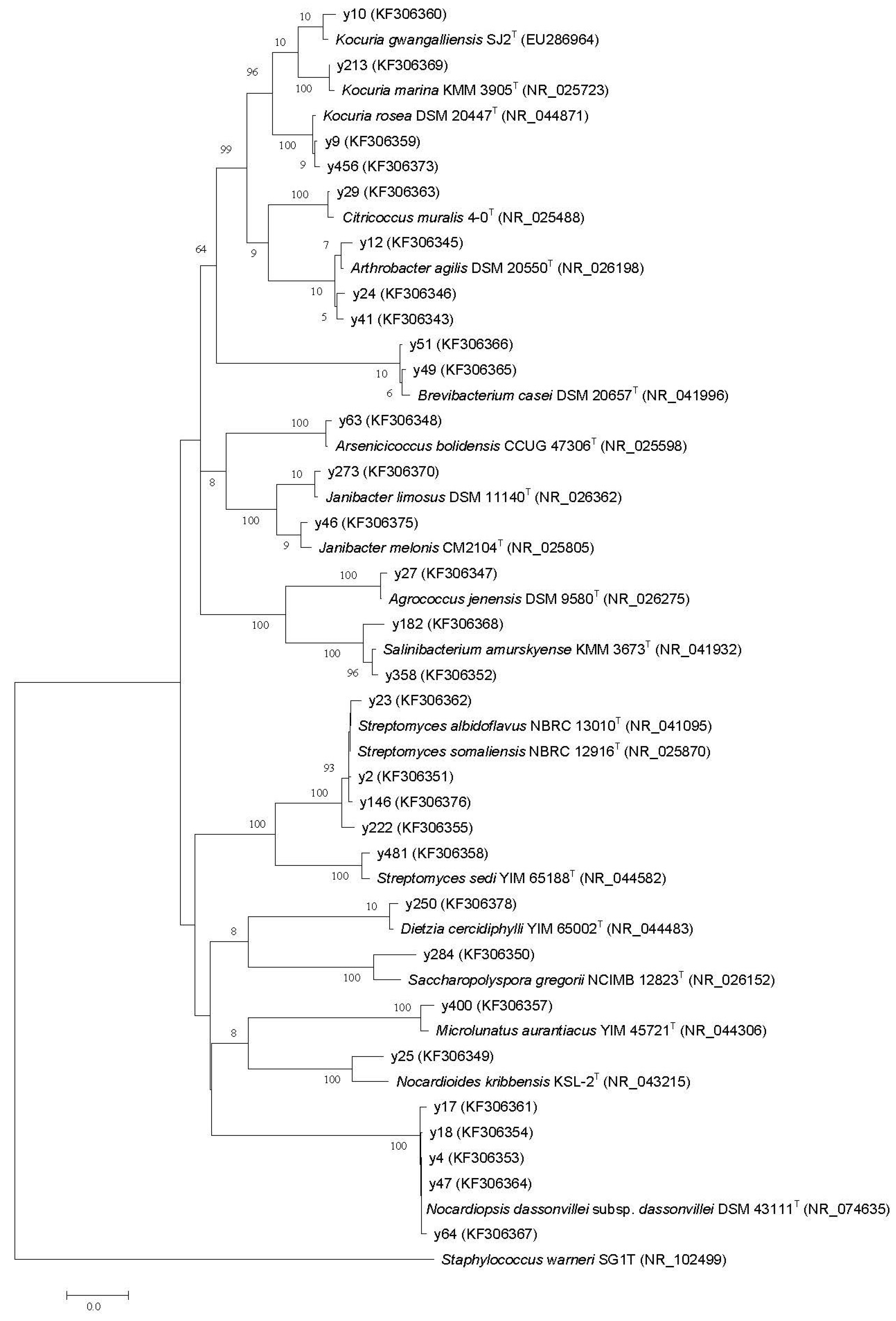
2.2. Phylogenetic Diversity

The nearly complete 16S rRNA gene sequences (1479 bp to 1506 bp) of the 30 representatives were PCR-amplified and sequenced. The results of the 16S rRNA gene sequences analysis are shown in Figure 1 and Table 1. Based on similarity criteria of 98% at the 16S rRNA gene, the 30 isolates were sorted into 18 phylotypes, belonging to 14 different genera, and fell into 10 family level groupings: Brevibacteriaceae, Dietziaceae, Intrasporangiaceae, Microbacteriaceae, Micrococcaceae, Nocardioidaceae, Nocardiopsaceae, Propionibacteriaceae, Pseudonocardiaceae and Streptomycetaceae. Twenty-eight of the 30 isolates shared 99.2% to 99.9% sequence similarities with the closest type strains. The other two strains, y25 and y284, quite likely represented novel species with less than 98% 16S rRNA gene sequence identity to the valid published species [21,22,23]. Interestingly, we found that strain y400 was assigned to the genus, Microlunatus, and was most closely related to the type strain of Microlunatus aurantiacus YIM 45721T isolated from a rhizosphere soil sample in Yunnan Province, China [24]. To our knowledge, this study is the first to report on the isolation of Microlunatus genus members from a marine environment. These data indicated the considerable diversity of Actinobacteria within the Chukchi Shelf sediments.
Table 1. Phylogenetic identity, seawater requirement, biosynthetic genes and antimicrobial activities in the actinobacterial isolates.
GenusIsolateThe Closest Type Strain
(% Identity)
Seawater RequirementBiosynthetic GenesAntimicrobial Activities *
PKS IPKS IINRPSphzEdTGDHaloCYPB. subtilisC. albicans
Brevibacteriaceae
Brevibacteriumy49Brevibacterium casei NCDO 2048T (99.3)++++
y51Brevibacterium casei NCDO 2048T (99.4)+++
Dietziaceae
Dietziay250Dietzia cercidiphylli YIM 65002T (99.6)++++
Intrasporangiaceae
Arsenicicoccusy63Arsenicicoccus bolidensis CCUG 47306T (99.8)++
Janibactery46Janibacter melonis CM2104T (99.5)+++
y273Janibacter limosus DSM 11140T (99.7)+++++
Microbacteriaceae
Agrococcusy27Agrococcus jenensis DSM 9580T (99.7)++
Salinibacteriumy182Salinibacterium amurskyense KMM 3673T (99.0)++++
y358Salinibacterium amurskyense KMM 3673T (99.7)++
Micrococcaceae
Arthrobactery12Arthrobacter agilis DSM 20550T (99.5)+++
y24Arthrobacter agilis DSM 20550T (99.3)+++
y41Arthrobacter agilis DSM 20550T (99.2)++++
Citricoccusy29Citricoccus muralis 4-0T (99.7)++++
Kocuriay9Kocuria rosea DSM 20447T (99.7)+++
y456Kocuria rosea DSM 20447T (99.7)++++
y10Kocuria gwangalliensis SJ2T (99.4)++
y213Kocuria marina KMM 3905T (99.9)++
Nocardioidaceae
Nocardioidesy25Nocardioides kribbensis KSL-2T (97.8)+++
Nocardiopsaceae
Nocardiopsisy4Nocardiopsis dassonvillei subsp. dassonvillei DSM 43111T (ABUI01000017)++++++
y17Nocardiopsis dassonvillei subsp. dassonvillei DSM 43111T (99.7)++++++
y18Nocardiopsis dassonvillei subsp. dassonvillei DSM 43111T (99.8)+++++++
y47Nocardiopsis dassonvillei subsp. dassonvillei DSM 43111T (99.9)++++
y64Nocardiopsis dassonvillei subsp. dassonvillei DSM 43111T (99.8)++++
Propionibacteriaceae
Microlunatusy400Microlunatus aurantiacus YIM 45721T (99.2)+++++
Pseudonocardiaceae
Saccharopolysporay284Saccharopolyspora gregorii NCIMB 12823T (97.8)++
Streptomycetaceae
Streptomycesy2 *Streptomyces somaliensis NBRC 12916T (99.9)++++++
y23Streptomyces somaliensis NBRC 12916T (99.7)+++++
y146 *Streptomyces somaliensis NBRC 12916T (99.9)++++++
y222Streptomyces albidoflavus DSM 40455T (99.5)+++++++
y481Streptomyces sedi YIM 65188T (99.4)++++++
Grew better on ISP2 media prepared with seawater than on those prepared with deionized water plus 3.2% NaCl and on those prepared with deionized water; * y146 showed anti-E. coli activity; y2 showed anti-S. aureus activity; no isolates showed activities against Gram-negative bacterium Pseudomonas aeruginosa; +, positive; −, negative.
Figure 1. The phylogenetic relationship of the actinobacterial isolates based on 16S rRNA gene homology. The tree was constructed using the neighbor-joining method with Kimura two-state parameters and pairwise-deletion model analyses, which were implemented in the Molecular Evolutionary Genetics Analysis (MEGA), version 5.0 program. The resultant tree topologies were evaluated by bootstrap analysis based on 1000 replicates. The numbers at nodes represent the percentage levels of bootstrap support (%). The GenBank accession numbers of 16S rRNA sequences are given in the parentheses. Bar = 2% sequence divergence.
Marinedrugs 12 01281 g001

2.3. Antimicrobial Activities and Detection of Biosynthetic Genes

Actinobacteria are well known for their ability to produce secondary metabolites, many of which are active against pathogenic microorganisms. Some of the closest neighbors of our isolates were found to produce biologically active compounds. Dietzia cercidiphylli was found to produce biosurfactants [25]. Helquinoline, a new tetrahydroquinoline antibiotic, was isolated from Janibacter melonis [26]. Two strains of Arthrobacter agilis produced potent antibacterial compounds with activity against Gram-positive bacteria and possibly related to novel cyclic thiazolyl peptides [27]. Fu et al. found that Nocardiopsis dassonvillei subsp. dassonvillei could produce α-pyrones [28]. Streptomyces albidoflavus was found to produce antifungal compounds, such as a non-polyene substance [29], Antimycin A18 [30], etc. The 30 selected actinobacterial isolates obtained from the Chukchi Shelf sediments, which belong to 14 actinobacterial genera, were tested for antimicrobial activities against Gram-positive bacteria Staphylococcus aureus and Bacillus subtilis, Gram-negative bacteria Escherichia coli and Pseudomonas aeruginosa and the yeast, Candida albicans. The results are presented in Table 1. Of the 30 isolates, 11 exhibited antibacterial and/or antifungal activity. Among the 11 antimicrobial isolates, seven showed activity against both Gram-positive bacterium Bacillus subtilis and yeast Candida albicans. Only one isolate, y146, and one isolate, y2, had activity against Gram-negative E. coli and Gram-positive bacteria Staphylococcus aureus, respectively. Furthermore, the data clearly show that strains that can produce antibiotic compounds belonging to three genera: Streptomyces, Nocardiopsis and Microlunatus.
A PCR-based screening for the presence of genes associated with biosynthetic pathways can rapidly predict the unknown strains that have the potential to produce secondary metabolites. Therefore, in parallel with antimicrobial activity testing, we attempted the PCR-based approach to detect type I polyketide synthase (PKS I), PKS II, nonribosomal peptide synthase (NRPS), aminodeoxyisochorismate synthase (phzE), dTDP-glucose-4, 6-dehydratase (dTGD), halogenase (Halo) and cytochrome P450 hydroxylase (CYP) genes. Several amplified gene fragments were cloned and sequenced, and subsequent analysis confirmed that these genes encode parts of the expected biosynthetic enzymes [31]. Thus, all isolates have at least two secondary-metabolite biosynthetic genes, and one-third of the isolates possess more than four biosynthetic genes (Table 1). The number of strains that exhibited a certain gene with the corresponding number of phylotypes they represent are as follows: PKS I loci, 21 (70%) strains and 13 phylotypes; PKS II loci, 11 (36.7%) strains and seven phylotypes; NRPS loci, 13 (43.3%) strains and eight phylotypes; phzE loci, six (20%) strains and four phylotypes; dTGD loci, 24 (80%) strains and 15 phylotypes; Halo loci, 12 (40%) strains and 11 phylotypes; and CYP loci, 10 (33.3%) strains and six phylotypes. These results suggested that the 6-deoxyhexoses glycosylation biosynthetic and type I PKS pathways were widespread in the Chukchi Shelf sediment-derived Actinobacteria. We successfully amplified secondary metabolite biosynthetic gene fragments from the genomes of these isolates with detectable antimicrobial activity. However, most of the non-active isolates have at least one kind of PKS I, PKS II, NRPS and phzE gene involved in the backbone biosynthesis of secondary metabolites and one of the dTGD and Halo genes encoding modification enzymes responsible for the introduction and generation of diversity and activity in several secondary metabolites [32,33]. The CYP gene, which encodes the key enzyme, cytochrome P450 hydroxylase, in polyene antibiotics biosynthesis [34] could be detected in several strains of Janibacter, Salinibacterium and Arthrobacter genera without antimicrobial activity. We also sequenced the PCR products of the ketosynthase (KS) domain of PKS II from five Nocardiopsis strains genomes. The KS sequences from these strains displayed the amino acid sequence identities (67% to 69%) of the KS domain of fabF involved in the biosynthesis of pradimicin, an antifungal antibiotic from Actinomadura hibisca [35], or had 68% similarity with TamM, which is related to the biosynthesis of Tetarimycin A, a tetracyclic methicillin-resistant Staphylococcus aureus (MRSA)-active antibiotic [36]. These results revealed that the Actinobacteria associated with the sediments of the Chukchi Shelf possess the potential ability to produce diversely bioactive secondary metabolites. We will attempt to use a variety of media and culture conditions to induce the expression of secondary metabolite gene clusters in these isolates. Other bioactivities of the isolates, e.g., anticancer activity or immunosuppressive activities, will also be analyzed. Furthermore, in contrast to the genus, Streptomyces, only a limited number of secondary metabolite biosynthesis gene clusters in rare Actinobacteria have been studied [37]. The biosynthetic gene, such as dTGD or Halo, from our rare actinobacterial isolates may provide novel material for genetic manipulation and combinatorial biosynthesis, which may lead to the generation of pharmaceutical compounds.

3. Experimental Section

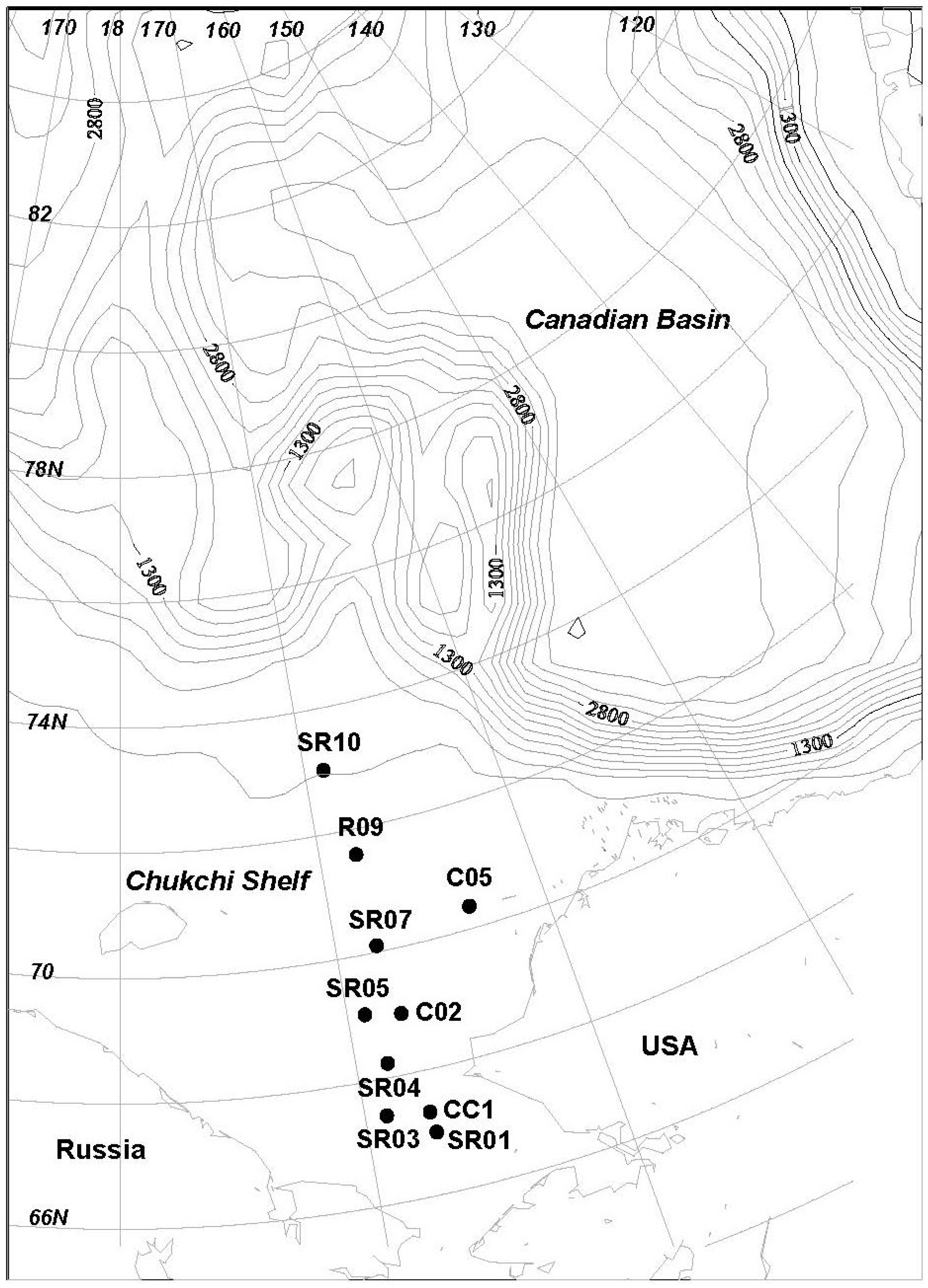
3.1. Sediments Sample

A total of 10 sediment samples were collected during the 4th Chinese National Arctic Research Expedition cruise of the icebreaker, Xue Long, into the Arctic Ocean during the summer of 2010 (Figure 2, Table 2). The sediments were sampled using the Ekman-bottom grab. Only the upper 3 cm of the sediment were stored in sterilized 500 mL plastic zip-lock bags and transported to the laboratory at temperatures between 0 °C and 4 °C.
Table 2. Characteristics of Chukchi Shelf sediments in the Arctic Ocean.
SamplesWater Depth (m)ColorCompositionBenthos
CC145Light greySilty clayBivalves
C0241GreyClayConch
C0526CinerousFine sandCrab, conch, sand dollar, Polychaeta
SR0141.5CinerousSilty sandConch, bivalve
SR0350.7CinerousClayPolychaeta, conch, sipunculoid
SR0448GreySilty clayNone
SR0546.7CinerousSilty clayConch
SR0729.7CinerousSilty mudNone
R0943.5GreyMud soilNone
SR1069.6CinerousSilty claySea star, conch
Figure 2. Sediment sampling locations.
Marinedrugs 12 01281 g002

3.2. Isolation of Actinobacterial Strains

The sediment samples were treated through DDC [38] and a water bath at 55 °C for 6 min. An aliquot of 200 μL of the pretreated 100, 10−1 and 10−2 dilution was spread over the surface of the isolation media in Petri dishes. The following four isolation media were used:
M1: 0.4 g peptone, 2 g yeast extract, malt extract 2 g, 15 g agar and 1 L sea water.
SC: 10 g soluble starch, 1 g casein, 2 g dipotassium phosphate, 0.02 g calcium carbonate, 2 g potassium nitrate, 2 g sodium chloride, 0.05 g magnesium sulfate heptahydrate, 0.01 g ferrous sulfate heptahydrate, 15 g agar and 1 L sea water.
ISP2: 4 g glucose, 4 g yeast extract, 10 g malt extract, 15 g agar and 1 L sea water.
MR2A: 0.5 g yeast extract, 0.5 g peptone, 0.5 g casein hydrolysate, 0.5 g glucose, 0.5 g soluble starch, 0.3 g sodium pyruvate, 15 g agar and 1 L sea water.
All isolation media were prepared with 1 L natural seawater and amended with filtered (0.2 μm pore size) nalidixic acid (25 μg/mL), nystatin (50 μg/mL) and cycloheximide (50 μg/mL) to inhibit the growth of Gram-negative bacteria and fungi. The isolation plates were incubated in the dark at 28 °C for 4 weeks. Colonies from various agar plates were selected based on differing colony morphologies. Isolates were obtained in pure culture after three successive transfers to fresh isolation media. Pure cultures were frozen in 20% glycerol at −80 °C for long-term storage.

3.3. Dereplication by Rep-Polymerase Chain Reaction (PCR)

Repetitive DNA fingerprinting was performed on the actinobacterial isolates from the same sample. Genomic DNA was prepared using a bacterial genomic DNA kit (TIANGENE, Beijing, China), according to the supplied protocol from each isolate. PCR amplification was performed using the BOXA1R primer (5′-CTA CGG CAA GGC GAC GCT GAC G-3′) [33]. The PCR reaction mixture (25 μL) contained: 1 μL genomic DNA extract, 2.5 μL 10× buffer, 2 μL deoxynucleotide triphosphate (dNTP) mixture (2.5 mM), 2 μL BOXA1R primer (25 μM) and 2 U Taq DNA polymerase. The reaction started with pre-denaturation at 95 °C for 8 min followed by 30 cycles of denaturation at 95 °C for 1 min, annealing at 53 °C for 1 min and 4 min of extension at 65 °C, with a final extension at 65 °C for 8 min. PCR products were separated by 2% agarose gel electrophoresis. The gels were analyzed using the program, Quantity one (Bio-Rad). The similarity between patterns was calculated using Pearson coefficients. The clustering pattern was generated using the unweighted pair group method with the arithmetic mean algorithm. If the strains formed a cluster defined at the 72% similarity level, they would be assigned to a single species [39,40].

3.4. Seawater Requirements

The dereplicated actinobacterial isolates were tested for the required seawater for growth. Frozen stored strains were inoculated onto the ISP2 agar medium prepared using seawater (MISP2). Once sufficient growth had occurred, a sterile wire loop was used to transfer cell material onto a new plate of the same medium prepared with seawater, a plate prepared with deionized water plus 3.2% NaCl and a plate prepared with deionized water. The plates were incubated at 28 °C for up to 4 weeks. Growth was monitored under a stereomicroscope at 64-fold magnification. If no growth was observed on the plate prepared with deionized water, that strain was considered to require seawater for growth.

3.5. 16S rRNA Gene Amplification and Phylogenetic Analysis

The 16S rRNA gene was amplified by PCR using primers 8F (5′-AGA GTT TGA TCC TGG CTC AG-3′) and 1492R (5′-GGT TAC CTT GTT ACG ACT T-3′) [41]. The reaction mixture (50 μL) contained: 2 μL genomic DNA extract, 5 μL 10× buffer, 4 μL BSA (1 mg/mL), 1 μL dNTP mixture (2.5 mM), 1 μL each primer (10 μM) and 3.5 U Taq DNA polymerase 3.5 U. PCR conditions were as follows: 6 min at 94 °C followed by 35 cycles of 45 s at 94 °C, 45 s at 55 °C and 1.5 min at 72 °C, followed by an 8-min extension at 72 °C. PCR amplification was performed in an Eppendorf Mastercycler Gradient. An ABI PRISM 3730 sequencer at Shanghai Majorbio Company was used to sequence the PCR products. The identification of phylogenetic neighbors and the calculation of pairwise 16S rDNA sequence similarities were achieved using the EzTaxon-e server [42,43]. In addition, the sequences were compared to the sequences within the NCBI database [44] using the BLASTN algorithm. Sequences were aligned, and a phylogenetic tree was constructed using the Molecular Evolutionary Genetics Analysis (MEGA) software, version 5.0 [45]. DNA sequences were deposited in GenBank under accession numbers: KF306343, KF306345–KF306355, KF306357–KF306370, KF306373, KF306375, KF306376 and KF306378.

3.6. Antimicrobial Activity Testing by Agar Diffusion (Inhibition Zones)

Actinobacterial isolates were transferred to the plates containing three different production media: PM3 (20 g oatmeal, 2.5 g glycerol, 0.1 mg FeSO4·7H2O, 0.1 mg MnCl2·4H2O, 0.1 mg ZnSO4·7H2O, 15 g agar and 1 L sea water) [15], PM4 (0.5 g glucose, 2.5 g glycerol, 5.0 g oatmeal, 5.0 g soybean meal, 0.5 g yeast extract, 2.0 g casamino acids, 10 g agar and 1 L sea water) [15] and GYM4 (10 g glucose, 4 g yeast extract, 4 g malt extract, 15 g agar, 1 L sea water and pH 7.2) [46]. The plates were incubated at 28 °C for 1 week to 4 weeks, depending on the growth rate of the isolates. Then, the agar blocks (diameter: 8 mm) that contained actinobacterial cells were excised and placed on the assay plates spread with test microbial strains. Two Gram-positive bacterial strains, Staphylococcus aureus ATCC 29213 and Bacillus subtilis CMCC 63501, two Gram-negative bacterial strains, Escherichia coli ATCC 44102 and Pseudomonas aeruginosa ATCC 27853, and one yeast strain, Candida albicans ATCC 10231, were used as test strains. The antimicrobial activities were expressed as inhibition zones after 16 h of incubation at 28 °C.

3.7. Amplification of Biosynthetic Gene Fragments

To amplify the genes for the KS domains of PKS I, the KS domains of PKS II, the adenylation domains of NRPS, the enzyme PhzE of the phenazine pathways, the enzyme dTGD of 6-deoxyhexoses glycosylation pathway, the enzyme Halo of halogenation pathway and the enzyme CYP in polyene polyketide biosynthesis, the degenerate primers as described previously were used (Table 3). The components and reaction conditions of the PCR mixture are as follows:
PKS I (50 μL): 2 μL template, 5 μL 10× buffer, 2.5 μL BSA (1 mg/mL), 2 μL dNTP (2.5 mM), 1 μL each primer (10 μM) and 2.5 U Taq DNA polymerase; 5 min at 95 °C, followed by 35 cycles of 1 min at 95 °C, 1 min at 60 °C and 2 min at 72 °C, followed by a 5-min extension at 72 °C.
PKS II (50 μL): 2 μL template, 5 μL 10× buffer (Mg2+ free), 2 μL MgCl2 (25 mM), 4 μL BSA (1 mg/mL), 4 μL dNTP (2.5 mM), 5 μL each primer (10 μM) and 2.5 U Taq DNA polymerase; 5 min at 95 °C, followed by 40 cycles of 1 min at 95 °C, 1 min at 64 °C and 1.5 min at 72 °C, followed by a 15-min extension at 72 °C.
NRPS (50 μL): 2 μL template, 5 μL 10× buffer, 2.5 μL BSA (1 mg/mL), 2 μL dNTP (2.5 mM), 1 μL each primer (25 μM) and 2.5 U Taq DNA polymerase; 5 min at 95 °C, followed by 35 cycles of 30 s at 95 °C, 2 min at 59 °C and 4 min at 72 °C, followed by a 10-min extension at 72 °C.
phzE (25 μL): 1 μL template, 2.5 μL 10× buffer, 2 μL BSA (1 mg/mL), 2 μL dNTP (2.5 mM), 0.5 μL each primer (10 μM) and 1 U Taq DNA polymerase; 2 min at 94 °C, followed by 36 cycles of 1 min at 94 °C, 1 min at 54.7 °C and 2 min at 72 °C, followed by a 7-min extension at 72 °C.
dTGD (20 μL): template 0.7 μL, 10× buffer 2.5 μL, BSA (1 mg/mL) 2 μL, dNTP (2.5 mM) 2 μL, each primer (10 μM) 1.5 μL and 3.5 U Taq DNA polymerase; 4 min at 98 °C, followed by 30 cycles of 40 s at 96 °C, 40 s at 50 °C and 1.5 min at 72 °C, followed by a 5-min extension at 72 °C.
Halo (50 μL): template 2 μL, 10× buffer 5 μL, BSA (1 mg/mL) 2.5 μL, dNTP (2.5 mM) 2 μL, each primer (25 μM) 1 μL and 2.5 U Taq DNA polymerase; 3 min at 94 °C, followed by 30 cycles of 1 min at 94 °C, 1.5 min at 58 °C and 1 min at 72 °C, followed by a 5-min extension at 72 °C.
CYP (50 μL): 1 μL template, 5 μL 10× buffer (Mg2+ free), 2 μL MgCl2 (25 mM), 5 μL BSA (1 mg/mL), 4 μL dNTP (2.5 mM), 1 μL each primer (20 μM) and 2.5 U Taq DNA polymerase; 5 min at 96 °C, followed by 45 cycles of 1 min at 96 °C, 30 s at 60 °C and 45 s at 72 °C, followed by a 5-min extension at 72 °C.
Table 3. Primers of secondary metabolite biosynthetic genes.
GenePrimerLength (bp)Reference
PKS I (KSMA-F, KSMB-R)5′-TSGCSATGGACCCSCAGCAG-3′
5′-CCSGTSCCGTGSGCCTCSAC-3′
700[47]
PKS II (540F, 1100R)5′-GGITGCACSTCIGGIMTSGAC-3′
5′-CCGATSGCICCSAGIGAGTG-3′
554[48]
NRPS (A3F, A7R)5′-GCSTACSYSATSTACACSTCSGG-3′
5′-SASGTCVCCSGTSCGGTAS-3′
700[49]
PhzE (phzEf, phzEr)5′-GAAGGCGCCAACTTCGTYATCAA-3′
5′-GCCYTCGATGAAGTACTCGGTGTG-3′
450[50]
Halo (FW, RV)5′-TTCCCSCGSTACCASATCGGSGAG-3′
5′-GSGGGATSWMCCAGWACCASCC-3′
500[32]
dTGD (dTGD-1, dTGD-2)5′-GSGGSGSSGCSGGSTTCATSGG-3′
5′-GGGWRCTGGYRSGGSCCGTAGTTG-3′
600[51]
CYP (PEH-1, PEH-2)5′-TGGATCGGCGACGACCGSVYCGT-3′
5′-CCGWASAGSAYSCCGTCGTACTT-3′
350[34]

4. Conclusions

A total of 73 actinobacterial strains have been isolated from the Chukchi Shelf sediments in the Arctic Ocean. We used repetitive DNA fingerprinting analysis to select 30 representatives for further investigation. An unexpected variety of actinomycete genera were isolated. The 30 representatives were sorted into 18 phylotypes belonging to 14 different genera. Moreover, 25 of the 30 representatives are rare actinomycetes. We also screened them for biosynthetic genes and antimicrobial activities. Of the 30 isolates, 11 exhibited antibacterial and/or antifungal activity, seven of which have activities against Bacillus subtilis and Candida albicans. All representatives have at least two biosynthetic genes, and one-third of these representatives possess more than four biosynthetic genes. The KS sequences from five Nocardiopsis strains with antimicrobial activity displayed the amino acid sequence identities (67% to 69%) of the KS domain of fabF involved in the biosynthesis of pradimicin or had 68% similarity with TamM, related to the biosynthesis of Tetarimycin A. This study demonstrates the significant diversity of Actinobacteria in the Chukchi Shelf sediment. The new strains might represent a valuable source of biologically active compounds with antimicrobial activity and genes for their biosynthesis.

Abbreviations

DDC
Dispersion and Differential Centrifugation
BSA
Albumin from Bovine Serum
NCBI
National Center of Biotechnology Information
BLASTN
Basic Local Alignment Search Tool of Nucleic acid
PKS
PolyKetide Synthase
NRPS
Nonribosomal Peptide Synthase
phzE
aminodeoxyisochorismate synthase
dTGD
dTDP-Glucose-4, 6-Dehydratase
Halo
Halogenase
CYP
Cytochrome P450 hydroxylase
FADH
Flavin Adenine Dinucleotide Hydrogen carrier

Acknowledgments

We thank the Chinese Arctic and Antarctic Administration (CAA) who assisted in the field work of this study. This study was supported by the National Natural Science Foundation of China (No. 41276175), the Natural Science Foundation of Shanghai (No. 11ZR1441000), the National High Technology Research and Development Program (“863” Program) of China (No. 2012AA021706) and the Chinese Polar Environment Comprehensive Investigation and Assessment Programs (No. CHINARE2013-02-01).

Conflicts of Interest

The authors declare no conflict of interest.

References

  1. Ventura, M.; Canchaya, C.; Tauch, A.; Chandra, G.; Fitzgerald, G.F.; Chater, K.F.; van Sinderen, D. Genomics of Actinobacteria: Tracing the evolutionary history of an ancient phylum. Microbiol. Mol. Biol. Rev. 2007, 71, 495–548. [Google Scholar] [CrossRef]
  2. Bérdy, J. Bioactive microbial metabolites. J. Antibiot. 2005, 58, 1–26. [Google Scholar] [CrossRef]
  3. Fiedler, H.-P.; Bruntner, C.; Bull, A.T.; Ward, A.C.; Goodfellow, M.; Potterat, O.; Puder, C.; Mihm, G. Marine actinomycetes as a source of novel secondary metabolites. Antonie van Leeuwenhoek 2005, 87, 37–42. [Google Scholar] [CrossRef]
  4. Lam, K.S. Discovery of novel metabolites from marine actinomycetes. Curr. Opin. Microbiol. 2006, 9, 245–251. [Google Scholar] [CrossRef]
  5. Bull, A.T.; Stach, J.E.M. Marine actinobacteria: New opportunities for natural product search and discovery. Trends Microbiol. 2007, 15, 491–499. [Google Scholar] [CrossRef]
  6. Stach, J.E.M.; Bull, A.T. Estimating and comparing the diversity of marine actinobacteria. Antonie van Leeuwenhoek 2005, 87, 3–9. [Google Scholar] [CrossRef]
  7. Goodfellow, M.; Fiedler, H.-P. A guide to successful bioprospecting: Informed by actinobacterial systematics. Antonie van Leeuwenhoek 2010, 98, 119–142. [Google Scholar] [CrossRef]
  8. Fenical1, W.; Jensen, P.R. Developing a new resource for drug discovery: Marine actinomycete bacteria. Nat. Chem. Biol. 2006, 2, 666–673. [Google Scholar] [CrossRef]
  9. Olano, C.; Méndez, C.; Salas, J.A. Antitumor compounds from marine actinomycetes. Mar. Drugs 2009, 7, 210–248. [Google Scholar] [CrossRef]
  10. Fattorusso, E.; Taglialatela-Scafati, O. Marine antimalarials. Mar. Drugs 2009, 7, 130–152. [Google Scholar] [CrossRef]
  11. Rahman, H.; Austin, B.; Mitchell, W.J.; Morris, P.C.; Jamieson, D.J.; Adams, D.R.; Spragg, A.M.; Schweizer, M. Novel anti-infective compounds from marine bacteria. Mar. Drugs 2010, 8, 498–518. [Google Scholar] [CrossRef]
  12. Xiong, Z.-Q.; Wang, J.-F.; Hao, Y.-Y.; Wang, Y. Recent advances in the discovery and development of marine microbial natural products. Mar. Drugs 2013, 11, 700–717. [Google Scholar] [CrossRef]
  13. Weyland, H. Distribution of Actinomycetes on the Sea Floor. In Actinomycetes: Proceedings of the Fourth International Symposium on Actinomycete Biology, Cologne, 3–7 September 1979; Gustav Fischer Verlag: Stuttgart, Germany, 1981; pp. 185–193. [Google Scholar]
  14. Yu, Y.; Li, H.; Zeng, Y.; Chen, B. Isolation and phylogenetic assignation of actinomycetes in the marine sediments from the Arctic Ocean. Acta Oceanol. Sin. 2005, 24, 135–142. [Google Scholar]
  15. Bredholdt, H.; Galatenko, O.A.; Engelhardt, K.; Tjaervik, E.; Terekhova, L.P.; Zotchev, S.B. Rare actinomycete bacteria from the shallow water sediments of the Trondheim fjord, Norway: Isolation, diversity and biological activity. Environ. Microbiol. 2007, 9, 2756–2764. [Google Scholar] [CrossRef]
  16. Bredholdt, H.; Tjaervik, E.; Johnsen, G.; Zotchev, S.B. Actinomycetes from sediments in the Trondheim fjord, Norway: Diversity and biological activity. Mar. Drugs 2008, 6, 12–24. [Google Scholar] [CrossRef]
  17. Jørgensen, H.; Fjærvik, E.; Hakvåg, S.; Bruheim, P.; Bredholt, H.; Klinkenberg, G.; Ellingsen, T.E.; Zotchev, S.B. Candicidin biosynthesis gene cluster is widely distributed among Streptomyces spp. isolated from the sediments and the neuston layer of the Trondheim fjord, Norway. Appl. Environ. Microbiol. 2009, 75, 3296–3303. [Google Scholar] [CrossRef]
  18. Jørgensen, H.; Degnes, K.F.; Dikiy, A.; Fjærvik, E.; Klinkenberg, G.; Zotchev, S.B. Insights into the evolution of macrolactam biosynthesis through cloning and comparative analysis of the biosynthetic gene cluster for a novel macrocyclic lactam, ML-449. Appl. Environ. Microbiol. 2010, 76, 283–293. [Google Scholar] [CrossRef]
  19. Engelhardt, K.; Degnes, K.F.; Kemmler, M.; Bredholt, H.; Fjærvik, E.; Klinkenberg, G.; Sletta, H.; Ellingsen, T.E.; Zotchev, S.B. Production of a new thiopeptide antibiotic, TP-1161, by a marine Nocardiopsis sspecies. Appl. Environ. Microbiol. 2010, 76, 4969–4976. [Google Scholar] [CrossRef] [Green Version]
  20. Hakvåg, S.; Fjærvik, E.; Josefsen, K.D.; Ian, E.; Ellingsen, T.E.; Zotchev, S.B. Characterization of Streptomyces spp. isolated from the sea surface microlayer in the Trondheim fjord, Norway. Mar. Drugs 2008, 6, 620–635. [Google Scholar] [CrossRef]
  21. Wayne, L.G.; Brenner, D.J.; Colwell, R.R.; Grimont, P.A.D.; Kandler, O.; Krichevsky, M.I.; Moore, L.H.; Moore, W.E.C.; Murray, R.G.E.; Stackebrandt, E.; et al. Report of the ad hoc committee on reconciliation of approaches to bacterial systematics. Int. J. Syst. Evol. Microbiol. 1987, 37, 463–464. [Google Scholar]
  22. Stackebrandt, E.; Goebel, B.M. Taxonomic note: A place for DNA–DNA reassociation and 16S rRNA sequence analysis in the present species definition in bacteriology. Int. J. Syst. Evol. Microbiol. 1994, 44, 846–849. [Google Scholar]
  23. Stackebrandt, E.; Ebers, J. Taxonomic parameters revisited: Tarnished gold standards. Microbiol. Today 2006, 33, 152–155. [Google Scholar]
  24. Wang, Y.X.; Cai, M.; Zhi, X.Y.; Zhang, Y.Q.; Tang, S.K.; Xu, L.H.; Cui, X.L.; Li, W.J. Microlunatus aurantiacus sp. nov., a novel actinobacterium isolated from a rhizosphere soil sample. Int. J. Syst. Evol. Microbiol. 2008, 58, 1873–1877. [Google Scholar] [CrossRef]
  25. Chen, L.; Li, S.U.; Xue, Y.F. Characterization of an alkaliphilic biosurfactant- producing strain of Dietzia. Microbiol. China 2012, 39, 1573–1579. [Google Scholar]
  26. Ratnakar, N.A.; Dirk, S.; Rolf, H.; Siegmund, L.; Irene, W.D.; Hartmut, L. Helquinoline, a new tetrahydroquinoline antibiotic from Janibacter limosus Hel 1+. J. Antibiot. 2004, 57, 17–23. [Google Scholar] [CrossRef]
  27. Rojas, J.L; Martín, J.; Tormo, J.R.; Vicente, F.; Brunati, M.; Ciciliato, I.; Losi, D.; Van, T.S.; Mergaert, J.; Swings, J.; et al. Bacterial diversity from benthic mats of Antarctic lakes as a source of new bioactive metabolites. Mar. Genomics 2009, 2, 33–41. [Google Scholar] [CrossRef]
  28. Fu, P.; Liu, P.; Gong, Q.H.; Wang, Y.; Wang, P.; Zhu, W.M. α-Pyrones from the marine-derived actinomycete Nocardiopsis dassonvillei subsp. dassonvillei XG-8-1. RSC Adv. 2013, 3, 20726–20731. [Google Scholar]
  29. Augustine, S.K.; Bhavsar, S.P.; Kapadnis, B.P. A non-polyene antifungal antibiotic from Streptomyces albidoflavus PU 23. J. Biosci. 2005, 30, 201–211. [Google Scholar] [CrossRef]
  30. Yan, L.L.; Han, N.N.; Zhang, Y.Q.; Yu, L.Y.; Chen, J.; Wei, Y.Z.; Li, Q.P.; Tao, L.; Zheng, G.H.; Yang, S.E.; et al. Antimycin A18 produced by an endophytic Streptomyces albidoflavus isolated from a mangrove plant. J. Antibiot. 2010, 63, 259–261. [Google Scholar] [CrossRef]
  31. Yuan, M.; Dong, N.; Li, H.R.; Yu, Y. Sequence-based analysis of secondary-metabolite biosynthesis in actinobacteria isolated from Chukchi shelf marine sediments. Adv. Polar Sci. 2014. submitted for publication. [Google Scholar]
  32. Hornung, A.; Bertazzo, M.; Dziarnowski, A.; Schneider, K.; Welzel, K.; Wohlert, S.E.; Holzenkämpfer, M.; Nicholson, G.J.; Bechthold, A.; Süssmuth, R.D.; et al. A Genomic screening approach to the structure-guided identification of drug candidates from natural sources. Chembiochem 2007, 8, 757–766. [Google Scholar] [CrossRef]
  33. Stockmann, M.; Piepersberg, W. Gene probes for the detection of 6-deoxyhexose metabolism in secondary metabolite-producing streptomycetes. FEMS Microbiol. Lett. 1992, 90, 185–189. [Google Scholar] [CrossRef]
  34. Lee, M.Y.; Myeong, J.S.; Park, H.J.; Han, K.; Kim, E.S. Isolation and partial characterization of a cryptic polyene gene cluster in Pseudonocardia autotrophica. J. Ind. Microbiol. Biotechnol. 2006, 33, 84–87. [Google Scholar] [CrossRef]
  35. Dairi, T.; Hamano, Y.; Igarashi, Y.; Furumai, T.; Oki, T. Cloning and nucleotide sequence of the putative polyketide synthase genes for pradimicin biosynthesis from Actinomadura hibisca. Biosci. Biotechnol. Biochem. 1997, 61, 1445–1453. [Google Scholar] [CrossRef]
  36. Kallifidas, D.; Kang, H.S.; Brady, S.F. Tetarimycin A, an MRSA-Active antibiotic identified through induced expression of environmental DNA gene clusters. J. Am. Chem. Soc. 2012, 134, 19552–19555. [Google Scholar] [CrossRef]
  37. Clardy, J.M.; Fischbach, A.; Walsh, C.T. New antibiotics from bacterial natural products. Nat. Biotechnol. 2006, 24, 1541–1550. [Google Scholar] [CrossRef]
  38. Hopkins, D.W.; McNaughton, S.J.; O’Donnell, A.G. A dispersion and differential centrifugation technique for representatively sampling microorganisms from soil. Soil Biol. Biochem. 1991, 23, 217–225. [Google Scholar] [CrossRef]
  39. Versalovic, J.; Schneider, M.; de Bruijn, F.J.; Lupski, J.R. Genomic fingerprinting of bacteria using repetitive sequencebased polymerase chain reaction. Methods Mol. Cell. Biol. 1994, 5, 24–40. [Google Scholar]
  40. Rademaker, J.L.W.; Hoste, B.; Louws, F.J.; Kersters, K.; Swings, J.; Vauterin, L.; Vauterin, P.; de Bruijn, F.J. Comparison of AFLP and rep-PCR genomic fingerprinting with DNA–DNA homology studies: Xanthomonas as a model system. Int. J. Syst. Evol. Microbiol. 2000, 50, 665–677. [Google Scholar] [CrossRef]
  41. Lane, D.J. 16S/23S rRNA Sequencing. In Nucleic Acid Techniques in Bacterial Systematics; Stackebrandt, E., Goodfellow, M., Eds.; Wiley: Chichester, UK, 1991; pp. 115–175. [Google Scholar]
  42. Kim, O.S.; Cho, Y.J.; Lee, K.; Yoon, S.H.; Kim, M.; Na, H.; Park, S.C.; Jeon, Y.S.; Lee, J.H.; Yi, H.; et al. Introducing EzTaxon-e: A prokaryotic 16S rRNA Gene sequence database with phylotypes that represent uncultured species. Int. J. Syst. Evol. Microbiol. 2012, 62, 716–721. [Google Scholar] [CrossRef]
  43. EzTaxon Server. version 2.1. Available online: http://eztaxon-e ezbiocloud.net (accessed on 1 November 2013).
  44. NCBI database. Available online: http://www.ncbi.nlm.nlh.gov (accessed on 1 November 2013).
  45. Tamura, K.; Peterson, D.; Peterson, N.; Stecher, G.; Nei, M.; Kumar, S. MEGA5: Molecular evolutionary genetics analysis using maximum likelihood, evolutionary distance, and maximum parsimony methods. Mol. Biol. Evol. 2011, 10, 2731–2739. [Google Scholar]
  46. Schneemann, I.; Nagel, K.; Kajahn, I.; Labes, A.; Wiese, J.; Imhoff, J.F. Comprehensive investigation of marine actinobacteria associated with the sponge Halichondria panacea. Appl. Environ. Microbiol. 2010, 76, 3702–3714. [Google Scholar] [CrossRef] [Green Version]
  47. Izumikawa, M.; Murata, M.; Tachibana, K.; Ebizuka, Y.; Fujii, I. Cloning of modular type I polyketide synthase genes from salinomycin producing strain of Streptomyces albus. Bioorg. Med. Chem. 2003, 11, 3401–3405. [Google Scholar] [CrossRef]
  48. Wawrik, B.; Kerkhof, L.; Zylstra, G.J.; Kukor, J.J. Identification of unique type II polyketide synthase genes in soil. Appl. Environ. Microbiol. 2005, 71, 2232–2238. [Google Scholar] [CrossRef]
  49. Ayuso-Sacido, A.; Genilloud, O. New PCR primers for the screening of NRPS and PKS-I systems in actinomycetes: Detection and distribution of these biosynthetic gene sequences in major taxonomic groups. Microb. Ecol. 2005, 49, 10–24. [Google Scholar] [CrossRef]
  50. Schneemann, I.; Wiese, J.; Kunz, A.L.; Imhoff, J.F. Genetic approach for the fast discovery of phenazine producing bacteria. Mar. Drugs 2011, 9, 772–789. [Google Scholar] [CrossRef] [Green Version]
  51. Du, Y.; Li, T.; Wang, Y.G.; Xia, H. Identification and functional analysis of dTDP-glucose-4, 6-dehydratase gene and its linked gene cluster in an aminoglycoside antibiotics producer of Streptomyces tenebrarius H6. Curr. Microbiol. 2004, 49, 99–107. [Google Scholar]

