Article

Voltammetric Detection of Damage to DNA by Arsenic Compounds at a DNA Biosensor

1
Department of Analytical Chemistry, Faculty of Chemical and Food Technology, Slovak University of Technology, 81237 Bratislava, Slovakia
2
UFZ Center for Environmental Research Leipzig-Halle, D-04318 Leipzig, Germany
*
Author to whom correspondence should be addressed.
Sensors 2005, 5(6), 411-423; https://doi.org/10.3390/s5060411
Submission received: 23 February 2005 / Accepted: 16 March 2005 / Published: 14 November 2005
(This article belongs to the Special Issue DNA Based Sensors)

Abstract

:
DNA biosensor can serve as a powerfull tool for simple in vitro tests of chemical toxicity. In this paper, damage to DNA attached to the surface of screen-printed carbon electrode by arsenic compounds in solution is described. Using the Co(III) complex with 1,10-phenanthroline, [Co(phen)3]3+, as an electrochemical DNA marker and the Ru(II) complex with bipyridyne, [Ru(bipy)3]2+, as a DNA oxidation catalyst, the portion of original dsDNA which survives an incubation of the biosensor in the cleavage medium was evaluated. The model cleavage mixture was composed of an arsenic compound at 10-3 mol/L concentration corresponding to real contaminated water, 2×10⁻⁴ mol/L Fe(II) or Cu(II) ions as the redox catalyst, and 1.5×10⁻² mol/L hydrogen peroxide. DNA damage by arsenite, dimethylarsinic acid as the metabolic product of inorganic arsenic and widely used herbicide, as well as phenylarsonic acid and p-arsanilic acid as the representatives of feed additives was found in difference to arsenate.

1. Introduction

Interactions of DNA with other molecules represent a fundamental issue in life sciences. They are, therefore, also a subject of investigations done with DNA-based biosensors. Comparing to biocomponents used typically as sensing parts of the biosensors, the DNA biopolymer is more utilized to the study of DNA itself including DNA association with low-molecular-weight compounds, DNA hybridization and DNA damage than to the conventional quantification of an analyte [1-8]. An evaluation of DNA interactions using the biosensors helps to understand action mechanisms of drugs as well as health risk chemicals, to develop compounds of desired activity and, on the other hand, to predict unwanted toxic effects and damage to DNA. Different particularly electrochemical DNA sensors have been used advantageously in recent years for rapid screening of various compounds [9-12].
Arsenic is well documented as both a carcinogen and an efficient chemotherapeutic agent (the paradox of arsenic). The majority of scientific evidence does not suggest that As is a mutagen in vivo although it has been shown to interact with DNA to cause damage and produce mutations in vitro. Increasing evidence suggests that As significantly affects specific signal transduction pathways involved in cell proliferation and/or apoptosis [13]. This can be relevant to carcinogenesis independently of, or in concert with, DNA damage [14, 15]. Low concentrations of As(III) interfere with DNA repair systems and can affect the mutagenicity of other carcinogens [16, 17].
An increased cancer risk is attributed mainly to the inorganic arsenic(III) which is predominant in drinking water from deep anaerobic wells. As(III) is oxidized to As(V) under aerobic conditions and reduced back to As(III) in cells [17]. Arsenic contamination of drinking water is a public health issue worldwide and, therefore, As is the most widely studied element in drinking water in the last few years [18]. Global natural emissions of As and its compounds are estimated to be 8000 t per year and antropogenic emissions are even three times higher. Arsenic is released to the atmosphere mainly as As2O3 and less often in form of its volatile organic compounds [19]. Typical As concentration, in ground and underground drinking water is below 10 μg/L (the EPA limit for drinking water). However, in regions of thermal activity, in the presence of As rich minerals and industrial or agrochemical contaminations, the As content could be dramatically enhanced up to tenth of mg/L.
Arsenic-induced apoptosis is related to an enhanced production of prooxidants and depletion of glutathion [20]. This is supported by the observation that arsenite induces proteins which are induced by and protect against oxidative stress. DNA damage induced by arsenite is mediated by reactive oxygen species, ROS. Glutathion elevation and antioxidants like Vitamine E, catalase and superoxide dismutase protect against arsenite genotoxicity [17]. Dimethylarsine as a minor metabolit of DMA(V) can react with molecular oxygen to produce corresponding radical and peroxyl radical together with superoxide anion and in the presence of iron or other metal ions also hydroxyl radical [21].
DNA immobilized at the voltammetric electrode surface was shown to be a substrate for oxidative damage by ROS. Fenton and Haber-Weiss reactions of transition metals with hydrogen peroxide in the presence of chemical reducing agent such as ascorbic acid are known and used widely as DNA cleavage reaction mixtures producing hydroxyl radicals [22, 23]. Moreover, the As2O3 association with DNA base was reported [24]. The aim of this study is in vitro investigation of effects of arsenic compounds in aqueous solution on changes of integrity of surface attached DNA. Arsenite, arsenate, pentavalent dimethylarsinic acid, as their metabolic product and widely used herbicide as well as phenylarsonic acid and p-arsanilic acid as the representatives of feed additives in some countries have been studied in relatively high concentration corresponding to real contaminated water and in a simple system of Fenton and Haber-Weiss type reactions. The arsenic compounds are presented in Scheme 1.

2. Experimental

2.1 Apparatus and Reagents

A computerized voltammetric analyzer ECA pol, model 110 (Istran, Bratislava, Slovakia) fitted with a screen-printed three-electrode assembly (FACH, Prešov, Slovakia) including a carbon working electrode (SPE, 25 mm2 geometric surface area), a silver/silver chloride reference electrode (Ag/AgCl/SPE with the potential of 0.284 V vs conventional Ag/AgCl/sat. KCl electrode) and a carbon counter electrode was used for voltammetric measurements. The working electrode without any electrochemical preconditioning was chemically modified ex situ by covering with 5 μl of the DNA stock solution and leaving the electrode to dry overnight. The measurements were carried out in a 10 ml glass one-compartment voltammetric cell at room temperature (22 °C).
Calf thymus dsDNA was obtained from Merck (1.24013.0100) and used as received. Its stock solution (0.1 mg/mL) was prepared in 1×10⁻² mol/L Tris-HCl and 1×10⁻³ mol/L EDTA solution of pH 8.0 and stored at –4 °C. The absorbance values ratio for 260 and 280 nm equal to 1.82 confirmed the absence of proteins [25]. The complex compound [Co(phen)3](ClO4)3 was synthesized in our laboratory according to [26] and checked by chemical analysis. The standard solutions of the inorganic arsenic species arsenite (0.05 mol/L) and arsenate (1000 mg/L) were from Merck. Stock solutions (each 1000 mg/L as As) of dimethylarsinic acid and p-arsanilic acid (both Sigma) and phenylarsonic acid (Fluka) were prepared. Deionized and double distilled water was used throughout.

2.2 Procedure

The modified procedure reported previously [27] was used for the measurement with the DNA redox marker. Briefly, the DNA/SPE sensor was pre-treated by immersing to 5×10⁻³ mol/L phosphate buffer pH 7.0 under stirring for 5 min, and then rinsed with water. The [Co(phen)3]3+ marker was accumulated from 5 ml of its 5×10⁻⁷ mol/L solution in 5×10⁻³ mol/L phosphate buffer under stirring for 120 s at an open circuit. The differential pulse voltammogram (DPV) was recorded immediately from +0.300 to –0.500 V at the pulse amplitude of 100 mV, 2 mV scan step and the scan rate of 10 mV/s. The marker peak current (I0) was obtained using the evaluation against a base-line by standard software and the correction subtracting the mean marker peak current measured at the unmodified SPE (n = 10) under the same conditions. Then, the DNA/SPE sensor was regenerated by a removal of the electrostatically accumulated [Co(phen)3]3+ ions from the DNA layer at treating in the buffer medium of higher ionic strength (1×10⁻¹ mol/L phosphate buffer pH 7.0) under stirring during 60 s. A negligible marker signal was checked by the DPV record in blank. The peak current I0 was obtained in triplicate.
To detect the damage to DNA, the same DNA sensor was incubated in a separate cell in the cleavage mixture in 1×10⁻² mol/L phosphate buffer solution pH 7.0 for 10 min under stirring, and then rinsed with water. The marker peak current (I) was obtained again in duplicate using the DPV measurement/biosensor regeneration scheme and the normalized (relative) signal I/I0 was calculated.
For the measurement of DNA oxidation signal using the Ru(bpy)3]2+ catalyst, the DP voltammograms were recorded at DNA/SPE in solution of 5×10⁻⁶ mol/L Ru(bpy)3]2+ in 2×10⁻¹ mol/L acetate buffer solution (pH = 5.40) containing 5×10⁻² mol/L NaCl from +0.500 to 1.100 V at the pulse amplitude of 100 mV, 2 mV scan step and the scan rate of 1 mV/s.

3. Results and Discussion

3.1 Optimization of detection conditions

To enhance sensitivity of the biosensor, some experimental conditions have been proved before the arsenic investigations. Electrode pre-activation was tested by determining the effective surface area of the bare working screen-printed electrode (SPE). Here, using the cyclic voltammetric peak current of 5×10⁻⁷ mol/L [Co(phen)3]3+ complex in 5×10⁻³ mol/L phosphate buffer solution at the scan rate of 100 mV/s and the value of D=2×10⁻⁶ cm2/s [22], the electrode surface area obtained from Randles-Sevcik equation was (31.0±1.0) mm2 and (37.2±0.7) mm2 before and after the electrochemical activation of the SPE at +1.4 V, respectively. Since the pre-activation step used before ex situ SPE modification by the DNA layer did not change significantly the final signal of DNA/SPE, this step was omitted to simplify the procedure.
For a chemical modification of SPE by an evaporation of the DNA solution (ex situ), several concentrations of DNA (5.0, 2.5, 1.0 and 0.1 mg/mL) were tested. The [Co(phen)3]3+ signals obtained at such DNA/SPE sensors were about 4, 4, 3 and 2 μA, respectively, comparing to about 0.2 μA at the bare electrode. The stability of marker signal at several repetitive regeneration / measurement cycles was satisfying (s = 0.1 μA) for 0.1 mg/mL DNA while a stepwise signal decrease by 25 % after 4 cycles with higher DNA concentrations was observed. The modification by the thin DNA layer possessed a relatively stable [Co(phen)3]3+ signal within 1 week and about 40 measurements at the same DNA/SPE.
A strong effect of electrolyte medium on the DNA marker signal was found using 2×10⁻² mol/L acetate buffer of pH 4.70 and 5.35 as well as 5×10⁻³ mol/L phosphate buffer solution pH 7.05 (all with approximately the same ionic strength of about 1×10⁻² mol/L given by the buffer salts only) for both the [Co(phen)3]3+ accumulation at +0.300 mV and the DPV measurement. The electrostatic marker binding to DNA is predominant under such relatively low ionic strength [22]. The corresponding [Co(phen)3]3+ signals at the same DNA/SPE were 0.20 μA, 0.70 μA and 2.3 μA, respectively, which indicate an influence of a pH value of the buffer medium on the DNA acidic dissociation and, consequently, on electrostatic binding and signal of [Co(phen)3]3+ [22, 25]. A stimulation of the [Co(phen)3]3+ accumulation at the negatively charged DNA backbone by a positive potential value of 0 to +0.300 V range was confirmed using the DPV scan from (+0.300 to –0.500) V. On the other hand, the signal decreases to less than 50 % for the accumulation at –0.300 V or –0.500 V as well as the scan from (–0.500 to +0.300) V.
To investigate the signal of the DNA guanine moiety, its direct and catalytic oxidation was tested at both in situ and ex situ prepared biosensors. Fig. 1 shows the catalytic oxidation of guanine moiety obtained in DNA solution at the DNA/SPE prepared in situ by the DNA adsorption on the activated SPE. It demonstrates the [Ru(bipy)3]2+ complex as an effective catalyst of the DNA oxidation which possesses the current signal higher than that observed for the direct guanine oxidation of at +0.7 V, e. g. 40 nA for 5 μg/mL DNA. The picture of catalytic oxidation does not change significantly using the acetate buffer concentration of 2×10⁻² mol/L or 2×10⁻¹ mol/L (in these experiments both acetate buffers contained also 5×10⁻² mol/L NaCl) indicating that a possible electrostatic accumulation of [Ru(bipy)3]2+ at the DNA backbone is eliminated due to high ionic strength. Similar signal enhancement was observed at the DNA/SPE prepared ex situ by the evaporation of DNA solution, i.e. from 0.2 μA at the bare SPE to about 3 μA at DNA/SPE.
In our opinion, the best practice of an indication of a change in DNA integrity at DNA/SPE prepared ex situ is based on the use of DNA marker [Co(phen)3]3+. This detection was checked after 10 min incubation of the biosensor in model cleavage mixtures. Fig. 2 shows a typical change in the DP voltammograms of the marker. In order to compensate the differences in the properties of individual DNA/SPE strips, a normalized (relative) signal I/I0 was used where I0 and I are the peak currents measured before and after the treatment of the DNA/SPE the cleavage mixture, respectively. Thus the relative marker signal reflects a portion of the original dsDNA which survives an incubation of the biosensor in the medium of cleavage mixture.
The relative [Co(phen)3]3+ signal (I/I0) depends on the cleavage mixture composition. It varies as follows: 0.26 for 2×10⁻⁴ mol/L Cu2+, 5×10⁻⁴ mol/L ascorbic acid and 1.5×10⁻² mol/L H2O2 (or bubbling air), 0.02 for 2×10⁻⁴ mol/L Cu2+, 1×10⁻³ mol/L ascorbic acid and 1.5×10⁻² mol/L H2O2, but 0.98 for 2×10⁻⁴ mol/L Fe2+, 5×10⁻⁴ mol/L ascorbic acid and 1.5×10⁻² mol/L H2O2 (or bubbling air) in 1×10⁻² mol/L phosphate buffer solution at 22 °C. These data confirm the sensitivity of the surface attached DNA to ROS produced in the model cleavage mixtures, particularly in the presence of the Cu2+ catalyst, as well as the sensitivity of the detection scheme to a portion of DNA which survives the treatment in cleavage mixture.

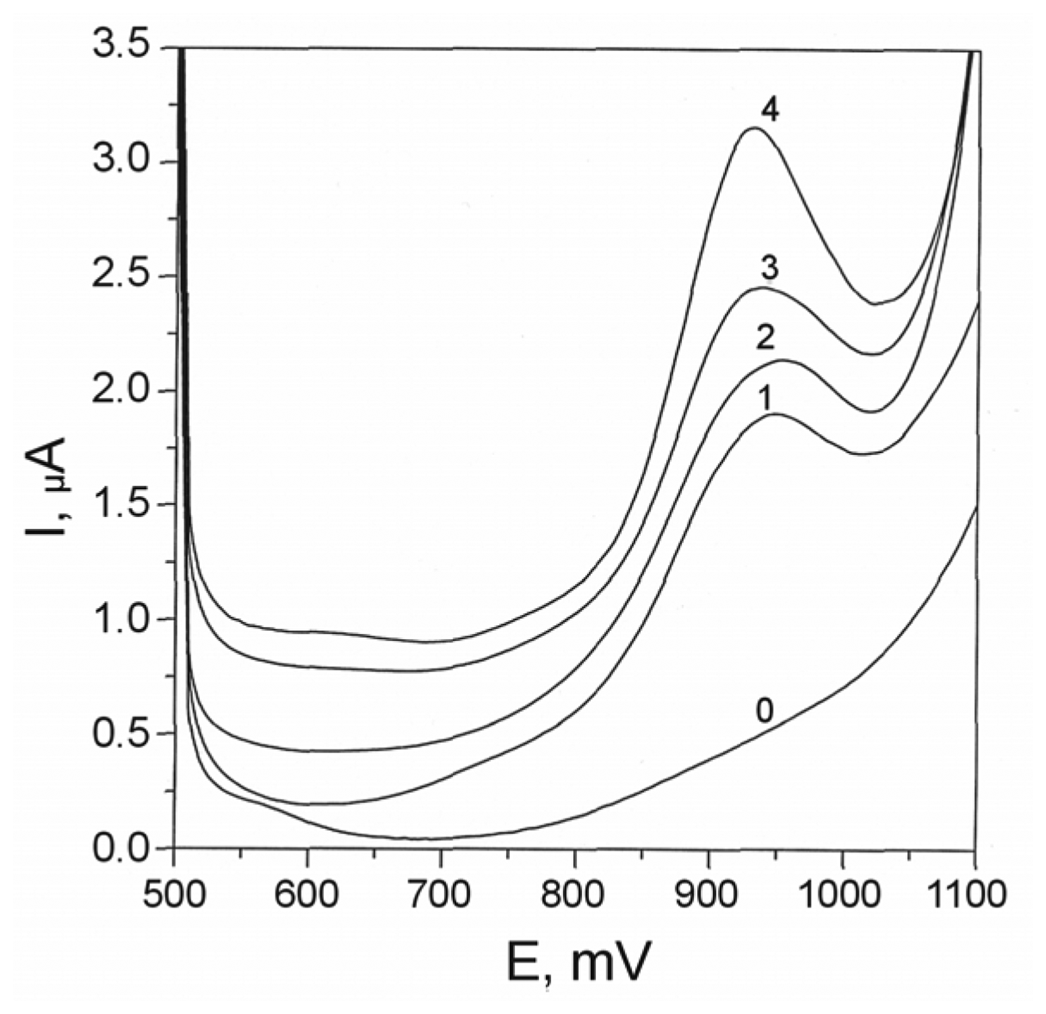
3.2 Cleavage effect of inorganic arsenic

Inorganic arsenic was investigated in the form of arsenite or arsenate present as the component of cleavage mixture (1×10⁻² mol/L phosphate buffer solution pH 7.0). Values of the relative DPV signal of the DNA marker are summarized in Tables 1 and 2. These data demonstrates some DNA degradation by arsenite at 22 °C in the presence of Fe2+ as well as Cu2+ ions as the redox catalysts.
No significant DNA damage was found by applying arsenate which evidently cannot fulfil the role of reducing agent for the metal catalyst. It seems, however, that the cleavage effect diminishes at 37 °C as well as using Fe3+ salt as the catalyst which indicates other reaction pathways or kinetics.
The cleavage effect of arsenite was confirmed also by a change in the catalytic current of the guanine moiety. Fig. 3 (curves 2 and 3) shows a dramatic change in the DNA anodic signal after the treatment of the DNA/SPE biosensor in the mixture with As(III).

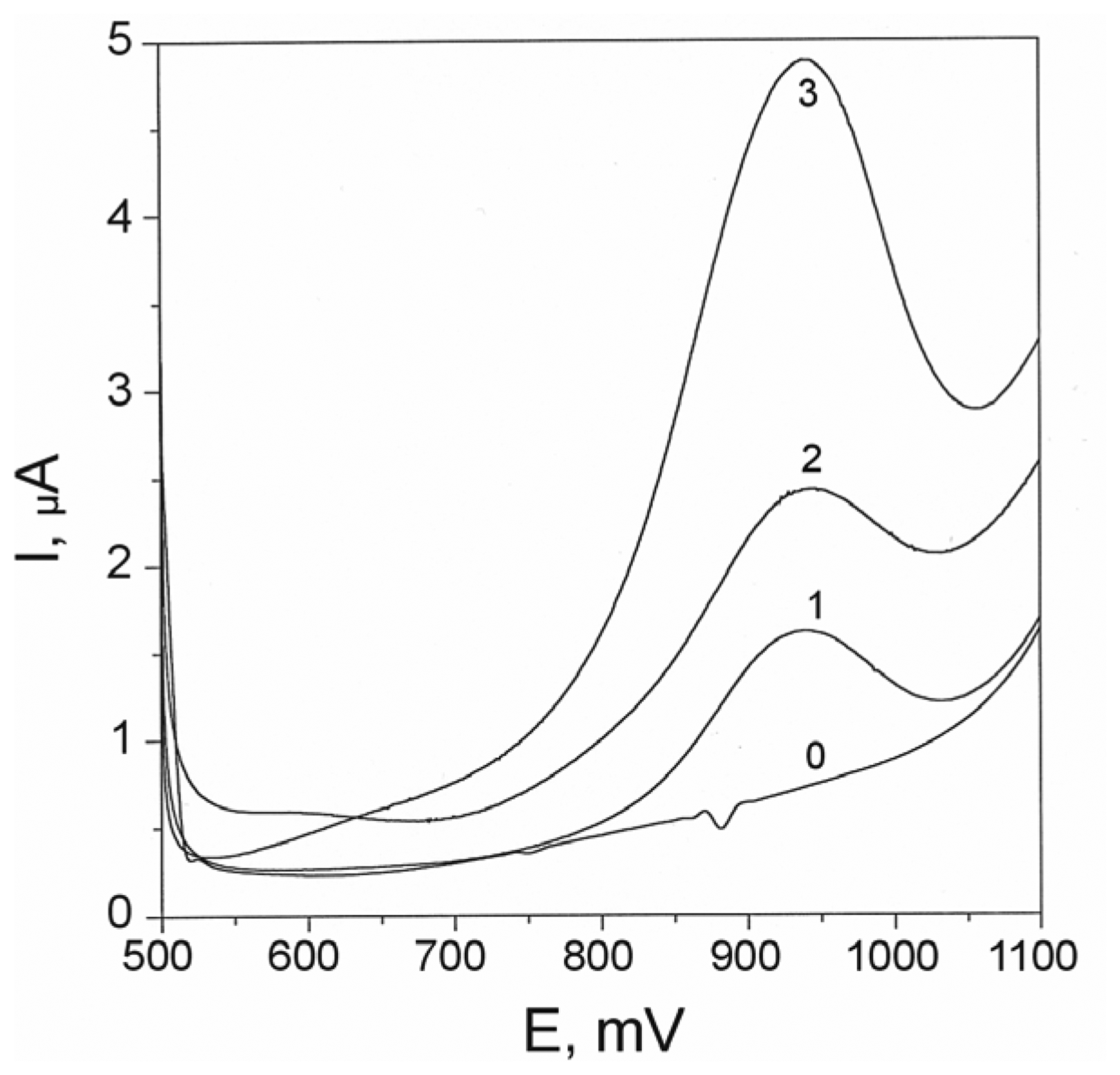
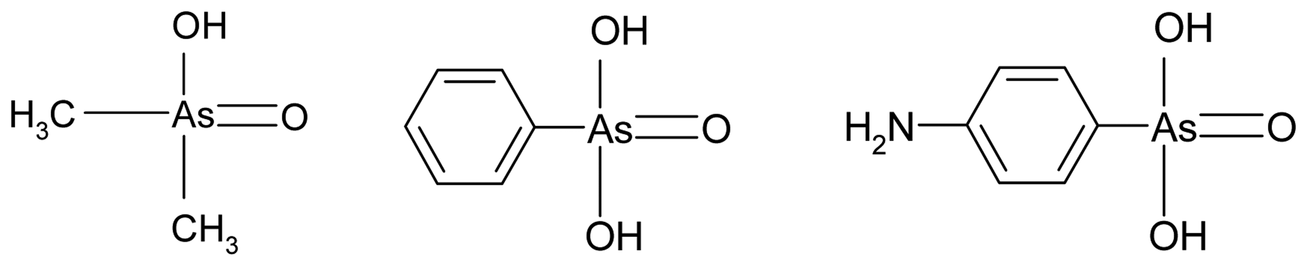
3.3 Cleavage effect of organic arsenic

Table 3 summarizes data on the relative DPV signal of the [Co(phen)3]3+ complex obtained at the DNA/SPE sensor after its incubation in the cleavage mixtures containing organic arsenic species. The data clearly show DNA damage by these organoarsenicals in the presence of metal catalyst. The contribution of simple solutions of Fe2+ and H2O2 as well as Cu2+ and H2O2 to the DNA damage was small (Tables 1 and 2).
The activity of the organic arsenic species is higher than that of inorganic ones. However, there is no significant difference in the effect of individual organic arsenic compounds. On the other hand, somewhat larger DNA damage was found in the presence of Fe2+ ions than in the presence of Cu2+.

Conclusions

Carcinogenicity of arsenic is generally supposed to be in relation to the production of ROS via activation of enzymes participating on the ROS formation or to the inhibition of DNA repair mechanism. Using the DNA based electrochemical biosensor, we have found damage to DNA under in vitro conditions at an incubation of the biosensor in reaction mixtures with arsenite and organic arsenic compounds. The redox potential of arsenic allows to consider it as a reducing agent in the Haber-Weiss system. The activity of organoarsenicals compounds may be related to a direct association interaction and exposure of a DNA base, such as guanine, to these arsenic species for oxidative damage. Differences in reaction mechanism of inorganic and organic arsenic species can be probably related also to the different effects of copper and iron ions present in the cleavage media.
It could be demonstrated that DNA based biosensor acts as an effective chemical toxicity sensor regarding serious environmental contaminant such as arsenic. Thanks to reversible binding and redox change of the [Co(phen)3]3+ complex, it can be used analytically repeatedly to detect an integrity of the original dsDNA, particularly regarding its ability to bind the positively charged marker particles. On the other hand, the direct and Ru(II)-catalyzed DNA oxidation is an irreverzible process based a possibility of an electron transfer from the inside of the dsDNA and to the electrode surface. Therefore, these anodic signals may be used to evaluate DNA association interactions with low molecular (arsenic) species and damage to DNA bases as well as various DNA structural changes from helix distorsion (opening) to strand breaks.
The rather complex behavior of arsenic compounds towards DNA observed even under in vitro conditions needs further study. The investigation of effects of natural arsenic co-reactants, such as glutathione and ascorbic acid, in the reaction medium at the biosensor incubation (together with a validation of the results by independent methods such as impedance spectroscopy at DNA/SPE and conventional electrophoresis in solution) are in progress.

Acknowledgments

This work was supported by the Grant Agency VEGA of the Slovak Republic (Grants No. 1/9253/02 and 1/2462/05) and by a joint project of the Ministry of Education of the Slovak Republic and DAAD, Germany (No. 5/2002).

References

  1. Palecek, E.; Fojta, M. Anal. Chem. 2001, 73, 74A–83A.
  2. Mascini, M.; Palchetti, I.; Marrazza, G. Fres. J. Anal. Chem. 2001, 369, 15–22.
  3. Wang, J. Anal. Chim. Acta 2002, 469, 63–71.
  4. Palecek, E.; Jelen, F. Crit. Rev. Anal. Chem. 2002, 32, 261–270.
  5. Ye, Y.; Ju, H. Sensors 2003, 3, 128–145.
  6. Wang, J. Anal. Chim. Acta 2003, 500, 247–257.
  7. Fojta, M. Collect. Czech. Chem. Commun. 2004, 69, 715–747.
  8. Labuda, J.; Fojta, M.; Jelen, F.; Paleček, E. Encyclopedia of Sensors; Grimes, C. A., Dickey, E., Eds.; American Scientidic Publishers: Stevenson Ranch, CA, in press.
  9. Abreu, F.C.; Goulart, M. O. F.; Oliveira Brett, A. M. Biosens. Bioelectron. 2002, 17, 913–919.
  10. Oliveira Brett, A. M.; da Silva, L. A.; Fujii, H.; Mataka, S.; Thiemann, T. J. Electroanal. Chem. 2003, 549, 91–99.
  11. Erdem, A.; Ozsoz, M. Electroanalysis 2002, 14, 965–974.
  12. Labuda, J.; Bučková, M.; Heilerová,, L'.; Šilhár, S.; Štepánek, I. Anal. Bioanal. Chem. 2003, 376, 168–173.
  13. Bode, A.M.; Dong, Z. Crit. Rev. Oncol../Hematol. 2002, 42, 5–24.
  14. Harris, G.K.; Shi, X. Mutation Res. 2003, 533, 183–200.
  15. Okayasu, R.; Takahashi, S.; Sato, H.; Kubota, Y.; Scolavino, S.; Bedford, J.S. DNA Repair 2003, 2, 309–314.
  16. Hartwig, A.; Blessing, H.; Schwerdtle, T.; Walter, I. Toxicol. 2003, 193, 161–169.
  17. Rossman, T.G. Mutation Res. 2003, 533, 37–65.
  18. Ng, J.C.; Wang, J.; Shraim, A. Chemosphere 2003, 52, 1353–1359.
  19. Gomez-Caminer, A.; Howe, P.; Hughes, M.; Kenyon, E.; Lewis, D. R.; Moore, M.; Ng, J.; Aitio, A.; Becking, G. Arsenic and Arsenic Compouns. In Environ.Health Criteria 224; WHO: Geneva, 2001. [Google Scholar]
  20. Hughes, M. F. Toxicol. Lett. 2002, 133, 1–16.
  21. Ahmad, S.; Kitchin, K. T.; Cullen, W. R. Arch. Biochem. Biophys. 2000, 382, 195–202.
  22. Labuda, J.; Bučková, M.; Vaníčková, M.; Mattusch, J.; Wennrich, R. Electroanalysis 1999, 11, 101–107.
  23. Fojta, M.; Staňková, V.; Paleček, E.; Koscielniak, P.; Mitáš, J. Talanta 1998, 46, 155–161.
  24. Ozsoz, M.; Erdem, A.; Kara, P.; Kerman, K.; Ozkan, D. Electroanalysis 2003, 15, 613–619.
  25. Pang, D. W.; Abruña, H. D. Anal. Chem. 1998, 70, 3162–3169.
  26. Dollimore, L. S.; Gillard, R. D. J. Chem. Soc. 1973, 78, 933–940.
  27. Labuda, J.; Bučková, M.; Heilerová,, L'.; Čaniová- Žiaková, A.; Brandšteterová, E.; Mattusch, J; Wennrich, R. Sensors 2002, 2, 1–10.
Figure 1. DP voltammograms of [Ru(bpy)3]2+ at SPE (1), DNA/SPE prepared in situ in DNA solution of 10 μg/mL (2), 20 μg/mL (3), 50 μg/mL (4). Curve (0) is blank. Conditions: 5×10⁻⁶ mol/L Ru(bpy)3]2+ in 2×10⁻¹ mol/L acetate buffer solution pH 5.40 with 5×10⁻² mol/L NaCl, scan rate 1 mV/s.
Sensors 05 00411f1
Figure 2. DP voltammograms of [Co(phen)3]3+ at SPE (1), ex situ prepared DNA/SPE before (2) and after (3) incubation in model cleavage mixture. Conditions: 5×10⁻⁷ mol/L [Co(phen)3]3+ in 5×10⁻³ mol/L phosphate buffer solution pH 7.0, 120 s accumulation at +0.300 V, scan rate 10 mV/s.
Sensors 05 00411f2
Figure 3. DP voltammograms of [Ru(bpy)3]2+ at SPE (1), ex situ prepared DNA/SPE before (2) and after (3) incubation in cleavage mixture of 6×10⁻³ mol/L arsenite, 2×10⁻⁴ mol/L Fe(II) and 1.5×10⁻² mol/L H2O2 for 10 min. Conditions: 5×10⁻⁶ mol/L Ru(bpy)3]2+ in 2×10⁻¹ mol/L acetate buffer solution pH 5.40 with 5×10⁻² mol/L NaCl, scan rate 1 mV/s.
Sensors 05 00411f3
Scheme 1. Structure of dimethylarsinic acid (DMA), phenylarsonic acid (PhA) and p-arsanilic acid (APhA)
Sensors 05 00411f4
Table 1. Effect of inorganic arsenic on DNA at 10 min incubation of DNA/SPE sensor in cleavage mixtures with Fe(II) ions at 22 °C (not indicated) and 37 °C. Conditions: 1×10⁻² mol/L phosphate buffer solution pH 7.0 under stirring.
Cleavage mixtureDNA marker signal (I/I0)
CompositionConcentration, mol/LNo AsAs(III)As(V)
Fe(II)
H2O2
2×10⁻⁴
1.5×10⁻²
0.90±0.05--
As
Fe(II)
H2O2
6×10⁻³
2×10⁻⁴
1.5×10⁻²
-0.83±0.030.90±0.04
As
Fe(II)
H2O2, 37 °C
6×10⁻³
2×10⁻⁴
1.5×10⁻²
-0.94±0.03-
As
Fe(III)
H2O2
6×10⁻³
2×10⁻⁴
1.5×10⁻²
-0.99±0.02-
Table 2. Effect of inorganic arsenic on DNA at 10 min incubation of DNA/SPE sensor in cleavage mixtures with Cu(II) ions at 22 °C (not indicated) and 37 °C. Conditions: 1×10⁻² mol/L phosphate buffer solution pH 7.0 under stirring
Cleavage mixtureDNA marker signal (I/I0)
CompositionConcentration, mol/LNo AsAs(III)As(V)
Cu(II)
H2O2
2×10⁻⁴
1.5×10⁻²
0.92±0.11--
As
Cu(II)
H2O2
6×10⁻³
2×10⁻⁴
1.5×10⁻²
-0.79±0.080.95±0.02
Cu(II)
H2O2, 37 °C
2×10⁻⁷
1.5×10⁻²
0.95±0.07--
As
Cu(II)
H2O2, 37 °C
6×10⁻³
2×10⁻⁷
1.5×10⁻²
-1.21±0.04-
Table 3. Effect of organic arsenic species on DNA at 10 min incubation of DNA/SPE sensor in cleavage mixtures with Fe(II) and Cu(II) ions. Conditions: 1×10⁻² mol/L phosphate buffer solution pH 7.0 under stirring.
Cleavage mixtureDNA marker signal (I/I0)
CompositionConcentration, mol/LDMAPhAAPhA
As1×10-30.90±0.011.00±0.050.76±0.04
As
Fe(II)
H2O2
1×10⁻³
2×10⁻⁴
1.5×10⁻²
0.58±0.040.66±0.110.61±0.05
As
Cu(II)
H2O2
1×10⁻³
2×10⁻⁴
1.5×10⁻²
0.67±0.080.73±0.030.83±0.03

