Review

Electrochemical DNA Sensors for Detection of DNA Damage

Departamento de Química, Faculdade de Ciências e Tecnologia, Universidade de Coimbra, 3004-535 Coimbra, Portugal
*
Author to whom correspondence should be addressed.
Sensors 2005, 5(6), 377-393; https://doi.org/10.3390/s5060377
Submission received: 3 February 2005 / Accepted: 14 March 2005 / Published: 14 November 2005
(This article belongs to the Special Issue DNA Based Sensors)

Abstract

:
Electrochemical devices have received particular attention due to their rapid detection and great sensitivity for the evaluation of DNA-hazard compounds interaction mechanisms. Several types of bioanalytical method use nucleic acids probes to detect DNA damage. This article reviews current directions and strategies in the development and applications of electrochemical DNA sensors for the detection of DNA damage.

1. Overview

Deoxyribonucleic acid (DNA) is a very important biomolecule that has an essential role in the determination of hereditary characteristics, storing the genetic information necessary for the replication of living organisms.
Due to its important chemical and biophysical properties, DNA has been the subject of many physical and chemical investigations, leading to the discovery of the genetic code, and more recently to genome sequencing. One of the most important results of the human genome-sequencing project is the identification of point mutations and their correlation with pathologies such as cancer. As carrier of genetic information, DNA is a major target for drug interaction. Many compounds bind and interact with DNA causing changes to the structure of DNA and the base sequences, leading to perturbations in DNA replication and cancer. Thus it is very important to explain the factors that determine affinity and selectivity in binding molecules to DNA, identify these chemical species and ascertain their potency so that human exposure to them can be minimized. From this point of view, the expansion of technologies and methodologies to detect DNA-drug interactions is very important. The necessity for stable, low cost, and easily adaptable analytical tools for the detection of DNA damage have been the driving force for DNA biosensor technology development.

2. DNA-electrochemical biosensors

DNA biosensors are integrated receptor-transducer devices that use DNA as biomolecular recognition element to measure specific binding processes with DNA, usually by electrical, thermal, or optical signal transduction [1].
Compared with other transducers, electrochemical ones received particular interest due to a rapid detection and great sensitivity. Combining the characteristics of DNA probes with the capacity of direct and label-free electrochemical detection represents an attractive solution in many different fields of application, such as rapid monitoring of pollutant agents or metals in the environment, investigation and evaluation of DNA-drug interaction mechanisms, detection of DNA base damage in clinical diagnosis, or detection of specific DNA sequences in human, viral, and bacterial nucleic acids [2-8]. Amongst the electrochemical transducers, carbon electrodes such as glassy carbon, carbon fibre, graphite, or carbon black exhibit several unique properties. The wide electrochemical potential window in the positive direction allows sensitive electrochemical detection of oxidative damage caused to DNA by monitoring the appearance of oxidation peaks of DNA bases.
To design efficient DNA-electrochemical biosensors, it is essential to know the structure and to understand the electrochemical characteristics of DNA molecules.
DNA consists of two antiparallel polynucleotide chains formed by monomeric nucleotide units. Each nucleotide is formed by three types of chemical components: a phosphate group, a sugar called deoxyribose, and four different nitrogen bases. The phosphate-deoxyribose sugar polymer represents the DNA backbone. The cellular genetic information is coded by the purine bases, adenine (A) and guanine (G), and the pyrimidine bases, cytosine (C) and thymine (T), as a function of their consecutive order in the chain. The two strands of nucleotides are twisted into a double helix, held together by hydrogen bonds between the A·T and G·C bases of each strand.
Electrochemical oxidation of DNA at a glassy carbon electrode [2, 3] using differential pulse voltammetry shows two current peaks due to the oxidation of guanine and adenine residues, Fig. 1. The electrochemical behaviour obtained with double stranded DNA (dsDNA) and single stranded DNA (ssDNA) illustrates the greater difficulty for the transfer of electrons from the inside of the double-stranded rigid form of DNA to the electrode surface, than from the flexible single stranded form of DNA where the bases are in close proximity to the electrode surface. The roughness of the solid electrode surface means that dsDNA has some difficulty in following the surface contours whereas unwound ssDNA molecules fit more easily into the grooves of the surface, owing to their greater flexibility.
However, it was a challenge to increase the sensitivity and to allow simultaneous electrochemical detection of all purine and pyrimidine DNA bases in natural samples at physiological pH, and to extend
the DNA voltammetric studies to nucleotides so as to make possible the simultaneous detection of all bases in single-stranded DNA (ssDNA) without the necessity of a previous hydrolysis step.
Mechanistic studies on the electrochemical behaviour of purine and pyrimidine derivatives have been performed during the past four decades. Electrochemical oxidation on carbon electrodes [8-12] showed that all bases - guanine, adenine, thymine and cytosine - could be oxidized following a pH dependent mechanism. For the first time equimolar mixtures of all DNA bases, nucleosides, and nucleotides were quantified by differential pulse voltammetry, Fig. 2 [12].
Electrochemical preconditioning of the glassy carbon electrode (GCE) enabled the achievement of a better peak separation and an enhancement of the oxidation peak currents for all four DNA bases in pH 7.4 phosphate buffer supporting electrolyte, close to physiological pH. Detection limits in the nano-and micromolar ranges were obtained for purine and pyrimidine bases, respectively, together in solution. The results also showed for the first time that the pyrimidine nucleosides and nucleotides are electroactive on glassy carbon electrodes and that, in addition to the easy detection of the purines, it was also possible to detect simultaneously the oxidation of pyrimidine residues in ssDNA.
When natural or synthetic DNA molecules interact with electrode surfaces, adsorption occurs. The adsorption of dsDNA is quasi-reversible, and its structure undergoes conformational modifications during immobilization on the electrodes. Observing DNA adsorption was the first step in developing DNA-modified electrodes, and thence DNA-electrochemical biosensors. DNA immobilization and the stabilization procedure of the DNA probe on the electrode surface represent key aspects in the manufacture of a DNA-electrochemical biosensor, influencing the characteristics of the DNA probe, the sensor response and its performance [13]. Different structural conformations adopted by DNA can lead to different interactions with small molecules and to changes in the accessibility of drugs to the DNA grooves. A good understanding of all these factors is essential in order to design surfaces exhibiting good biological activity and selectivity.
Different immobilization procedures of monolayer and multilayer DNA films on highly oriented pyrolytic graphite (HOPG) electrodes have been characterized by Magnetic AC Mode Atomic Force Microscopy (MAC AFM) [14-17]. HOPG provides a particularly smooth electrode surface, Fig. 3A, which enables the detection of topographical modifications when DNA molecules are adsorbed. Direct observation of the electrode surface demonstrated that the characteristics of the DNA film and the optimal electrochemical response of the DNA-biosensor depend on pH, buffer composition, ionic strength, immobilization procedure, adsorption time, applied potential, type and concentration of DNA.
At small DNA concentrations, thin film dsDNA and ssDNA layers are formed, that appear as large-scale network structures uniformly covering the HOPG surface, Figs. 3B and 4. Large pores in the film leave parts of the electrode uncovered. Thicker films prepared by different adsorption immobilization procedures show uniform and complete coverage of the electrode, with uniformly distributed peaks and valleys, Fig. 3C and D. DNA films grown in more acid buffer solutions always present greater DNA surface coverage, due to overlaying and superposition of DNA molecules, with respect to films formed in neutral buffer solutions, Fig. 4. The DNA-electrode surface interactions are stronger and the DNA films more stable when a potential is applied during adsorption. Therefore the DNA immobilization procedure must be a compromise between the degree of electrode coverage and strong adsorption due to the applied potential.
The dsDNA networks created during the formation of thin layers define different active surface areas on the DNA-electrochemical biosensor and form a biomaterial matrix to attach and study interaction with other molecules such as drugs. Due to the existence of pores in the DNA structure, some areas of the electrode surface are not covered by the molecular film and may act as a system of microelectrodes with nanometer or micrometer dimensions. These pores can cause misleading results when using a DNA electrochemical biosensor for detection of DNA-drug interactions. The fact that the electrode is not completely covered may allow the diffusion of drug molecules from bulk solution to the surface and their non-specific adsorption on the uncovered areas, leading to two contributions to the electrochemical signal, one from the simple adsorbed drug and the other due to damage of immobilized dsDNA, it not being easy to distinguish between the two signals. By using thick film DNA-biosensors, where the undesired binding of drug molecules to the electrode surface is impossible due to complete coverage of the electrode surface, Fig. 3 C and D, the DNA-biosensor response is only determined by interaction of the compound with the dsDNA in the film. Knowledge of the morphology of adsorbed DNA, controlled by the appropriate choice of DNA solution concentration, pH and applied potential, helps in the improvement and development of specific DNA electrochemical biosensors.

3. DNA oxidative biomarker

Oxidative DNA damage caused by oxygen-free radicals leads to multiple modifications in DNA, including base-free sites and oxidized bases. The damage caused to DNA bases is potentially mutagenic [18, 19]. The DNA oxidative damage can be enzymatically repaired.
The major product of DNA oxidative damage is 8-oxo-7,8-dihydroguanine (8-oxoGua) which is the oxidation product of guanine, the most easily oxidized base in DNA. This modified base is highly mutagenic [20], that potentially mispairs with adenine during DNA replication, causing GC→TA transversion and loss of base-pairing specificity [21]. Since it was first reported two decades ago, 8-oxoGua, in particular its deoxynucleoside, 8-oxo-7,8-dihydro-2′-deoxyguanosine (8-oxodGuo) has been the subject of intensive investigation and has become widely accepted as a biomarker of oxidative
DNA damage and cellular oxidative stress [22, 23]. Also, 8-oxoG and 8-oxodGuo are major products of DNA oxidative damage, being excreted into urine.
At present, HPLC with electrochemical detection (HPLC-ECD) is the most commonly used technique to assess urinary 8-oxodGuo, but HPLC with tandem mass spectrometry (HPLC-MS/MS) is also used, showing high sensitivity and better specificity. However, HPLC-ECD is easier to use and less demanding of resources. There is a significant lack of HPLC-ECD based methods for 8-oxoGua detection in human urine, despite 8-oxoGua also being electrochemically detectable [24] at a lower potential than 8-oxodGuo [25]. A selective method based on HPLC-ECD was developed to enable simultaneous detection of 8-oxo-7,8-dihydroguanine and 8-oxo-7,8-dihydro-2′-deoxyguanosine, products of DNA oxidative damage, after uric acid (UA) elimination by uricase, consisting of HPLC isocratic elution with amperometric detection, enabling a detection limit for 8-oxoGua and 8-oxodGuo lower than 1 nM in standard mixtures [26, 27]. Despite the complexity and inter-individual variability of urine samples, the method was successfully tested with urine samples from children with metabolic disorders and it was confirmed that at the applied working potential, hypoxanthine, present in urine, does not give any electrochemical signal and does not interfere at all in the 8-oxoGua signal.
The oxidation of the other DNA bases is much more difficult due to their high oxidation potentials [8, 12]. Chemical modification of each of the DNA bases causes molecular disturbance to the genetic machinery that leads to cell malfunction. Oxidative damage to DNA by free radicals and exposure to ionizing radiation generates several products within the double helix besides 8-oxoGua, such as 2,8-dihydroxyadenine, 5-formyluracil, 5-hydroxycytosine, etc., which are mutagenic [19].
Hence, there is great interest in the sensitive determination and full characterization of the mechanism involved in oxidative damage to DNA bases. Electrochemical methods are very promising for the study of DNA oxidative damage as well as in the investigation of the mechanisms of interactions of drugs with DNA. In particular, electrochemical DNA-biosensors have proved to be excellent tools for investigating the effect of various endogenous and exogenous sources of hazard to genomic material, allowing quick and low cost determination of DNA damage.

4. Electrochemical biosensors for detection of DNA damage

There are hundreds of compounds that bind and interact with DNA: toxic chemicals damage DNA and can cause human cancer. The factors that determine affinity and selectivity in binding molecules to DNA need to be explained, because a quantitative understanding of the reasons that determine selection of DNA reaction sites is useful in designing sequence-specific DNA binding molecules for application in chemotherapy and in explaining the mechanism of action of neoplasic drugs [29].
Metal ions exist in body fluids and the nucleic acids and nucleotides occur as complexes coordinated [30] with these ions. Some of these divalent ions are recognized for their carcinogenicity since they damage DNA and alter the fidelity of DNA synthesis [31]. Although the possible relation between oxidative damage and metal concentrations is not completely clear, it is generally accepted that metals react with superoxide anion (O2−•) and H2O2 to produce highly reactive species such as hydroxyl free radicals (OH•) and metal oxygen complexes in biological systems, resulting in metal-mediated oxidative DNA damage.
The cytostatic activity of various platinum drugs has shown that platinum coordination complexes cause irreversible inhibition of DNA synthesis due to covalent binding with DNA. This other causes the treatment to be accompanied by adverse reactions. The DNA-biosensor was successfully used for the electrochemical determination of carboplatin in serum samples of patients with ovarian cancer undergoing treatment [32]. The mechanism of action of carboplatin with DNA clearly demonstrates that for low concentrations, carboplatin interacts preferentially with adenine rather than guanine groups and this can contribute to clarifying the mechanisms of interaction of platinum anti-cancer compounds with DNA.
Metals are considered to act not only as carcinogens but also to activate carcinogenic chemicals. A number of aromatic compounds induce oxidative DNA damage through metal catalyzed reactive oxygen species (ROS) generation. ROS produced in vivo react with DNA and its precursors modifying them, therefore, giving rise to the so-called oxidative stress. It is thought that the modification of DNA (DNA lesions) leads to the formation of incorrect base pairs, changes in the genetic information, which induces mutagenesis and carcinogenesis. Therefore, in a health preventing perspective, there is a deep interest in identifying free radical scavengers or anti-oxidants that inhibit oxidative DNA damage. Owing to their polyphenolic nature, flavonoids, compounds found in rich abundance in all land plants, often exhibit strong antioxidant properties [33, 34, 35, 36, 37]. Initially, flavonoids were investigated as potential chemopreventive agents against certain carcinogens. Previous intake of a large quantity of flavonoid inhibited the incidence of ROS produced damages to DNA. In sharp contrast with their commonly accepted role, there is also considerable evidence that flavonoids themselves are mutagenic and have DNA damaging ability [34, 35].
One striking example reported in the literature is that of quercetin, which in the presence of transition metals acts as a pro-oxidant and has mutagenic activity [38, 39]. It was shown that some flavonoids are able to reduce transition metals, a process during which the highly oxidizing radical OH• is formed and could therefore damage DNA.
In an electrochemical study of the DNA-Cu(II)-quercetin interaction [40], several situations were studied using a bare GCE in a solution containing dsDNA incubated with quercetin or quercetin-Cu(II) complex. Whereas a very weak interaction between quercetin and DNA was found to take place in solution, the addition of Cu(II) ions to the DNA-quercetin solution showed that a deep degradation of the DNA helix takes place. Using spectrophotometric techniques as a complement to electrochemical methods, it was observed that dsDNA damage occurred over time which suggests that the quercetin- Cu(II) complex intercalates with dsDNA and slowly interacts with it causing breaking of the hydrogen bonds.
Electrochemical voltammetric in situ sensing of dsDNA oxidative damage caused by oxidized quercetin incorporated into dsDNA was possible using a DNA-electrochemical biosensor [41]. The results indicate that quercetin binds to the dsDNA and can undergo oxidation at Epa = + 0.32 V, Fig. 5 Sensors 05 00377i1
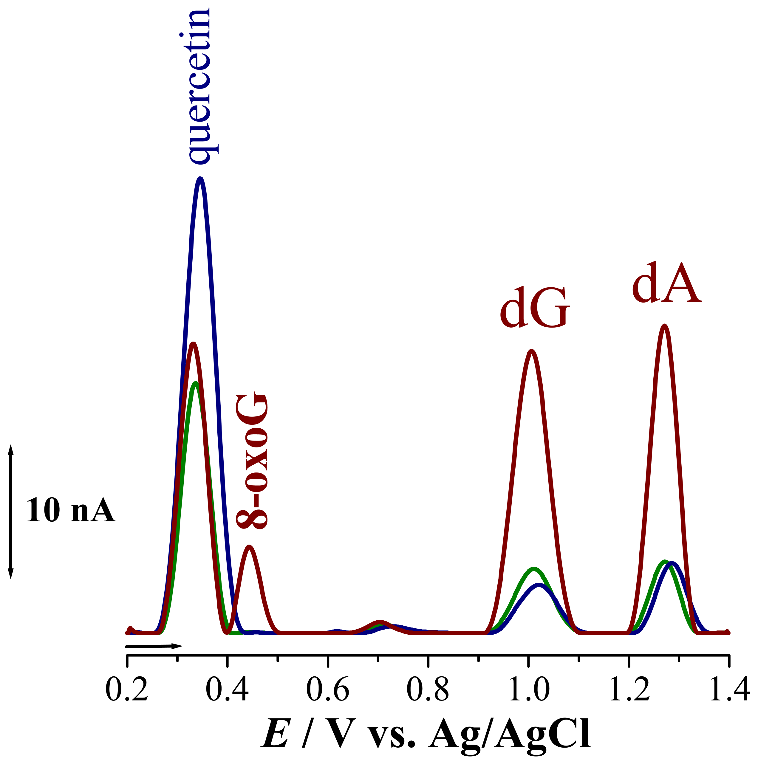
. It is known that oxidation of quercetin is a complex process during which superoxide radicals are formed. These radicals readily attack the dsDNA disrupting the helix, causing contact between the electrode surface and the DNA nucleotides guanosine, Epa = + 1.02 V, and adenosine, Epa= +1.28 V, such that their oxidation peaks increase, Fig. 5 Sensors 05 00377i2. On the other hand, the radicals formed during the oxidation of quercetin incorporated into dsDNA lead to the formation of 8-oxoguanine, with peak at Epa = + 0.45 V. In order to prove the involvement of the oxygen radicals in DNA damage during oxidation of intercalated quercetin, experiments were also carried out in solutions saturated with N2, Fig. 5 Sensors 05 00377i3. The differential pulse voltammogram obtained showed only small guanosine and adenosine oxidation peaks, demonstrating that no DNA damage had occurred. Also, no additional peak, at
+ 0.450 V (8-oxoGua) was observed although a smaller quercetin oxidation peak 1 appeared. A model for this electrochemically observed in situ oxidative damage has been proposed and may be used to explain the oxidant-antioxidant effect of quercetin.
In a similar way, the detection of DNA damage involving strand breaks was observed using a hanging mercury drop electrode (HMDE) [42]. Extensive cleavage of electrode-confined DNA by reactive oxygen species (ROS) was obtained in the absence of chemical reductants when redox cycling of metal (iron/DNA) complex was controlled. The cleavage agents were detected and the DNA cleavage was modulated, by generating the DNA-damaging species electrochemically.
The detection of chemicals that cause irreversible damage to DNA is also very important, and a short-time screening test for carcinogens based on ac-voltammetric measurements was developed in order to study in vitro damage to DNA caused by the action of alkylating mutagens [43]. Nitroimidazoles are among the most important nitroheterocyclic drugs of interest in cancer chemotherapy. It was observed that adenine and guanine interact with intermediates generated during nitroimidazole reduction, causing irreversible damage to DNA and suggesting mutagenic properties of these compounds.
The mechanism of reduction of a group of nitroimidazoles was investigated using the DNA-biosensor [44, 45, 46]. The analyte was pre-concentrated on the electrode surface containing DNA and either the reduction or the oxidation of the reduction products retained on the electrode surface was studied. It was possible to follow their reduction, the reversible oxidation of the hydroxylamine derivative formed (RNHOH) to the corresponding nitroso derivative (RNO), the condensation reaction between the hydroxylamine and nitroso derivatives to form the azoxycompound (RNO:NR) and interaction with DNA.
Conventional cancer chemotherapy is as toxic as it is beneficial. The DNA interacting drugs prevent cell growth, but not only cancer cell growth; the cytotoxic effect also blocks the growth of normal cells. The lack of selectivity of cancer drugs is one of the main problems in cancer chemotherapy [47, 48]. The DNA-biosensors are an important tool for the investigation of the electrochemical and biological mechanism of drugs active against cancer [4, 7, 28, 32, 44, 49].
Electrochemical voltammetric in situ detection of dsDNA oxidative damage caused by reduced adriamycin [7], an antibiotic of the family of anthracyclines, intercalated into DNA, was carried out using a DNA-biosensor. Oxidation and reduction of adriamycin molecules intercalated in dsDNA were investigated in order to understand the in vivo mechanism of action of DNA with this anti-neoplasic drug. The results showed that the interaction of adriamycin with dsDNA is potential-dependent. A mechanism for adriamycin reduction and oxidation in situ when intercalated in dsDNA immobilized onto the glassy carbon electrode surface was proposed. This mechanism leads to the formation of the mutagenic 8-oxoGua, whose redox behaviour was studied.
Thiophene-S-oxides are a new group of compounds with biological activity against a number of cancer cells, only recently isolated and characterized in their pure form [50]. The in situ electrochemical detection of dsDNA damage caused by the reduced thiophene-S-oxide was detected using a glassy carbon electrode modified with a thiophene-S-oxide/dsDNA mixture. The results indicate that the reduced thiophene-S-oxide interacts with dsDNA, causing damage with possible strand breaks and that the thiophene-S-oxide adduct formed with dsDNA can still undergo reduction. The potential use of a compound/dsDNA film-modified glassy carbon electrode for understanding dsDNA interaction with molecules insoluble in water was confirmed.
The halogenated salicylanilides are a large group of compounds, which have been developed mainly due to their antiparasitic activity in animals. The investigation of the niclosamide-/DNA interaction [51] using an electrochemical DNA-biosensor showed for the first time clear evidence of interaction with DNA and suggested that niclosamide toxicity can be caused by this interaction, after reductive activation.

5. Conclusions

The development of electrochemical DNA-biosensors has opened a wide perspective using particularly sensitive and selective methods for the detection of specific interactions. Uniform coverage of the electrode surface by DNA is a must, since non-uniform coverage allows the adsorption of the hazard compound on the electrode surface, leading to contributions from both simple adsorbed analyte and from products of damage to immobilized DNA, which need to be carefully distinguished.
The DNA-electrochemical biosensor enables pre-concentration of the hazard compounds investigated onto the sensor surface and in situ electrochemical generation of radical intermediates, which cause damage to the DNA immobilized on the electrode surface and can be detected electrochemically. Voltammetric methods are an inexpensive and fast detection procedure that can contribute to elucidation of the mechanism through which DNA is oxidatively damaged by hazard compounds, in an approach to the real scenario that occurs in the living cell, and are a complementary tool for the study of biomolecular interaction mechanisms.

Acknowledgments

Financial support from Fundação para a Ciência Tecnologia (FCT), Post-Doctoral Grants SFRH /BPD/14425/2003 (A.-M.C.P.) and SFRH/BPD/18824/2004 (V.C.D.), POCTI (co-financed by the European Community Fund FEDER) and ICEMS (Research Unit 103) is gratefully acknowledged.

References

  1. Thevenot, D. R.; Toth, K.; Durst, R. A.; Wilson, G. S. Electrochemical biosensors: Recommended definitions and classification. Pure Appl. Chem. 1999, 71, 2333–2348. [Google Scholar]
  2. Oliveira Brett, A. M.; Serrano, S. H. P.; Piedade, J. A. P. Electrochemistry of DNA, Comprehensive Chemical Kinetics, Applications of Kinetic Modeling; Compton, R. G., Hancock, G., Eds.; Elsevier: Oxford, UK, 1999; vol. 37, chapter 3; pp. 91–119. [Google Scholar]
  3. Oliveira Brett, A. M. DNA-based biosensors, Comprehensive Analytical Chemistry. Biosensors and Modern Specific Analytical Techniques. 2005, vol. 44, 179–208. [Google Scholar]
  4. Palecek, E. Past, present and future of nucleic acids electrochemistry. Talanta 2002, 56, 809–819. [Google Scholar]
  5. Palecek, E.; Fojta, M.; Tomschik, M.; Wang, J. Electrochemical biosensors for DNA hybridization and DNA damage. Biosens. Bioelectron. 1998, 13, 621–628. [Google Scholar]
  6. Mascini, M.; Palchetti, I.; Marrazza, G. DNA electrochemical biosensors. Fresenius J. Anal. Chem. 2001, 369, 15–22. [Google Scholar]
  7. Oliveira-Brett, A. M.; Vivan, M.; Fernandes, I. R.; Piedade, J. A. P. Electrochemical detection of in situ adriamycin oxidative damage to DNA. Talanta 2002, 56, 959–970. [Google Scholar]
  8. Oliveira Brett, A. M.; Matysik, F. M. Voltammetric and sonovoltammetric studies on the oxidation of thymine and cytosine at a glassy carbon electrode. J. Electroanal. Chem. 1997, 429, 95–99. [Google Scholar]
  9. Oliveira Brett, A. M.; Matysik, F. M. Sonoelectrochemical studies of guanine and guanosine. Bioelectrochem. Bioenerg. 1997, 42, 111–116. [Google Scholar]
  10. Oliveira-Brett, A. M; Silva, L. A.; Brett, C. M. A. Adsorption of guanine, guanosine, and adenine at electrodes studied by differential pulse voltammetry and electrochemical impedance. Langmuir 2002, 18, 2326–2330. [Google Scholar]
  11. Oliveira Brett, A. M.; Diculescu, V.; Piedade, J. A. P. Electrochemical oxidation mechanism of guanine and adenine using a glassy carbon microelectrode. Bioelectrochem 2002, 55, 61–62. [Google Scholar]
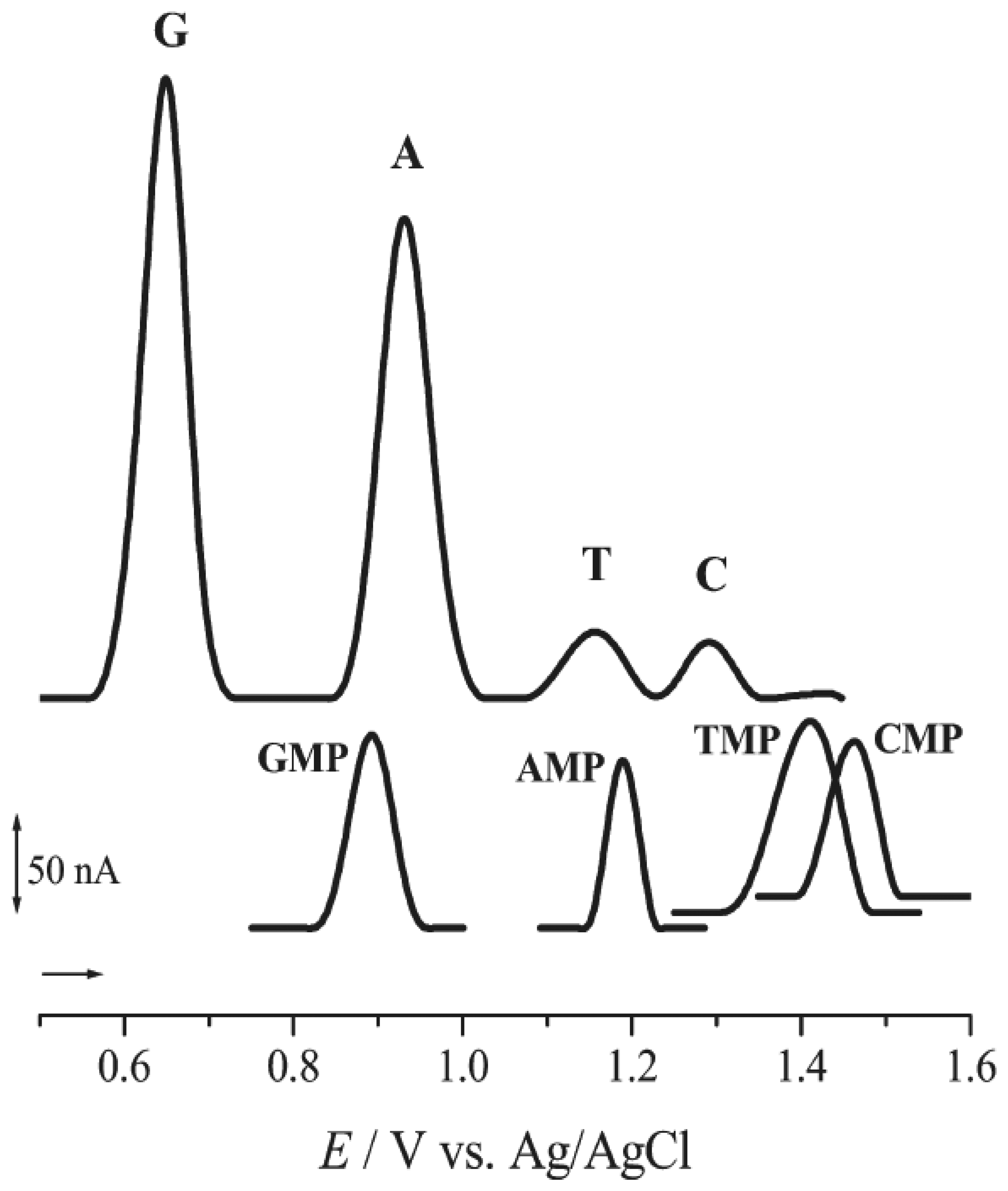
  12. Oliveira Brett, A. M.; Piedade, J. A.; Silva, L. A.; Diculescu, V. C. Voltammetric determination of all DNA nucleotides. Anal. Biochem. 2004, 332, 321–329. [Google Scholar]
  13. Pividori, M. I.; Merkoçi, A.; Alegret, S. Electrochemical genosensor design: immobilization of oligonucleotides onto transducer surfaces and detection methods. Biosens. Bioelectron. 2000, 15, 291–303. [Google Scholar]
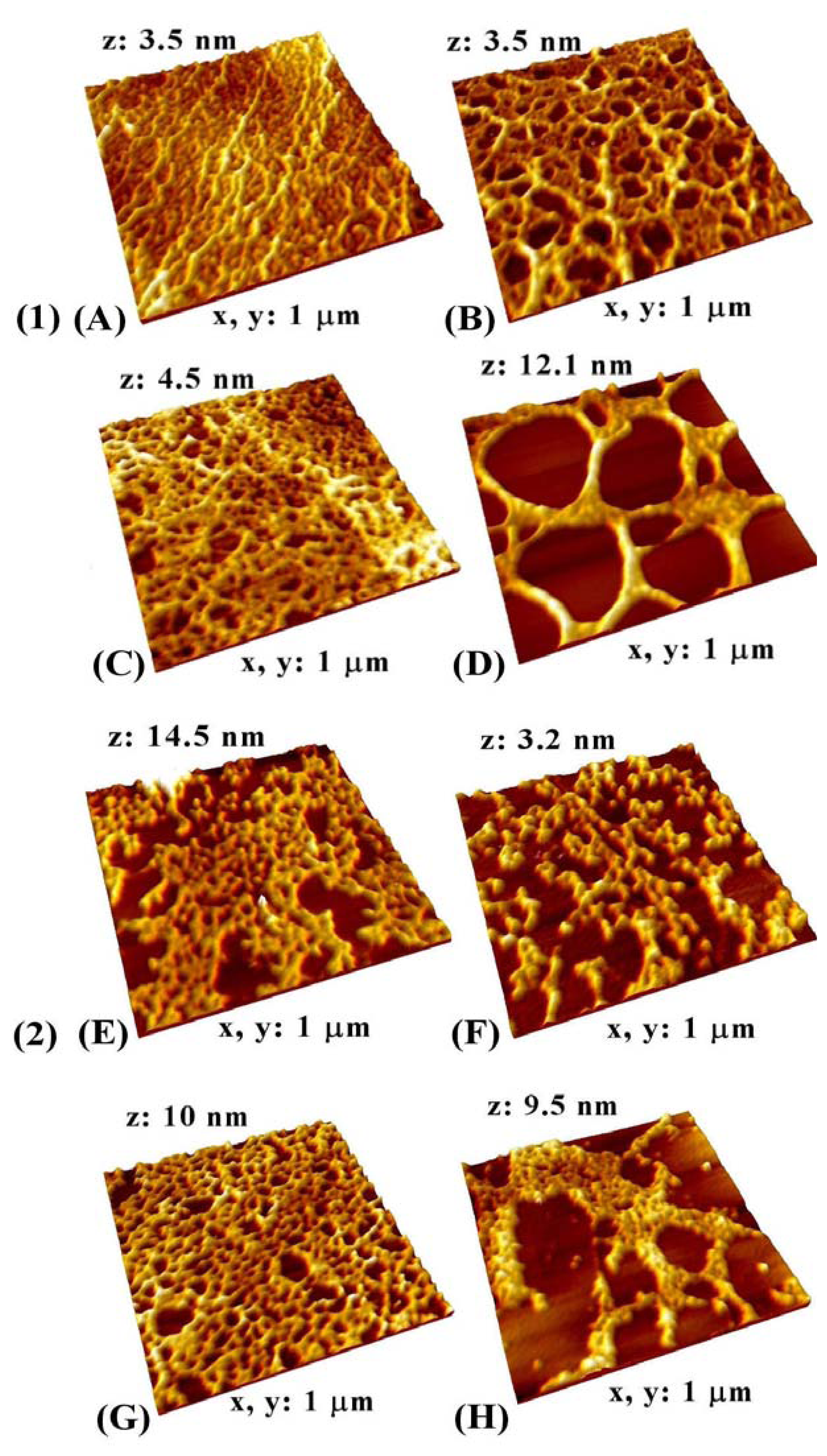
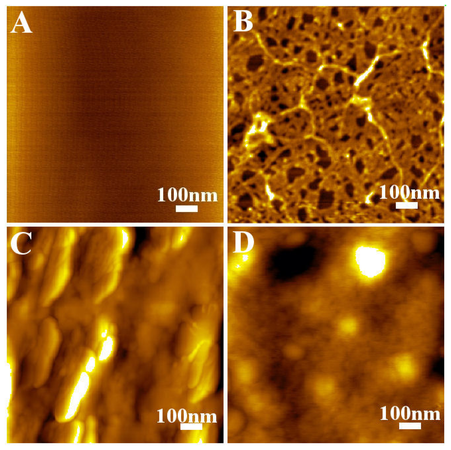
  14. Oliveira-Brett, A. M.; Chiorcea, A.-M. Effect of pH and applied potential on the adsorption of DNA on highly oriented pyrolytic graphite electrodes. Atomic force microscopy surface characterisation. Electrochem. Commun. 2003, 5, 178–183. [Google Scholar]
  15. Oliveira-Brett, A. M.; Chiorcea, A.-M. Atomic force microscopy of DNA immobilized onto a highly oriented pyrolytic graphite electrode surface. Langmuir 2003, 19, 3830–3839. [Google Scholar]
  16. Oliveira-Brett, A. M.; Chiorcea, A.-M. DNA imaged on a HOPG electrode surface by AFM with controlled potential. Bioelectrochem. 2004, in press. [Google Scholar]
  17. Chiorcea, A.-M.; Oliveira-Brett, A. M. Atomic force microscopy characterisation of an electrochemical DNA–biosensor. Bioelectrochem. 2003, 63, 229–232. [Google Scholar]
  18. Halliwell, B.; Gutteridge, J. M. C. Biologically relevant metal ion-dependent hydroxyl radical generation. FEBS Letters 1992, 307, 108–112. [Google Scholar]
  19. Halliwell, B.; Gutteridge, J. M. C. Free Radicals in Biology and Medicine, 3rd ed.; Oxford University Press: UK, 1999. [Google Scholar]
  20. Shibutani, S.; Takeshita, M.; Grollman, A. P. Insertion of specific bases during DNA synthesis past the oxidation-damaged base 8-oxodG. Nature 1991, 349, 431–434. [Google Scholar]
  21. Cysewski, P. An ab initio study of the tautomeric and coding of 8-oxo-guanine. J. Chem. Soc., Faraday. Trans. 1998, 94, 3117–3125. [Google Scholar]
  22. Kasai, H. Analysis of a form of oxidative DNA damage, 8-hydroxy-2′-deoxyguanosine, as a marker of cellular oxidative stress during carcinogenesis. Mutat. Res. 1997, 387, 147–163. [Google Scholar]
  23. Halliwell, B. Oxygen and nitrogen are pro-carcinogens. Damage to DNA by reactive oxygen, chlorine and nitrogen species: measurement, mechanism and the effects of nutrition. Mutat. Res. 1999, 443, 37–52. [Google Scholar]
  24. Oliveira Brett, A. M.; Piedade, J. A.; Serrano, S. H. P. Electrochemical oxidation of 8-oxoguanine. Electroanal 2000, 12, 969–973. [Google Scholar]
  25. Floyd, R. A.; Watson, J. J.; Harris, J.; West, M.; Wong, P. K. Formation of 8-hydroxydeoxyguanosine, hydroxyl free radical adduct of DNA in granulocytes exposed to the tumor promoter, tetradeconylphorbolacetate. Biochem. Biophys. Res. Comm. 1987, 137, 841–846. [Google Scholar]
  26. Rebelo, I. A.; Piedade, J. A. P.; Oliveira-Brett, A. M. Development of an HPLC method with electrochemical detection of femtomoles of 8-oxo-7,8-dihydroguanine and 8-oxo-7,8-dihydro-2′-deoxyguanosine in the presence of uric acid. Talanta 2004, 63, 323–331. [Google Scholar]
  27. Rebelo, I. A.; Piedade, J. A. P.; Oliveira-Brett, A. M. Electrochemical determination of 8-oxoguanine in presence of uric acid. Bioelectrochem. 2004, 63, 267–270. [Google Scholar]
  28. Fojta, M. Electrochemical sensors for DNA interactions and damage. Electroanal 2002, 14, 1449–1463. [Google Scholar]
  29. Larsen, I. K. A Textbook of drug design and development; Krosgaard-Larse, P., Bundgaard, H., Eds.; Harwood Academic Publisher: UK, 1991; p. p. 192. [Google Scholar]
  30. Saenger, W. (Ed.) Principles of Nucleic Acids Structure; Springer-Verlag: New York, 1984; p. p. 201.
  31. Eichorn, G. L. Metal Ions in Genetic Information Transfer; Eichorn, G. L., Marzilli, L. G., Eds.; Elsevier: New York, 1981; p. p1. [Google Scholar]
  32. Oliveira Brett, A. M.; Serrano, S. H. P.; Macedo, T. A.; Raimundo, D.; Marques, M. H.; LaScalea, M. A. Electrochemical determination of carboplatin in serum using DNA-modified glassy carbon electrode. Electroanal. 1996, 8, 992–997. [Google Scholar]
  33. Rice-Evans, C. A.; Miller, N. J.; Paganga, G. Structure-antioxidant activity relationships of flavonoids and phenolic acids. Free Rad. Biol. Med. 1996, 20, 933–956. [Google Scholar]
  34. Johnson, M. K.; Loo, G. Effects of epigallocatechin gallate and quercetin on oxidative damage to cellular DNA. Mut. Res./ DNA Repair. 2000, 459, 211–218. [Google Scholar]
  35. Ohshima, H.; Yoshie, Y.; Auriol, S.; Gilibert, I. Antioxidant and pro-oxidant actions of flavonoids: effects on DNA damage induced by nitric oxide, peroxynitrite and nitroxyl anion. Free Rad. Biol. Med. 1998, 25, 1057–1065. [Google Scholar]
  36. Labuda, J.; Bučková, M.; Heilerová, L.; Šilhár, S.; Štepánek, I. Evaluation of the redox properties and anti/pro-oxidant effects of selected flavonoids by means of a DNA-based electrochemical biosensor. Anal Bioanal Chem 2003, 376, 168–173. [Google Scholar]
  37. Korbut, O.; Buckova, M.; Labuda, J.; Grundler, P. Voltammetric detection of anti-oxidative properties of flavonoids using electrically heated DNA modified carbon paste electrodes. Sensors 2003, 3, 1–10. [Google Scholar]
  38. Sahu, S. C.; Washington, M. C. Effects of antioxidants on quercetin-induced nuclear DNA damage and lipid peroxidation. Cancer Lett. 1991, 60, 259–264. [Google Scholar]
  39. Duthie, S. J.; Johnson, W.; Dobson, V. L. The effect of dietary flavonoids on DNA damage (strand breaks and oxidized pyrimdines (sic)) and growth in human cells. Mutat. Res. 1997, 390, 141–151. [Google Scholar]
  40. Oliveira Brett, A. M.; Diculescu, V. C. Electrochemical study of quercetin-DNA interaction: Part I. Analysis in incubated solutions. Bioelectrochem. 2004, 64, 133–141. [Google Scholar]
  41. Oliveira Brett, A. M.; Diculescu, V. C. Electrochemical study of quercetin-DNA interaction: Part II. In situ sensing with DNA biosensor. Bioelectrochem. 2004, 64, 143–150. [Google Scholar]
  42. Fojta, M.; Kubicarova, T.; Palecek., E. Electrode potential-modulated cleavage of surface-confined DNA by hydroxyl radicals detected by an electrochemical biosensor. Biosens. Bioelectron. 2000, 15, 107–115. [Google Scholar]
  43. Seqúaris, J. M.; Valenta, P. AC voltammetry: a control method for the damage to DNA caused in vitro by alkylating mutagens. J. Electroanal. Chem. 1987, 227, 11–20. [Google Scholar]
  44. Oliveira Brett, A. M.; Macedo, T. R. A.; Raimundo, D.; Marques, M. H.; Serrano, S. H. P. Voltammetric behaviour of mitoxantrone at a DNA-biosensor. Biosens. Bioelectron. 1998, 13, 861–867. [Google Scholar]
  45. Oliveira Brett, A. M.; Serrano, S. H. P.; Gutz, I.; La-Scalea, M. A. Electrochemical reduction of metronidazole at a DNA-modified glassy carbon electrode. Bioelectrochem. Bioenerg. 1997, 42, 175–178. [Google Scholar]
  46. LaScalea, M. A.; Serrano, S. H. P.; Ferreira, E. I.; Oliveira Brett, A. M. Voltammetric behavior of benznidazole at a DNA electrochemical biosensor. J. Pharm. Biomed Anal. 2002, 29, 561–568. [Google Scholar]
  47. The Chemotherapy Source Book; Perry, M.C. (Ed.) Williams and Wilkins: Baltimore, USA, 1991.
  48. Nucleic Acids in Chemistry and BiologyBlackburn, C.M.; Gait, J. (Eds.) , 2nd Edition; OUP: UK, 1996.
  49. Rusling, J. F. Sensors for toxicity of chemicals and oxidative stress based on electrochemical catalytic DNA oxidation. Biosens. Bioelectroni. 2004, 20, 1022–1028. [Google Scholar]
  50. Oliveira Brett, A. M.; da Silva, L. A.; Fujii, H.; Mataka, S.; Thiemann, T. Detection of the damage caused to DNA by a thiophene-S-oxide using an electrochemical DNA-biosensor. J. Electroanal. Chem. 2003, 549, 91–99. [Google Scholar]
  51. Abreu, F. C.; Goulart, M. O. F.; Oliveira Brett, A. M. Detection of the damage caused to DNA by niclosamide using an electrochemical DNA-biosensor. Biosens. Bioelectron. 2002, 17, 913–919. [Google Scholar]
Figure 1. Differential pulse voltammogram obtained with GCE in a solution of pH 4.5 0.1 M acetate buffer of 60 μg ml-1: ssDNA - ( Sensors 05 00377i4) 1st voltammogram and dsDNA - (▪▪▪▪) 1st and ( Sensors 05 00377i5) 40th voltammogram [From Ref [7] with permission].
Sensors 05 00377f1
Figure 2. Baseline-corrected differential pulse voltammograms obtained for a 20 μM equimolar mixture of guanine (G), adenine (A), thymine (T), and cytosine (C), 20 μM guanosine 5-monophosphate (GMP), 20 μM adenosine 5-monophosphate (AMP), 500 μM thymidine 5-monophosphate (TMP), and 500 μM cytidine 5-monophosphate (CMP) in pH 7.4, 0.1 M phosphate buffer supporting electrolyte. Pulse amplitude 50 mV; pulse width 70 ms; scan rate 5 mVs-1. [From Ref [12] with permission].
Sensors 05 00377f2
Figure 3. MAC Mode AFM topographical images in air of: (A) clean HOPG electrode; (B) thin film dsDNA-biosensor surface, prepared onto HOPG by 3 min free adsorption from 60 μg/mL dsDNA in pH 4.5 0.1 M acetate buffer; (C) thick film dsDNA-biosensor, prepared onto HOPG by evaporation of 3 consecutive drops each containing 5 μL of 50 μg/mL dsDNA in pH 4.5 0.1 M acetate buffer; (D) thick film dsDNA-biosensor, prepared onto HOPG by evaporation from 37.5 mg/mL dsDNA in pH 4.5 0.1 M acetate buffer. [From Ref [17] with permission].
Sensors 05 00377f3
Fig. 4. MAC mode AFM topographical images in air of the DNA-biosensor surface prepared onto HOPG by 3 min adsorption in 0.1 M electrolyte solution containing: (1) 60 μg/ml dsDNA; (A, B) free adsorption in (A) pH 5.3 acetate buffer and (B) pH 7.0 phosphate buffer; (C, D) at a deposition potential of +300 mV (vs. AgQRE) in (C) pH 5.3 acetate buffer and (D) pH 7.0 phosphate buffer; (2) 5 μg/ml ssDNA; (E, F) free adsorption in (E) pH 5.3 acetate buffer and (F) pH 7.0 phosphate buffer; (G, H) at a deposition potential of +300 mV (vs. AgQRE) in (G) pH 5.3 acetate buffer and (H) pH 7.0 phosphate buffer [From Ref [14] with permission].
Sensors 05 00377f4
Figure 5. Differential pulse voltammograms in pH 4.3 0.1 M acetate buffer obtained with a DNA biosensor incubated in 100 μM quercetin ( Sensors 05 00377i6
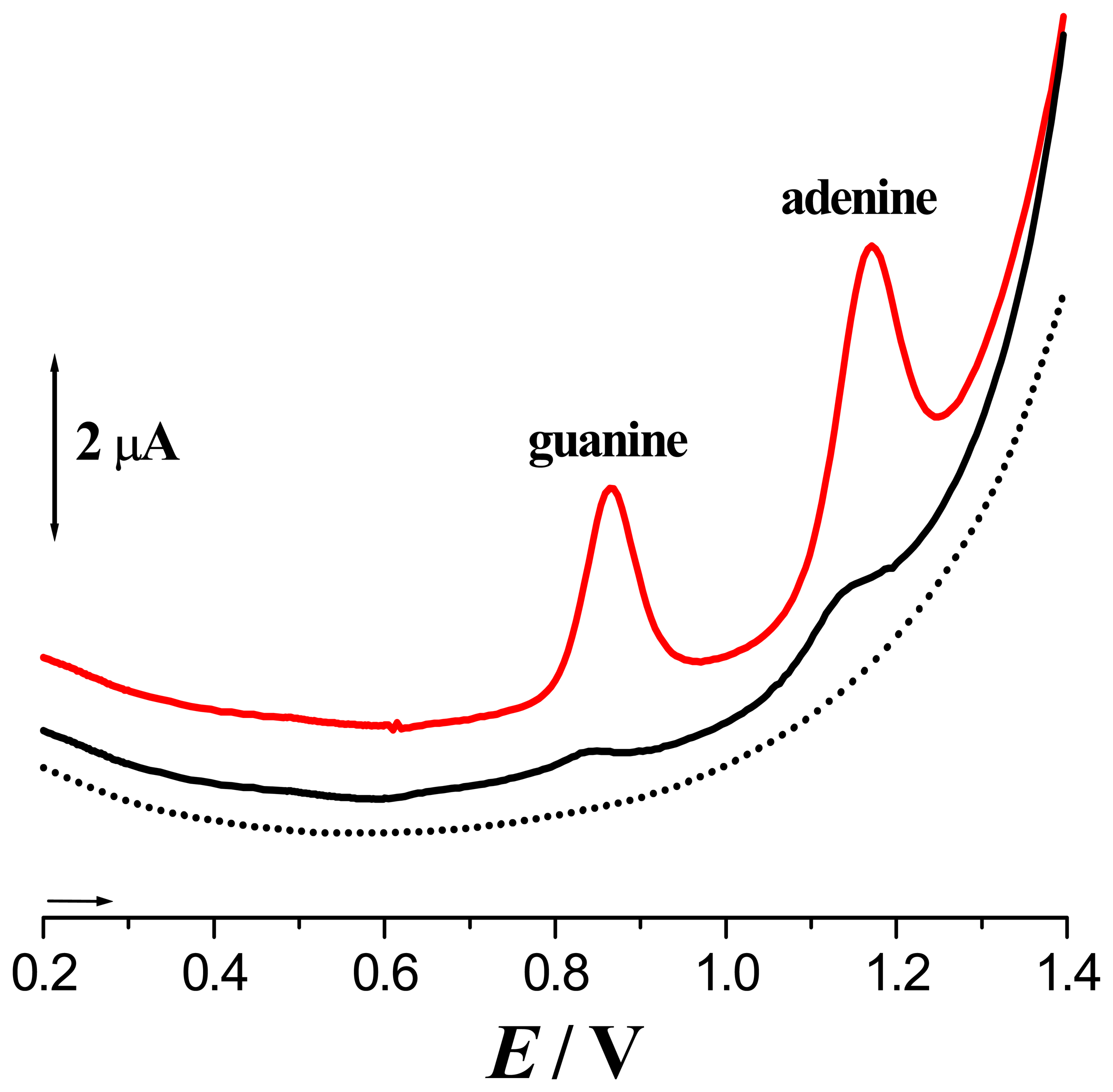
) before and after applying +0.400 V during 300 s in ( Sensors 05 00377i7) normal atmosphere and in ( Sensors 05 00377i8) N2 saturated buffer solution. [From Ref [40] with permission]
Sensors 05 00377f5

