<?xml version="1.0" encoding="UTF-8"?>
<!DOCTYPE article PUBLIC "-//NLM//DTD Journal Publishing DTD v2.3 20070202//EN" "journalpublishing.dtd">
<article xmlns:mml="http://www.w3.org/1998/Math/MathML" xmlns:xlink="http://www.w3.org/1999/xlink" xml:lang="en" article-type="review-article">
<front>
<journal-meta>
<journal-id journal-id-type="nlm-ta">Sensors</journal-id>
<journal-title>Sensors</journal-title>
<issn pub-type="epub">1424-8220</issn>
<publisher>
<publisher-name>Molecular Diversity Preservation International (MDPI)</publisher-name></publisher></journal-meta>
<article-meta>
<article-id pub-id-type="doi">10.3390/s121115036</article-id>
<article-id pub-id-type="publisher-id">sensors-12-15036</article-id>
<article-categories>
<subj-group>
<subject>Review</subject></subj-group></article-categories>
<title-group>
<article-title>Interfacial Structures and Properties of Organic Materials for Biosensors: An Overview</article-title></title-group>
<contrib-group>
<contrib contrib-type="author">
<name><surname>Zhou</surname><given-names>Yan</given-names></name><xref ref-type="aff" rid="af1-sensors-12-15036"><sup>1</sup></xref></contrib>
<contrib contrib-type="author">
<name><surname>Chiu</surname><given-names>Cheng-Wei</given-names></name><xref ref-type="aff" rid="af2-sensors-12-15036"><sup>2</sup></xref></contrib>
<contrib contrib-type="author">
<name><surname>Liang</surname><given-names>Hong</given-names></name><xref ref-type="aff" rid="af1-sensors-12-15036"><sup>1</sup></xref><xref ref-type="aff" rid="af2-sensors-12-15036"><sup>2</sup></xref><xref ref-type="corresp" rid="c1-sensors-12-15036"><sup>*</sup></xref></contrib></contrib-group>
<aff id="af1-sensors-12-15036">
<label>1</label> Materials Science and Engineering, Texas A&amp;M University, College Station, TX 77843, USA; E-Mail: <email>yanzhou@tamu.edu</email></aff>
<aff id="af2-sensors-12-15036">
<label>2</label> Department of Mechanical Engineering, Texas A&amp;M University, College Station, TX 77843, USA; E-Mail: <email>helloweiwei6824@tamu.edu</email></aff>
<author-notes>
<corresp id="c1-sensors-12-15036">
<label>*</label> Author to whom correspondence should be addressed; E-Mail: <email>hliang@tamu.edu</email>; Tel.: +1-979-862-2623; Fax: +1-979-845-3081.</corresp></author-notes>
<pub-date pub-type="collection">
<year>2012</year></pub-date>
<pub-date pub-type="epub">
<day>06</day>
<month>11</month>
<year>2012</year></pub-date>
<volume>12</volume>
<issue>11</issue>
<fpage>15036</fpage>
<lpage>15062</lpage>
<history>
<date date-type="received">
<day>21</day>
<month>09</month>
<year>2012</year></date>
<date date-type="rev-recd">
<day>31</day>
<month>10</month>
<year>2012</year></date>
<date date-type="accepted">
<day>02</day>
<month>11</month>
<year>2012</year></date></history>
<permissions>
<copyright-statement>© 2012 by the authors; licensee MDPI, Basel, Switzerland.</copyright-statement>
<copyright-year>2012</copyright-year>
<license>
<p>This article is an open access article distributed under the terms and conditions of the Creative Commons Attribution license (http://creativecommons.org/licenses/by/3.0/).</p></license></permissions>
<abstract>
<p>The capabilities of biosensors for bio-environmental monitoring have profound influences on medical, pharmaceutical, and environmental applications. This paper provides an overview on the background and applications of the state-of-the-art biosensors. Different types of biosensors are summarized and sensing mechanisms are discussed. A review of organic materials used in biosensors is given. Specifically, this review focuses on self-assembled monolayers (SAM) due to their high sensitivity and high versatility. The kinetics, chemistry, and the immobilization strategies of biomolecules are discussed. Other representative organic materials, such as graphene, carbon nanotubes (CNTs), and conductive polymers are also introduced in this review.</p></abstract>
<kwd-group>
<kwd>biosensor</kwd>
<kwd>interfacial properties</kwd>
<kwd>organic materials</kwd></kwd-group></article-meta></front>
<body>
<sec sec-type="intro">
<label>1.</label>
<title>Introduction</title>
<p>
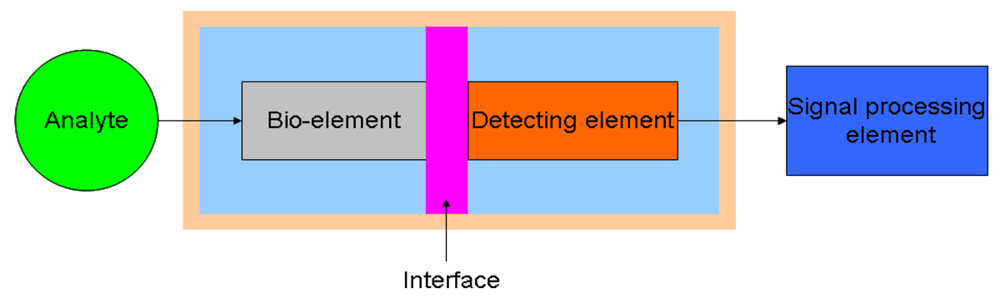
A biosensor is a device for transforming biological signals into analytical ones. It combines a bio-component with a physical element that is mainly used for converting the complex biologically derived message to quantitative information. A biosensing device has a wide range of application in the fields of environmental monitoring, drug development, and biomolecular interactions. The representation of a biosensor consists of three major components: a sensitive bio-element, a detecting element, and a signal processing element. The bio-element can be enzymes, living cells, or microorganisms, <italic>etc.</italic> [<xref ref-type="bibr" rid="b1-sensors-12-15036">1</xref>–<xref ref-type="bibr" rid="b3-sensors-12-15036">3</xref>], which recognizes the target analyte. The detecting element can be used to monitor the variation of electric current and potential [<xref ref-type="bibr" rid="b4-sensors-12-15036">4</xref>–<xref ref-type="bibr" rid="b7-sensors-12-15036">7</xref>], impedance [<xref ref-type="bibr" rid="b8-sensors-12-15036">8</xref>–<xref ref-type="bibr" rid="b11-sensors-12-15036">11</xref>], optical intensity [<xref ref-type="bibr" rid="b12-sensors-12-15036">12</xref>–<xref ref-type="bibr" rid="b15-sensors-12-15036">15</xref>], and electromagnetic radiation [<xref ref-type="bibr" rid="b16-sensors-12-15036">16</xref>,<xref ref-type="bibr" rid="b17-sensors-12-15036">17</xref>], among others. The bio-element directly interfaces to a signal transducer (detecting element), which together relate the variation of the analyte to a measurable response. Different constitutions of a bio-element coupled to a detecting element lead to a variety of applications. <xref ref-type="fig" rid="f1-sensors-12-15036">Figure 1</xref> is the schematic diagram that represents the concept of a biosensor.</p></sec>
<sec>
<label>2.</label>
<title>Types of Biosensors</title>
<p>Due to the different signal detecting mechanisms, biosensors can be categorized into various types, including resonant, photometric, thermal detection, ion-sensitive field-effect transistors (ISFETs), and electrochemical sensors. In the following sections, the types used for biosensors and their sensing mechanisms will be discussed.</p>
<sec>
<label>2.1.</label>
<title>Resonant Biosensors</title>
<p>A resonant biosensor is generally used to detect bio-relevant molecules in aqueous media. The sensing focuses on the change of mass, viscosity, or conductivity of the substrate surface. Many biosensors fall into this category, which includes surface acoustic wave sensors, magnetoelastic sensors, quartz crystal (piezoelectric) biosensors, <italic>etc</italic>.</p>
<p>
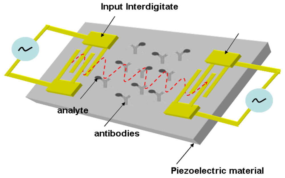
The prototype of a surface acoustic wave sensor (SAW) was established based on a strongly confined acoustic energy detected by interdigitated transducers [<xref ref-type="bibr" rid="b4-sensors-12-15036">4</xref>,<xref ref-type="bibr" rid="b19-sensors-12-15036">19</xref>,<xref ref-type="bibr" rid="b20-sensors-12-15036">20</xref>]. A SAW sensor has one piezoelectric material positioned in between two transducers. An input transducer electrically excites acoustic wave of the piezoelectric material, in which the acoustic wave is received at the output transducer. The wave energy radiates into the aqueous bulk due to the perpendicular displacement of the wave propagation in aqueous environment. This causes a high attenuation of the received signal, which hinders the application transferred into a biosensor [<xref ref-type="bibr" rid="b4-sensors-12-15036">4</xref>,<xref ref-type="bibr" rid="b21-sensors-12-15036">21</xref>,<xref ref-type="bibr" rid="b22-sensors-12-15036">22</xref>]. The shear-horizontal (SH) surface wave, generally referred to as a Love wave, is generated using a deposited elastic layer to guide the direction of the acoustic resonance. The elastic material significantly reduces the spreading loss of acoustic energy [<xref ref-type="bibr" rid="b23-sensors-12-15036">23</xref>–<xref ref-type="bibr" rid="b25-sensors-12-15036">25</xref>]. Now the operation of a SAW-based biosensor is driven by the coupled wave transducer and antibody on a piezoelectric substrate. The antibody used as a bio-element is immobilized on the device that catches analytes from the aqueous medium. The bonded analytes will change the velocity of the SAW, which alters the output signal generated by the integrated electronics. The variation of the output signal can be used to evaluate the concentration of the analytes. The schematic setup of a SAW-based biosensor is presented in <xref ref-type="fig" rid="f2-sensors-12-15036">Figure 2</xref> [<xref ref-type="bibr" rid="b26-sensors-12-15036">26</xref>].</p>
<p>The theoretical principal of quartz crystal biosensors relies on the piezoelectric effect of the crystal. When a piezoelectric material is subjected to an AC potential, a mechanical oscillation of the material is excited. The frequency change of such oscillation is correlated to the mass change on the material surface. The mathematical relationship (<xref rid="FD1" ref-type="disp-formula">Equation (1)</xref>) between the resonant frequency and the mass variation was firstly described by Sauerbrey [<xref ref-type="bibr" rid="b27-sensors-12-15036">27</xref>]:
<disp-formula id="FD1">
<label>(1)</label>
<mml:math id="mm1" display="block">
<mml:semantics id="sm1">
<mml:mrow>
<mml:mo>Δ</mml:mo>
<mml:msub>
<mml:mi>f</mml:mi>
<mml:mi>m</mml:mi></mml:msub>
<mml:mo>=</mml:mo>
<mml:mfrac>
<mml:mrow>
<mml:msup>
<mml:mrow>
<mml:msub>
<mml:mi>f</mml:mi>
<mml:mn>0</mml:mn></mml:msub></mml:mrow>
<mml:mn>2</mml:mn></mml:msup></mml:mrow>
<mml:mrow>
<mml:msub>
<mml:mi>F</mml:mi>
<mml:mi>q</mml:mi></mml:msub>
<mml:msub>
<mml:mi>ρ</mml:mi>
<mml:mi>q</mml:mi></mml:msub></mml:mrow></mml:mfrac>
<mml:msub>
<mml:mi>m</mml:mi>
<mml:mi>s</mml:mi></mml:msub></mml:mrow></mml:semantics></mml:math></disp-formula> where <italic>f<sub>0</sub></italic> is the resonance frequency of the quartz resonator, F<sub>q</sub> is the constant of crystal frequency, ρ<sub>q</sub> is the material density, and m<sub>s</sub> is the mass/area. In the quartz biosensor, the adsorption of target analytes onto the quartz surface causes the shift of resonant frequency that can derive information for quantitative analysis.</p>
<p>The operation of magnetoelastic sensors relies on the mechanical vibration of a magnetoelastic material when the material is subjected to a magnetic field. Such responses of the sensor not only can be detected acoustically, but also magnetically. The vibration generates an elastic wave within the magnetoelastic material that results in a detectable magnetic flux [<xref ref-type="bibr" rid="b28-sensors-12-15036">28</xref>]. In a magnetoelastic sensor, the vibrational frequency (<italic>f</italic>) can be expressed as[<xref ref-type="bibr" rid="b29-sensors-12-15036">29</xref>]:
<disp-formula id="FD2">
<label>(2)</label>
<mml:math id="mm2" display="block">
<mml:semantics id="sm2">
<mml:mrow>
<mml:mi>f</mml:mi>
<mml:mo>=</mml:mo>
<mml:mfrac>
<mml:mn>1</mml:mn>
<mml:mrow>
<mml:mn>2</mml:mn>
<mml:mi>L</mml:mi></mml:mrow></mml:mfrac>
<mml:msqrt>
<mml:mrow>
<mml:mfrac>
<mml:mi>E</mml:mi>
<mml:mi>ρ</mml:mi></mml:mfrac></mml:mrow></mml:msqrt></mml:mrow></mml:semantics></mml:math></disp-formula> where L is the length, E is elasticity, and ρ is the density. When a magnetoelastic sensor is subjected to a mass change (Δm), the variation of frequency can be expressed as[<xref ref-type="bibr" rid="b29-sensors-12-15036">29</xref>]:
<disp-formula id="FD3">
<label>(3)</label>
<mml:math id="mm3" display="block">
<mml:semantics id="sm3">
<mml:mrow>
<mml:mo>Δ</mml:mo>
<mml:mi>f</mml:mi>
<mml:mo>=</mml:mo>
<mml:mo>−</mml:mo>
<mml:msub>
<mml:mi>f</mml:mi>
<mml:mn>0</mml:mn></mml:msub>
<mml:mfrac>
<mml:mrow>
<mml:mo>Δ</mml:mo>
<mml:mi>m</mml:mi></mml:mrow>
<mml:mrow>
<mml:mn>2</mml:mn>
<mml:msub>
<mml:mi>m</mml:mi>
<mml:mn>0</mml:mn></mml:msub></mml:mrow></mml:mfrac></mml:mrow></mml:semantics></mml:math></disp-formula> where f<sub>0</sub> is the initial resonance frequency. When the sensor is subjected to a damping effect created by the surrounding medium, the shift in resonance frequency can be expressed as [<xref ref-type="bibr" rid="b30-sensors-12-15036">30</xref>]:
<disp-formula id="FD4">
<label>(4)</label>
<mml:math id="mm4" display="block">
<mml:semantics id="sm4">
<mml:mrow>
<mml:mo>Δ</mml:mo>
<mml:mi>f</mml:mi>
<mml:mo>=</mml:mo>
<mml:mfrac>
<mml:mrow>
<mml:msqrt>
<mml:mrow>
<mml:mi>π</mml:mi>
<mml:msub>
<mml:mi>f</mml:mi>
<mml:mn>0</mml:mn></mml:msub></mml:mrow></mml:msqrt></mml:mrow>
<mml:mrow>
<mml:mn>2</mml:mn>
<mml:mi>π</mml:mi>
<mml:msub>
<mml:mi>ρ</mml:mi>
<mml:mi>s</mml:mi></mml:msub>
<mml:mi>d</mml:mi></mml:mrow></mml:mfrac>
<mml:msqrt>
<mml:mrow>
<mml:mi>η</mml:mi>
<mml:msub>
<mml:mi>ρ</mml:mi>
<mml:mn>1</mml:mn></mml:msub></mml:mrow></mml:msqrt></mml:mrow></mml:semantics></mml:math></disp-formula> where η is viscosity of medium, s<sub>l</sub> is the density of medium, ρ<sub>s</sub> is the density of sensor material, and d is the thickness of the sensor. At present, the characteristics of magnetoelastic materials have been employed in different kinds of bio-sensing such as glucose, avidin, <italic>Escherichia coli</italic>, and platelet-rich plasma [<xref ref-type="bibr" rid="b30-sensors-12-15036">30</xref>–<xref ref-type="bibr" rid="b33-sensors-12-15036">33</xref>], among others.</p></sec>
<sec>
<label>2.2.</label>
<title>Thermal Detection Biosensors</title>
<p>Surprisingly, even with their poor reputation for weak sensitivity and non-specific heating effects, the applications of thermal biosensors still draws considerable attention. A thermal biosensor is a promising analytical tool due to the following advantages:
<list list-type="simple">
<list-item>
<label>-</label>
<p>No chemical contact between transducer and sample leading to long-term stability.</p></list-item>
<list-item>
<label>-</label>
<p>Economical bulk products and quick response.</p></list-item>
<list-item>
<label>-</label>
<p>Measurements are not interfered by sample characteristics.</p></list-item></list></p>
<p>Novel thermal biosensors based on enzymatic conversion have been developed for monitoring enzyme reactions. This is mainly because most biochemical reactions have an exothermic character. The principle of thermal biosensor measurement is based on the first law of thermodynamics:
<disp-formula id="FD5">
<label>(5)</label>
<mml:math id="mm5" display="block">
<mml:semantics id="sm5">
<mml:mrow>
<mml:mtext>Q</mml:mtext>
<mml:mo>=</mml:mo>
<mml:mo>−</mml:mo>
<mml:msub>
<mml:mtext>n</mml:mtext>
<mml:mtext>p</mml:mtext></mml:msub>
<mml:mo>∑</mml:mo>
<mml:mo>Δ</mml:mo>
<mml:mtext>H</mml:mtext></mml:mrow></mml:semantics></mml:math></disp-formula>
<disp-formula id="FD6">
<label>(6)</label>
<mml:math id="mm6" display="block">
<mml:semantics id="sm6">
<mml:mrow>
<mml:mtext>Q</mml:mtext>
<mml:mo>=</mml:mo>
<mml:mtext>Cp</mml:mtext>
<mml:mo>Δ</mml:mo>
<mml:mtext>T</mml:mtext></mml:mrow></mml:semantics></mml:math></disp-formula> where Q is the total energy of heat, H is the enthalpy, Cp is the heat capacity. The measureable local temperature shift (ΔT) generated by the heat production is dependent on the heat capacity of the system [<xref ref-type="bibr" rid="b34-sensors-12-15036">34</xref>]:
<disp-formula id="FD7">
<label>(7)</label>
<mml:math id="mm7" display="block">
<mml:semantics id="sm7">
<mml:mrow>
<mml:mo>Δ</mml:mo>
<mml:mtext>T</mml:mtext>
<mml:mo>=</mml:mo>
<mml:mo>−</mml:mo>
<mml:mo>Δ</mml:mo>
<mml:msub>
<mml:mrow>
<mml:mtext>Hn</mml:mtext></mml:mrow>
<mml:mtext>p</mml:mtext></mml:msub>
<mml:mo>/</mml:mo>
<mml:mtext>Cp</mml:mtext></mml:mrow></mml:semantics></mml:math></disp-formula></p>
<p>
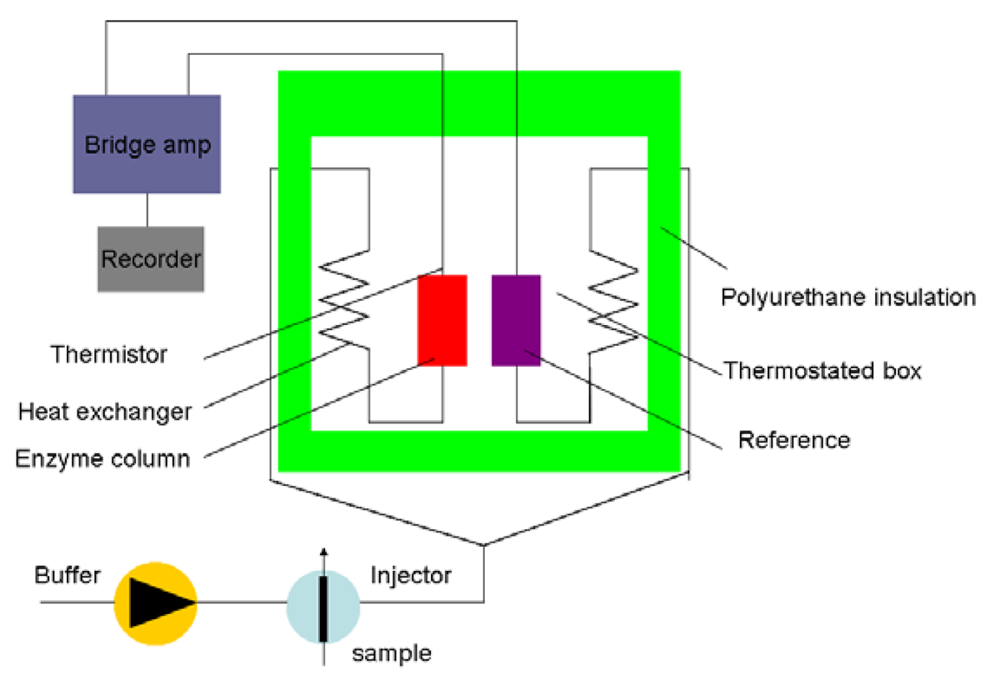
Generally, the Cp of most organic media is lower than that of the aqueous solvent [<xref ref-type="bibr" rid="b35-sensors-12-15036">35</xref>]. The sensitivity and detection limit of the sensor are determined by the organic solvents. <xref ref-type="fig" rid="f3-sensors-12-15036">Figure 3</xref> shows a schematic diagram of the principle set-up for an enzyme thermistor (ET). The thermostated box controls the physiological temperature. Samples and the buffer are injected in the ET where the aluminum thermostates the buffer stream. The heat generated by the enzymatic conversion reduces thermistor resistance and the bridge amplifier registers the signal.</p></sec>
<sec>
<label>2.3.</label>
<title>Photometric Biosensors</title>
<p>
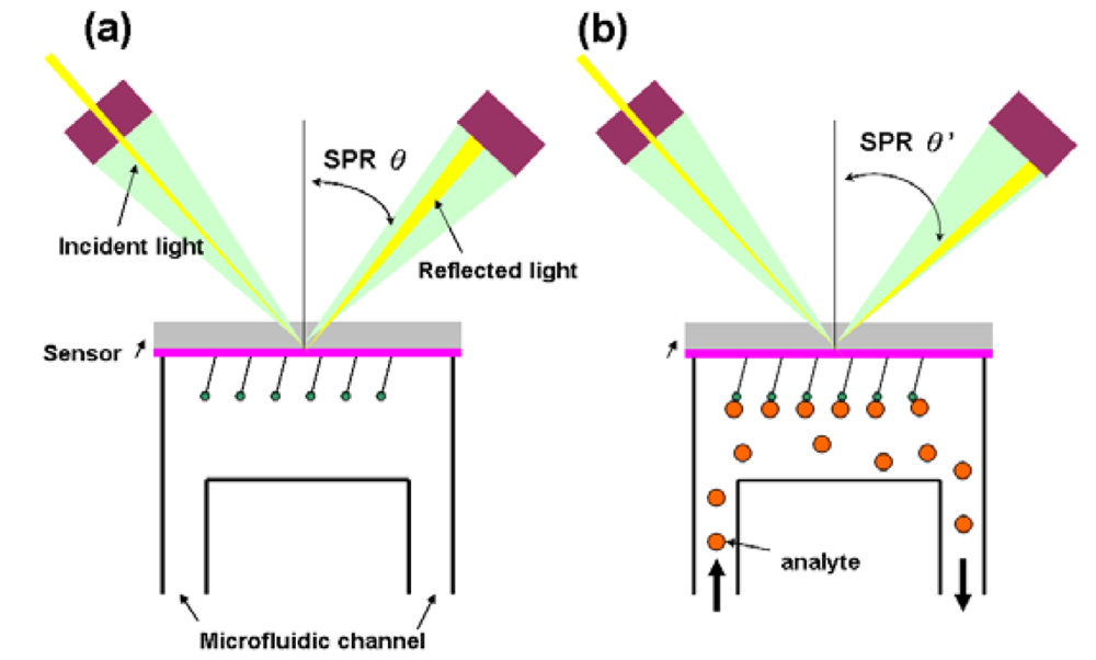
In this type of biosensor, the measured output transduced signal is the light intensity. More than 75% of the research papers for optical biosensors focus on using the surface plasmon resonance (SPR) [<xref ref-type="bibr" rid="b37-sensors-12-15036">37</xref>] effect. The SPR essentially is a diffraction anomaly due to the surface excited plasma [<xref ref-type="bibr" rid="b38-sensors-12-15036">38</xref>]. The electrons resonate when the wavelength of oscillating mobile electrons (plasma) matches the wave vector of incident light. The resonating plasma is associated with the electromagnetic waves propagating in a direction parallel to the interface of two media, and decaying evanescently, <italic>i.e.</italic>, evanescent wave. Due to the limited propagation length of a surface plasma wave (SPW), the detection of SPR sensor is conducted where the SPW is excited by the incident light source. A SPR sensor is constituted by an incident light, a transducer with a gold side contacted with the detection apparatus and the other side contacted with microfluidic system (flow side), and an electronic system for processing the output signal. Fixed wavelength is shot to the gold side and is reflected, which induces an evanescent wave penetrating into the flow side. During the measurement, the analyte is introduced through the microfluidic channel and bound with the sensor, which changes the dielectric constant of the medium. This will lead to the changes of refractive index near the surface hence affecting the refracting SPR angle (<xref ref-type="fig" rid="f4-sensors-12-15036">Figure 4</xref>) [<xref ref-type="bibr" rid="b39-sensors-12-15036">39</xref>–<xref ref-type="bibr" rid="b41-sensors-12-15036">41</xref>].</p>
<p>The sensing mechanims of other photometric biosensors are based on the optical properties of analytes, which include absorbance/scattering and fluorescence. The utilization of the absorbance properties of analytes is to detect the amount of laser light being blocked or transmitted by the target cells [<xref ref-type="bibr" rid="b43-sensors-12-15036">43</xref>,<xref ref-type="bibr" rid="b44-sensors-12-15036">44</xref>]. Such a measuring system provides a rapid, simple, repeatable, and label-free assay for the immobilized analytes. Due to the limited sensitivity of the conventional methods in absorbance studies, an optical waveguide has been introduced in order to enhance the efficiency of absorbance biosensor. Optic fiber probes with different geometries have been employeed in absorbance biosensors that include coiled and tapered fibers, among others [<xref ref-type="bibr" rid="b45-sensors-12-15036">45</xref>,<xref ref-type="bibr" rid="b46-sensors-12-15036">46</xref>]. The fluorescent staining technique is also introduced in photometric biosensors. Such a technique is usually requred in microfludic-platform biosensors for the purpose of bacterial counting [<xref ref-type="bibr" rid="b47-sensors-12-15036">47</xref>].</p></sec>
<sec>
<label>2.4.</label>
<title>Ion-Sensitive FETs Biosensors</title>
<p>The development of ISFETs, usually used for pH and ion concentration measurements [<xref ref-type="bibr" rid="b48-sensors-12-15036">48</xref>–<xref ref-type="bibr" rid="b50-sensors-12-15036">50</xref>], started in the 1970s. To integrate the sensing circuit, most ISFETs were produced through MEMS fabrication based on silicon substrates. This also makes ISFETs a perfect transducing element for biosensing. The reactive silanol (SiOH) groups on the SiO<sub>2</sub> surface provide a stem for covalent attachment of bio-molecules by using H<sup>+</sup> and OH<sup>+</sup> as binding sites:
<disp-formula id="FD8">
<mml:math id="mm8" display="block">
<mml:semantics id="sm8">
<mml:mrow>
<mml:mtable columnalign="left">
<mml:mtr columnalign="left">
<mml:mtd columnalign="left">
<mml:mrow>
<mml:mrow>
<mml:mtext>SiOH</mml:mtext>
<mml:mo>‐</mml:mo>
<mml:mo>‐</mml:mo>
<mml:mo>‐</mml:mo>
<mml:mo>‐</mml:mo>
<mml:mo>‐</mml:mo>
<mml:mo>‐</mml:mo>
<mml:mo>‐</mml:mo>
<mml:mo>‐</mml:mo>
<mml:mo>‐</mml:mo>
<mml:mo>‐</mml:mo>
<mml:mo>‐</mml:mo></mml:mrow>
<mml:msup>
<mml:mrow>
<mml:mtext>SiO</mml:mtext></mml:mrow>
<mml:mo>−</mml:mo></mml:msup>
<mml:mo>+</mml:mo>
<mml:msup>
<mml:mtext>H</mml:mtext>
<mml:mo>+</mml:mo></mml:msup></mml:mrow></mml:mtd></mml:mtr>
<mml:mtr columnalign="left">
<mml:mtd columnalign="left">
<mml:mrow>
<mml:mtext>SiOH</mml:mtext>
<mml:mo>+</mml:mo>
<mml:msup>
<mml:mtext>H</mml:mtext>
<mml:mo>+</mml:mo></mml:msup>
<mml:msup>
<mml:mrow>
<mml:mrow>
<mml:mo>‐</mml:mo>
<mml:mo>‐</mml:mo>
<mml:mo>‐</mml:mo>
<mml:mo>‐</mml:mo>
<mml:mo>‐</mml:mo>
<mml:mo>‐</mml:mo>
<mml:mo>‐</mml:mo>
<mml:mo>‐</mml:mo>
<mml:mo>‐</mml:mo>
<mml:mtext>SiOH</mml:mtext></mml:mrow></mml:mrow>
<mml:mrow>
<mml:mn>2</mml:mn>
<mml:mo>+</mml:mo></mml:mrow></mml:msup></mml:mrow></mml:mtd></mml:mtr></mml:mtable></mml:mrow></mml:semantics></mml:math></disp-formula></p>
<p>
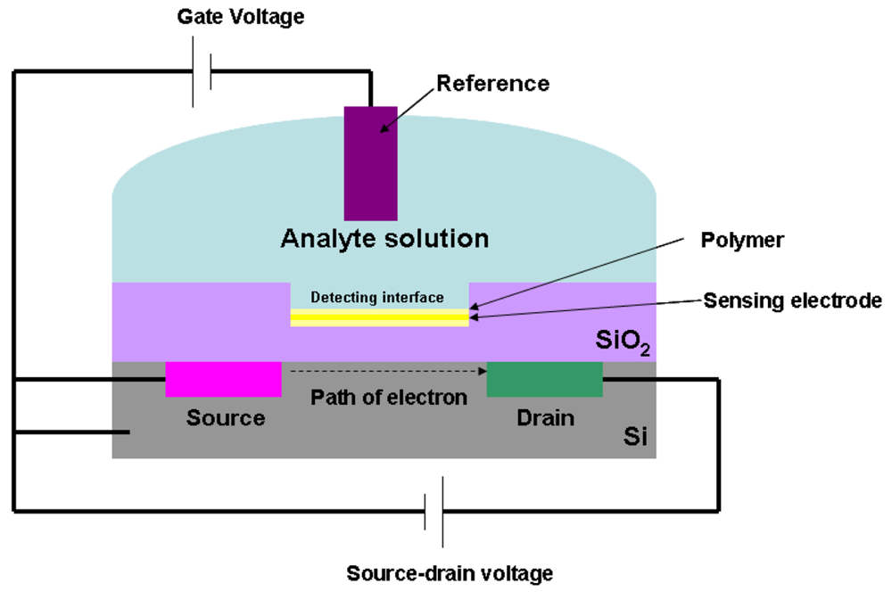
Through silicon surface modification, the analytes can be successfully immobilized onto the gate surface [<xref ref-type="bibr" rid="b51-sensors-12-15036">51</xref>–<xref ref-type="bibr" rid="b55-sensors-12-15036">55</xref>]. <xref ref-type="fig" rid="f5-sensors-12-15036">Figure 5</xref> outlines the schematic configuration of ISFETs.</p>
<p>An ISFET consists of a sensing electrode coated with a polymer selectively permeable to analyte ions, and a field effect transistor structure. The current flow in the gate voltage is regulated by the potential difference between source and drain. When the analyte ions diffuse through the ion-sensitive polymer, the charged biomolecules cause the depletion or accumulation of charge carriers. This will lead to the potential difference at the detecting interface. This interfacial potential difference is regulated by the ions concentration in the solution; therefore, the change of the current in the transistor is a measure of analyte concentration.</p></sec>
<sec>
<label>2.5.</label>
<title>Electrochemical Biosensors</title>
<p>
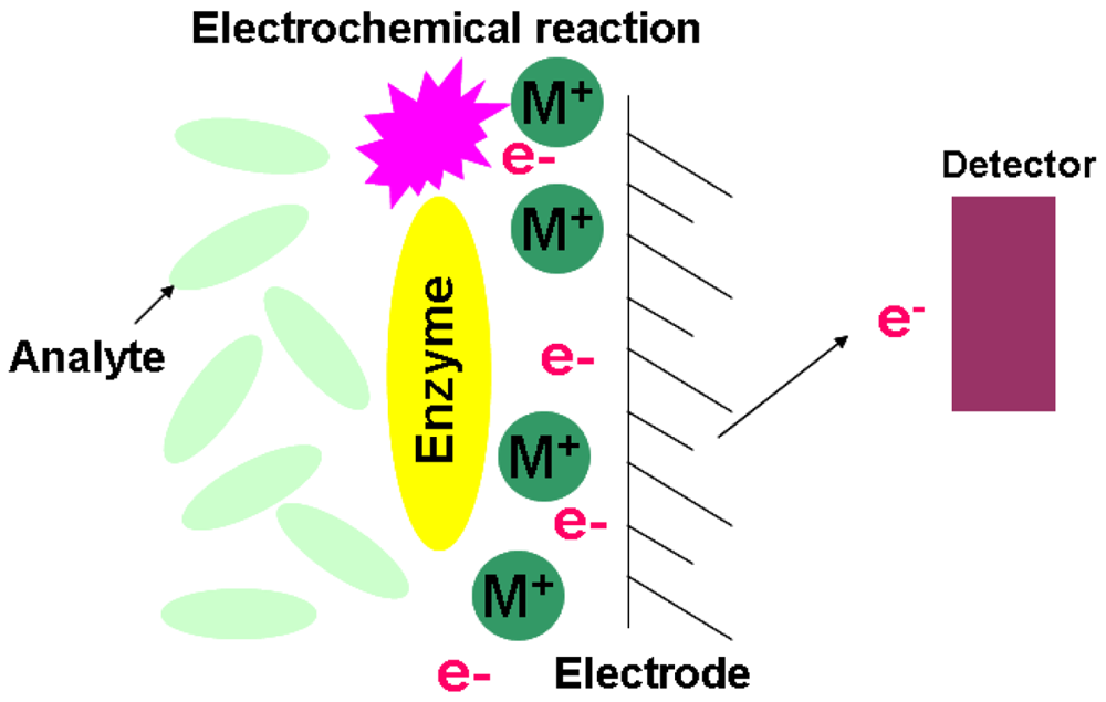
The constitutional concept of an electrochemical biosensor is based on a matrix bound bioactive material coupled with an electrochemical transducer. Essentially, it is a surface modified electrical conductor for different electrochemical functions. This type of sensor targets those biological reactions that derive ionic production and consumption. This will cause the charge transfer across the double layer of the physio-chemical transducer that generates the measureable signal [<xref ref-type="bibr" rid="b56-sensors-12-15036">56</xref>–<xref ref-type="bibr" rid="b58-sensors-12-15036">58</xref>]. Based on the measured signal characterizations, the electrochemical biosensor has three main classifications of silicon based chips. <xref ref-type="fig" rid="f6-sensors-12-15036">Figure 6</xref> is the illustration of the electrochemical biosensor.</p>
<sec>
<label>2.5.1.</label>
<title>Potentiometric</title>
<p>The principle of the potentiometric biosensor is based on an ISFET. The output signal is generated by the potential differences of oxidation/reduction reactions. The electrochemical reaction generated ions are accumulated at the ion-sensitive membrane of the ISFET interface. When this potential is applied to the electrode, it modulates the current flow through the FET leading to a measurable potential of the detector.</p></sec>
<sec>
<label>2.5.2.</label>
<title>Amperometric</title>
<p>The high sensitivity of an amperometric sensor provides the sensing ability to detect electroactive substances in biological samples. By applying a constant potential between the sensing and auxiliary electrode, the conversion of electroactive species takes place at the electrode. This will result in electron transfer, and the current is directly correlated to the bulk concentration of tested electroactive species [<xref ref-type="bibr" rid="b56-sensors-12-15036">56</xref>,<xref ref-type="bibr" rid="b57-sensors-12-15036">57</xref>].</p></sec>
<sec>
<label>2.5.3.</label>
<title>Impedimetric</title>
<p>The chemical reactions resulting in either ion production or consumption will change the conductivity of the solution. The measure of solution's impedance (Z) change is introduced for this type of sensor. The sensitivity of an impedimetric sensor is relatively low since the measure of conductance is essentially non-specific. This drawback can be overcome by targeting the specific defined geometry of enzymatic reactions in a microelectronic cell [<xref ref-type="bibr" rid="b56-sensors-12-15036">56</xref>].</p>
<p>The types of biosensors, sensing mechanisms, and the applications are summarized in <xref ref-type="table" rid="t1-sensors-12-15036">Table 1</xref>. Beside discussed above, hybrid sensors have emerged in recent years [<xref ref-type="bibr" rid="b59-sensors-12-15036">59</xref>–<xref ref-type="bibr" rid="b62-sensors-12-15036">62</xref>]. These sensors contain types and mechanisms that are expected to grow in the near future.</p></sec></sec></sec>
<sec>
<label>3.</label>
<title>Self-Assembled Monolayer</title>
<p>This section focuses on the organic materials used in biosensors. Emphasis will be given to the self-assembled monolayer (SAM), which provides molecular level control over the density and position of assembled molecules. SAM is capable of packing different types of molecules in an orderly fashion at the molecular level, which generates a multifunctional surface for multitasks. SAM is advantageous due to its simplicity of preparation, high sensitivity, and few limitations in the detection range of an analyte, and most importantly, the versatility of modification that no other organic materials could match. The assembling kinetics, the chemistry of SAM, and the immobilization strategy of biomolecules onto SAM will be discussed.</p>
<sec>
<label>3.1.</label>
<title>Introduction</title>
<p>By definition, SAM is a single layer of biological and/or chemical molecules formed through self-assembly. It is a part of molecular nanotechnology that attracts interest due to its simple production and versatility in molecular and reaction selection. The first study on SAM was reported in 1946 by Bigelow <italic>et al.</italic> who used a metal surface to absorb surfactant molecules and made a layer with monomolecules [<xref ref-type="bibr" rid="b63-sensors-12-15036">63</xref>].</p></sec>
<sec>
<label>3.2.</label>
<title>Structure and Assembling Kinetics</title>
<p>
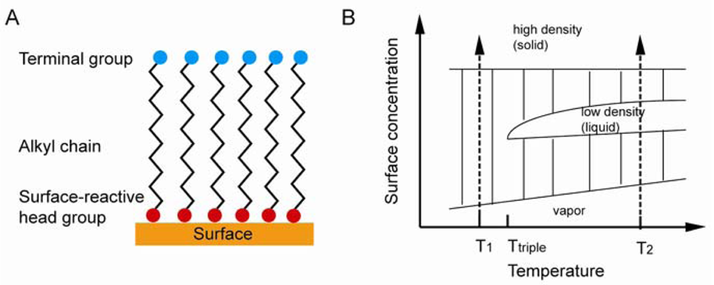
Under suitable conditions, SAMs can be directly formed on a sensor substrate. A substrate is placed in a solution containing assembling molecules, where SAM forms spontaneously. In some cases, UV irradiation, heat, voltage, and other conditions need to be applied in order to complete SAM formation. Self-assembling molecules contain three parts: the head group, which is open for bond formation with surface substrate; the alkyl chain, which is important in stabilization and order, interacts with neighboring chains through van der Waals and electrostatic forces; the functional terminal, which is open for functionalization and is used to determine the chemistry properties of SAM surface with the outer environment [<xref ref-type="bibr" rid="b64-sensors-12-15036">64</xref>]. The idealized SAM system is shown in <xref ref-type="fig" rid="f7-sensors-12-15036">Figure 7(A)</xref>.</p>
<p>As shown in <xref ref-type="fig" rid="f7-sensors-12-15036">Figure 7(B)</xref>, there are four phases during the SAM formation, including vapor phase, intermediate phase, low density phase, and high density phase. In the vapor phase, the surface is randomly deposited with isolated molecules. In the intermediate phase, the surface is deposited by adsorbate molecules with disordered conformation. In the low density (liquid) phase, adsorbate molecules are lying down on the surface. In the high density (solid) phase, molecules are standing in order and packed with tilting angle less than 30°. SAM formation would first go through the vapor phase when the temperature is lower than the triple point, followed by the intermediate phase, and the high density solid phase last. Otherwise, the formation would incorporate a low density liquid phase if the temperature is higher than the triple point. Both processes involve the formation of solid phase islands surrounded by isolated vapor phase molecules and the nucleation and growth of these islands till the entire surface is covered. The Langmuir model <xref rid="FD9" ref-type="disp-formula">Equation (8)</xref>) shown below is used to explain SAM formation:
<disp-formula id="FD9">
<label>(8)</label>
<mml:math id="mm9" display="block">
<mml:semantics id="sm9">
<mml:mrow>
<mml:mfrac>
<mml:mrow>
<mml:mi>d</mml:mi>
<mml:mi>θ</mml:mi></mml:mrow>
<mml:mrow>
<mml:mi>d</mml:mi>
<mml:mi>t</mml:mi></mml:mrow></mml:mfrac>
<mml:mo>=</mml:mo>
<mml:mi>k</mml:mi>
<mml:mo stretchy="false">(</mml:mo>
<mml:mn>1</mml:mn>
<mml:mo>−</mml:mo>
<mml:mi>θ</mml:mi>
<mml:mo stretchy="false">)</mml:mo></mml:mrow></mml:semantics></mml:math></disp-formula> where <italic>θ</italic> is the proportional coverage of the surface, and k is the rate constant [<xref ref-type="bibr" rid="b65-sensors-12-15036">65</xref>]. The equation indicates that the SAM formation rate is proportional to the uncovered surface. The initial formation of alkanethiol SAMs is 80–90% complete during the first several minutes, and molecules reorganize themselves over 12–16 hours [<xref ref-type="bibr" rid="b66-sensors-12-15036">66</xref>]; however, changes may still happen weeks later [<xref ref-type="bibr" rid="b67-sensors-12-15036">67</xref>].</p></sec>
<sec>
<label>3.3.</label>
<title>Chemistry of SAM</title>
<p>Increasing numbers of SAM systems are presented due to the accumulating knowledge in chemistry. SAM systems are separated into four types based on their chemical characteristics (<xref ref-type="table" rid="t2-sensors-12-15036">Table 2</xref>). They are alkanethiol and organosulfur on a metal surface, organosilane on a hydroxylated oxide surface, alkyne and alkene on a hydride-silicon surface, and aryl diazomium salts on a carbon, metal, metal oxide, silicon surface, <italic>etc</italic>. In the following sections, they will be discussed in detail.</p>
<sec>
<label>3.3.1.</label>
<title>Alkanethiol and Organosulfur</title>
<p>In the early days, the work of SAM was focused on the formation of organosulfur SAM on planar gold and silver surfaces through solution or vapor deposition [<xref ref-type="bibr" rid="b67-sensors-12-15036">67</xref>]. Metal surface and assembling molecules had to be transformed into a reactive substrate to allow the formation of SAM. An increasing numbers of compound ligands and metal substrates can be used in SAM formation. However, limitations still exist in the type of ligands that can be matched to the metal at certain oxidation states. Gold is the most studied substrate due to several reasons: gold is an inert element; gold thin film is convenient to obtain and is a standard material for several characterization methods; gold shows low toxicity to biology systems and has high biocompatibility. It is also straightforward to pattern gold through lithography and chemical etching [<xref ref-type="bibr" rid="b67-sensors-12-15036">67</xref>]. Thiol groups form gold-thiol bonds on gold surfaces with gold-thiol energies as low as 170 kJ/mol [<xref ref-type="bibr" rid="b68-sensors-12-15036">68</xref>]. It is believed that the van der Waals force plays an important role in stabilizing SAM structures. Other commonly used and studied metals are silver, copper [<xref ref-type="bibr" rid="b69-sensors-12-15036">69</xref>], and palladium [<xref ref-type="bibr" rid="b70-sensors-12-15036">70</xref>]. The alkane chains in the SAM adopt trans-conformation and tilt ∼30° from normal on gold surfaces, ∼10° on silver surfaces, 12° on copper surfaces [<xref ref-type="bibr" rid="b69-sensors-12-15036">69</xref>], and 0° on mercury [<xref ref-type="bibr" rid="b71-sensors-12-15036">71</xref>]. Sulfonates and sulfinates, oxidized metal-thiol bonds, are formed when SAM is exposed to air. The weaker bonds between oxidized species and metal substrate lead to molecules falling from the gold surface [<xref ref-type="bibr" rid="b72-sensors-12-15036">72</xref>]. It is shown that the length of an alkane chain is a factor in determining the rate of oxidation of a thiol group in SAM. The shorter the chain, the easier the oxidation occurs [<xref ref-type="bibr" rid="b73-sensors-12-15036">73</xref>].</p></sec>
<sec>
<label>3.3.2.</label>
<title>Organosilane Based Layers</title>
<p>
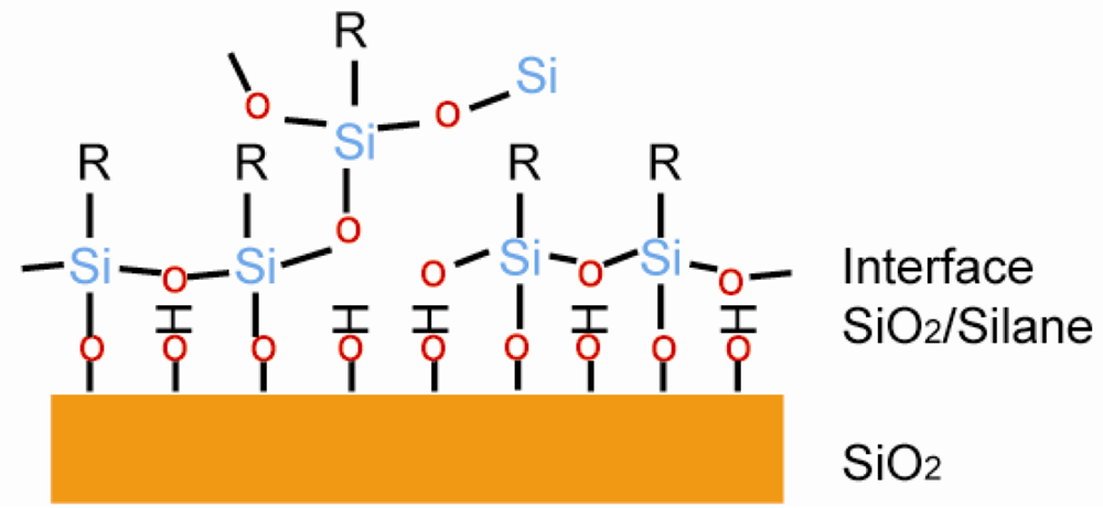
Organosilane based monolayers are one of the most studied SAM systems. The substrate has to be a hydroxylated oxide surface, which includes silicon dioxide and other metal oxides [<xref ref-type="bibr" rid="b64-sensors-12-15036">64</xref>]. A typical structure is shown in <xref ref-type="fig" rid="f8-sensors-12-15036">Figure 8</xref>, where organosilane SAM is connected to the hydroxylated surface through the S–O bond. Sagiv reported the octadecyltrichlorosilane (OTS) SAM on a hydroxylated surface. The SAM formed through a condensation reaction between the hydrolyzed OTS and the hydroxylated surface[<xref ref-type="bibr" rid="b74-sensors-12-15036">74</xref>]. The silicon dioxide has to go through a hydrophilic treatment before usage, otherwise the uniformity of SAM would drop dramatically [<xref ref-type="bibr" rid="b75-sensors-12-15036">75</xref>]. Less than 20% of the molecules formed S–O bonds on the hydroxylated surface, and the rest were connected to the neighboring molecules to form SAM [<xref ref-type="bibr" rid="b81-sensors-12-15036">81</xref>].</p></sec>
<sec>
<label>3.3.3.</label>
<title>Hydrosilylation</title>
<p>
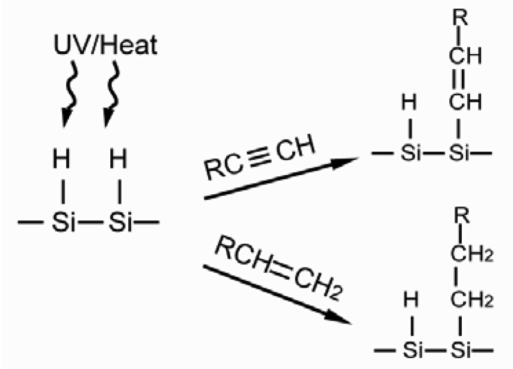
In the preparation of SAM through the hydrosilylation reaction, the silicon surface is pretreated with UV or heat to generate the S–H radicals in order for the surface to react with alkyl chains presenting 1-alkyne and 1-alkene terminals, as seen in <xref ref-type="fig" rid="f9-sensors-12-15036">Figure 9</xref>. When the reaction is completed, the silicon surface is linked with alkyl chains by S–C bond and generates alkene and alkane accordingly [<xref ref-type="bibr" rid="b82-sensors-12-15036">82</xref>]. SAM prepared by this method does not show the multilayer defect, but it has superior stability due to the non-polar bond of S–C. However, the silicon oxide largely affects the formation of the S-C bond hence reducing the quality of SAM. Thus, the SAM preparation has to be performed using oxide free silicon in an atmosphere with no oxygen [<xref ref-type="bibr" rid="b78-sensors-12-15036">78</xref>].</p></sec>
<sec>
<label>3.3.4.</label>
<title>Aryl Diazonium</title>
<p>Pinson first reported a SAM based on the aryl diazonium reaction in 1992 [<xref ref-type="bibr" rid="b79-sensors-12-15036">79</xref>]. It involves the reduction of aryl diazonium (<xref ref-type="fig" rid="f10-sensors-12-15036">Figure 10</xref>), which functionalizes the carbon surface with an aromatic group, which is then open to classical chemistry reactions. This method is of interest due to SAM's capability of being applied to all carbon, silicon, metals, and metal oxides substrates. In this mechanism, it is believed that an aryl radical forms an aryl diazonium species with the release of N<sub>2</sub>, then a covalent bond forms between the aryl group and the substrate [<xref ref-type="bibr" rid="b80-sensors-12-15036">80</xref>]. The resultant SAM shows higher stability, however, control over the reaction is limited.</p></sec></sec>
<sec>
<label>3.4.</label>
<title>Attachment of Biomolecules to SAM Biosensor Systems</title>
<p>Biomolecules can be attached to the functional terminals of modified electrodes by covalent and non-covalent bonds, as summarized in <xref ref-type="table" rid="t3-sensors-12-15036">Table 3</xref>. Non-covalent bonds, which includes hydrogen bonds and electrostatic interactions, are widely applied in attachment of biomolecules. The attachment is relatively weak compared to a covalent bond. Nevertheless, it only needs simple reaction steps and usually is reagentless. Covalent bonds provide stronger immobilization, but are restricted to certain reactions.</p>
<sec>
<label>3.4.1.</label>
<title>Immobilization of Biomolecules to SAM Biosensor Systems by Non-Covalent Bond</title>
<p>
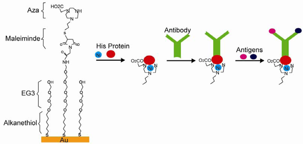
Electrostatic, hydrogen, and chelation interactions are non-covalent bonds between SAM and immobilized biomolecules. Cytochrome was successfully immobilized on carboxylic terminals of alkanethiol-gold SAM through electrostatic interactions. The immobilization reaches maximum at pH values of 3.5∼5.5, where cytochrome is positively charged and attracted by negatively charged carboxylic terminals [<xref ref-type="bibr" rid="b84-sensors-12-15036">84</xref>]. More than one species of biomolecules were immobilized through electrostatic interactions. Carboxylic terminated SAM was designed to attract lipid-DNA complexes (LDc), which showed positive charges due to excessive cationic lipids. Anionic plasmid DNA was then absorbed after the SAMs surface charge was reversed to cationic after LDc absorption, and the resulting SAM succeeded in gene transfer [<xref ref-type="bibr" rid="b85-sensors-12-15036">85</xref>]. Hydrogen bonds were used in biomolecules immobilization, which was reported by Gomes and others. They showed a SAM system with glycan-modified surfaces and immobilized proteins through hydrogen bonds [<xref ref-type="bibr" rid="b87-sensors-12-15036">87</xref>]. Histidine (His) modified proteins were attached to the surface through chelation interactions. For instance, nitrilotriacetic acid (NTA) was used to pretreat the quartz surface. Divalent metals such as Cu, Ni were then applied to fill tetradentate chelator sites, which formed hexagonal complexes and left two unoccupied sites for His bonding. Proteins with His tags would then be immobilized [<xref ref-type="bibr" rid="b88-sensors-12-15036">88</xref>]. In another system showed in <xref ref-type="fig" rid="f11-sensors-12-15036">Figure 11</xref>, tri(ethylene glycol) and maleimide modified alkanethiol molecules were used to form SAM on a Au surface. The SAM surface was then decorated with triazacyclononane (aza) or NTA ligands. His tagged proteins were then immobilized on SAM in the presence of divalent metals followed by IgG antibodies anchored on His tagged proteins. An immunoassay was carried out based on this system, which proved His tagged proteins, IgG antibodies, and antibody specific antigens were present [<xref ref-type="bibr" rid="b89-sensors-12-15036">89</xref>]. This type of reaction can be reversed after adding EDTA, which makes it ideal for biosensor applications [<xref ref-type="bibr" rid="b101-sensors-12-15036">101</xref>].</p></sec>
<sec>
<label>3.4.2.</label>
<title>Immobilization of Biomolecules to SAM Biosensor Systems by Covalent Bonds</title>
<p>When non-covalent bonds are used in immobilizing biomolecules on biosensor surfaces, the orientations of biomolecules tend to be random. Stronger covalent bonds provide more control over the orientation. Dehydration synthesis between carboxyl groups and amino groups helps protein array to achieve certain orientation. It is a widely used strategy due to its biocompatibility, simplicity of operation, and easy access to carbodiimide agents [<xref ref-type="bibr" rid="b64-sensors-12-15036">64</xref>]. Amino-modified SAM would react with C-termini of proteins and <italic>vice versa</italic>. For example, poly(ethylene glycol) (PEG) thiol SAM has Cys N-termini, which reacts with the C-termini of proteins through dehydration and forms amide bonds. The resulting immobilized proteins showed specific orientation, as seen in <xref ref-type="fig" rid="f12-sensors-12-15036">Figure 12</xref> [<xref ref-type="bibr" rid="b102-sensors-12-15036">102</xref>]. In another study, Herrwerth designed a PEG-alkanethiol SAM with carboxy termini in order to covalently couple to IgG antibodies upon chemical activation [<xref ref-type="bibr" rid="b90-sensors-12-15036">90</xref>].</p>
<p>
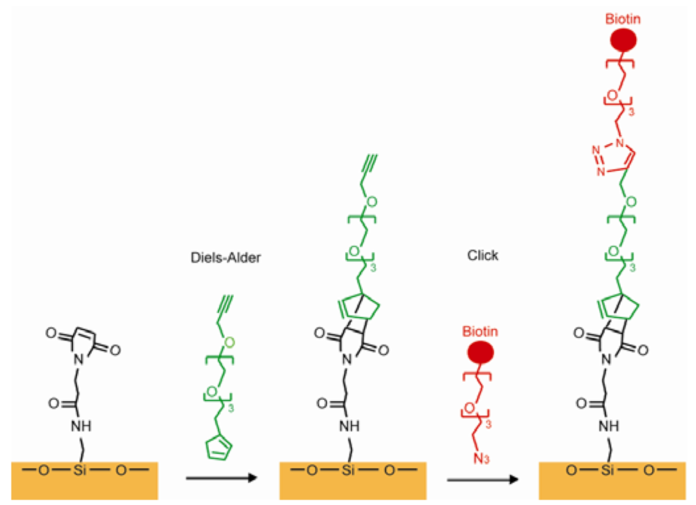
Using the reaction between maleimide and thiol residues of Cys is another common way in covalently immobilizing proteins. A hydroxylated glass surface was connected to the amino-terminated silane, and the amino group was subsequently linked to the N-succinimidyl-6-maleimidocaproate (EMCS). The thioether was produced to bind the maleimide group of EMCS on the surface and the thiol group on the Cys in the protein [<xref ref-type="bibr" rid="b92-sensors-12-15036">92</xref>]. The “click” reaction forms covalent immobilization, which exists between alkyne and azide through cycloaddition using Cu(I) as a catalyst. This method leads to high yield with little unwanted byproducts because azido and ethynyl are rare in nature and cannot be presented as contaminations. A SAM with alkyne termini was linked with an azido modified peptide through the “click” reaction and the resulting SAM was used for a cell adhesion study [<xref ref-type="bibr" rid="b93-sensors-12-15036">93</xref>]. Azide-terminated carbohydrates can be used to connect SAMs presenting terminal alkynes through the “click” reaction [<xref ref-type="bibr" rid="b94-sensors-12-15036">94</xref>,<xref ref-type="bibr" rid="b95-sensors-12-15036">95</xref>]. The Diels-Alder reaction is used to form a cyclohexene between a diene and a dieneophile. Houseman reported a process using the Diels-Alder reaction to connect a saccharide-cyclopentadiene to a benzoquinone group on the SAM. A carbohydrate array was successfully made and were capable of identifying specific lectins [<xref ref-type="bibr" rid="b97-sensors-12-15036">97</xref>,<xref ref-type="bibr" rid="b98-sensors-12-15036">98</xref>]. Chaikof combined the Diels-Alder reaction and the “click” reaction. A glass surface was functionalized with N-(e-maleimidocaproyl) (EMC). A PEG linker was synthesized that had a cyclopentadiene on one side and an alkyne on the other side. The cyclopentadiene was used to connect EMC by the Diels-Alder reaction and the alkyne end was designed to link the peptide or the carbohydrate by their functionalized azide group [<xref ref-type="bibr" rid="b103-sensors-12-15036">103</xref>], as shown in <xref ref-type="fig" rid="f13-sensors-12-15036">Figure 13</xref>. In another synthesis route, an aldehyde-terminated alkanethiol SAM on a gold surface was used to immobilize proteins through its amine groups. The imine product was reduced to a secondary amine due to the instability of the imine group in the air [<xref ref-type="bibr" rid="b99-sensors-12-15036">99</xref>]. This amine-aldehyde reaction was widely used in protein microarray fabrications [<xref ref-type="bibr" rid="b100-sensors-12-15036">100</xref>]. An amine-modified single strand DNA (ssDNA) was immobilized on a SAM having an epoxy surface through a coupling reaction. However, proteins would be denatured because of the high ionic requirement of the coupling reaction [<xref ref-type="bibr" rid="b104-sensors-12-15036">104</xref>]. Alkyne coupling was utilized as a synthesis method for connecting molecules to a SAM, which is presented by Bedyzk. An idiophenyl acetylene functionalized surface was designed to link with bromophenyl acetylene by connecting two phenyl groups with an acetylene bond [<xref ref-type="bibr" rid="b105-sensors-12-15036">105</xref>].</p>
<p>
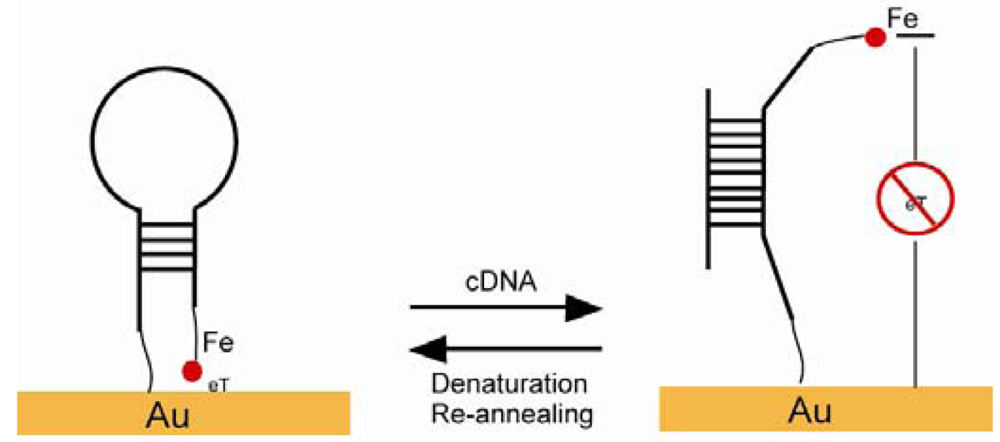
A maleimide-derived glass surface was functionalized with EMC. The Diels-Alder reaction and the “click” reaction happened sequentially and ended with biotin termini [<xref ref-type="bibr" rid="b103-sensors-12-15036">103</xref>]. Fan constructed a stem-loop oligonucleotide with one terminal thiol and one terminal ferrocene (<xref ref-type="fig" rid="f14-sensors-12-15036">Figure 14</xref>). Once the oligonucleotide self-assembled onto a gold electrode surface through a thiol group, the stem-loop structure held the ferrocene at proximity to the gold, which makes the electron transfer easy between the ferrocene and the gold. Upon target nucleotide sequence hybridization, the stem-loop structure was disrupted and a rod-like rigid structure was formed, which separated the ferrocene away from the gold electrode and caused the electron transfer abolished [<xref ref-type="bibr" rid="b106-sensors-12-15036">106</xref>]. A similar SAM was developed with thiolated ssDNA mixed with alkanethiols as a diluent. ssDNA lied across the surface of the SAMs and prevented ions from reaching the electrode. ssDNA turned into a rod-like rigid structure upon targeting sequence hybridization and freed the space for ions to reach the electrode [<xref ref-type="bibr" rid="b107-sensors-12-15036">107</xref>]. An excellent example of a pH sensor was presented by Wrighton and co-workers. They produced a SAM by using pH insensitive ferrocene and pH sensitive quinone. The system showed a linear response to pH changes monitored by voltammetry, where ferrocene as a reference and quinone as an indicator in addition to a large surface area as a counter electrode [<xref ref-type="bibr" rid="b108-sensors-12-15036">108</xref>].</p></sec></sec></sec>
<sec sec-type="materials">
<label>4.</label>
<title>Other Materials</title>
<p>Other widely used organic biosensing materials are: graphene, carbon nanotubes (CNTs), and electrogenerated polymers. The interfaces between the organic materials of biosensors and the corresponding inorganic electrodes are highlighted.</p>
<sec>
<label>4.1.</label>
<title>Graphene</title>
<p>Graphene was first discovered by Novoselov in 2004 [<xref ref-type="bibr" rid="b109-sensors-12-15036">109</xref>]. The superior electron mobility, biocompatibility, and flexibility of graphene qualify it as an ideal material for biosensors. In a glucose biosensor application, graphene was modified by polyvinylpyrrolidone (PVP) and added to a certain ionic liquid (IL). The solution of PVP-graphene-IL was dropped on a glass carbon electrode (GCE) and was allowed to dry before detecting glucose. A linear response for glucose detection from 2 mM to 14 mM was recorded [<xref ref-type="bibr" rid="b110-sensors-12-15036">110</xref>]. In a similar report, a graphene-chitosan solution was dropped onto a GCE and allowed to dry. Glucose oxidase was then coated on the graphene-chitosan-GCE. The glucose detection range was from 0.08 mM up to 12 mM [<xref ref-type="bibr" rid="b111-sensors-12-15036">111</xref>]. Zeng functionalized graphene with sodium dodecylbenzenesulphonate (SDBS). Horseradish peroxidase (HRP) and SDBS-graphene self-assembled on the surface of a GCE. The resulting sensor showed high sensitivity and a H<sub>2</sub>O<sub>2</sub> linear response [<xref ref-type="bibr" rid="b112-sensors-12-15036">112</xref>]. Another graphene-based biosensor for H<sub>2</sub>O<sub>2</sub> detection incorporated metallic nanoparticles. The solution, which contained graphene, HRP, and chitosan, was casted on a GCE. Au was electrodeposited on the surface of the modified GCE and clusters of Au nanoparticles were later formed. The H<sub>2</sub>O<sub>2</sub> detection range was from 0.005 mM to 5.13 mM with a detection limit as low as 1.7 μM [<xref ref-type="bibr" rid="b113-sensors-12-15036">113</xref>]. A screen printed electrode (SPE) was used as a disposable sensor system shown by Song (<xref ref-type="fig" rid="f15-sensors-12-15036">Figure 15</xref>). 1-Pyrenebutanoic acid succinimidyl ester (PASE) has a pyrenyl group, which interacted strongly with graphene, and a succinimidyl ester group, which reacted highly with the amines substitution. Tyrosinase functionalized Au NPs were mixed with PASE modified graphene oxide (GO). The solution of the mixture was dropped on the working SPE for catechol monitoring. The resulting sensor showed high stability and sensitivity [<xref ref-type="bibr" rid="b114-sensors-12-15036">114</xref>].</p>
<p>Some graphene biosensors are electrodeless. ssDNA molecules were connected to certain dyes. GO bonded to the ssDNA labeled dye and quenched the dye's fluorescence. When the targeted ssDNA hybridized to the ssDNA-dye-GO complex, the double stranded DNA-dye was released from the GO and the fluorescence was enhanced [<xref ref-type="bibr" rid="b115-sensors-12-15036">115</xref>]. In a biological environment, it is difficult to differentiate dopamine from its coexisting ascorbic acid (AA) and uric acid (UA) because of the overlapping voltammetric responses. A graphene nanoflake film was synthesized on a Si substrate by microwave plasma enhanced CVD. The film was shown to be capable of determining dopamine in the presence of AA and UA with high sensitivity [<xref ref-type="bibr" rid="b116-sensors-12-15036">116</xref>].</p></sec>
<sec>
<label>4.2.</label>
<title>Carbon Nanotube</title>
<p>
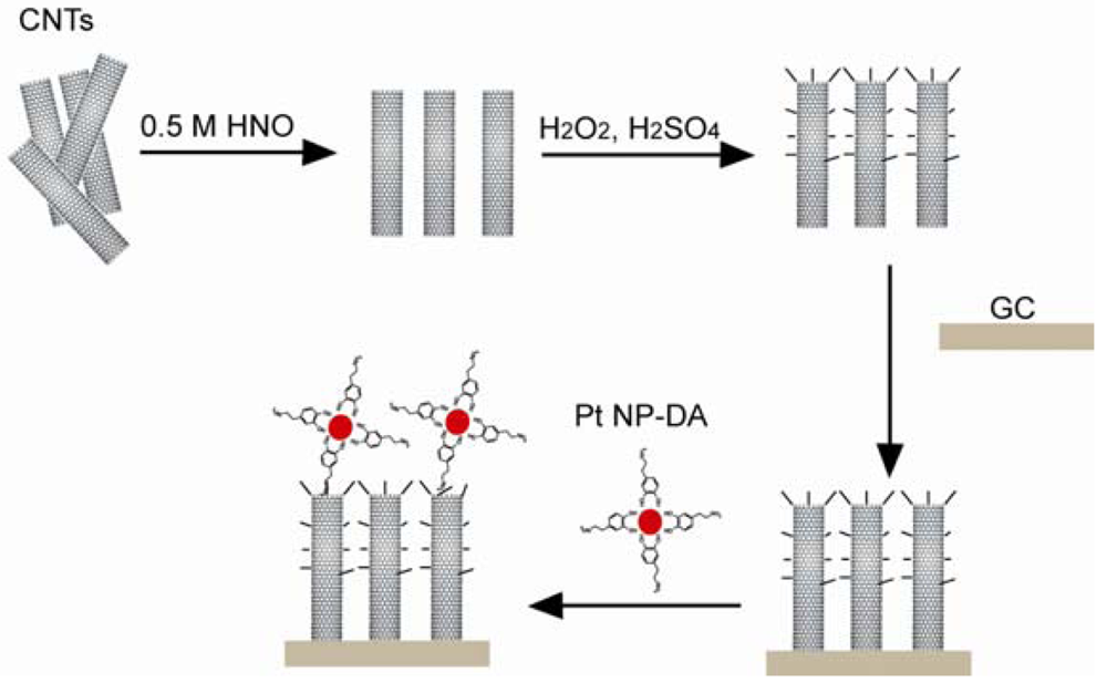
CNTs are another group of organic materials used in biosensors due to their low detection limit and good electron transfer properties. In the case of a CNT glucose biosensor, the CNTs were conjugated with glucose oxidase and was then mixed with polypyrroles (PPy). After the electropolymerization of PPy on a GCE, the biosensor showed a linear response to the glucose up to 50 mM [<xref ref-type="bibr" rid="b117-sensors-12-15036">117</xref>]. The CNT-sulfuric acid solution was dropped on a GCE and allowed to dry. The NADH was detected through cyclic voltammograms [<xref ref-type="bibr" rid="b118-sensors-12-15036">118</xref>]. The mixture of CNTs, chitosan, MDB, and glutamate dehydrogenase was dropped onto the surface of a GCE and left to dry before use. Thus, the resulting glutamate sensor is sensitive and stable [<xref ref-type="bibr" rid="b119-sensors-12-15036">119</xref>]. In the complex H<sub>2</sub>O<sub>2</sub> sensor system presented in <xref ref-type="fig" rid="f16-sensors-12-15036">Figure 16</xref>, activated CNTs with carboxylic groups were coated on a GCE. Dopamine functionalized Pt NPs were connected to the surface of the activated CNTs [<xref ref-type="bibr" rid="b120-sensors-12-15036">120</xref>]. Berti synthesized a CNT thin film using the CVD method and covalently connected the CNT surface with ssDNA probes. In this specific case, the ssDNA probe was inosine-modified and guanine free. After hybridization, the guanine from the target ssDNA was easily oxidized and the oxidation signal was detected [<xref ref-type="bibr" rid="b121-sensors-12-15036">121</xref>]. A single wall CNT-FET was reported for detecting bacterial cells. The CVD method was used to generate single wall CNT connections between catalyst islands patterned on a Si substrate. The conduction of CNT was shown to drop 50% when an <italic>E. coli</italic> cell stayed on the CNT [<xref ref-type="bibr" rid="b122-sensors-12-15036">122</xref>,<xref ref-type="bibr" rid="b123-sensors-12-15036">123</xref>]. In a biosensor for biotin-connected molecule detection, the pyrolytic graphite electrode was oxidized in order to generate carboxylic groups. Poly-L-lysine, CNT, and anti-biotin were covalently connected to the carboxylic groups. An amperometric signal was detected when biotin-connected molecules were bound to the sensor [<xref ref-type="bibr" rid="b124-sensors-12-15036">124</xref>]. A paste electrode was fabricated using specpure carbon, CNT, Cu<sub>2</sub>O, and paraffin oil, which was used to differentiate amino acids with a signal to a noise ratio of 3 [<xref ref-type="bibr" rid="b125-sensors-12-15036">125</xref>].</p></sec>
<sec>
<label>4.3.</label>
<title>Conductive Polymers</title>
<p>Conductive polymers are widely used in biosensors due to their sensitivity, selectivity, and the ability of integration for low-cost microfabrication [<xref ref-type="bibr" rid="b126-sensors-12-15036">126</xref>,<xref ref-type="bibr" rid="b127-sensors-12-15036">127</xref>]. In 1977, the high conductivity of halogen derivatives of polyacetylene was reported by Heeger, MacDiarmid, and Shirakawa [<xref ref-type="bibr" rid="b128-sensors-12-15036">128</xref>]. The alternating singles and double bonds in conjugated polymers provide delocalized electrons, and the charge is carried by the same. The common conductive polymers include polyaniline, polythiophene, polyacetylene, and PPy [<xref ref-type="bibr" rid="b129-sensors-12-15036">129</xref>]. The porous structure of polymers can be used as a bio-analyte immobilization matrix coupled with an electronic conduit. The most common technique for conductive polymer film fabrication is electrochemical polymerization, where the process is carried out in a monomer and bio-active species solution. In research done by Wallace, the negatively charged bio-active species were entrapped in the polymer during the electrochemical oxidation [<xref ref-type="bibr" rid="b127-sensors-12-15036">127</xref>]. The enzyme, which serves as glucose oxidize, immobilized in the polymer matrix triggered the redox reaction of glucose. The electronic signal generated by the glucose oxidation/reduction can be relayed back to the detector through the conductive matrix. In a neuron recording application, the polymer SU-8 was used in the electrode fabrication in order to increase biocompatibility and to eliminate the tissue-electrode gap filled with a passivation layer [<xref ref-type="bibr" rid="b130-sensors-12-15036">130</xref>]. In order to anchor the biotinylated proteins and the DNA, the NTA functionalized PPy has been coated on a Pt electrode. This reaction was proven to be as efficient as biotin-avidin reaction but avoiding the avidin layer [<xref ref-type="bibr" rid="b131-sensors-12-15036">131</xref>]. In the example shown in <xref ref-type="fig" rid="f17-sensors-12-15036">Figure 17</xref>, either glucose hydrogenase or glucose oxidase was entrapped in PPy electropolymerized film on a graphite substrate. The linear detection limit of alkaline phosphate was 10<sup>−6</sup> nM for glucose hydrogenase and 10<sup>−3</sup> nM for glucose oxidase [<xref ref-type="bibr" rid="b132-sensors-12-15036">132</xref>]. In another enzyme entrapment electrode, sulfite oxidase was immobilized in PPy film on a Pt electrode. The detection range for sulfite was from 0.9 to 400 mM [<xref ref-type="bibr" rid="b133-sensors-12-15036">133</xref>]. PPy was electrogenerated on a GCE. Graphene and glucose oxidase were incorporated in the film matrix. It is shown that the enzyme-doped graphene sensor had high sensitivity towards glucose [<xref ref-type="bibr" rid="b134-sensors-12-15036">134</xref>].</p></sec></sec>
<sec sec-type="conclusions">
<label>5.</label>
<title>Conclusions</title>
<p>This review has discussed types of biosensors and applications of organic materials in biosensor systems, emphasizing SAM due to its versatility and molecular control. The interfacial properties with sensor materials of types of organic materials, such as graphene, CNTs, and electroactive polymers were discussed. Several representative applications, such as glucose sensor, pH sensor, and DNA sensor were introduced. Biosensors have attracted great attention in research due to their importance in surface science and applications. It is expected that new biosensors with novel applications will emerge in the coming years.</p></sec></body>
<back>
<ref-list>
<title>References</title>
<ref id="b1-sensors-12-15036"><label>1.</label><citation citation-type="journal"><person-group person-group-type="author"><name><surname>Held</surname><given-names>M.</given-names></name><name><surname>Schuhmann</surname><given-names>W.</given-names></name><name><surname>Jahreis</surname><given-names>K.</given-names></name><name><surname>Schmidt</surname><given-names>H.-L.</given-names></name></person-group><article-title>Microbial biosensor array with transport mutants of <italic>Escherichia coli</italic> K12 for the simultaneous determination of mono-and disaccharides</article-title><source>Biosens. Bioelectr.</source><year>2002</year><volume>17</volume><fpage>1089</fpage><lpage>1094</lpage><pub-id pub-id-type="doi">10.1016/S0956-5663(02)00103-3</pub-id></citation></ref>
<ref id="b2-sensors-12-15036"><label>2.</label><citation citation-type="journal"><person-group person-group-type="author"><name><surname>Tlili</surname><given-names>C.</given-names></name><name><surname>Reybier</surname><given-names>K.</given-names></name><name><surname>Géloën</surname><given-names>A.</given-names></name><name><surname>Ponsonnet</surname><given-names>L.</given-names></name><name><surname>Martelet</surname><given-names>C.</given-names></name><name><surname>Ouada</surname><given-names>H.B.</given-names></name><name><surname>Lagarde</surname><given-names>M.</given-names></name><name><surname>Jaffrezic-Renault</surname><given-names>N.</given-names></name></person-group><article-title>Fibroblast cells: A sensing bioelement for glucose detection by impedance spectroscopy</article-title><source>Anal. Chem.</source><year>2003</year><volume>75</volume><fpage>3340</fpage><lpage>3344</lpage><pub-id pub-id-type="doi">10.1021/ac0340861</pub-id><pub-id pub-id-type="pmid">14570182</pub-id></citation></ref>
<ref id="b3-sensors-12-15036"><label>3.</label><citation citation-type="journal"><person-group person-group-type="author"><name><surname>Vianello</surname><given-names>F.</given-names></name><name><surname>Ragusa</surname><given-names>S.</given-names></name><name><surname>Cambria</surname><given-names>M.T.</given-names></name><name><surname>Rigo</surname><given-names>A.</given-names></name></person-group><article-title>A high sensitivity amperometric biosensor using laccase as biorecognition element</article-title><source>Biosens. Bioelectr.</source><year>2006</year><volume>21</volume><fpage>2155</fpage><lpage>2160</lpage><pub-id pub-id-type="doi">10.1016/j.bios.2005.10.006</pub-id></citation></ref>
<ref id="b4-sensors-12-15036"><label>4.</label><citation citation-type="journal"><person-group person-group-type="author"><name><surname>Andle</surname><given-names>J.C.</given-names></name><name><surname>Vetelino</surname><given-names>J.F.</given-names></name></person-group><article-title>Acoustic wave biosensors</article-title><source>Sens. Actuators A Phys.</source><year>1994</year><volume>44</volume><fpage>167</fpage><lpage>176</lpage><pub-id pub-id-type="doi">10.1016/0924-4247(94)00801-9</pub-id></citation></ref>
<ref id="b5-sensors-12-15036"><label>5.</label><citation citation-type="journal"><person-group person-group-type="author"><name><surname>Sethi</surname><given-names>R.S.</given-names></name></person-group><article-title>Transducer aspects of biosensors</article-title><source>Biosens. Bioelectr.</source><year>1994</year><volume>9</volume><fpage>243</fpage><lpage>264</lpage><pub-id pub-id-type="doi">10.1016/0956-5663(94)80127-4</pub-id></citation></ref>
<ref id="b6-sensors-12-15036"><label>6.</label><citation citation-type="journal"><person-group person-group-type="author"><name><surname>Ivnitski</surname><given-names>D.</given-names></name><name><surname>Wilkins</surname><given-names>E.</given-names></name><name><surname>Tien</surname><given-names>H.T.</given-names></name><name><surname>Ottova</surname><given-names>A.</given-names></name></person-group><article-title>Electrochemical biosensor based on supported planar lipid bilayers for fast detection of pathogenic bacteria</article-title><source>Electrochem. Commun.</source><year>2000</year><volume>2</volume><fpage>457</fpage><lpage>460</lpage><pub-id pub-id-type="doi">10.1016/S1388-2481(00)00060-6</pub-id></citation></ref>
<ref id="b7-sensors-12-15036"><label>7.</label><citation citation-type="journal"><person-group person-group-type="author"><name><surname>Pal</surname><given-names>S.</given-names></name><name><surname>Alocilja</surname><given-names>E.C.</given-names></name><name><surname>Downes</surname><given-names>F.P.</given-names></name></person-group><article-title>Nanowire labeled direct-charge transfer biosensor for detecting Bacillus species</article-title><source>Biosens. Bioelectr.</source><year>2007</year><volume>22</volume><fpage>2329</fpage><lpage>2336</lpage><pub-id pub-id-type="doi">10.1016/j.bios.2007.01.013</pub-id></citation></ref>
<ref id="b8-sensors-12-15036"><label>8.</label><citation citation-type="journal"><person-group person-group-type="author"><name><surname>Lillie</surname><given-names>G.</given-names></name><name><surname>Payne</surname><given-names>P.</given-names></name><name><surname>Vadgama</surname><given-names>P.</given-names></name></person-group><article-title>Electrochemical impedance spectroscopy as a platform for reagentless bioaffinity sensing</article-title><source>Sens. Actuators B Chem.</source><year>2001</year><volume>78</volume><fpage>249</fpage><lpage>256</lpage><pub-id pub-id-type="doi">10.1016/S0925-4005(01)00821-8</pub-id></citation></ref>
<ref id="b9-sensors-12-15036"><label>9.</label><citation citation-type="journal"><person-group person-group-type="author"><name><surname>K'Owino</surname><given-names>I.O.</given-names></name><name><surname>Sadik</surname><given-names>O.A.</given-names></name></person-group><article-title>Impedance spectroscopy: A powerful tool for rapid biomolecular screening and cell culture monitoring</article-title><source>Electroanalysis</source><year>2005</year><volume>17</volume><fpage>2101</fpage><lpage>2113</lpage><pub-id pub-id-type="doi">10.1002/elan.200503371</pub-id></citation></ref>
<ref id="b10-sensors-12-15036"><label>10.</label><citation citation-type="journal"><person-group person-group-type="author"><name><surname>Rodriguez</surname><given-names>M.C.</given-names></name><name><surname>Kawde</surname><given-names>A.-N.</given-names></name><name><surname>Wang</surname><given-names>J.</given-names></name></person-group><article-title>Aptamer biosensor for label-free impedance spectroscopy detection of proteins based on recognition-induced switching of the surface charge</article-title><source>Chem. Commun.</source><year>2005</year><fpage>4267</fpage><lpage>4269</lpage></citation></ref>
<ref id="b11-sensors-12-15036"><label>11.</label><citation citation-type="journal"><person-group person-group-type="author"><name><surname>Varshney</surname><given-names>M.</given-names></name><name><surname>Li</surname><given-names>Y.</given-names></name></person-group><article-title>Interdigitated array microelectrodes based impedance biosensors for detection of bacterial cells</article-title><source>Biosens. Bioelectr.</source><year>2009</year><volume>24</volume><fpage>2951</fpage><lpage>2960</lpage><pub-id pub-id-type="doi">10.1016/j.bios.2008.10.001</pub-id></citation></ref>
<ref id="b12-sensors-12-15036"><label>12.</label><citation citation-type="journal"><person-group person-group-type="author"><name><surname>Gautier</surname><given-names>S.M.</given-names></name><name><surname>Blum</surname><given-names>L.J.</given-names></name><name><surname>Coulet</surname><given-names>P.R.</given-names></name></person-group><article-title>Fibre-optic biosensor based on luminescence and immobilized enzymes: Microdetermination of sorbitol, ethanol and oxaloacetate</article-title><source>J. Biolumin. Chemilumin.</source><year>1990</year><volume>5</volume><fpage>57</fpage><lpage>63</lpage><pub-id pub-id-type="doi">10.1002/bio.1170050112</pub-id><pub-id pub-id-type="pmid">2316395</pub-id></citation></ref>
<ref id="b13-sensors-12-15036"><label>13.</label><citation citation-type="journal"><person-group person-group-type="author"><name><surname>McCurley</surname><given-names>M.F.</given-names></name></person-group><article-title>An optical biosensor using a fluorescent, swelling sensing element</article-title><source>Biosens. Bioelectr.</source><year>1994</year><volume>9</volume><fpage>527</fpage><lpage>533</lpage><pub-id pub-id-type="doi">10.1016/0956-5663(94)90015-9</pub-id></citation></ref>
<ref id="b14-sensors-12-15036"><label>14.</label><citation citation-type="journal"><person-group person-group-type="author"><name><surname>Potyrailo</surname><given-names>R.A.</given-names></name><name><surname>Conrad</surname><given-names>R.C.</given-names></name><name><surname>Ellington</surname><given-names>A.D.</given-names></name><name><surname>Hieftje</surname><given-names>G.M.</given-names></name></person-group><article-title>Adapting Selected Nucleic Acid Ligands (Aptamers) to Biosensors</article-title><source>Anal. Chem.</source><year>1998</year><volume>70</volume><fpage>3419</fpage><lpage>3425</lpage><pub-id pub-id-type="doi">10.1021/ac9802325</pub-id><pub-id pub-id-type="pmid">9726167</pub-id></citation></ref>
<ref id="b15-sensors-12-15036"><label>15.</label><citation citation-type="journal"><person-group person-group-type="author"><name><surname>Choudhury</surname><given-names>B.</given-names></name><name><surname>Shinar</surname><given-names>R.</given-names></name><name><surname>Shinar</surname><given-names>J.</given-names></name></person-group><article-title>Glucose biosensors based on organic light-emitting devices structurally integrated with a luminescent sensing element</article-title><source>J. Appl. Phys.</source><year>2004</year><volume>96</volume><fpage>2949</fpage><lpage>2954</lpage><pub-id pub-id-type="doi">10.1063/1.1778477</pub-id></citation></ref>
<ref id="b16-sensors-12-15036"><label>16.</label><citation citation-type="journal"><person-group person-group-type="author"><name><surname>Haes</surname><given-names>A.</given-names></name><name><surname>Zou</surname><given-names>S.</given-names></name><name><surname>Schatz</surname><given-names>G.</given-names></name><name><surname>Van Duyne</surname><given-names>R.</given-names></name></person-group><article-title>A nanoscale optical biosensor: The long range distance dependence of the localized surface plasmon resonance of noble metal nanoparticles</article-title><source>J. Phys. Chem. B</source><year>2004</year><volume>108</volume><fpage>109</fpage><lpage>116</lpage><pub-id pub-id-type="doi">10.1021/jp0361327</pub-id></citation></ref>
<ref id="b17-sensors-12-15036"><label>17.</label><citation citation-type="journal"><person-group person-group-type="author"><name><surname>Haes</surname><given-names>A.J.</given-names></name><name><surname>Chang</surname><given-names>L.</given-names></name><name><surname>Klein</surname><given-names>W.L.</given-names></name><name><surname>Van Duyne</surname><given-names>R.P.</given-names></name></person-group><article-title>Detection of a biomarker for Alzheimer's disease from synthetic and clinical samples using a nanoscale optical biosensor</article-title><source>J. Am. Chem. Soc.</source><year>2005</year><volume>127</volume><fpage>2264</fpage><lpage>2271</lpage><pub-id pub-id-type="doi">10.1021/ja044087q</pub-id><pub-id pub-id-type="pmid">15713105</pub-id></citation></ref>
<ref id="b18-sensors-12-15036"><label>18.</label><citation citation-type="journal"><person-group person-group-type="author"><name><surname>Mohanty</surname><given-names>S.P.</given-names></name><name><surname>Kougianos</surname><given-names>E.</given-names></name></person-group><article-title>Biosensors: A tutorial review</article-title><source>Potent. IEEE</source><year>2006</year><volume>25</volume><fpage>35</fpage><lpage>40</lpage></citation></ref>
<ref id="b19-sensors-12-15036"><label>19.</label><citation citation-type="journal"><person-group person-group-type="author"><name><surname>Grate</surname><given-names>J.W.</given-names></name><name><surname>Frye</surname><given-names>G.C.</given-names></name></person-group><article-title>Acoustic wave sensors</article-title><source>Sens. Update</source><year>1996</year><volume>2</volume><fpage>37</fpage><lpage>83</lpage><pub-id pub-id-type="doi">10.1002/1616-8984(199610)2:1&lt;37::AID-SEUP37&gt;3.0.CO;2-F</pub-id></citation></ref>
<ref id="b20-sensors-12-15036"><label>20.</label><citation citation-type="journal"><person-group person-group-type="author"><name><surname>White</surname><given-names>R.M.</given-names></name><name><surname>Voltmer</surname><given-names>F.W.</given-names></name></person-group><article-title>Direct piezoelectric coupling to surface elastic waves</article-title><source>Appl. Phys. Lett.</source><year>1965</year><volume>7</volume><fpage>314</fpage><lpage>316</lpage><pub-id pub-id-type="doi">10.1063/1.1754276</pub-id></citation></ref>
<ref id="b21-sensors-12-15036"><label>21.</label><citation citation-type="journal"><person-group person-group-type="author"><name><surname>Drobe</surname><given-names>H.</given-names></name><name><surname>Leidl</surname><given-names>A.</given-names></name><name><surname>Rost</surname><given-names>M.</given-names></name><name><surname>Ruge</surname><given-names>I.</given-names></name></person-group><article-title>Acoustic sensors based on surface-localized HPSWs for measurements in liquids</article-title><source>Sens. Actuators A Phys.</source><year>1993</year><volume>37–38</volume><fpage>141</fpage><lpage>148</lpage></citation></ref>
<ref id="b22-sensors-12-15036"><label>22.</label><citation citation-type="journal"><person-group person-group-type="author"><name><surname>Calabrese</surname><given-names>G.S.</given-names></name><name><surname>Wohltjen</surname><given-names>H.</given-names></name><name><surname>Roy</surname><given-names>M.K.</given-names></name></person-group><article-title>Surface acoustic wave devices as chemical sensors in liquids. Evidence disputing the importance of Rayleigh wave propagation</article-title><source>Anal. Chem.</source><year>1987</year><volume>59</volume><fpage>833</fpage><lpage>837</lpage><pub-id pub-id-type="doi">10.1021/ac00133a010</pub-id></citation></ref>
<ref id="b23-sensors-12-15036"><label>23.</label><citation citation-type="journal"><person-group person-group-type="author"><name><surname>Gizeli</surname><given-names>E.</given-names></name><name><surname>Stevenson</surname><given-names>A.C.</given-names></name><name><surname>Goddard</surname><given-names>N.J.</given-names></name><name><surname>Lowe</surname><given-names>C.R.</given-names></name></person-group><article-title>A novel love-plate acoustic sensor utilizing polymer overlayers</article-title><source>IEEE Trans. Ultrason. Ferroelectr. Freq. Control</source><year>1992</year><volume>39</volume><fpage>657</fpage><lpage>659</lpage><pub-id pub-id-type="doi">10.1109/58.156185</pub-id><pub-id pub-id-type="pmid">18267678</pub-id></citation></ref>
<ref id="b24-sensors-12-15036"><label>24.</label><citation citation-type="journal"><person-group person-group-type="author"><name><surname>Kovacs</surname><given-names>G.</given-names></name><name><surname>Venema</surname><given-names>A.</given-names></name></person-group><article-title>Theoretical comparison of sensitivities of acoustic shear wave modes for (bio)chemical sensing in liquids</article-title><source>Appl. Phys. Lett.</source><year>1992</year><volume>61</volume><fpage>639</fpage><lpage>641</lpage><pub-id pub-id-type="doi">10.1063/1.107807</pub-id></citation></ref>
<ref id="b25-sensors-12-15036"><label>25.</label><citation citation-type="journal"><person-group person-group-type="author"><name><surname>Wang</surname><given-names>Z.</given-names></name><name><surname>Jen</surname><given-names>C.K.</given-names></name><name><surname>Cheeke</surname><given-names>J.D.N.</given-names></name></person-group><article-title>Mass sensitivity of two-layer shear horizontal plate wave sensors</article-title><source>Ultrasonics</source><year>1994</year><volume>32</volume><fpage>209</fpage><lpage>215</lpage><pub-id pub-id-type="doi">10.1016/0041-624X(94)90035-3</pub-id></citation></ref>
<ref id="b26-sensors-12-15036"><label>26.</label><citation citation-type="journal"><person-group person-group-type="author"><name><surname>Länge</surname><given-names>K.</given-names></name><name><surname>Rapp</surname><given-names>B.</given-names></name><name><surname>Rapp</surname><given-names>M.</given-names></name></person-group><article-title>Surface acoustic wave biosensors: A review</article-title><source>Anal. Bioanal. Chem.</source><year>2008</year><volume>391</volume><fpage>1509</fpage><lpage>1519</lpage><pub-id pub-id-type="doi">10.1007/s00216-008-1911-5</pub-id><pub-id pub-id-type="pmid">18265962</pub-id></citation></ref>
<ref id="b27-sensors-12-15036"><label>27.</label><citation citation-type="journal"><person-group person-group-type="author"><name><surname>Sauerbrey</surname><given-names>G.</given-names></name></person-group><article-title>Verwendung von Schwingquarzen zur Wägung dünner Schichten und zur Mikrowägung</article-title><source>Z. Phys.</source><year>1959</year><volume>155</volume><fpage>206</fpage><lpage>222</lpage><pub-id pub-id-type="doi">10.1007/BF01337937</pub-id></citation></ref>
<ref id="b28-sensors-12-15036"><label>28.</label><citation citation-type="journal"><person-group person-group-type="author"><name><surname>Grimes</surname><given-names>C.A.</given-names></name><name><surname>Roy</surname><given-names>S.C.</given-names></name><name><surname>Rani</surname><given-names>S.</given-names></name><name><surname>Cai</surname><given-names>Q.</given-names></name></person-group><article-title>Theory, instrumentation and applications of magnetoelastic resonance sensors: A review</article-title><source>Sensors</source><year>2011</year><volume>11</volume><fpage>2809</fpage><lpage>2844</lpage><pub-id pub-id-type="doi">10.3390/s110302809</pub-id><pub-id pub-id-type="pmid">22163768</pub-id></citation></ref>
<ref id="b29-sensors-12-15036"><label>29.</label><citation citation-type="book"><person-group person-group-type="author"><name><surname>Landau</surname><given-names>L.D</given-names></name><name><surname>Lifshitz</surname><given-names>E.M.</given-names></name></person-group><source>Theory of Elasticity</source><edition>3rd ed.</edition><publisher-name>Pergamon Press</publisher-name><publisher-loc>New York, NY, USA</publisher-loc><year>1986</year></citation></ref>
<ref id="b30-sensors-12-15036"><label>30.</label><citation citation-type="journal"><person-group person-group-type="author"><name><surname>Stoyanov</surname><given-names>P.G.</given-names></name><name><surname>Grimes</surname><given-names>C.A.</given-names></name></person-group><article-title>A remote query magnetostrictive viscosity sensor</article-title><source>Sens. Actuators A Phys.</source><year>2000</year><volume>80</volume><fpage>8</fpage><lpage>14</lpage><pub-id pub-id-type="doi">10.1016/S0924-4247(99)00288-5</pub-id><pub-id pub-id-type="pmid">12143885</pub-id></citation></ref>
<ref id="b31-sensors-12-15036"><label>31.</label><citation citation-type="journal"><person-group person-group-type="author"><name><surname>Ruan</surname><given-names>C.</given-names></name><name><surname>Zeng</surname><given-names>K.</given-names></name><name><surname>Varghese</surname><given-names>O.K.</given-names></name><name><surname>Grimes</surname><given-names>C.A.</given-names></name></person-group><article-title>A magnetoelastic bioaffinity-based sensor for avidin</article-title><source>Biosens. Bioelectr.</source><year>2004</year><volume>19</volume><fpage>1695</fpage><lpage>1701</lpage><pub-id pub-id-type="doi">10.1016/j.bios.2004.01.005</pub-id></citation></ref>
<ref id="b32-sensors-12-15036"><label>32.</label><citation citation-type="journal"><person-group person-group-type="author"><name><surname>Ruan</surname><given-names>C.</given-names></name><name><surname>Zeng</surname><given-names>K.</given-names></name><name><surname>Varghese</surname><given-names>O.K.</given-names></name><name><surname>Grimes</surname><given-names>C.A.</given-names></name></person-group><article-title>A staphylococcal enterotoxin B magnetoelastic immunosensor</article-title><source>Biosens. Bioelectr.</source><year>2004</year><volume>20</volume><fpage>585</fpage><lpage>591</lpage><pub-id pub-id-type="doi">10.1016/j.bios.2004.03.003</pub-id></citation></ref>
<ref id="b33-sensors-12-15036"><label>33.</label><citation citation-type="journal"><person-group person-group-type="author"><name><surname>Roy</surname><given-names>S.C.</given-names></name><name><surname>Werner</surname><given-names>J.R.</given-names></name><name><surname>Mambrini</surname><given-names>G.</given-names></name><name><surname>Grimes</surname><given-names>C.A.</given-names></name></person-group><article-title>Use of magnetoelastic sensors for quantifying platelet aggregation II: Distinguishing contributions of fibrin and thrombin to the coagulation kinetics of whole blood</article-title><source>Sens. Lett.</source><year>2008</year><volume>6</volume><fpage>285</fpage><lpage>289</lpage><pub-id pub-id-type="doi">10.1166/sl.2008.035</pub-id></citation></ref>
<ref id="b34-sensors-12-15036"><label>34.</label><citation citation-type="journal"><person-group person-group-type="author"><name><surname>Ackermann</surname><given-names>T.</given-names></name></person-group><article-title>Book review: Analytical solution calorimetry</article-title><source>Angew. Chem. Int. Ed. Engl.</source><year>1986</year><volume>25</volume><fpage>482</fpage><lpage>483</lpage></citation></ref>
<ref id="b35-sensors-12-15036"><label>35.</label><citation citation-type="journal"><person-group person-group-type="author"><name><surname>Flygare</surname><given-names>L.</given-names></name><name><surname>Danielsson</surname><given-names>B.</given-names></name></person-group><article-title>Advantages of organic solvents in thermometric and optoacoustic enzymic analysis</article-title><source>Ann. N. Y. Acad. Sci.</source><year>1988</year><volume>542</volume><fpage>485</fpage><lpage>496</lpage><pub-id pub-id-type="doi">10.1111/j.1749-6632.1988.tb25876.x</pub-id></citation></ref>
<ref id="b36-sensors-12-15036"><label>36.</label><citation citation-type="journal"><person-group person-group-type="author"><name><surname>Zheng</surname><given-names>Y.-H.</given-names></name><name><surname>Hua Sun</surname><given-names>D.-W.</given-names></name><name><surname>Xiao</surname><given-names>J.-J.</given-names></name><name><surname>Xu</surname><given-names>F.</given-names></name><name><surname>Wang</surname><given-names>F.-F.</given-names></name></person-group><article-title>Detection of dichlorvos residue by flow injection calorimetric biosensor based on immobilized chicken liver esterase</article-title><source>J. Food Eng.</source><year>2006</year><volume>74</volume><fpage>24</fpage><lpage>29</lpage><pub-id pub-id-type="doi">10.1016/j.jfoodeng.2005.02.009</pub-id></citation></ref>
<ref id="b37-sensors-12-15036"><label>37.</label><citation citation-type="journal"><person-group person-group-type="author"><name><surname>Homola</surname><given-names>J.</given-names></name><name><surname>Yee</surname><given-names>S.</given-names></name><name><surname>Gauglitz</surname><given-names>G.</given-names></name></person-group><article-title>Surface plasmon resonance sensors: Review</article-title><source>Sens. Actuators B Chem.</source><year>1999</year><volume>54</volume><fpage>3</fpage><lpage>15</lpage><pub-id pub-id-type="doi">10.1016/S0925-4005(98)00321-9</pub-id></citation></ref>
<ref id="b38-sensors-12-15036"><label>38.</label><citation citation-type="journal"><person-group person-group-type="author"><name><surname>Wood</surname><given-names>R.W.</given-names></name></person-group><article-title>On a remarkable case of uneven distribution of light in a diffraction grating spectrum</article-title><source>Proc. Phys. Soc. Lond.</source><year>1902</year><volume>18</volume><fpage>269</fpage><lpage>275</lpage><pub-id pub-id-type="doi">10.1088/1478-7814/18/1/325</pub-id></citation></ref>
<ref id="b39-sensors-12-15036"><label>39.</label><citation citation-type="journal"><person-group person-group-type="author"><name><surname>Liedberg</surname><given-names>B.</given-names></name><name><surname>Nylander</surname><given-names>C.</given-names></name><name><surname>Lunström</surname><given-names>I.</given-names></name></person-group><article-title>Surface plasmon resonance for gas detection and biosensing</article-title><source>Sens. Actuators</source><year>1983</year><volume>4</volume><fpage>299</fpage><lpage>304</lpage><pub-id pub-id-type="doi">10.1016/0250-6874(83)85036-7</pub-id></citation></ref>
<ref id="b40-sensors-12-15036"><label>40.</label><citation citation-type="journal"><person-group person-group-type="author"><name><surname>Zhang</surname><given-names>L.M.</given-names></name><name><surname>Uttamchandani</surname><given-names>D.</given-names></name></person-group><article-title>Optical chemical sensing employing surface plasmon resonance</article-title><source>Electr. Lett.</source><year>1988</year><volume>24</volume><fpage>1469</fpage><lpage>1470</lpage><pub-id pub-id-type="doi">10.1049/el:19881004</pub-id></citation></ref>
<ref id="b41-sensors-12-15036"><label>41.</label><citation citation-type="journal"><person-group person-group-type="author"><name><surname>Vukusic</surname><given-names>P.S.</given-names></name><name><surname>Bryan-Brown</surname><given-names>G.P.</given-names></name><name><surname>Sambles</surname><given-names>J.R.</given-names></name></person-group><article-title>Surface plasmon resonance on gratings as a novel means for gas sensing</article-title><source>Sens. Actuators B Chem.</source><year>1992</year><volume>8</volume><fpage>155</fpage><lpage>160</lpage><pub-id pub-id-type="doi">10.1016/0925-4005(92)80173-U</pub-id></citation></ref>
<ref id="b42-sensors-12-15036"><label>42.</label><citation citation-type="journal"><person-group person-group-type="author"><name><surname>Achilleos</surname><given-names>C.</given-names></name><name><surname>Tailhardat</surname><given-names>M.</given-names></name><name><surname>Courtellemont</surname><given-names>P.</given-names></name><name><surname>Varlet</surname><given-names>B.L.</given-names></name><name><surname>Dupont</surname><given-names>D.</given-names></name></person-group><article-title>Investigation of surface plasmon resonance biosensor for skin sensitizers studies</article-title><source>Toxicol. Vitro</source><year>2009</year><volume>23</volume><fpage>308</fpage><lpage>318</lpage><pub-id pub-id-type="doi">10.1016/j.tiv.2008.11.007</pub-id></citation></ref>
<ref id="b43-sensors-12-15036"><label>43.</label><citation citation-type="journal"><person-group person-group-type="author"><name><surname>Acharya</surname><given-names>G.</given-names></name><name><surname>Chang</surname><given-names>C.-L.</given-names></name><name><surname>Savran</surname><given-names>C.</given-names></name></person-group><article-title>An optical biosensor for rapid and label-free detection of cells</article-title><source>J. Am. Chem. Soc.</source><year>2006</year><volume>128</volume><fpage>3862</fpage><lpage>3863</lpage><pub-id pub-id-type="doi">10.1021/ja057490l</pub-id><pub-id pub-id-type="pmid">16551065</pub-id></citation></ref>
<ref id="b44-sensors-12-15036"><label>44.</label><citation citation-type="journal"><person-group person-group-type="author"><name><surname>Haddock</surname><given-names>H.S.</given-names></name><name><surname>Shankar</surname><given-names>P.M.</given-names></name><name><surname>Mutharasan</surname><given-names>R.</given-names></name></person-group><article-title>Evanescent sensing of biomolecules and cells</article-title><source>Sens. Actuators B Chem.</source><year>2003</year><volume>88</volume><fpage>67</fpage><lpage>74</lpage><pub-id pub-id-type="doi">10.1016/S0925-4005(02)00310-6</pub-id></citation></ref>
<ref id="b45-sensors-12-15036"><label>45.</label><citation citation-type="journal"><person-group person-group-type="author"><name><surname>Sai</surname><given-names>V.V.R.</given-names></name><name><surname>Kundu</surname><given-names>T.</given-names></name><name><surname>Deshmukh</surname><given-names>C.</given-names></name><name><surname>Titus</surname><given-names>S.</given-names></name><name><surname>Kumar</surname><given-names>P.</given-names></name><name><surname>Mukherji</surname><given-names>S.</given-names></name></person-group><article-title>Label-free fiber optic biosensor based on evanescent wave absorbance at 280 nm</article-title><source>Sens. Actuators B Chem.</source><year>2010</year><volume>143</volume><fpage>724</fpage><lpage>730</lpage><pub-id pub-id-type="doi">10.1016/j.snb.2009.10.021</pub-id></citation></ref>
<ref id="b46-sensors-12-15036"><label>46.</label><citation citation-type="journal"><person-group person-group-type="author"><name><surname>DeGrandpre</surname><given-names>M.D.</given-names></name><name><surname>Burgess</surname><given-names>L.W.</given-names></name></person-group><article-title>Long path fiber-optic sensor for evanescent field absorbance measurements</article-title><source>Anal. Chem.</source><year>1988</year><volume>60</volume><fpage>2582</fpage><lpage>2586</lpage><pub-id pub-id-type="doi">10.1021/ac00174a011</pub-id></citation></ref>
<ref id="b47-sensors-12-15036"><label>47.</label><citation citation-type="journal"><person-group person-group-type="author"><name><surname>Bao</surname><given-names>N.</given-names></name><name><surname>Jagadeesan</surname><given-names>B.</given-names></name><name><surname>Bhunia</surname><given-names>A.K.</given-names></name><name><surname>Yao</surname><given-names>Y.</given-names></name><name><surname>Lu</surname><given-names>C.</given-names></name></person-group><article-title>Quantification of bacterial cells based on autofluorescence on a microfluidic platform</article-title><source>J. Chromatogr. A</source><year>2008</year><volume>1181</volume><fpage>153</fpage><lpage>158</lpage><pub-id pub-id-type="doi">10.1016/j.chroma.2007.12.048</pub-id><pub-id pub-id-type="pmid">18187141</pub-id></citation></ref>
<ref id="b48-sensors-12-15036"><label>48.</label><citation citation-type="journal"><person-group person-group-type="author"><name><surname>Moss</surname><given-names>S.D.</given-names></name><name><surname>Janata</surname><given-names>J.</given-names></name><name><surname>Johnson</surname><given-names>C.C.</given-names></name></person-group><article-title>Potassium ion-sensitive field effect transistor</article-title><source>Anal. Chem.</source><year>1975</year><volume>47</volume><fpage>2238</fpage><lpage>2243</lpage><pub-id pub-id-type="doi">10.1021/ac60363a005</pub-id></citation></ref>
<ref id="b49-sensors-12-15036"><label>49.</label><citation citation-type="journal"><person-group person-group-type="author"><name><surname>Duroux</surname><given-names>P.</given-names></name><name><surname>Emde</surname><given-names>C.</given-names></name><name><surname>Bauerfeind</surname><given-names>P.</given-names></name><name><surname>Francis</surname><given-names>C.</given-names></name><name><surname>Grisel</surname><given-names>A.</given-names></name><name><surname>Thybaud</surname><given-names>L.</given-names></name><name><surname>Arstrong</surname><given-names>D.</given-names></name><name><surname>Depeursinge</surname><given-names>C.</given-names></name><name><surname>Blum</surname><given-names>A.L.</given-names></name></person-group><article-title>The ion sensitive field effect transistor (ISFET) pH electrode: A new sensor for long term ambulatory pH monitoring</article-title><source>Gut</source><year>1991</year><volume>32</volume><fpage>240</fpage><lpage>245</lpage><pub-id pub-id-type="doi">10.1136/gut.32.3.240</pub-id><pub-id pub-id-type="pmid">2013417</pub-id></citation></ref>
<ref id="b50-sensors-12-15036"><label>50.</label><citation citation-type="journal"><person-group person-group-type="author"><name><surname>Esashi</surname><given-names>M.</given-names></name><name><surname>Matsuo</surname><given-names>T.</given-names></name></person-group><article-title>Integrated Micro Multi Ion Sensor Using Field Effect of Semiconductor</article-title><source>IEEE Trans. Biomed. Eng.</source><year>1978</year><volume>BME-25</volume><fpage>184</fpage><lpage>192</lpage><pub-id pub-id-type="doi">10.1109/TBME.1978.326245</pub-id></citation></ref>
<ref id="b51-sensors-12-15036"><label>51.</label><citation citation-type="journal"><person-group person-group-type="author"><name><surname>Kharitonov</surname><given-names>A.B.</given-names></name><name><surname>Zayats</surname><given-names>M.</given-names></name><name><surname>Lichtenstein</surname><given-names>A.</given-names></name><name><surname>Katz</surname><given-names>E.</given-names></name><name><surname>Willner</surname><given-names>I.</given-names></name></person-group><article-title>Enzyme monolayer-functionalized field-effect transistors for biosensor applications</article-title><source>Sens. Actuators B Chem.</source><year>2000</year><volume>70</volume><fpage>222</fpage><lpage>231</lpage><pub-id pub-id-type="doi">10.1016/S0925-4005(00)00573-6</pub-id></citation></ref>
<ref id="b52-sensors-12-15036"><label>52.</label><citation citation-type="journal"><person-group person-group-type="author"><name><surname>Lenigk</surname><given-names>R.</given-names></name><name><surname>Carles</surname><given-names>M.</given-names></name><name><surname>Ip</surname><given-names>N.Y.</given-names></name><name><surname>Sucher</surname><given-names>N.J.</given-names></name></person-group><article-title>Surface characterization of a silicon-chip-based DNA microarray</article-title><source>Langmuir</source><year>2001</year><volume>17</volume><fpage>2497</fpage><lpage>2501</lpage><pub-id pub-id-type="doi">10.1021/la001355z</pub-id></citation></ref>
<ref id="b53-sensors-12-15036"><label>53.</label><citation citation-type="journal"><person-group person-group-type="author"><name><surname>Bent</surname><given-names>S.F.</given-names></name></person-group><article-title>Organic functionalization of group IV semiconductor surfaces: Principles, examples, applications, and prospects</article-title><source>Surf. Sci.</source><year>2002</year><volume>500</volume><fpage>879</fpage><lpage>903</lpage><pub-id pub-id-type="doi">10.1016/S0039-6028(01)01553-9</pub-id></citation></ref>
<ref id="b54-sensors-12-15036"><label>54.</label><citation citation-type="journal"><person-group person-group-type="author"><name><surname>Wang</surname><given-names>Y.</given-names></name><name><surname>Lieberman</surname><given-names>M.</given-names></name></person-group><article-title>Growth of ultrasmooth octadecyltrichlorosilane self-assembled monolayers on SiO<sub>2</sub></article-title><source>Langmuir</source><year>2003</year><volume>19</volume><fpage>1159</fpage><lpage>1167</lpage><pub-id pub-id-type="doi">10.1021/la020697x</pub-id></citation></ref>
<ref id="b55-sensors-12-15036"><label>55.</label><citation citation-type="journal"><person-group person-group-type="author"><name><surname>Midwood</surname><given-names>K.S.</given-names></name><name><surname>Carolus</surname><given-names>M.D.</given-names></name><name><surname>Danahy</surname><given-names>M.P.</given-names></name><name><surname>Schwarzbauer</surname><given-names>J.E.</given-names></name><name><surname>Schwartz</surname><given-names>J.</given-names></name></person-group><article-title>Easy and Efficient Bonding of Biomolecules to an Oxide Surface of Silicon</article-title><source>Langmuir</source><year>2004</year><volume>20</volume><fpage>5501</fpage><lpage>5505</lpage><pub-id pub-id-type="doi">10.1021/la049506b</pub-id><pub-id pub-id-type="pmid">15986692</pub-id></citation></ref>
<ref id="b56-sensors-12-15036"><label>56.</label><citation citation-type="confproc"><person-group person-group-type="author"><name><surname>Sethi</surname><given-names>R.S.</given-names></name><name><surname>Lowe</surname><given-names>C.R.</given-names></name></person-group><article-title>Electrochemical microbiosensors</article-title><conf-name>Proceedings of IEE Colloquium on Microsensors</conf-name><conf-loc>London, UK</conf-loc><conf-date>4 April 1990</conf-date></citation></ref>
<ref id="b57-sensors-12-15036"><label>57.</label><citation citation-type="journal"><person-group person-group-type="author"><name><surname>Thévenot</surname><given-names>D.R.</given-names></name><name><surname>Toth</surname><given-names>K.</given-names></name><name><surname>Durst</surname><given-names>R.A.</given-names></name><name><surname>Wilson</surname><given-names>G.S.</given-names></name></person-group><article-title>Electrochemical biosensors: Recommended definitions and classification</article-title><source>Biosens. Bioelectr.</source><year>2001</year><volume>16</volume><fpage>121</fpage><lpage>131</lpage><pub-id pub-id-type="doi">10.1016/S0956-5663(01)00115-4</pub-id></citation></ref>
<ref id="b58-sensors-12-15036"><label>58.</label><citation citation-type="journal"><person-group person-group-type="author"><name><surname>Mioslav</surname><given-names>P.</given-names></name><name><surname>Petr</surname><given-names>S.</given-names></name></person-group><article-title>Electrochemical biosensors-principles and applications</article-title><source>J. Appl. Biomed.</source><year>2008</year><volume>6</volume><fpage>57</fpage><lpage>64</lpage></citation></ref>
<ref id="b59-sensors-12-15036"><label>59.</label><citation citation-type="journal"><person-group person-group-type="author"><name><surname>Lee</surname><given-names>H.</given-names></name><name><surname>Cooper</surname><given-names>R.</given-names></name><name><surname>Wang</surname><given-names>K.</given-names></name><name><surname>Liang</surname><given-names>H.</given-names></name></person-group><article-title>Nano-scale characterization of a piezoelectric polymer (polyvinylidene difluoride, PVDF)</article-title><source>Sensors</source><year>2008</year><volume>8</volume><fpage>7359</fpage><lpage>7368</lpage><pub-id pub-id-type="doi">10.3390/s8117359</pub-id></citation></ref>
<ref id="b60-sensors-12-15036"><label>60.</label><citation citation-type="journal"><person-group person-group-type="author"><name><surname>Lee</surname><given-names>H.</given-names></name><name><surname>Cooper</surname><given-names>R.</given-names></name><name><surname>Mika</surname><given-names>B.</given-names></name><name><surname>Clayton</surname><given-names>D.</given-names></name><name><surname>Garg</surname><given-names>R.</given-names></name><name><surname>González</surname><given-names>J.M.</given-names></name><name><surname>Vinson</surname><given-names>S.B.</given-names></name><name><surname>Khatri</surname><given-names>S.</given-names></name><name><surname>Liang</surname><given-names>H.</given-names></name></person-group><article-title>Polymeric sensors to monitor cockroach locomotion</article-title><source>IEEE Sens. J.</source><year>2007</year><volume>7</volume><fpage>1698</fpage><lpage>1702</lpage><pub-id pub-id-type="doi">10.1109/JSEN.2007.909624</pub-id></citation></ref>
<ref id="b61-sensors-12-15036"><label>61.</label><citation citation-type="journal"><person-group person-group-type="author"><name><surname>Cooper</surname><given-names>R.</given-names></name><name><surname>Lee</surname><given-names>H.</given-names></name><name><surname>Butler</surname><given-names>J.</given-names></name><name><surname>Gonzalez</surname><given-names>J.</given-names></name><name><surname>Yi</surname><given-names>J.</given-names></name><name><surname>Vinson</surname><given-names>B.</given-names></name><name><surname>Liang</surname><given-names>H.</given-names></name></person-group><article-title>Stress-resolved and cockroach-friendly piezoelectric sensors</article-title><source>Proc. SPIE</source><year>2008</year><volume>6943</volume><pub-id pub-id-type="doi">10.1117/12.777624</pub-id></citation></ref>
<ref id="b62-sensors-12-15036"><label>62.</label><citation citation-type="journal"><person-group person-group-type="author"><name><surname>Mika</surname><given-names>B.</given-names></name><name><surname>Lee</surname><given-names>H.</given-names></name><name><surname>González</surname><given-names>J.M.</given-names></name><name><surname>Vinson</surname><given-names>S.B.</given-names></name><name><surname>Liang</surname><given-names>H.</given-names></name></person-group><article-title>Studying insect motion with piezoelectric sensors</article-title><source>Proc. SPIE</source><year>2007</year><volume>6528</volume><pub-id pub-id-type="doi">10.1117/12.715885</pub-id></citation></ref>
<ref id="b63-sensors-12-15036"><label>63.</label><citation citation-type="journal"><person-group person-group-type="author"><name><surname>Bigelow</surname><given-names>W.</given-names></name><name><surname>Pickett</surname><given-names>D.</given-names></name><name><surname>Zisman</surname><given-names>W.</given-names></name></person-group><article-title>Oleophobic monolayers. I. Films adsorbed from solution in non-polar liquids</article-title><source>J. Colloid Sci.</source><year>1946</year><volume>1</volume><fpage>513</fpage><lpage>538</lpage><pub-id pub-id-type="doi">10.1016/0095-8522(46)90059-1</pub-id></citation></ref>
<ref id="b64-sensors-12-15036"><label>64.</label><citation citation-type="journal"><person-group person-group-type="author"><name><surname>Gooding</surname><given-names>J.J.</given-names></name><name><surname>Ciampi</surname><given-names>S.</given-names></name></person-group><article-title>The molecular level modification of surfaces: From self-assembled monolayers to complex molecular assemblies</article-title><source>Chem. Soc. Rev.</source><year>2011</year><volume>40</volume><fpage>2704</fpage><lpage>2718</lpage><pub-id pub-id-type="doi">10.1039/c0cs00139b</pub-id><pub-id pub-id-type="pmid">21290036</pub-id></citation></ref>
<ref id="b65-sensors-12-15036"><label>65.</label><citation citation-type="journal"><person-group person-group-type="author"><name><surname>Schwartz</surname><given-names>D.K.</given-names></name></person-group><article-title>Mechanisms and kinetics of self-assembled monolayer formation</article-title><source>Ann. Rev. Phys. Chem.</source><year>2001</year><volume>52</volume><fpage>107</fpage><lpage>137</lpage><pub-id pub-id-type="doi">10.1146/annurev.physchem.52.1.107</pub-id></citation></ref>
<ref id="b66-sensors-12-15036"><label>66.</label><citation citation-type="journal"><person-group person-group-type="author"><name><surname>Bain</surname><given-names>C.D.</given-names></name><name><surname>Troughton</surname><given-names>E.B.</given-names></name><name><surname>Tao</surname><given-names>Y.T.</given-names></name><name><surname>Evall</surname><given-names>J.</given-names></name><name><surname>Whitesides</surname><given-names>G.M.</given-names></name><name><surname>Nuzzo</surname><given-names>R.G.</given-names></name></person-group><article-title>Formation of monolayer films by the spontaneous assembly of organic thiols from solution onto gold</article-title><source>J. Am. Chem. Soc.</source><year>1989</year><volume>111</volume><fpage>321</fpage><lpage>335</lpage><pub-id pub-id-type="doi">10.1021/ja00183a049</pub-id></citation></ref>
<ref id="b67-sensors-12-15036"><label>67.</label><citation citation-type="journal"><person-group person-group-type="author"><name><surname>Love</surname><given-names>J.C.</given-names></name><name><surname>Estroff</surname><given-names>L.A.</given-names></name><name><surname>Kriebel</surname><given-names>J.K.</given-names></name><name><surname>Nuzzo</surname><given-names>R.G.</given-names></name><name><surname>Whitesides</surname><given-names>G.M.</given-names></name></person-group><article-title>Self-assembled monolayers of thiolates on metals as a form of nanotechnology</article-title><source>Chem. Rev.</source><year>2005</year><volume>105</volume><fpage>1103</fpage><lpage>1170</lpage><pub-id pub-id-type="doi">10.1021/cr0300789</pub-id><pub-id pub-id-type="pmid">15826011</pub-id></citation></ref>
<ref id="b68-sensors-12-15036"><label>68.</label><citation citation-type="journal"><person-group person-group-type="author"><name><surname>Dubois</surname><given-names>L.H.</given-names></name><name><surname>Nuzzo</surname><given-names>R.G.</given-names></name></person-group><article-title>Synthesis, structure, and properties of model organic surfaces</article-title><source>Ann. Rev. Phys. Chem.</source><year>1992</year><volume>43</volume><fpage>437</fpage><lpage>463</lpage><pub-id pub-id-type="doi">10.1146/annurev.pc.43.100192.002253</pub-id></citation></ref>
<ref id="b69-sensors-12-15036"><label>69.</label><citation citation-type="journal"><person-group person-group-type="author"><name><surname>Laibinis</surname><given-names>P.E.</given-names></name><name><surname>Whitesides</surname><given-names>G.M.</given-names></name><name><surname>Allara</surname><given-names>D.L.</given-names></name><name><surname>Tao</surname><given-names>Y.T.</given-names></name><name><surname>Parikh</surname><given-names>A.N.</given-names></name><name><surname>Nuzzo</surname><given-names>R.G.</given-names></name></person-group><article-title>Comparison of the structures and wetting properties of self-assembled monolayers of n-alkanethiols on the coinage metal surfaces, copper, silver, and gold</article-title><source>J. Am. Chem. Soc.</source><year>1991</year><volume>113</volume><fpage>7152</fpage><lpage>7167</lpage><pub-id pub-id-type="doi">10.1021/ja00019a011</pub-id></citation></ref>
<ref id="b70-sensors-12-15036"><label>70.</label><citation citation-type="journal"><person-group person-group-type="author"><name><surname>Love</surname><given-names>J.C.</given-names></name><name><surname>Wolfe</surname><given-names>D.B.</given-names></name><name><surname>Chabinyc</surname><given-names>M.L.</given-names></name><name><surname>Paul</surname><given-names>K.E.</given-names></name><name><surname>Whitesides</surname><given-names>G.M.</given-names></name></person-group><article-title>Self-assembled monolayers of alkanethiolates on palladium are good etch resists</article-title><source>J. Am. Chem. Soc.</source><year>2002</year><volume>124</volume><fpage>1576</fpage><lpage>1577</lpage><pub-id pub-id-type="doi">10.1021/ja012569l</pub-id><pub-id pub-id-type="pmid">11853422</pub-id></citation></ref>
<ref id="b71-sensors-12-15036"><label>71.</label><citation citation-type="journal"><person-group person-group-type="author"><name><surname>Muskal</surname><given-names>N.</given-names></name><name><surname>Mandler</surname><given-names>D.</given-names></name></person-group><article-title>Thiol self-assembled monolayers on mercury surfaces: The adsorption and electrochemistry of [omega]-mercaptoalkanoic acids</article-title><source>Electrochim. Acta</source><year>1999</year><volume>45</volume><fpage>537</fpage><lpage>548</lpage><pub-id pub-id-type="doi">10.1016/S0013-4686(99)00232-7</pub-id></citation></ref>
<ref id="b72-sensors-12-15036"><label>72.</label><citation citation-type="journal"><person-group person-group-type="author"><name><surname>Yang</surname><given-names>G.</given-names></name><name><surname>Amro</surname><given-names>N.A.</given-names></name><name><surname>Starkewolfe</surname><given-names>Z.B.</given-names></name><name><surname>Liu</surname><given-names>G.</given-names></name></person-group><article-title>Molecular-level approach to inhibit degradations of alkanethiol self-assembled monolayers in aqueous media</article-title><source>Langmuir</source><year>2004</year><volume>20</volume><fpage>3995</fpage><lpage>4003</lpage><pub-id pub-id-type="doi">10.1021/la0499160</pub-id><pub-id pub-id-type="pmid">15969390</pub-id></citation></ref>
<ref id="b73-sensors-12-15036"><label>73.</label><citation citation-type="journal"><person-group person-group-type="author"><name><surname>Hutt</surname><given-names>D.A.</given-names></name><name><surname>Leggett</surname><given-names>G.J.</given-names></name></person-group><article-title>Influence of adsorbate ordering on rates of UV photooxidation of self-assembled monolayers</article-title><source>J. Phys. Chem.</source><year>1996</year><volume>100</volume><fpage>6657</fpage><lpage>6662</lpage><pub-id pub-id-type="doi">10.1021/jp952734h</pub-id></citation></ref>
<ref id="b74-sensors-12-15036"><label>74.</label><citation citation-type="journal"><person-group person-group-type="author"><name><surname>Sagiv</surname><given-names>J.</given-names></name></person-group><article-title>Organized monolayers by adsorption. 1. Formation and structure of oleophobic mixed monolayers on solid surfaces</article-title><source>J. Am. Chem. Soc.</source><year>1980</year><volume>102</volume><fpage>92</fpage><lpage>98</lpage><pub-id pub-id-type="doi">10.1021/ja00521a016</pub-id></citation></ref>
<ref id="b75-sensors-12-15036"><label>75.</label><citation citation-type="journal"><person-group person-group-type="author"><name><surname>Kobayashi</surname><given-names>S.</given-names></name><name><surname>Nishikawa</surname><given-names>T.</given-names></name><name><surname>Takenobu</surname><given-names>T.</given-names></name><name><surname>Mori</surname><given-names>S.</given-names></name><name><surname>Shimoda</surname><given-names>T.</given-names></name><name><surname>Mitani</surname><given-names>T.</given-names></name><name><surname>Shimotani</surname><given-names>H.</given-names></name><name><surname>Yoshimoto</surname><given-names>N.</given-names></name><name><surname>Ogawa</surname><given-names>S.</given-names></name><name><surname>Iwasa</surname><given-names>Y.</given-names></name></person-group><article-title>Control of carrier density by self-assembled monolayers in organic field-effect transistors</article-title><source>Nat. Mater.</source><year>2004</year><volume>3</volume><fpage>317</fpage><lpage>322</lpage><pub-id pub-id-type="doi">10.1038/nmat1105</pub-id><pub-id pub-id-type="pmid">15064756</pub-id></citation></ref>
<ref id="b76-sensors-12-15036"><label>76.</label><citation citation-type="journal"><person-group person-group-type="author"><name><surname>Tian</surname><given-names>R.</given-names></name><name><surname>Seitz</surname><given-names>O.</given-names></name><name><surname>Li</surname><given-names>M.</given-names></name><name><surname>Hu</surname><given-names>W.</given-names></name><name><surname>Chabal</surname><given-names>Y.J.</given-names></name><name><surname>Gao</surname><given-names>J.</given-names></name></person-group><article-title>Infrared characterization of interfacial Si–O bond formation on silanized flat SiO<sub>2</sub>/Si surfaces</article-title><source>Langmuir</source><year>2010</year><volume>26</volume><fpage>4563</fpage><lpage>4566</lpage><pub-id pub-id-type="doi">10.1021/la904597c</pub-id><pub-id pub-id-type="pmid">20180563</pub-id></citation></ref>
<ref id="b77-sensors-12-15036"><label>77.</label><citation citation-type="journal"><person-group person-group-type="author"><name><surname>Buriak</surname><given-names>J.M.</given-names></name></person-group><article-title>Organometallic chemistry on silicon surfaces: Formation of functional monolayers bound through Si–C bonds</article-title><source>Chem. Commun.</source><year>1999</year><pub-id pub-id-type="doi">10.1039/A900108E</pub-id></citation></ref>
<ref id="b78-sensors-12-15036"><label>78.</label><citation citation-type="journal"><person-group person-group-type="author"><name><surname>Ciampi</surname><given-names>S.</given-names></name><name><surname>Harper</surname><given-names>J.B.</given-names></name><name><surname>Gooding</surname><given-names>J.J.</given-names></name></person-group><article-title>Wet chemical routes to the assembly of organic monolayers on silicon surfaces via the formation of Si–C bonds: Surface preparation, passivation and functionalization</article-title><source>Chem. Soc. Rev.</source><year>2010</year><volume>39</volume><fpage>2158</fpage><lpage>2183</lpage><pub-id pub-id-type="doi">10.1039/b923890p</pub-id><pub-id pub-id-type="pmid">20393648</pub-id></citation></ref>
<ref id="b79-sensors-12-15036"><label>79.</label><citation citation-type="journal"><person-group person-group-type="author"><name><surname>Delamar</surname><given-names>M.</given-names></name><name><surname>Hitmi</surname><given-names>R.</given-names></name><name><surname>Pinson</surname><given-names>J.</given-names></name><name><surname>Saveant</surname><given-names>J.M.</given-names></name></person-group><article-title>Covalent modification of carbon surfaces by grafting of functionalized aryl radicals produced from electrochemical reduction of diazonium salts</article-title><source>J. Am. Chem. Soc.</source><year>1992</year><volume>114</volume><fpage>5883</fpage><lpage>5884</lpage><pub-id pub-id-type="doi">10.1021/ja00040a074</pub-id></citation></ref>
<ref id="b80-sensors-12-15036"><label>80.</label><citation citation-type="journal"><person-group person-group-type="author"><name><surname>Saby</surname><given-names>C.</given-names></name><name><surname>Ortiz</surname><given-names>B.</given-names></name><name><surname>Champagne</surname><given-names>G.Y.</given-names></name><name><surname>Bélanger</surname><given-names>D.</given-names></name></person-group><article-title>Electrochemical modification of glassy carbon electrode using aromatic diazonium salts. 1. Blocking effect of 4-nitrophenyl and 4-carboxyphenyl groups</article-title><source>Langmuir</source><year>1997</year><volume>13</volume><fpage>6805</fpage><lpage>6813</lpage><pub-id pub-id-type="doi">10.1021/la961033o</pub-id></citation></ref>
<ref id="b81-sensors-12-15036"><label>81.</label><citation citation-type="journal"><person-group person-group-type="author"><name><surname>Tian</surname><given-names>R.</given-names></name><name><surname>Seitz</surname><given-names>O.</given-names></name><name><surname>Li</surname><given-names>M.</given-names></name><name><surname>Hu</surname><given-names>W.</given-names></name><name><surname>Chabal</surname><given-names>Y.J.</given-names></name><name><surname>Gao</surname><given-names>J.</given-names></name></person-group><article-title>Infrared Characterization of Interfacial Si–O Bond Formation on Silanized Flat SiO2/Si Surfaces</article-title><source>Langmuir</source><year>2010</year><volume>26</volume><fpage>4563</fpage><lpage>4566</lpage><pub-id pub-id-type="doi">10.1021/la904597c</pub-id><pub-id pub-id-type="pmid">20180563</pub-id></citation></ref>
<ref id="b82-sensors-12-15036"><label>82.</label><citation citation-type="journal"><person-group person-group-type="author"><name><surname>Buriak</surname><given-names>J.M.</given-names></name></person-group><article-title>Organometallic chemistry on silicon surfaces: Formation of functional monolayers bound through Si–C bonds</article-title><source>Chem. Commun.</source><year>1999</year><pub-id pub-id-type="doi">10.1039/A900108E</pub-id></citation></ref>
<ref id="b83-sensors-12-15036"><label>83.</label><citation citation-type="journal"><person-group person-group-type="author"><name><surname>Gooding</surname><given-names>J.J.</given-names></name></person-group><article-title>Advances in interfacial design for electrochemical biosensors and sensors: Aryl diazonium salts for modifying carbon and metal electrodes</article-title><source>Electroanalysis</source><year>2008</year><volume>20</volume><fpage>573</fpage><lpage>582</lpage><pub-id pub-id-type="doi">10.1002/elan.200704124</pub-id></citation></ref>
<ref id="b84-sensors-12-15036"><label>84.</label><citation citation-type="journal"><person-group person-group-type="author"><name><surname>de Groot</surname><given-names>M.T.</given-names></name><name><surname>Evers</surname><given-names>T.H.</given-names></name><name><surname>Merkx</surname><given-names>M.</given-names></name><name><surname>Koper</surname><given-names>M.T.M.</given-names></name></person-group><article-title>Electron transfer and ligand binding to cytochrome c'immobilized on self-assembled monolayers</article-title><source>Langmuir</source><year>2007</year><volume>23</volume><fpage>729</fpage><lpage>736</lpage><pub-id pub-id-type="doi">10.1021/la062308v</pub-id><pub-id pub-id-type="pmid">17209627</pub-id></citation></ref>
<ref id="b85-sensors-12-15036"><label>85.</label><citation citation-type="journal"><person-group person-group-type="author"><name><surname>Yamauchi</surname><given-names>F.</given-names></name><name><surname>Koyamatsu</surname><given-names>Y.</given-names></name><name><surname>Kato</surname><given-names>K.</given-names></name><name><surname>Iwata</surname><given-names>H.</given-names></name></person-group><article-title>Layer-by-layer assembly of cationic lipid and plasmid DNA onto gold surface for stent-assisted gene transfer</article-title><source>Biomaterials</source><year>2006</year><volume>27</volume><fpage>3497</fpage><lpage>3504</lpage><pub-id pub-id-type="doi">10.1016/j.biomaterials.2006.02.004</pub-id><pub-id pub-id-type="pmid">16488471</pub-id></citation></ref>
<ref id="b86-sensors-12-15036"><label>86.</label><citation citation-type="journal"><person-group person-group-type="author"><name><surname>Boozer</surname><given-names>C.</given-names></name><name><surname>Ladd</surname><given-names>J.</given-names></name><name><surname>Chen</surname><given-names>S.</given-names></name><name><surname>Jiang</surname><given-names>S.</given-names></name></person-group><article-title>DNA-directed protein immobilization for simultaneous detection of multiple analytes by surface plasmon resonance biosensor</article-title><source>Anal. Chem.</source><year>2006</year><volume>78</volume><fpage>1515</fpage><lpage>1519</lpage><pub-id pub-id-type="doi">10.1021/ac051923l</pub-id><pub-id pub-id-type="pmid">16503602</pub-id></citation></ref>
<ref id="b87-sensors-12-15036"><label>87.</label><citation citation-type="journal"><person-group person-group-type="author"><name><surname>Gomes</surname><given-names>I.</given-names></name><name><surname>Di Paolo</surname><given-names>R.E.</given-names></name><name><surname>Pereira</surname><given-names>P.M.</given-names></name><name><surname>Pereira</surname><given-names>I.A.C.</given-names></name><name><surname>Saraiva</surname><given-names>L.M.</given-names></name><name><surname>Penadés</surname><given-names>S.</given-names></name><name><surname>Franco</surname><given-names>R.</given-names></name></person-group><article-title>Spectroelectrochemistry of Type II Cytochrome c3 on a Glycosylated Self-Assembled Monolayer</article-title><source>Langmuir</source><year>2006</year><volume>22</volume><fpage>9809</fpage><lpage>9811</lpage><pub-id pub-id-type="doi">10.1021/la061392b</pub-id><pub-id pub-id-type="pmid">17106964</pub-id></citation></ref>
<ref id="b88-sensors-12-15036"><label>88.</label><citation citation-type="journal"><person-group person-group-type="author"><name><surname>Schmid</surname><given-names>E.L.</given-names></name><name><surname>Keller</surname><given-names>T.A.</given-names></name><name><surname>Dienes</surname><given-names>Z.</given-names></name><name><surname>Vogel</surname><given-names>H.</given-names></name></person-group><article-title>Reversible oriented surface immobilization of functional proteins on oxide surfaces</article-title><source>Anal. Chem.</source><year>1997</year><volume>69</volume><fpage>1979</fpage><lpage>1985</lpage><pub-id pub-id-type="doi">10.1021/ac9700033</pub-id><pub-id pub-id-type="pmid">9183172</pub-id></citation></ref>
<ref id="b89-sensors-12-15036"><label>89.</label><citation citation-type="journal"><person-group person-group-type="author"><name><surname>Patrie</surname><given-names>S.M.</given-names></name><name><surname>Mrksich</surname><given-names>M.</given-names></name></person-group><article-title>Self-assembled monolayers for MALDI-TOF mass spectrometry for immunoassays of human protein antigens</article-title><source>Anal. Chem.</source><year>2007</year><volume>79</volume><fpage>5878</fpage><lpage>5887</lpage><pub-id pub-id-type="doi">10.1021/ac0701738</pub-id><pub-id pub-id-type="pmid">17602570</pub-id></citation></ref>
<ref id="b90-sensors-12-15036"><label>90.</label><citation citation-type="journal"><person-group person-group-type="author"><name><surname>Herrwerth</surname><given-names>S.</given-names></name><name><surname>Rosendahl</surname><given-names>T.</given-names></name><name><surname>Feng</surname><given-names>C.</given-names></name><name><surname>Fick</surname><given-names>J.</given-names></name><name><surname>Eck</surname><given-names>W.</given-names></name><name><surname>Himmelhaus</surname><given-names>M.</given-names></name><name><surname>Dahint</surname><given-names>R.</given-names></name><name><surname>Grunze</surname><given-names>M.</given-names></name></person-group><article-title>Covalent coupling of antibodies to self-assembled monolayers of carboxy-functionalized poly (ethylene glycol): protein resistance and specific binding of biomolecules</article-title><source>Langmuir</source><year>2003</year><volume>19</volume><fpage>1880</fpage><lpage>1887</lpage><pub-id pub-id-type="doi">10.1021/la026327q</pub-id></citation></ref>
<ref id="b91-sensors-12-15036"><label>91.</label><citation citation-type="journal"><person-group person-group-type="author"><name><surname>Camarero</surname><given-names>J.A.</given-names></name><name><surname>Kwon</surname><given-names>Y.</given-names></name><name><surname>Coleman</surname><given-names>M.A.</given-names></name></person-group><article-title>Chemoselective attachment of biologically active proteins to surfaces by expressed protein ligation and its application for protein chip fabrication</article-title><source>J. Am. Chem. Soc.</source><year>2004</year><volume>126</volume><fpage>14730</fpage><lpage>14731</lpage><pub-id pub-id-type="doi">10.1021/ja0456611</pub-id><pub-id pub-id-type="pmid">15535692</pub-id></citation></ref>
<ref id="b92-sensors-12-15036"><label>92.</label><citation citation-type="journal"><person-group person-group-type="author"><name><surname>Hong</surname><given-names>H.G.</given-names></name><name><surname>Bohn</surname><given-names>P.W.</given-names></name><name><surname>Sligar</surname><given-names>S.G.</given-names></name></person-group><article-title>Optical determination of surface density in oriented metalloprotein nanostructures</article-title><source>Anal. Chem.</source><year>1993</year><volume>65</volume><fpage>1635</fpage><lpage>1638</lpage><pub-id pub-id-type="doi">10.1021/ac00059a025</pub-id></citation></ref>
<ref id="b93-sensors-12-15036"><label>93.</label><citation citation-type="journal"><person-group person-group-type="author"><name><surname>Gallant</surname><given-names>N.D.</given-names></name><name><surname>Lavery</surname><given-names>K.A.</given-names></name><name><surname>Amis</surname><given-names>E.J.</given-names></name><name><surname>Becker</surname><given-names>M.L.</given-names></name></person-group><article-title>Universal gradient substrates for click biofunctionalization</article-title><source>Adv. Mater.</source><year>2007</year><volume>19</volume><fpage>965</fpage><lpage>969</lpage><pub-id pub-id-type="doi">10.1002/adma.200602221</pub-id></citation></ref>
<ref id="b94-sensors-12-15036"><label>94.</label><citation citation-type="journal"><person-group person-group-type="author"><name><surname>Zhang</surname><given-names>Y.</given-names></name><name><surname>Luo</surname><given-names>S.</given-names></name><name><surname>Tang</surname><given-names>Y.</given-names></name><name><surname>Yu</surname><given-names>L.</given-names></name><name><surname>Hou</surname><given-names>K.Y.</given-names></name><name><surname>Cheng</surname><given-names>J.P.</given-names></name><name><surname>Zeng</surname><given-names>X.</given-names></name><name><surname>Wang</surname><given-names>P.G.</given-names></name></person-group><article-title>Carbohydrate-protein interactions by “clicked” carbohydrate self-assembled monolayers</article-title><source>Anal. Chem.</source><year>2006</year><volume>78</volume><fpage>2001</fpage><lpage>2008</lpage><pub-id pub-id-type="doi">10.1021/ac051919+</pub-id><pub-id pub-id-type="pmid">16536439</pub-id></citation></ref>
<ref id="b95-sensors-12-15036"><label>95.</label><citation citation-type="journal"><person-group person-group-type="author"><name><surname>Miura</surname><given-names>Y.</given-names></name><name><surname>Yamauchi</surname><given-names>T.</given-names></name><name><surname>Sato</surname><given-names>H.</given-names></name><name><surname>Fukuda</surname><given-names>T.</given-names></name></person-group><article-title>The self-assembled monolayer of saccharide via click chemistry: Formation and protein recognition</article-title><source>Thin Solid Films</source><year>2008</year><volume>516</volume><fpage>2443</fpage><lpage>2449</lpage><pub-id pub-id-type="doi">10.1016/j.tsf.2007.04.061</pub-id></citation></ref>
<ref id="b96-sensors-12-15036"><label>96.</label><citation citation-type="journal"><person-group person-group-type="author"><name><surname>Houseman</surname><given-names>B.T.</given-names></name><name><surname>Huh</surname><given-names>J.H.</given-names></name><name><surname>Kron</surname><given-names>S.J.</given-names></name><name><surname>Mrksich</surname><given-names>M.</given-names></name></person-group><article-title>Peptide chips for the quantitative evaluation of protein kinase activity</article-title><source>Nat. Biotechnol.</source><year>2002</year><volume>20</volume><fpage>270</fpage><lpage>274</lpage><pub-id pub-id-type="doi">10.1038/nbt0302-270</pub-id><pub-id pub-id-type="pmid">11875428</pub-id></citation></ref>
<ref id="b97-sensors-12-15036"><label>97.</label><citation citation-type="journal"><person-group person-group-type="author"><name><surname>Houseman</surname><given-names>B.T.</given-names></name><name><surname>Mrksich</surname><given-names>M.</given-names></name></person-group><article-title>Carbohydrate arrays for the evaluation of protein binding and enzymatic modification</article-title><source>Chem. Biol.</source><year>2002</year><volume>9</volume><fpage>443</fpage><lpage>454</lpage><pub-id pub-id-type="doi">10.1016/S1074-5521(02)00124-2</pub-id><pub-id pub-id-type="pmid">11983333</pub-id></citation></ref>
<ref id="b98-sensors-12-15036"><label>98.</label><citation citation-type="journal"><person-group person-group-type="author"><name><surname>Houseman</surname><given-names>B.T.</given-names></name><name><surname>Gawalt</surname><given-names>E.S.</given-names></name><name><surname>Mrksich</surname><given-names>M.</given-names></name></person-group><article-title>Maleimide-functionalized self-assembled monolayers for the preparation of peptide and carbohydrate biochips</article-title><source>Langmuir</source><year>2003</year><volume>19</volume><fpage>1522</fpage><lpage>1531</lpage><pub-id pub-id-type="doi">10.1021/la0262304</pub-id></citation></ref>
<ref id="b99-sensors-12-15036"><label>99.</label><citation citation-type="journal"><person-group person-group-type="author"><name><surname>Peelen</surname><given-names>D.</given-names></name><name><surname>Kodoyianni</surname><given-names>V.</given-names></name><name><surname>Lee</surname><given-names>J.</given-names></name><name><surname>Zheng</surname><given-names>T.</given-names></name><name><surname>Shortreed</surname><given-names>M.R.</given-names></name><name><surname>Smith</surname><given-names>L.M.</given-names></name></person-group><article-title>Specific capture of mammalian cells by cell surface receptor binding to ligand immobilized on gold thin films</article-title><source>J. Proteome Res.</source><year>2006</year><volume>5</volume><fpage>1580</fpage><lpage>1585</lpage><pub-id pub-id-type="doi">10.1021/pr050467e</pub-id><pub-id pub-id-type="pmid">16823965</pub-id></citation></ref>
<ref id="b100-sensors-12-15036"><label>100.</label><citation citation-type="journal"><person-group person-group-type="author"><name><surname>MacBeath</surname><given-names>G.</given-names></name><name><surname>Schreiber</surname><given-names>S.L.</given-names></name></person-group><article-title>Printing proteins as microarrays for high-throughput function determination</article-title><source>Science</source><year>2000</year><volume>289</volume><fpage>1760</fpage><lpage>1763</lpage><pub-id pub-id-type="pmid">10976071</pub-id></citation></ref>
<ref id="b101-sensors-12-15036"><label>101.</label><citation citation-type="journal"><person-group person-group-type="author"><name><surname>Haddour</surname><given-names>N.</given-names></name><name><surname>Cosnier</surname><given-names>S.</given-names></name><name><surname>Gondran</surname><given-names>C.</given-names></name></person-group><article-title>Electrogeneration of a poly (pyrrole)-NTA chelator film for a reversible oriented immobilization of histidine-tagged proteins</article-title><source>J. Am. Chem. Soc.</source><year>2005</year><volume>127</volume><fpage>5752</fpage><lpage>5753</lpage><pub-id pub-id-type="doi">10.1021/ja050390v</pub-id><pub-id pub-id-type="pmid">15839649</pub-id></citation></ref>
<ref id="b102-sensors-12-15036"><label>102.</label><citation citation-type="journal"><person-group person-group-type="author"><name><surname>Camarero</surname><given-names>J.A.</given-names></name><name><surname>Kwon</surname><given-names>Y.</given-names></name><name><surname>Coleman</surname><given-names>M.A.</given-names></name></person-group><article-title>Chemoselective attachment of biologically active proteins to surfaces by expressed protein ligation and its application for “protein chip” fabrication</article-title><source>J. Am. Chem. Soc.</source><year>2004</year><volume>126</volume><fpage>14730</fpage><lpage>14731</lpage><pub-id pub-id-type="doi">10.1021/ja0456611</pub-id><pub-id pub-id-type="pmid">15535692</pub-id></citation></ref>
<ref id="b103-sensors-12-15036"><label>103.</label><citation citation-type="journal"><person-group person-group-type="author"><name><surname>Sun</surname><given-names>X.L.</given-names></name><name><surname>Stabler</surname><given-names>C.L.</given-names></name><name><surname>Cazalis</surname><given-names>C.S.</given-names></name><name><surname>Chaikof</surname><given-names>E.L.</given-names></name></person-group><article-title>Carbohydrate and protein immobilization onto solid surfaces by sequential Diels-Alder and azide-alkyne cycloadditions</article-title><source>Bioconj. Chem.</source><year>2006</year><volume>17</volume><fpage>52</fpage><lpage>57</lpage><pub-id pub-id-type="doi">10.1021/bc0502311</pub-id></citation></ref>
<ref id="b104-sensors-12-15036"><label>104.</label><citation citation-type="journal"><person-group person-group-type="author"><name><surname>Li</surname><given-names>F.</given-names></name><name><surname>Chen</surname><given-names>W.</given-names></name><name><surname>Zhang</surname><given-names>S.</given-names></name></person-group><article-title>Development of DNA electrochemical biosensor based on covalent immobilization of probe DNA by direct coupling of sol-gel and self-assembly technologies</article-title><source>Biosens. Bioelectr.</source><year>2008</year><volume>24</volume><fpage>781</fpage><lpage>786</lpage><pub-id pub-id-type="doi">10.1016/j.bios.2008.06.052</pub-id></citation></ref>
<ref id="b105-sensors-12-15036"><label>105.</label><citation citation-type="journal"><person-group person-group-type="author"><name><surname>Lin</surname><given-names>J.C.</given-names></name><name><surname>Kim</surname><given-names>J.H.</given-names></name><name><surname>Kellar</surname><given-names>J.A.</given-names></name><name><surname>Hersam</surname><given-names>M.C.</given-names></name><name><surname>Nguyen</surname><given-names>S.B.T.</given-names></name><name><surname>Bedzyk</surname><given-names>M.J.</given-names></name></person-group><article-title>Building Conjugated Organic Structures on Si (111) Surfaces via Microwave-Assisted Sonogashira Coupling</article-title><source>Langmuir</source><year>2010</year><volume>26</volume><fpage>3771</fpage><lpage>3773</lpage><pub-id pub-id-type="doi">10.1021/la903578r</pub-id><pub-id pub-id-type="pmid">20158279</pub-id></citation></ref>
<ref id="b106-sensors-12-15036"><label>106.</label><citation citation-type="journal"><person-group person-group-type="author"><name><surname>Fan</surname><given-names>C.</given-names></name><name><surname>Plaxco</surname><given-names>K.W.</given-names></name><name><surname>Heeger</surname><given-names>A.J.</given-names></name></person-group><article-title>Electrochemical interrogation of conformational changes as a reagentless method for the sequence-specific detection of DNA</article-title><source>Proc. Nat. Acad. Sci. USA</source><year>2003</year><volume>100</volume><fpage>9134</fpage><lpage>9137</lpage><pub-id pub-id-type="doi">10.1073/pnas.1633515100</pub-id><pub-id pub-id-type="pmid">12867594</pub-id></citation></ref>
<ref id="b107-sensors-12-15036"><label>107.</label><citation citation-type="journal"><person-group person-group-type="author"><name><surname>Gooding</surname><given-names>J.J.</given-names></name><name><surname>Chou</surname><given-names>A.</given-names></name><name><surname>Mearns</surname><given-names>F.J.</given-names></name><name><surname>Jericho</surname><given-names>K.L.</given-names></name></person-group><article-title>The ion gating effect: Using a change in flexibility to allow label free electrochemical detection of DNA hybridisation</article-title><source>Chem. Commun.</source><year>2003</year><pub-id pub-id-type="doi">10.1039/B305798B</pub-id></citation></ref>
<ref id="b108-sensors-12-15036"><label>108.</label><citation citation-type="journal"><person-group person-group-type="author"><name><surname>Hickman</surname><given-names>J.J.</given-names></name><name><surname>Ofer</surname><given-names>D.</given-names></name><name><surname>Laibinis</surname><given-names>P.E.</given-names></name><name><surname>Whitesides</surname><given-names>G.M.</given-names></name><name><surname>Wrighton</surname><given-names>M.S.</given-names></name></person-group><article-title>Molecular self-assembly of two-terminal, voltammetric microsensors with internal references</article-title><source>Science</source><year>1991</year><volume>252</volume><fpage>688</fpage><lpage>691</lpage><pub-id pub-id-type="doi">10.1126/science.252.5006.688</pub-id><pub-id pub-id-type="pmid">17746667</pub-id></citation></ref>
<ref id="b109-sensors-12-15036"><label>109.</label><citation citation-type="journal"><person-group person-group-type="author"><name><surname>Novoselov</surname><given-names>K.</given-names></name><name><surname>Geim</surname><given-names>A.</given-names></name><name><surname>Morozov</surname><given-names>S.</given-names></name><name><surname>Jiang</surname><given-names>D.</given-names></name><name><surname>Zhang</surname><given-names>Y.</given-names></name><name><surname>Dubonos</surname><given-names>S.</given-names></name><name><surname>Grigorieva</surname><given-names>I.</given-names></name><name><surname>Firsov</surname><given-names>A.</given-names></name></person-group><article-title>Electric field effect in atomically thin carbon films</article-title><source>Science</source><year>2004</year><volume>306</volume><fpage>666</fpage><lpage>669</lpage><pub-id pub-id-type="doi">10.1126/science.1102896</pub-id><pub-id pub-id-type="pmid">15499015</pub-id></citation></ref>
<ref id="b110-sensors-12-15036"><label>110.</label><citation citation-type="journal"><person-group person-group-type="author"><name><surname>Shan</surname><given-names>C.</given-names></name><name><surname>Yang</surname><given-names>H.</given-names></name><name><surname>Song</surname><given-names>J.</given-names></name><name><surname>Han</surname><given-names>D.</given-names></name><name><surname>Ivaska</surname><given-names>A.</given-names></name><name><surname>Niu</surname><given-names>L.</given-names></name></person-group><article-title>Direct electrochemistry of glucose oxidase and biosensing for glucose based on graphene</article-title><source>Anal.Chem.</source><year>2009</year><volume>81</volume><fpage>2378</fpage><lpage>2382</lpage><pub-id pub-id-type="doi">10.1021/ac802193c</pub-id><pub-id pub-id-type="pmid">19227979</pub-id></citation></ref>
<ref id="b111-sensors-12-15036"><label>111.</label><citation citation-type="journal"><person-group person-group-type="author"><name><surname>Kang</surname><given-names>X.</given-names></name><name><surname>Wang</surname><given-names>J.</given-names></name><name><surname>Wu</surname><given-names>H.</given-names></name><name><surname>Aksay</surname><given-names>I.A.</given-names></name><name><surname>Liu</surname><given-names>J.</given-names></name><name><surname>Lin</surname><given-names>Y.</given-names></name></person-group><article-title>Glucose Oxidase–graphene–chitosan modified electrode for direct electrochemistry and glucose sensing</article-title><source>Biosens. Bioelectr.</source><year>2009</year><volume>25</volume><fpage>901</fpage><lpage>905</lpage><pub-id pub-id-type="doi">10.1016/j.bios.2009.09.004</pub-id></citation></ref>
<ref id="b112-sensors-12-15036"><label>112.</label><citation citation-type="journal"><person-group person-group-type="author"><name><surname>Zeng</surname><given-names>Q.</given-names></name><name><surname>Cheng</surname><given-names>J.</given-names></name><name><surname>Tang</surname><given-names>L.</given-names></name><name><surname>Liu</surname><given-names>X.</given-names></name><name><surname>Liu</surname><given-names>Y.</given-names></name><name><surname>Li</surname><given-names>J.</given-names></name><name><surname>Jiang</surname><given-names>J.</given-names></name></person-group><article-title>Self-assembled graphene–enzyme hierarchical nanostructures for electrochemical biosensing</article-title><source>Adv. Funct. Mater.</source><year>2010</year><volume>20</volume><fpage>3366</fpage><lpage>3372</lpage><pub-id pub-id-type="doi">10.1002/adfm.201000540</pub-id></citation></ref>
<ref id="b113-sensors-12-15036"><label>113.</label><citation citation-type="journal"><person-group person-group-type="author"><name><surname>Zhou</surname><given-names>K.</given-names></name><name><surname>Zhu</surname><given-names>Y.</given-names></name><name><surname>Yang</surname><given-names>X.</given-names></name><name><surname>Luo</surname><given-names>J.</given-names></name><name><surname>Li</surname><given-names>C.</given-names></name><name><surname>Luan</surname><given-names>S.</given-names></name></person-group><article-title>A novel hydrogen peroxide biosensor based on Au–graphene–HRP–chitosan biocomposites</article-title><source>Electrochim. Acta</source><year>2010</year><volume>55</volume><fpage>3055</fpage><lpage>3060</lpage><pub-id pub-id-type="doi">10.1016/j.electacta.2010.01.035</pub-id></citation></ref>
<ref id="b114-sensors-12-15036"><label>114.</label><citation citation-type="journal"><person-group person-group-type="author"><name><surname>Song</surname><given-names>W.</given-names></name><name><surname>Li</surname><given-names>D.-W.</given-names></name><name><surname>Li</surname><given-names>Y.-T.</given-names></name><name><surname>Li</surname><given-names>Y.</given-names></name><name><surname>Long</surname><given-names>Y.-T.</given-names></name></person-group><article-title>Disposable biosensor based on graphene oxide conjugated with tyrosinase assembled gold nanoparticles</article-title><source>Biosens. Bioelectr.</source><year>2011</year><volume>26</volume><fpage>3181</fpage><lpage>3186</lpage><pub-id pub-id-type="doi">10.1016/j.bios.2010.12.022</pub-id></citation></ref>
<ref id="b115-sensors-12-15036"><label>115.</label><citation citation-type="journal"><person-group person-group-type="author"><name><surname>Lu</surname><given-names>C.-H.</given-names></name><name><surname>Yang</surname><given-names>H.-H.</given-names></name><name><surname>Zhu</surname><given-names>C.-L.</given-names></name><name><surname>Chen</surname><given-names>X.</given-names></name><name><surname>Chen</surname><given-names>G.-N.</given-names></name></person-group><article-title>A graphene platform for sensing biomolecules</article-title><source>Angew. Chem. Int. Ed.</source><year>2009</year><volume>48</volume><fpage>4785</fpage><lpage>4787</lpage><pub-id pub-id-type="doi">10.1002/anie.200901479</pub-id></citation></ref>
<ref id="b116-sensors-12-15036"><label>116.</label><citation citation-type="journal"><person-group person-group-type="author"><name><surname>Shang</surname><given-names>N.G.</given-names></name><name><surname>Papakonstantinou</surname><given-names>P.</given-names></name><name><surname>McMullan</surname><given-names>M.</given-names></name><name><surname>Chu</surname><given-names>M.</given-names></name><name><surname>Stamboulis</surname><given-names>A.</given-names></name><name><surname>Potenza</surname><given-names>A.</given-names></name><name><surname>Dhesi</surname><given-names>S.S.</given-names></name><name><surname>Marchetto</surname><given-names>H.</given-names></name></person-group><article-title>Catalyst-free efficient growth, orientation and biosensing properties of multilayer graphene nanoflake films with sharp edge planes</article-title><source>Adv. Funct. Mat.</source><year>2008</year><volume>18</volume><fpage>3506</fpage><lpage>3514</lpage><pub-id pub-id-type="doi">10.1002/adfm.200800951</pub-id></citation></ref>
<ref id="b117-sensors-12-15036"><label>117.</label><citation citation-type="journal"><person-group person-group-type="author"><name><surname>Wang</surname><given-names>J.</given-names></name><name><surname>Musameh</surname><given-names>M.</given-names></name></person-group><article-title>Carbon-nanotubes doped polypyrrole glucose biosensor</article-title><source>Anal. Chim. Acta</source><year>2005</year><volume>539</volume><fpage>209</fpage><lpage>213</lpage><pub-id pub-id-type="doi">10.1016/j.aca.2005.02.059</pub-id></citation></ref>
<ref id="b118-sensors-12-15036"><label>118.</label><citation citation-type="journal"><person-group person-group-type="author"><name><surname>Musameh</surname><given-names>M.</given-names></name><name><surname>Wang</surname><given-names>J.</given-names></name><name><surname>Merkoci</surname><given-names>A.</given-names></name><name><surname>Lin</surname><given-names>Y.</given-names></name></person-group><article-title>Low-potential stable NADH detection at carbon-nanotube-modified glassy carbon electrodes</article-title><source>Electrochem. Commun.</source><year>2002</year><volume>4</volume><fpage>743</fpage><lpage>746</lpage><pub-id pub-id-type="doi">10.1016/S1388-2481(02)00451-4</pub-id></citation></ref>
<ref id="b119-sensors-12-15036"><label>119.</label><citation citation-type="journal"><person-group person-group-type="author"><name><surname>Chakraborty</surname><given-names>S.</given-names></name><name><surname>Retna Raj</surname><given-names>C.</given-names></name></person-group><article-title>Amperometric biosensing of glutamate using carbon nanotube based electrode</article-title><source>Electrochem. Commun.</source><year>2007</year><volume>9</volume><fpage>1323</fpage><lpage>1330</lpage><pub-id pub-id-type="doi">10.1016/j.elecom.2007.01.039</pub-id></citation></ref>
<ref id="b120-sensors-12-15036"><label>120.</label><citation citation-type="journal"><person-group person-group-type="author"><name><surname>Guzmán</surname><given-names>C.</given-names></name><name><surname>Orozco</surname><given-names>G.</given-names></name><name><surname>Verde</surname><given-names>Y.</given-names></name><name><surname>Jiménez</surname><given-names>S.</given-names></name><name><surname>Godínez</surname><given-names>L.A.</given-names></name><name><surname>Juaristi</surname><given-names>E.</given-names></name><name><surname>Bustos</surname><given-names>E.</given-names></name></person-group><article-title>Hydrogen peroxide sensor based on modified vitreous carbon with multiwall carbon nanotubes and composites of Pt nanoparticles-dopamine</article-title><source>Electrochim. Acta</source><year>2009</year><volume>54</volume><fpage>1728</fpage><lpage>1732</lpage><pub-id pub-id-type="doi">10.1016/j.electacta.2008.09.072</pub-id></citation></ref>
<ref id="b121-sensors-12-15036"><label>121.</label><citation citation-type="journal"><person-group person-group-type="author"><name><surname>He</surname><given-names>P.</given-names></name><name><surname>Dai</surname><given-names>L.</given-names></name></person-group><article-title>Aligned carbon nanotube-DNA electrochemical sensors</article-title><source>Chem. Commun.</source><year>2004</year><pub-id pub-id-type="doi">10.1039/B313030B</pub-id></citation></ref>
<ref id="b122-sensors-12-15036"><label>122.</label><citation citation-type="journal"><person-group person-group-type="author"><name><surname>So</surname><given-names>H.-M.</given-names></name><name><surname>Park</surname><given-names>D.-W.</given-names></name><name><surname>Jeon</surname><given-names>E.-K.</given-names></name><name><surname>Kim</surname><given-names>Y.-H.</given-names></name><name><surname>Kim</surname><given-names>B.S.</given-names></name><name><surname>Lee</surname><given-names>C.-K.</given-names></name><name><surname>Choi</surname><given-names>S.Y.</given-names></name><name><surname>Kim</surname><given-names>S.C.</given-names></name><name><surname>Chang</surname><given-names>H.</given-names></name><name><surname>Lee</surname><given-names>J.-O.</given-names></name></person-group><article-title>Detection and titer estimation of <italic>Escherichia coli</italic> using aptamer-functionalized single-walled carbon-nanotube field-effect transistors</article-title><source>Small</source><year>2008</year><volume>4</volume><fpage>197</fpage><lpage>201</lpage><pub-id pub-id-type="doi">10.1002/smll.200700664</pub-id><pub-id pub-id-type="pmid">18214875</pub-id></citation></ref>
<ref id="b123-sensors-12-15036"><label>123.</label><citation citation-type="journal"><person-group person-group-type="author"><name><surname>Kong</surname><given-names>J.</given-names></name><name><surname>Soh</surname><given-names>H.T.</given-names></name><name><surname>Cassell</surname><given-names>A.M.</given-names></name><name><surname>Quate</surname><given-names>C.F.</given-names></name><name><surname>Dai</surname><given-names>H.</given-names></name></person-group><article-title>Synthesis of individual single-walled carbon nanotubes on patterned silicon wafers</article-title><source>Nature</source><year>1998</year><volume>395</volume><fpage>878</fpage><lpage>881</lpage><pub-id pub-id-type="doi">10.1038/27632</pub-id></citation></ref>
<ref id="b124-sensors-12-15036"><label>124.</label><citation citation-type="journal"><person-group person-group-type="author"><name><surname>Cataldo</surname><given-names>V.</given-names></name><name><surname>Vaze</surname><given-names>A.</given-names></name><name><surname>Rusling</surname><given-names>J.F.</given-names></name></person-group><article-title>Improved detection limit and stability of amperometric carbon nanotube-based immunosensors by crosslinking antibodies with polylysine</article-title><source>Electroanalysis</source><year>2008</year><volume>20</volume><fpage>115</fpage><lpage>122</lpage><pub-id pub-id-type="doi">10.1002/elan.200704040</pub-id><pub-id pub-id-type="pmid">18846266</pub-id></citation></ref>
<ref id="b125-sensors-12-15036"><label>125.</label><citation citation-type="journal"><person-group person-group-type="author"><name><surname>Dong</surname><given-names>S.</given-names></name><name><surname>Zhang</surname><given-names>S.</given-names></name><name><surname>Chi</surname><given-names>L.</given-names></name><name><surname>He</surname><given-names>P.</given-names></name><name><surname>Wang</surname><given-names>Q.</given-names></name><name><surname>Fang</surname><given-names>Y.</given-names></name></person-group><article-title>Electrochemical behaviors of amino acids at multiwall carbon nanotubes and Cu<sub>2</sub>O modified carbon paste electrode</article-title><source>Anal. Biochem.</source><year>2008</year><volume>381</volume><fpage>199</fpage><lpage>204</lpage><pub-id pub-id-type="doi">10.1016/j.ab.2008.05.011</pub-id><pub-id pub-id-type="pmid">18522798</pub-id></citation></ref>
<ref id="b126-sensors-12-15036"><label>126.</label><citation citation-type="book"><person-group person-group-type="author"><name><surname>Lammers</surname><given-names>F.</given-names></name><name><surname>Scheper</surname><given-names>T.</given-names></name></person-group><article-title>Thermal Biosensors in Biotechnology</article-title><source>Thermal Biosensors, Bioactivity, Bioaffinitty</source><person-group person-group-type="editor"><name><surname>Scheper</surname><given-names>T.</given-names></name></person-group><publisher-name>Springer</publisher-name><publisher-loc>Berlin/Heidelberg, Germany</publisher-loc><year>1999</year><fpage>35</fpage><lpage>67</lpage></citation></ref>
<ref id="b127-sensors-12-15036"><label>127.</label><citation citation-type="journal"><person-group person-group-type="author"><name><surname>Wallace</surname><given-names>G.G.</given-names></name><name><surname>Smyth</surname><given-names>M.</given-names></name><name><surname>Zhao</surname><given-names>H.</given-names></name></person-group><article-title>Conducting electroactive polymer-based biosensors</article-title><source>TrAC Trends Anal. Chem.</source><year>1999</year><volume>18</volume><fpage>245</fpage><lpage>251</lpage><pub-id pub-id-type="doi">10.1016/S0165-9936(98)00113-7</pub-id></citation></ref>
<ref id="b128-sensors-12-15036"><label>128.</label><citation citation-type="journal"><person-group person-group-type="author"><name><surname>Shirakawa</surname><given-names>H.</given-names></name><name><surname>Louis</surname><given-names>E.J.</given-names></name><name><surname>MacDiarmid</surname><given-names>A.G.</given-names></name><name><surname>Chiang</surname><given-names>C.K.</given-names></name><name><surname>Heeger</surname><given-names>A.J.</given-names></name></person-group><article-title>Synthesis of electrically conducting organic polymers: Halogen derivatives of polyacetylene, (CH)</article-title><source>J. Chem. Soc., Chem. Commun.</source><year>1977</year><pub-id pub-id-type="doi">10.1039/C39770000578</pub-id></citation></ref>
<ref id="b129-sensors-12-15036"><label>129.</label><citation citation-type="journal"><person-group person-group-type="author"><name><surname>Nambiar</surname><given-names>S.</given-names></name><name><surname>Yeow</surname><given-names>J.T.W.</given-names></name></person-group><article-title>Conductive polymer-based sensors for biomedical applications</article-title><source>Biosens. Bioelectr.</source><year>2011</year><volume>26</volume><fpage>1825</fpage><lpage>1832</lpage><pub-id pub-id-type="doi">10.1016/j.bios.2010.09.046</pub-id></citation></ref>
<ref id="b130-sensors-12-15036"><label>130.</label><citation citation-type="journal"><person-group person-group-type="author"><name><surname>Altuna</surname><given-names>A.</given-names></name><name><surname>Menendez de la Prida</surname><given-names>L.</given-names></name><name><surname>Bellistri</surname><given-names>E.</given-names></name><name><surname>Gabriel</surname><given-names>G.</given-names></name><name><surname>Guimerá</surname><given-names>A.</given-names></name><name><surname>Berganzo</surname><given-names>J.</given-names></name><name><surname>Villa</surname><given-names>R.</given-names></name><name><surname>Fernández</surname><given-names>L.J.</given-names></name></person-group><article-title>SU-8 based microprobes with integrated planar electrodes for enhanced neural depth recording</article-title><source>Biosens. Bioelectr.</source><year>2012</year><volume>37</volume><fpage>1</fpage><lpage>5</lpage><pub-id pub-id-type="doi">10.1016/j.bios.2012.03.039</pub-id></citation></ref>
<ref id="b131-sensors-12-15036"><label>131.</label><citation citation-type="journal"><person-group person-group-type="author"><name><surname>Baur</surname><given-names>J.</given-names></name><name><surname>Holzinger</surname><given-names>M.</given-names></name><name><surname>Gondran</surname><given-names>C.</given-names></name><name><surname>Cosnier</surname><given-names>S.</given-names></name></person-group><article-title>Immobilization of biotinylated biomolecules onto electropolymerized poly(pyrrole-nitrilotriacetic acid)–Cu<sup>2+</sup> film</article-title><source>Electrochem. Commun.</source><year>2010</year><volume>12</volume><fpage>1287</fpage><lpage>1290</lpage><pub-id pub-id-type="doi">10.1016/j.elecom.2010.07.001</pub-id></citation></ref>
<ref id="b132-sensors-12-15036"><label>132.</label><citation citation-type="journal"><person-group person-group-type="author"><name><surname>Santiago</surname><given-names>L.M.</given-names></name><name><surname>Bejarano-Nosas</surname><given-names>D.</given-names></name><name><surname>Lozano-Sanchez</surname><given-names>P.</given-names></name><name><surname>Katakis</surname><given-names>I.</given-names></name></person-group><article-title>Screen-printed microsystems for the ultrasensitive electrochemical detection of alkaline phosphatase</article-title><source>Analyst</source><year>2010</year><volume>135</volume><fpage>1276</fpage><lpage>1281</lpage><pub-id pub-id-type="doi">10.1039/c0an00001a</pub-id><pub-id pub-id-type="pmid">20396818</pub-id></citation></ref>
<ref id="b133-sensors-12-15036"><label>133.</label><citation citation-type="journal"><person-group person-group-type="author"><name><surname>Ameer</surname><given-names>Q.</given-names></name><name><surname>Adeloju</surname><given-names>S.B.</given-names></name></person-group><article-title>Galvanostatic entrapment of sulfite oxidase into ultrathin polypyrrole films for improved amperometric biosensing of sulfite</article-title><source>Electroanalysis</source><year>2008</year><volume>20</volume><fpage>2549</fpage><lpage>2556</lpage><pub-id pub-id-type="doi">10.1002/elan.200804357</pub-id></citation></ref>
<ref id="b134-sensors-12-15036"><label>134.</label><citation citation-type="journal"><person-group person-group-type="author"><name><surname>Alwarappan</surname><given-names>S.</given-names></name><name><surname>Liu</surname><given-names>C.</given-names></name><name><surname>Kumar</surname><given-names>A.</given-names></name><name><surname>Li</surname><given-names>C.-Z.</given-names></name></person-group><article-title>Enzyme-doped graphene nanosheets for enhanced glucose biosensing</article-title><source>J. Phys. Chem. C</source><year>2010</year><volume>114</volume><fpage>12920</fpage><lpage>12924</lpage><pub-id pub-id-type="doi">10.1021/jp103273z</pub-id></citation></ref></ref-list>
<sec sec-type="display-objects">
<title>Figures and Tables</title>
<fig id="f1-sensors-12-15036" position="float">
<label>Figure 1.</label>
<caption>
<p>Schematic representation of the basic concept of a biosensor (adapted from [<xref ref-type="bibr" rid="b18-sensors-12-15036">18</xref>]).</p></caption>
<graphic xlink:href="sensors-12-15036f1.gif"/></fig>
<fig id="f2-sensors-12-15036" position="float">
<label>Figure 2.</label>
<caption>
<p>Schematic representation of a SAW-based biosensor (adapted from [<xref ref-type="bibr" rid="b26-sensors-12-15036">26</xref>]).</p></caption>
<graphic xlink:href="sensors-12-15036f2.gif"/></fig>
<fig id="f3-sensors-12-15036" position="float">
<label>Figure 3.</label>
<caption>
<p>Schematic representation of a thermal biosensor (adapted from [<xref ref-type="bibr" rid="b36-sensors-12-15036">36</xref>]).</p></caption>
<graphic xlink:href="sensors-12-15036f3.gif"/></fig>
<fig id="f4-sensors-12-15036" position="float">
<label>Figure 4.</label>
<caption>
<p>Schematic representation of a SPR biosensor (adapted from [<xref ref-type="bibr" rid="b42-sensors-12-15036">42</xref>]).</p></caption>
<graphic xlink:href="sensors-12-15036f4.gif"/></fig>
<fig id="f5-sensors-12-15036" position="float">
<label>Figure 5.</label>
<caption>
<p>Schematic configuration of an ISFETs biosensor (adapted from [<xref ref-type="bibr" rid="b51-sensors-12-15036">51</xref>]).</p></caption>
<graphic xlink:href="sensors-12-15036f5.gif"/></fig>
<fig id="f6-sensors-12-15036" position="float">
<label>Figure 6.</label>
<caption>
<p>Schematic configuration of an electrochemical biosensor.</p></caption>
<graphic xlink:href="sensors-12-15036f6.gif"/></fig>
<fig id="f7-sensors-12-15036" position="float">
<label>Figure 7.</label>
<caption>
<p>(<bold>A</bold>) Idealized SAM system (adapted from [<xref ref-type="bibr" rid="b64-sensors-12-15036">64</xref>]), (<bold>B</bold>) Equilibrium phase diagram (adapted from [<xref ref-type="bibr" rid="b65-sensors-12-15036">65</xref>]).</p></caption>
<graphic xlink:href="sensors-12-15036f7.gif"/></fig>
<fig id="f8-sensors-12-15036" position="float">
<label>Figure 8.</label>
<caption>
<p>Structure of organosilane based layer. Organosilane SAM is connected to hydroxylated silicon dioxide surface through S–O bond. Some organosilane molecules were connected to the neighboring molecules (adapted from [<xref ref-type="bibr" rid="b81-sensors-12-15036">81</xref>]).</p></caption>
<graphic xlink:href="sensors-12-15036f8.gif"/></fig>
<fig id="f9-sensors-12-15036" position="float">
<label>Figure 9.</label>
<caption>
<p>Alkyl chains of 1-alkyne and 1-alkene terminals are connected to the S-H radicals on the silicone surface (adapted from [<xref ref-type="bibr" rid="b82-sensors-12-15036">82</xref>]).</p></caption>
<graphic xlink:href="sensors-12-15036f9.gif"/></fig>
<fig id="f10-sensors-12-15036" position="float">
<label>Figure 10.</label>
<caption>
<p>The reaction mechanism for aryl diazonium reaction based SAM (adapted from [<xref ref-type="bibr" rid="b83-sensors-12-15036">83</xref>]).</p></caption>
<graphic xlink:href="sensors-12-15036f10.gif"/></fig>
<fig id="f11-sensors-12-15036" position="float">
<label>Figure 11.</label>
<caption>
<p>The application of non-covalent bonding in a SAM immunoassay (adapted from [<xref ref-type="bibr" rid="b89-sensors-12-15036">89</xref>]).</p></caption>
<graphic xlink:href="sensors-12-15036f11.gif"/></fig>
<fig id="f12-sensors-12-15036" position="float">
<label>Figure 12.</label>
<caption>
<p>The Cys N-termini of SAM's reacts with the C-termini of protein through dehydration and forms an amide bond (adapted from [<xref ref-type="bibr" rid="b102-sensors-12-15036">102</xref>]).</p></caption>
<graphic xlink:href="sensors-12-15036f12.gif"/></fig>
<fig id="f13-sensors-12-15036" position="float">
<label>Figure 13.</label>
<caption>
<p>An example of the combination between the Diels-Alder reaction and the “click” reaction (adapted from [<xref ref-type="bibr" rid="b103-sensors-12-15036">103</xref>]).</p></caption>
<graphic xlink:href="sensors-12-15036f13.gif"/></fig>
<fig id="f14-sensors-12-15036" position="float">
<label>Figure 14.</label>
<caption>
<p>A stem-loop structure with a terminal ferrocene tag. The hybridization of target DNA changed the stem-loop structure and the electron transfer stopped, hence, the corresponding cyclic voltammogram changed (adapted from [<xref ref-type="bibr" rid="b106-sensors-12-15036">106</xref>]).</p></caption>
<graphic xlink:href="sensors-12-15036f14.gif"/></fig>
<fig id="f15-sensors-12-15036" position="float">
<label>Figure 15.</label>
<caption>
<p>Modified GO was coated on SPEs (adapted from [<xref ref-type="bibr" rid="b114-sensors-12-15036">114</xref>]).</p></caption>
<graphic xlink:href="sensors-12-15036f15.gif"/></fig>
<fig id="f16-sensors-12-15036" position="float">
<label>Figure 16.</label>
<caption>
<p>The schematic of the fabrication of a CNT based H<sub>2</sub>O<sub>2</sub> sensor, (A-C) CNTs were separated, cleaned, and attached to the surface of a GC; dopamine (DA) functionalized Pt nanoparticles were connected to CNTs (adapted from [<xref ref-type="bibr" rid="b120-sensors-12-15036">120</xref>]).</p></caption>
<graphic xlink:href="sensors-12-15036f16.gif"/></fig>
<fig id="f17-sensors-12-15036" position="float">
<label>Figure 17.</label>
<caption>
<p>Enzyme was embedded in a PPy surface matrix coated on a GCE (adapted from [<xref ref-type="bibr" rid="b132-sensors-12-15036">132</xref>]).</p></caption>
<graphic xlink:href="sensors-12-15036f17.gif"/></fig>
<table-wrap id="t1-sensors-12-15036" position="float">
<label>Table 1.</label>
<caption>
<p>Summary of biosensors.</p></caption>
<table frame="hsides" rules="groups">
<thead>
<tr>
<th align="left" valign="middle"><bold>Type</bold></th>
<th align="left" valign="middle"><bold>Sensing mechanism</bold></th>
<th align="left" valign="middle"><bold>Transducer</bold></th>
<th align="left" valign="middle"><bold>Measured property</bold></th>
<th align="center" valign="middle"><bold>References</bold></th></tr></thead>
<tbody>
<tr>
<td align="left" valign="middle">Resonant</td>
<td align="left" valign="middle">The change of the viscosity, mass leads to the change of resonant frequency of the acoustic wave</td>
<td align="left" valign="middle">Mass sensitive</td>
<td align="left" valign="middle">Resonant frequency</td>
<td align="center" valign="middle">[<xref ref-type="bibr" rid="b18-sensors-12-15036">18</xref>–<xref ref-type="bibr" rid="b25-sensors-12-15036">25</xref>]</td></tr>
<tr>
<td align="left" valign="middle">Thermal detection</td>
<td align="left" valign="middle">Bio-reaction results in exothermic character</td>
<td align="left" valign="middle">Thermal</td>
<td align="left" valign="middle">Heat of reaction or adsorption</td>
<td align="center" valign="middle">[<xref ref-type="bibr" rid="b27-sensors-12-15036">27</xref>]</td></tr>
<tr>
<td align="left" valign="middle">Photometric</td>
<td align="left" valign="middle">The change of refractive index of the solution leads to the change of refractive angle of the incident light</td>
<td align="left" valign="middle">Optical</td>
<td align="left" valign="middle">Surface Plasmon resonance angle</td>
<td align="center" valign="middle">[<xref ref-type="bibr" rid="b29-sensors-12-15036">29</xref>–<xref ref-type="bibr" rid="b32-sensors-12-15036">32</xref>]</td></tr>
<tr>
<td align="left" valign="middle">ISFETs</td>
<td align="left" valign="middle">The ionic analyte diffuses into the membrane hence change the potential difference at the detecting interface</td>
<td align="left" valign="middle">Ion-selective membrane</td>
<td align="left" valign="middle">Surface potential</td>
<td align="center" valign="middle">[<xref ref-type="bibr" rid="b33-sensors-12-15036">33</xref>–<xref ref-type="bibr" rid="b40-sensors-12-15036">40</xref>]</td></tr>
<tr>
<td align="left" valign="middle">Electrochemical</td>
<td align="left" valign="middle">Bio-reaction resulting in ions production or consumption will create the charge transfer across the double layer of the transducer.</td>
<td align="left" valign="middle">Electrochemical</td>
<td align="left" valign="middle">Potentiometric Amperometric Impedimetric</td>
<td align="center" valign="middle">[<xref ref-type="bibr" rid="b41-sensors-12-15036">41</xref>–<xref ref-type="bibr" rid="b43-sensors-12-15036">43</xref>]</td></tr></tbody></table></table-wrap>
<table-wrap id="t2-sensors-12-15036" position="float">
<label>Table 2.</label>
<caption>
<p>Four types of SAM systems.</p></caption>
<table frame="hsides" rules="groups">
<thead>
<tr>
<th align="left" valign="middle"><bold>Basic types (surface substrate)</bold></th>
<th align="left" valign="middle"><bold>Bond</bold></th>
<th align="left" valign="middle"><bold>Advantages</bold></th>
<th align="left" valign="middle"><bold>Disadvantages</bold></th>
<th align="center" valign="middle"><bold>References</bold></th></tr></thead>
<tbody>
<tr>
<td align="left" valign="middle">Alkanethiol and organosulfur (metal)</td>
<td align="left" valign="middle">Metal–thiolate</td>
<td align="left" valign="middle">Good control; most studied; gold is standard</td>
<td align="left" valign="middle">Ligands match metal oxidation state; low stability</td>
<td align="center" valign="middle">[<xref ref-type="bibr" rid="b67-sensors-12-15036">67</xref>–<xref ref-type="bibr" rid="b73-sensors-12-15036">73</xref>]</td></tr>
<tr>
<td align="left" valign="middle">Organosilane (oxides surface; silicon dioxide, metal oxide)</td>
<td align="left" valign="middle">S–O</td>
<td align="left" valign="middle">Easy to handle</td>
<td align="left" valign="middle">Multilayer defect</td>
<td align="center" valign="middle">[<xref ref-type="bibr" rid="b64-sensors-12-15036">64</xref>,<xref ref-type="bibr" rid="b74-sensors-12-15036">74</xref>–<xref ref-type="bibr" rid="b76-sensors-12-15036">76</xref>]</td></tr>
<tr>
<td align="left" valign="middle">Alkyne and alkene (hydride-silicon surface)</td>
<td align="left" valign="middle">Si–C</td>
<td align="left" valign="middle">Superior stability</td>
<td align="left" valign="middle">Oxide-free silicon is hard to obtain; multilayer defect</td>
<td align="center" valign="middle">[<xref ref-type="bibr" rid="b77-sensors-12-15036">77</xref>,<xref ref-type="bibr" rid="b78-sensors-12-15036">78</xref>]</td></tr>
<tr>
<td align="left" valign="middle">Aryl diazonium salts (carbon, metal, metal oxide, silicon surface)</td>
<td align="left" valign="middle">Aryl–surface</td>
<td align="left" valign="middle">Superior stability</td>
<td align="left" valign="middle">Limited control</td>
<td align="center" valign="middle">[<xref ref-type="bibr" rid="b79-sensors-12-15036">79</xref>,<xref ref-type="bibr" rid="b80-sensors-12-15036">80</xref>]</td></tr></tbody></table></table-wrap>
<table-wrap id="t3-sensors-12-15036" position="float">
<label>Table 3.</label>
<caption>
<p>Immobilization of biomolecules to biosensor systems.</p></caption>
<table frame="hsides" rules="groups">
<thead>
<tr>
<th align="left" valign="top"><bold>Reaction</bold></th>
<th align="left" valign="top"><bold>Bond</bold></th>
<th align="left" valign="top"><bold>Example</bold></th></tr></thead>
<tbody>
<tr>
<td align="left" valign="middle">Electrostatic</td>
<td align="left" valign="middle">Positively (negatively) charged functional terminal and negatively (positively) charged biomolecules</td>
<td align="left" valign="middle">Protein [<xref ref-type="bibr" rid="b84-sensors-12-15036">84</xref>], Lipid+DNA [<xref ref-type="bibr" rid="b85-sensors-12-15036">85</xref>]</td></tr>
<tr>
<td align="left" valign="middle">Hydrogen</td>
<td align="left" valign="middle">Hydrogen-electronegative atom</td>
<td align="left" valign="middle">DNA [<xref ref-type="bibr" rid="b86-sensors-12-15036">86</xref>], Glycan+protein [<xref ref-type="bibr" rid="b87-sensors-12-15036">87</xref>]</td></tr>
<tr>
<td align="left" valign="middle">Chelation</td>
<td align="left" valign="middle">His-NTA-Cu/Ni</td>
<td align="left" valign="middle">His-tagged Protein [<xref ref-type="bibr" rid="b88-sensors-12-15036">88</xref>,<xref ref-type="bibr" rid="b89-sensors-12-15036">89</xref>]</td></tr>
<tr>
<td align="left" valign="middle">Dehydration</td>
<td align="left" valign="middle">Amino SAM react with protein C-termini and vice versa, amide bond</td>
<td align="left" valign="middle">Protein [<xref ref-type="bibr" rid="b90-sensors-12-15036">90</xref>,<xref ref-type="bibr" rid="b91-sensors-12-15036">91</xref>], Carbohydrate</td></tr>
<tr>
<td align="left" valign="middle">Maleimide-derivated</td>
<td align="left" valign="middle">Maleimide-thiol</td>
<td align="left" valign="middle">Cys-protein [<xref ref-type="bibr" rid="b92-sensors-12-15036">92</xref>]</td></tr>
<tr>
<td align="left" valign="middle">Click</td>
<td align="left" valign="middle">Alkyne and azide, cycloaddition</td>
<td align="left" valign="middle">Protein [<xref ref-type="bibr" rid="b93-sensors-12-15036">93</xref>], Carbohydrate [<xref ref-type="bibr" rid="b94-sensors-12-15036">94</xref>,<xref ref-type="bibr" rid="b95-sensors-12-15036">95</xref>]</td></tr>
<tr>
<td align="left" valign="middle">Diels-Alder</td>
<td align="left" valign="middle">Diene and dieneophile</td>
<td align="left" valign="middle">Protein [<xref ref-type="bibr" rid="b96-sensors-12-15036">96</xref>], Carbohydrate [<xref ref-type="bibr" rid="b97-sensors-12-15036">97</xref>,<xref ref-type="bibr" rid="b98-sensors-12-15036">98</xref>]</td></tr>
<tr>
<td align="left" valign="middle">Amine-aldehyde</td>
<td align="left" valign="middle">Secondary amine</td>
<td align="left" valign="middle">Protein [<xref ref-type="bibr" rid="b99-sensors-12-15036">99</xref>,<xref ref-type="bibr" rid="b100-sensors-12-15036">100</xref>]</td></tr></tbody></table></table-wrap></sec></back></article>
