Communication

A Lanthanide-Based Chemosensor for Bioavailable Fe3+ Using a Fluorescent Siderophore: An Assay Displacement Approach

1
Department of Marine Science, University of Southern Mississippi, 1020 Balch Blvd. Stennis Space Center, MS 39529, USA
2
Department of Chemistry and Biochemistry, University of Southern Mississippi, 118 College Drive, Hattiesburg, MS 39406, USA
*
Author to whom correspondence should be addressed.
Sensors 2010, 10(2), 1326-1337; https://doi.org/10.3390/s100201326
Submission received: 24 December 2009 / Revised: 28 January 2010 / Accepted: 5 February 2010 / Published: 11 February 2010
(This article belongs to the Special Issue Fluorescent Chemosensors)

Abstract

:
The measurement of trace analytes in aqueous systems has become increasingly important for understanding ocean primary productivity. In oceanography, iron (Fe) is a key element in regulating ocean productivity, microplankton assemblages and has been identified as a causative element in the development of some harmful algal blooms. The chemosenor developed in this study is based on an indicator displacement approach that utilizes time-resolved fluorescence and fluorescence resonance energy transfer as the sensing mechanism to achieve detection of Fe3+ ions as low as 5 nM. This novel approach holds promise for the development of photoactive chemosensors for ocean deployment.

Graphical Abstract

1. Introduction

Currently, there is a need for ultra-sensitive, real-time monitoring and detection technologies in marine science. The measurement of trace analytes in aqueous systems has become increasingly important for understanding the controls of ocean primary productivity. In oceanography, iron (Fe) (total iron i.e., Fe2+ and Fe3+) is a key element in regulating the efficiency of the biological pump, the CO2 absorbing mechanism of the ocean [1]. The development of a sensitive photoactive chemosensor for biologically available Fe provides a model to build sensors for other biologically relevant analytes, such as other trace metals.
Biologically available Fe is the form that is nutritionally available to microorganisms. The majority of this fraction of Fe in seawater is coordinated to organic ligands. The fungal siderophore Desferrioxamine B (DFB) specifically complexes Fe over all other bioactive metals and can extract Fe from seawater [24]. DFB has a high binding affinity (log K ∼30.6) for Fe3+ and when added to natural seawater, it dramatically decreases the ability of microplankton to take up Fe [24]. Thus, DFB binds Fe that was previously nutritionally available to microplankton. This is the fraction of Fe that will be the target analyte in this chemosensor application and the DFB molecule will serve as the reactive interface.
Elemental Fe is a key regulator in oceanic primary productivity, microbial community assemblages and has been identified as an agent in the development of some harmful algal blooms. Although there are several methodologies to measure total Fe in seawater, there are few analytical methods for measuring ultra-trace biologically available Fe fraction in seawater [5]. Recently, novel approaches have been developed for assessing biologically available Fe in aqueous systems. For example, bioreporters, engineered cells that emit light under iron deficiency, are a direct approach to assessing the bioavailability of iron to heterotrophic bacteria and cyanobacteria [614]. Bioreporters were first demonstrated in fresh water systems using engineered bacterial and cyanobacterial cells [7,12] but they also have been used in marine systems to measure biologically available iron [6,11,15]. While these systems provide a measure of biologically available iron to prokaryotes, they cannot provide a measure of biologically available iron to eukaryotic phytoplankton. Another approach to determine the bioavailability of iron is to monitor the fluorescent signal using encapsulated bacterial parabactin sensor. This approach uses flow cell technology equipped with a sol-gel film with detection limits as low as 40 pM [16]. It takes approximately 20 minutes for a measurement-regeneration cycle. Another approach miniaturized a bulk liquid membrane system to a liposome-based nanodevice to sequester siderophore bound iron, demonstrating the feasibility of a nanostructured approach [17].
Fluorescence detection of Fe in cells and biological fluids use fluorescent siderophores [18,19]. These molecules can be fluorescent bacterial siderophores such as parabactin or azotobactin, DFB derivatives and reversed siderophores [18]. Fluorescent DFB siderophores are produced by a bioconjugate technique that links a fluorophore group to the amine group on the pendant arm of DFB. A chemically derived DFB siderophore called N-Methyanthranyl desferrioxamine (MA-DFB) has been used by Palanche et al. and has shown promise as a possible environmental chemosensor in natural waters with a reported detection limit of 1.1 ng/mL [19].
This paper presents preliminary evidence that a fluorescent siderophore can be used to sensitize a lanthanide ion, that is displaced upon the addition of Fe(III). The initial studies show that the present study has lowered the detection limit to 0.28 ng/mL in relation to previously reported systems, thus increasing the sensitivity at least four fold.

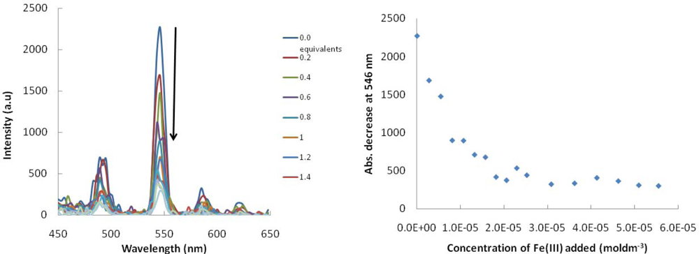
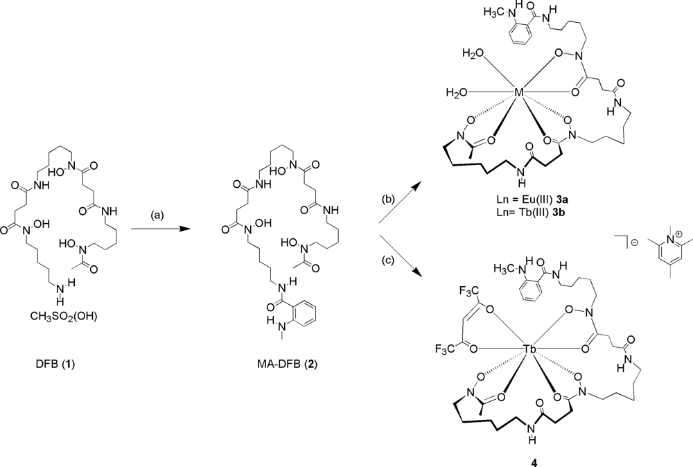
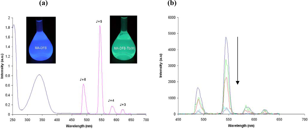
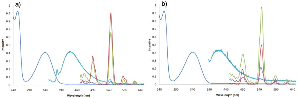
2. Results and Discussion

2.1. Design Criteria

Indicator displacement sensing, also referred to as an indicator displacement assay (IDA), has become an attractive method to detect analytes [2023]. In a typical IDA approach, the indicator competes with the substrate for the same binding site via rapid reversible interactions. The indicator sits within the cavity of the receptor, with a particular λmax, indicative of the microenvironment of the receptor. Upon addition of a guest, the indicator is displaced into free solution and an alternative wavelength is observed (Figure 1). As with any host–guest system design, there needs to be careful consideration of the target guest. Factors such as shape, size, charge, and hydrogen bonding donor/acceptor characteristics need to be taken into account. The binding sites for the particular guest need to be complimentary to the binding sites of the host. The more favorable the interactions, the more stable the host–guest complex.
The host-guest system described in this paper is such that the indicator of choice is a lanthanide ion (Eu3+ or Tb3+) coordinated to MA-DFB (compound 2- the host). It is well known that lanthanide complexes undergo rapid ligand exchange [24]. Lanthanide3+ ions are hard Lewis acids that have a tendency to form stable complexes with hard Lewis bases, those that contain oxygen atoms. Therefore the nature of the bonding in these complexes is predominately ionic and highly stable. However, lanthanide complexes in general, are also kinetically labile [25]. This is in contrast to the ferric ion siderophore coordination species whereby the complex is thermodynamically stable and kinetically inert. Taking this into consideration, the rationale behind the sensor design described in this paper was to utilize and synthesize an ID sensor, whereby the host-indicator (Ln-MA-DFB) was prepared (scheme 1) and Fe3+ acting as the guest.
It is well known that lanthanide ions, in particular Eu(III) and Tb(III) salts, have poor luminescence properties, due to f-f forbidden transitions, and as a consequence, lanthanide ions are not usually excited directly. Instead, they require a fluorophore (antenna) attached to a chelating functionality that incorporates the lanthanide metal [26,27]. The fluorophore (known as a sensitizer molecule) is typically an organic functional group. The organic fluorophore acts as an antenna absorbing light and transferring energy (via fluorescent transfer) to the lanthanide ion. An advantage of using lanthanide ions as part of a sensing application is their well-defined narrow excitation and emission bands and their long (micro- to millisecond) fluorescence, making them excellent candidates for molecular sensors. The long lifetimes (>200 μs) of Ln luminescence allow for discrete signal detection without background fluorescence, providing temporal selectivity for the lanthanide ion.
The advantage of this type of host-guest receptor is the sensing mechanism employed in the system, being a combination of Time-Resolved Fluorescence (TRF) and Fluorescent Resonance Energy Transfer (FRET). This union allows for greater analytical sensitivity because of the use of rare earth ions (lanthanides). A more correct description of energy transfer using lanthanides is Lanthanide Resonance Energy Transfer (LRET). LRET has a number of technical advantages compared to conventional FRET but is based on a similar mechanism [28]. For simplicity, the term FRET will be used to include LRET and FRET in this paper. The Ln ions absorb light poorly and require a sensitizer molecule for luminescence. The sensitizer molecule, (N-Methyanthranyl) will act as the antenna absorbing light and transferring that energy to the Ln ion.

2.2. Synthesis and Photophysical Properties

Three Ln(III) receptors have been prepared (compounds 3a, 3b and 4, Scheme 1) adapted from known literature procedures [29,30]. In order to test whether a MA-DFB (2) ligand can act as an antenna to sensitize the Eu(III) metal center, compound 3a was synthesized and isolated by reacting MA-DFB (2) with EuCl3•6H2O with triethylamine and the Eu(III). Europium(III) complexes typically show an intense luminescence signal between 610 and 620 nm [31]. The energy transfer (triplet state) from the 3An-Ln to An-Ln* (An = antenna) can occur via two mechanisms; the Dexter mechanism or the Förster energy transfer mechanism. The formation of the lanthanide excited state (Ar-Ln*) is a reversible process, and luminescence is dependent on a number of criterion; (1) how well the T1 excited state is populated, (2) The energy difference between the excited state of the antenna and the 5D excited state of Ln i.e., is the energy gap large enough such that the lanthanide emission cannot be quenched via back transfer to the antenna triplet state (3Ar-Ln), (3) The distance between the antenna and the lanthanide which follows a r6 dependence, and (4) The number of coordinated water molecules, as the fourth overtone of the H2O oscillator is lower in energy than the 5D state, and therefore decrease the quantum yield of the metal luminescence, (figure 2; boxed in region). Unfortunately, no luminescence emission was observed for compound 3a. The choice of antenna is very important in the receptor design, the lowest excited states of Eu(III) and Tb(III) are 17,200 cm−1 and 20,500 cm−1, respectively, due to point (2) mentioned above. For efficient population to the lanthanide excited state the energy of the triplet excited state needs to be at least 1,700 cm−1 above the excited state of the lanthanide ion, to prevent a thermally initiated back energy-transfer process, from An-Ln* to 3Ar-Ln and hence a signal response. If, however, the energy gap is less than 1,500 cm−1, such thermally activated back-energy transfer competes to repopulate the triplet state of the antenna and as a consequence no lanthanide luminescence is observed.
Therefore we switched the lanthanide ion to Tb3+ and prepared compound 3b. Upon the excitation of compound 3b at 340 nm, four distinct luminescence emissions are measured (transitions are 5D4-7FJ whereby J = 6,5,4 and 3) (also seen visually) in organic solvents such as ethyl acetate (Figure 3a). The initial studies showed that luminescence emission for compound 3b was seen in a variety of solvents (ethyl acetate (Figure 3a), acetone, dichloromethane, chloroform, acetonitrile, and diethyl ether) and we have demonstrated that the fluorescence is quenched upon the addition of FeCl3, (ethyl acetate Figure 3b). However, like the Eu(III) complex, the luminescence of the Terbium is quenched by protic polar solvents such as MeOH and solvents that are hydroscopic, for example DMSO, when water occupies the vacant metal coordination sites (compounds 3a and 3b).
In order for our system to work as a sensor in oceanographic applications, it is essential that the signal response is not quenched by water molecules. This would require a luminescence signal in solvents such as methanol (MeOH), ethanol (EtOH) or dimethyl sulfoxide (DMSO), organic solvents that are miscible with water. In this paper we demonstrate that indeed a luminescence signal is observed for compound 3b, in 100% MeOH and a 50:50% MeOH:dH2O mix (Figure 4a). As noted, the luminescence signal is quenched in 100% dH2O; because O-H, (like C-H moieties), is a higher-energy oscillator (vide supra) it significantly quenches the Ln luminescence, (Figure 4a purple line). We therefore modified the design of our molecular receptor to incorporate a “blocking” ligand that would prevent water molecules from coordinating to the Tb(III) centre. β-Diketonates have been used as sensitizers for lanthanide complexes [29,3235]. β-Diketonate ligands coordinate via the two oxygen atoms leading to neutral species in a 3:1 β-diketonate:lanthanide ratio, the complexes have been shown to be stable in aqueous solutions [35]. Therefore these molecules have been used in many applications from sensing [36], antibody labeling [37], new materials such as liquid crystals [38], near IR-LEDs, [39] sol-gel glasses [40], and polymers [40].
Incorporating a commercially available β-diketonate, for example, 1,1,1,5,5,5-hexafluoro-2, 4-pentanediione (Hhfac) ligand into our molecular receptor we synthesized compound 4, (Scheme 1). Again the same luminescence experiments were carried out in 100% MeOH, 50:50% MeOH:dH2O and 100% dH2O (pH 8) and in all three solvent systems a luminescence signal, is observed, (Figure 4b).
We have shown that the displacement of the lanthanide with FeCl3 is achieved in organic solvents, ethyl acetate, (Figure 3b). The same titration experiment has been carried out with compound 4 and FeCl3 in MeOH and dH2O (50:50%) and the same trend is seen, the luminescence is quenched rapidly upon the addition of small aliquots of Fe3+. The concentration of the Fe(III) equals 2.5 × 10⁻⁵ moldm−3 at the point where there is no more quenching (Figure 5).
This preliminary data provides us with a good understanding of the system and evidence that this theoretical approach will work for development into an iron chemosensor. This data clearly demonstrate that with a slight modification of the molecular receptor, one can observe luminescence in 100% dH2O and that this luminescence decreases as a function of increasing [Fe(III)]. Our initial studies provide an effective detection limit, based on the precision of the luminescent signal as low as 5 nM (assuming a 1L sample), an environmentally relevant concentration.

3. Experimental Section

Materials. Chemicals: All chemicals were purchased from Sigma-Aldrich Chemical Co. (St.Louis, MO) and used without further purification. N-methylisatoic anhydride was purchased from TCI America. Solvents used were all of UV-spectroscopic grade and purchased from Sigma-Aldrich, or VWR.
1H and 13C NMR spectra were recorded on a Bruker UltraShield plus 400 MHz spectrometer in DMSO-d6. Chemical shifts are reported in parts per million (d) downfield from tetramethylsilane (0 ppm) as the internal standard and coupling constants (J) are recorded in Hertz (Hz). The multiplicities in the 1H NMR are reported as (br) broad, (s) singlet, (d) doublet, (dd) doublet of doublets, (ddd) doublet of doublet of doublets, (t) triplet, (sp) septet, (m) multiplet. All spectra are recorded at ambient temperatures. IR was taken using a Nicolet Nexus 470 FT-IR paired with a Smart Orbit ATR attachment, with the characteristic functional groups reported in wavenumbers (cm−1). UV-vis experiments were performed on a Beckman DU-70 UV-vis spectrometer. A stock solution (2.9 × 10⁻⁴ moldm−3) of compound 2 was prepared by dissolving 2.0 mg in 10 mL of MeOH and 1 mL transferred to the UV-vis cell. To which 2,4,6 trimethylpyridine (1μL, 26 equivalents) was added to compound 2. A second stock solution of TbCl3•6H2O salt was prepared by dissolving 7.1 mg of Tb3+ salt in 5 mL of MeOH (3.8 mmol), from the Tb3+ salt stock solution a 2.9 × 10⁻³ mol dm−3 solution was prepared by transferring 152 μL to a 2 mL cell. Aliquots of the Tb3+ was then added incrementally up to 10 equivalents. Fluorescence experiments were carried out on a QuantaMaster 40 Intensity Based spectrofluorometer; steady-state (slitwidths 1.00 mm); λEx = 340 λEm = 360 to 600 nm and gated emission (slit widths 3.0 mm, delay time 200 μs, λEx = 340 λEm = 450 to 650 nm). MA-DFB and the Lanthanide salts were diluted to 2.25 × 10⁻⁶ moldm−3 and 2.2 × 10⁻⁴ moldm−3 respectively and used for the fluorescence studies. A FeCl3 solution (2.2 × 10⁻⁴ M) was prepared in MeOH and aliquots of 10 μL was titrated into the UV-vis or fluorescence cell. The dilution was taken into consideration when calculating the equivalents of host:guest solution.
Synthesis. Compound 2 (MA-DFB): The synthesis of MA-DFB was adopted from Loyevsky et al. [41]. Desferrioxamine (330 mg, 0.5 mmol) was dissolved in dimethylformamide (1 mL) and triethylamine (73 mg, 0.730 mmol). The solution was allowed to stir for 15 minutes, to which N-methylisatoic anhydride (90 mg, 0.5 mmol) was added to it, the resulting mixture allowed then to stir overnight at STP under argon. The supernatant was removed by centrifugation and the white solid was washed several times with ddH2O, then washed with diethyl ether and dried in vacuum for 24 h. Melting point: 177–181 °C; UV (methanol): 340 nm (ε = 2398 M−1 cm−1) and 250 nm (ε = 4659 M−1 cm−1); IR (neat ATR): 3304 (NH amide), 3092 (OH oxime), 1617 (CO-N) cm−1; 1H-NMR (DMSO-d6): 9.65 (br, s 3H OH oxime); disappears on D2O shake at room temperature, 8.27 (t, 1H, NH amide); disappears on D2O shake at 60 °C, 7.80 (m, 2H, NH amide) disappears on D2O shake at 60 °C, 7.61 (m, 1H, 2° NH amine); disappears on D2O shake at room temperature, 7.50 (m, 1H, ArH), 7.30 (m, 1H, ArH), 6.60 (m, 2H, ArH), 3.49 (m, 6H, CH2), 3.17 (m, 2H, CH2), 3.00 (m, 6H CH2), 2.75 (m, 4H, CH2), 2.55 (m, 4H, CH2CH2), 2.25 (m, 4H, CH2CH2), 2.34 (s, 3H, NCH3), 1.95 (s, 3H COCH3), 1.50–1.23 (12H, CH2CH2CH2); 13C-NMR (DMSO-d6): 172.5, 172.0, 170.7, 169.8, 150.2, 132.8, 128.7, 115.9, 114.4, 110.0, 49.1, 47.6, 47.3, 38.9, 30.3, 29.8, 29.3, 28.0, 27.2, 26.5, 26.2, 24.0, 23.3, 20.9 ppm.

4. Conclusions

We describe a novel approach that utilizes an indicator displacement assay to develop a chemosensor for the biologically available iron fraction in aquatic systems. This approach combines TRF and FRET with the unique photophysical properties of a lanthanide ion to increase the sensitivity of a fluorescent siderophore derivative, MA-DFB. The siderophore is derived from a natural siderophore, desferrrioxamine B, a strong fungal chelator. Currently there are very limited means to measure iron that is biologically available to eukaryotic phytoplankton. The development of this chemosensor will help build a better understanding of this fraction of iron in marine systems. The capability of measuring labile iron in situ with a photoactive chemosensor will lead to a more comprehensive understanding of primary production and the ocean carbon cycle.

Acknowledgments

This work was supported by startup grants to KMO and KJW from the University of Southern Mississippi and NSF0840390 for the acquisition of a 400 MHz NMR. The authors would also like to acknowledge support from the U.S. Department of Education GAANN Fellowship program (GR02619) for W. Scott Jones and the NSF MRSECenter for Responsive Driven Films at USM (DMR-0213883) for providing the REU (Research Experience for Undergraduates) stipend for David Schrock.

References and Notes

  1. Chisholm, S.W. Oceanography: Stirring times in the Southern Ocean. Nature 2000, 407, 685–687. [Google Scholar]
  2. Hutchins, D.A.; Franck, V.M.; Brzezinski, M.A.; Bruland, K.W. Inducing iron limitation in iron-replete coastal waters with a strong chelating ligand. Limnol. Oceanogr 1999, 44, 1009–1018. [Google Scholar]
  3. Wells, M.L. Manipulating iron availability in nearshore waters. Limnol. Oceanogr 1999, 44, 1002–1008. [Google Scholar]
  4. Wells, M.L.; Trick, C.G. Controlling iron availability to phytoplankton in iron-replete coastal waters. Mar. Chem 2004, 86, 1–13. [Google Scholar]
  5. Well, M.L.; Price, N.M.; Bruland, K.W. Iron chemistry in seawater and its relationship to phytoplankton: a workshop report. Mar. Chem 1995, 48, 157–182. [Google Scholar]
  6. Boyanapalli, R.; Bullerjahn, G.S.; Pohl, C.; Croot, P.L.; Boyd, P.W.; Michael, R.; McKay, R.M.L. Luminescent whole-cell cyanobacterial bioreporter for measuring Fe availability in diverse marine environments. Appl. Environ. Microbiol 2007, 73, 1019–1024. [Google Scholar]
  7. Durham, K.A.; Porta, D.; Twiss, M.R.; McKay, M.L.; Bullerjahn, G.S. Construction and initial characterization of a luminescent Synechoccus sp. PCC 7942 Fe-dependant bioreporter. FEMS Microbiol. Lett 2002, 209, 215–221. [Google Scholar]
  8. Hassler, C.S.; Wiss, M.R.; McKay, R.M.L.; Bullerjahn, G.S. Optimization of iron-dependant cyanobacterial (Synechoccocus, Cyanophyceae) bioreporters to measure iron availability. J. Phycol 2006, 42, 324–335. [Google Scholar]
  9. McKay, R.M.L.; Bullerjahn, G.S.; Porta, D.; Brown, E.T.; Sherrel, R.M.; Smutka, T.M.; Sterner, R.W.; Twiss, M.R.; Wilhelm, S.W. Consideration of the bioavailability of iron in the North American Great Lakes: Development of novel approaches toward understanding iron biogeochemistry. Aquat. Ecosyst. Health Manag 2004, 7, 475–490. [Google Scholar]
  10. McKay, R.M.L.; Porta, D.; Bullerjahn, G.S.; Al-Rshaidat, M.M.D.; Kilmowicz, J.A.; Sterner, R.W.; Smutka, T.M.; Brown, E.T.; Sherrel, R.M. Bioavailable iron in oligotrophic Lake Superior assessed using biological reporters. J. Plankton Res 2005, 27, 1033–1044. [Google Scholar]
  11. Mioni, C.E.; Handy, S.M.; Ellwood, M.J.; Twiss, M.R.; McKay, R.M.L.; Boyd, P.W.; Wilhelm, S.W. Tracking changes in bioavailable Fe within high-nutrient low-chlorophyll oceanic waters: a first estimate using a heterotrophic bacterial bioreporter. Global Biogeochem. Cycles 2005, 19, 1–10. [Google Scholar]
  12. Mioni, C.E.; Howard, A.M.; Debruyn, J.M.; Bright, N.G.; Twiss, M.R.; Applegate, B.M.; Wilhelm, S.W. Characterization and field trials of a bioluminescent bacterial reporter of iron availability. Mar. Chem 2003, 83, 31–46. [Google Scholar]
  13. Mioni, C.E.; Poorvin, L.; Wilhelm, S.W. Virus and siderophore-mediated transfer of available Fe between heterotrophic bacteria: characterization using an Fe-specific reporter. Aquat. Microb. Ecol 2005, 41, 233–245. [Google Scholar]
  14. Porta, D.; Bullerjahn, G.S.; Wilhelm, S.W.; Twiss, M.R.; McKay, R.M.L. Physiological characterization of Synechococcus sp. (cyanophyceae) strain PCC 7942 iron-dependant bioreporter for freshwater environments. J. Phycol 2003, 39, 64–73. [Google Scholar]
  15. Mioni, C.E.; Pakulski, J.D.; Poorvin, L.; Baldwin, A.; Twiss, M.R.; Jeffrey, W.H.; Wilhelm, S.W. Variability in the in situ bioavailability of Fe to bacterioplankton communities in the eastern subtropical Pacific Ocean. Aquat. Microb. Ecol 2007, 46, 239–251. [Google Scholar]
  16. Chung Chun Lam, C.K.S.; Jickells, T.D.; Richardson, D.J.; Russell, D.A. Fluorescence-based siderophore biosensor for the determination of bioavailable iron in oceanic waters. Anal. Chem 2006, 78, 5040–5045. [Google Scholar]
  17. Orcutt, K.M.; L, W.M. A liposome-based nanodevice for sequestering siderophore-bound Fe. J. Membrane Sci 2007, 288, 247–254. [Google Scholar]
  18. Espósito, B.P.; Epsztejn, S.; Breuer, W.; Cabantchik, Z.I. A review of fluorescence methods for assessing labile iron in cells and biological fluids. Anal. Biochem 2002, 304, 1–18. [Google Scholar]
  19. Palanché, B.P.; Marmolle, F.; Abraham, M.A.; Shanzer, A.; Albrecht-Gray, A.M. Fluorescent siderophore-based chemosensors: iron (III) quantitative determinations. J. Biol. Inorg. Chem 1999, 4, 188–198. [Google Scholar]
  20. de Silva, A.P.; Gunaratne, H.Q.N.; Gunnlaugsson, T.; Huxley, A.J.M.; McCoy, C.P.; Rademacher, J.T.; Rice, T.E. Signaling recognition events with fluorescent sensors and switches. Chem. Rev 1997, 97, 1515–1566. [Google Scholar]
  21. Houk, R.J.T.; Wallace, K.J.; Hewage, H.S.; Anslyn, E.V. A colorimetric chemodosimeter for Pd(II): a method for detecting residual palladium in cross-coupling reactions. Tetrahedron 2008, 64, 8271–8278. [Google Scholar]
  22. Wallace, K.J.; Nguyen, B.T.; Anslyn, E.V. Indicator Displacement Assay (IDA). In Encyclopedia of Supramolecular Chemistry, 1st ed; Atwood, J.L., Steed, J.W., Eds.; Taylor & Francis: Baca Raton, FL, USA, 2006. [Google Scholar]
  23. Wiskur, S.L.; Ait-Haddou, H.; Lavigne, J.J.; Anslyn, E.V. Teaching old indicators new tricks. Acc. Chem. Res 2001, 34, 963–972. [Google Scholar]
  24. Graeppi, N.; Powell, G.H.; Laurenczy, G.; Zékány, L.; Merbach, A. Coordination equilibria and water exchange kinetics of lanthanide(III) propylenediaminetetraacetates and other magnetic resonance imaging related complexes. Inorg Chimi. Acta 1995, 235, 311–326. [Google Scholar]
  25. Goshe, A.J.; Steele, I.M.; Ceccarelli, C.; Rheingold, A.L.; Bosnich, B. Supramolecular recognition: On the kinetic lability of thermodynamically stable host-guest association complexes. Proc. Natl. Acad. Sci 2002, 99, 4823–4829. [Google Scholar]
  26. Bunzli, J.C.G.; Piguet, C. Lanthanide-Containing molecular and supramolecular polymetallic functional assemblies. Chem. Rev 2002, 102, 1897–1928. [Google Scholar]
  27. Tsukube, H.; Shinoda, S. Lanthanide complexes in molecular recognition and chirality sensing of biological substrates. Chem. Rev 2002, 102, 2389–2403. [Google Scholar]
  28. Selvin, P.R. Principles and biophysical applications of lanthanide-based probes. Annu. Rev. Biophys. Biomol. Struct 2002, 31, 275–302. [Google Scholar]
  29. De Silva, C.R.; Maeyer, J.R.; Wang, R.; Nichol, G.S.; Zheng, Z. Adducts of europium b-diketonates with nitrogen p,p′-disubstituted bipyridine and phenanthroline ligands: Synthesis structural characterization, and luminescence studies. Inorg. Chimi. Acta 2007, 360, 3543–3552. [Google Scholar]
  30. Lytton, S.D.; Cabantchik, Z.I.; Libman, J.; Shanzer, A. Reversed siderophores as antimalarial agents. II. Selective scavenging of Fe (III) from parasitized erythrocytes by a fluorescent derivitive of Desferal. Mol. Pharmacol 1991, 40, 584–590. [Google Scholar]
  31. Moore, E.G.; Xu, J.; Jocher, C.J.; Werner, E.J.; Raymond, K.N. “Cymothoe sangaris”: An extremely stable and highly luminescent 1,2-hydroxypyridinonate chelate of Eu(III). J. Am. Chem. Soc 2006, 128, 10648–10649. [Google Scholar]
  32. Bassett, A.P.; Magennis, S.W.; Glover, P.B.; Lewis, D.J.; Spencer, N.; Parsons, S.; Williams, R.M.; De Cola, L.; Pikramenou, Z. Hghly Luminescent, Triple- and quadruple-stranded dinuclear Eu, Nd, and Sm(III) lanthanide complexes based on bis-diketonates? J. Am. Chem. Soc 2004, 126, 9413–9424. [Google Scholar]
  33. Batista, H.J.; de Andrade, A.V.M.; Longo, R.L.; Simas, M.; de Sá, G.D.; Ito, N.K.; Thompson, L.C. Synthesis, X-ray structure, spectroscopic characterization, and theoretical prediction of the Structure and Electronic Spectrum of Eu (btfa)3·bipy and an assesessment of the effect of fluorine as β-diketone substituent on the ligand-metal energy transfer proces. Inorg. Chem 1998, 37, 3542–3547. [Google Scholar]
  34. dos Santos, C.M.G.; Harte, A.J.; Quinn, S.J.; Gunnlaugsson, T. “Recent developments in the field of supramolecular lanthanide luminescent sensors and self-assemblies”. Coord. Chem. Rev 2008, 252, 2512–2527. [Google Scholar]
  35. Tsukube, H.; Shinoda, S.; Uenishi, J.; Kanatani, T.; Itoch, H.; Shiode, M.; Iwachido, T.; Yonemitsu, O. “Molecular Recognition with lanthanide(III) tris(β-diketonate) complexes: extraction, transport, and chiral recognition of unprotected amino acids”. Inorg. Chem 1998, 37, 1585–1591. [Google Scholar]
  36. Thibon, A.; Pierre, V.C. A highly selective luminescent sensor for the time-gated detection of potassium. J. Am. Chem. Soc 2009, 131, 434–435. [Google Scholar]
  37. Terenghi, M.; Elviri, L.; Careri, M.; Mangia, A.; Lobinski, R. Multiplexed determination of protein biomarkers using metal-tagged antibodies and size exclusion chromatography-inductively coupled plasma mass spectrometry. Anal. Chem 2009, 81, 9440–9448. [Google Scholar]
  38. Binnemans, K. Luminescence of metallomesogens in the liquid crystal state. J. Mater. Chem 2009, 19, 448–453. [Google Scholar]
  39. Guo, X.; Guo, H.; Fu, L.; Carlos, L.D.; Ferreira, R.A.S.; Sun, L.; Deng, R.; Zhang, H. Novel near-infrared luminescent hybrid materials covalently linking with lanthanide [Nd(III), Er(III), Yb(III), and Sm(III)] complexes via a primary β-Diketone ligand: synthesis and photophysical studies. J. Phys. Chem. C 2009, 113, 12538–12545. [Google Scholar]
  40. Binnemans, K. Lanthanide-Based Luminescent Hybrid Materials. Chem. Rev 2009, 109, 4283–4374. [Google Scholar]
  41. Loyevsky, M.; Lytton, S.D.; Mester, B.; Libman, J.; Shanzer, A.; Cabantchik, Z.I. The Antimalarial action of desferal involves a direct access route to erythrocytic (Plasmodium Falciparum) parasites. J. Clin. Invest 1993, 91, 218–224. [Google Scholar]
Figure 1. Cartoon representation of the Indicator Displace Assay (indicator = lanthanide, analyte = Fe3+ antenna = N-Methyanthranyl moiety).
Sensors 10 01326f1
Figure 2. Electronic energy level diagrams for Tb3+ and Eu3+. Radiationless energy transfer competes with the radioactive process through coupling of the emissive states to the O-H vibrational overtones of the coordinated water molecules.
Sensors 10 01326f2
Figure 3. (a) The UV-Vis (340 nm) and the corresponding Luminescence spectrum of compound 3b (Ex = 340 nm) in ethyl acetate (1 × 10⁻⁸ M) and (b) The UV-Vis (340 nm) and the corresponding decreasing Luminescence of compound 3b (Ex = 340 nm) in ethyl acetate (1 × 10⁻⁸ M) with increasing amounts of FeCl3 added.
Sensors 10 01326f3
Figure 4. (a) The spectra showing absorbance (dark blue 100% MeOH, compound 2), steady-state (light blue100 % MeOH, compound 3b) and gated luminescence (red 100% MeOH (3b), green 50:50% MeOH:H2O (3b), purple 100% H2O (3b)). (b) The spectra showing absorbance (dark blue 100% MeOH, compound 2), steady-state (light blue100 % MeOH, compound 4) and gated luminescence (red 100% MeOH (4), green 50:50% MeOH:H2O (4), purple 100% H2O (4)). Conditions: Ex 340 nm, Em 350–650 nm, slit width 2.25 mm, delay 200 μs.
Sensors 10 01326f4
Figure 5. Fluorescence titration with 4 and FeCl3 (0 to 5 equivalents) MeOH:H2O (50:50). RHS: Binding isotherm obtained showing the decrease in absorbance at 546 nm on the addition of Fe(III).
Sensors 10 01326f5
Scheme 1. Synthesis of MA-DFB-Ln(III) (a) N-methylisatoic anhydride and Et3N (b) LnCl3•6H2O (Ln = Eu(III) or Tb(III), 2,4,6 trimethylpyridine (c) TbCl3•6H2O, 1,1,1,5,5,5-hexafluoro-2, 4-pentanediione (Hhfac) and 2,4,6 trimethylpyridine.
Sensors 10 01326f6

