Bioconjugation Strategies for Microtoroidal Optical Resonators

Abstract
:
1. Introduction
2. Experimental Procedures
- low optical absorption at the wavelength of interest,
- length-scale compatible with the evanescent field,
- high density packing,
- specificity to only the target ligand,
- minimal reagent use, and
- high stability of the probe molecule to storage in air.
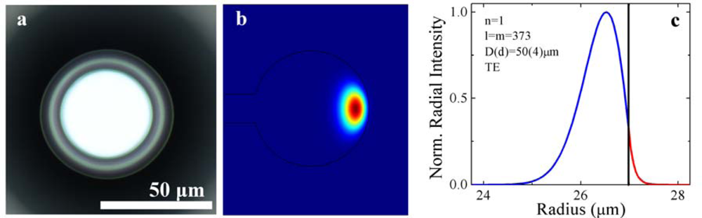
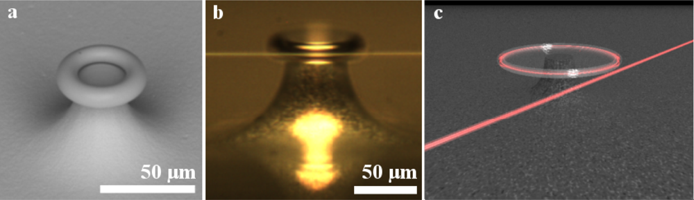
2.1. Device Fabrication
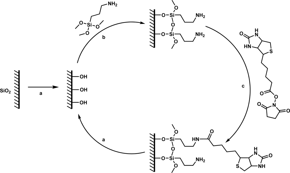
2.2. Device Functionalization Protocols
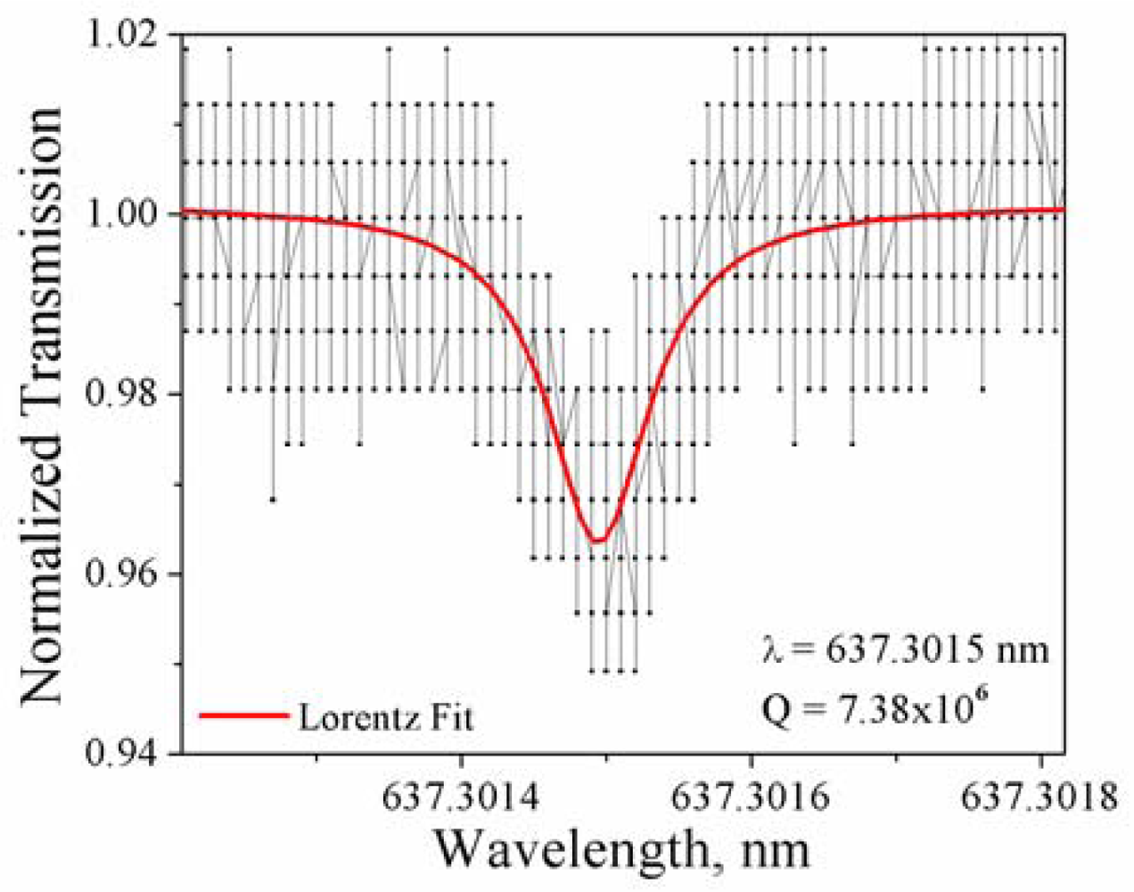
2.3. Device Characterization Protocols
3. Results and Discussion
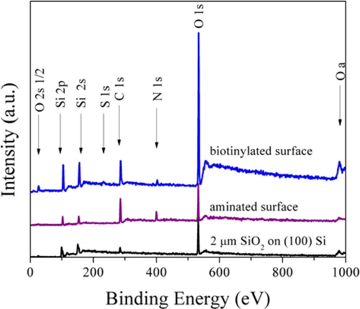
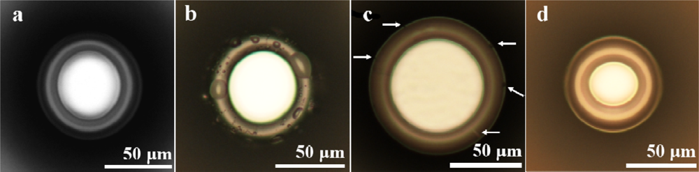
3.1. Analysis of the Surface Functionalization
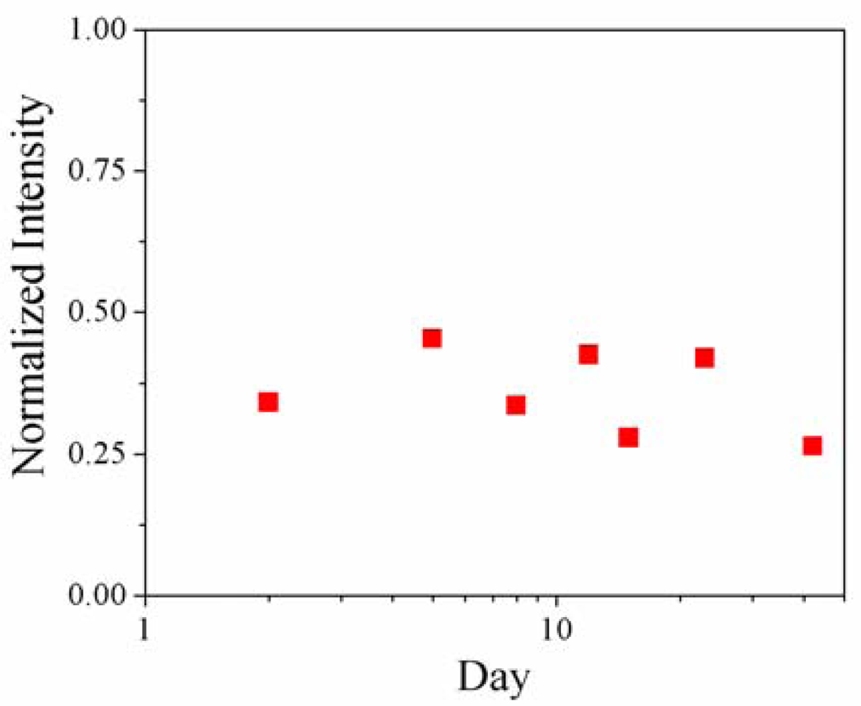
3.2. Stability of the Surface Chemistry
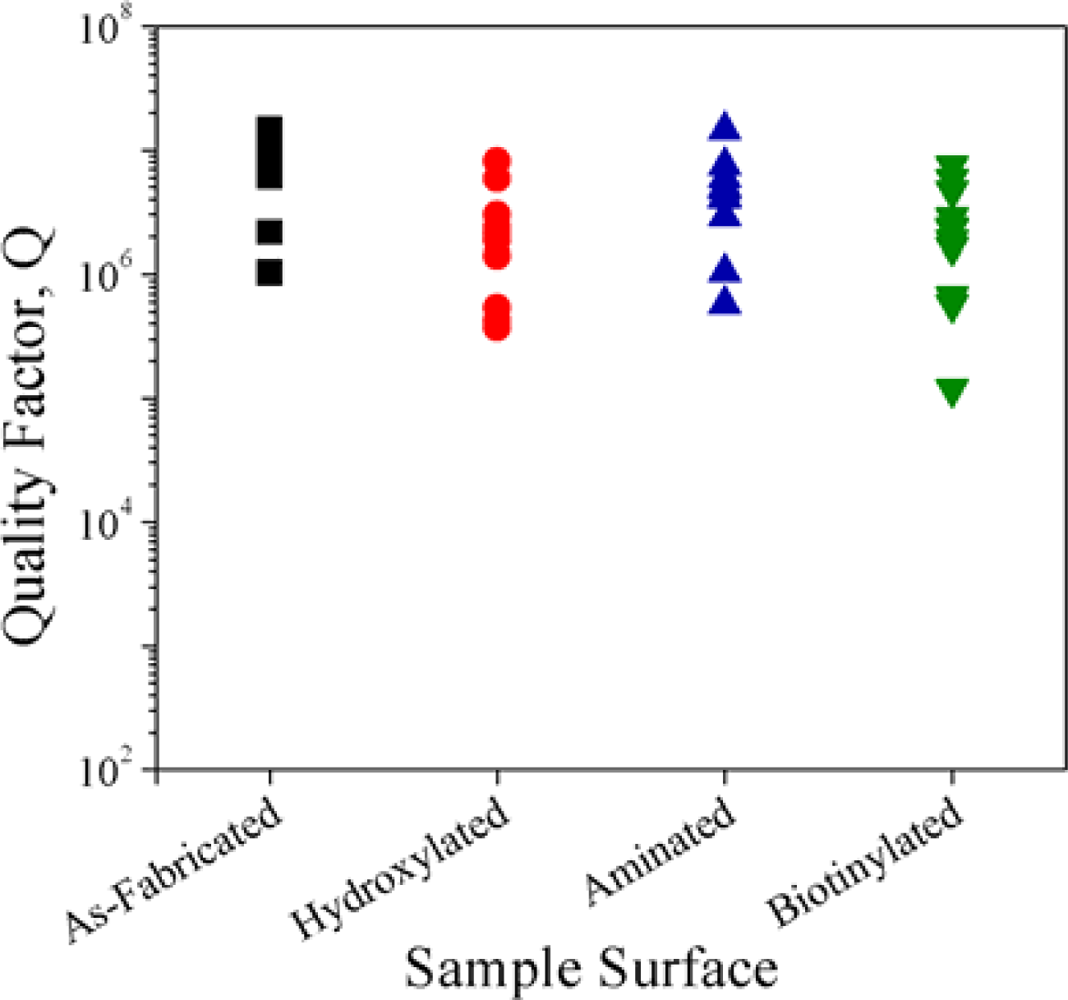
3.3. Quality Factor Study
4. Conclusion and Future Outlook
Acknowledgments
References and Notes
- Luppa, PB; Sokoll, LJ; Chan, DW. Immunosensors—Principles and applications to clinical chemistry. Clin. Chim. Acta 2001, 314, 1–26. [Google Scholar]
- Ambrose, WP; Goodwin, PM; Jett, JH; van Orden, A; Werner, JH; Keller, RA. Single molecule fluorescence spectroscopy at ambient temperature. Chem. Rev 1999, 99, 2929–2956. [Google Scholar]
- Funatsu, T; Harada, Y; Higuchi, H; Tokunaga, M; Saito, K; Ishii, Y; Vale, RD; Yanagida, T. Imaging and nano-manipulation of single biomolecules. Biophys. Chem 1997, 68, 63–72. [Google Scholar]
- Wormke, S; Mackowski, S; Brotosudarmo, THP; Jung, C; Zumbusch, A; Ehrl, M; Scheer, H; HofMann, E; Hiller, RG; Brauchle, C. Monitoring fluorescence of individual chromophores in peridininchlorophyll-protein complex using single molecule spectroscopy. Biochim. Biophys. Acta-Bioenerg 2007, 1767, 956–964. [Google Scholar]
- Xie, SN. Single-molecule approach to enzymology. Single Mol 2001, 2, 229–236. [Google Scholar]
- Zhang, JK; Dong, SM; Lu, JH; Turner, APF; Fan, QJ; Jia, SR; Yang, HJ; Qiao, CS; Zhou, H; He, GW. A label free electrochemical nanobiosensor study. Anal. Lett 2009, 42, 2905–2913. [Google Scholar]
- Mao, S; Lu, GH; Yu, KH; Chen, JH. Specific biosensing using carbon nanotubes functionalized with gold nanoparticle-antibody conjugates. Carbon 2010, 48, 479–486. [Google Scholar]
- Campbell, GA; Mutharasan, R. Detection and quantification of proteins using self-excited PZT-glass millimeter-sized cantilever. Biosens. Bioelectron 2005, 21, 597–607. [Google Scholar]
- Campbell, GA; Mutharasan, R. Detection of pathogen Escherichia coli O157 : H7 using self-excited PZT-glass microcantilevers. Biosens. Bioelectron 2005, 21, 462–473. [Google Scholar]
- Datar, R; Kim, S; Jeon, S; Hesketh, P; Manalis, S; Boisen, A; Thundat, T. Cantilever Sensors: Nanomechanical Tools for Diagnostics. MRS Bull 2009, 34, 449–454. [Google Scholar]
- Horvath, R; Pedersen, HC; Skivesen, N; Selmeczi, D; Larsen, NB. Optical waveguide sensor for on-line monitoring of bacteria. Optics Lett 2003, 28, 1233–1235. [Google Scholar]
- Dumais, P; Callender, CL; Noad, JP; Ledderhof, CJ. Silica-on-silicon optical sensor based on integrated waveguides and microchannels. IEEE Photonic. Techn. Lett 2005, 17, 441–443. [Google Scholar]
- Andras, S; Adanyi, N; Szekacs, I; Majer-Baranyi, K; Istvan, S. Optical waveguide light-mode spectroscopy immunosensors for environmental monitoring. Appl. Opt 2009, 48, B151–B158. [Google Scholar]
- Shankaran, DR; Gobi, KVA; Miura, N. Recent advancements in surface plasmon resonance immunosensors for detection of small molecules of biomedical, food and environmental interest. Sens. Actuat. B-Chem 2007, 121, 158–177. [Google Scholar]
- Sundberg, F; Karlsson, R. Rapid detection and characterization of immune responses using label-free biacore immunoassays. Immunology 2007, 120, 46–47. [Google Scholar]
- Kumbhat, S; Shankaran, DR; Kim, SJ; Gobi, KV; Joshi, V; Miura, N. Surface plasmon resonance biosensor for dopamine using D3 dopamine receptor as a biorecognition molecule. Biosens. Bioelectron 2007, 23, 421–427. [Google Scholar]
- Nagel, T; Ehrentreich-Forster, E; Singh, M; Schmitt, K; Brandenburg, A; Berka, A; Bier, FF. Direct detection of tuberculosis infection in blood serum using three optical label-free approaches. Sens. Actuat. B-Chem 2008, 129, 934–940. [Google Scholar]
- Tetz, KA; Pang, L; Fainman, Y. High-resolution surface plasmon resonance sensor based on linewidth-optimized nanohole array transmittance. Opt. Lett 2006, 31, 1528–1530. [Google Scholar]
- Chen, HM; Pang, L; Kher, A; Fainman, Y. Three-dimensional composite metallodielectric nanostructure for enhanced surface plasmon resonance sensing. Appl. Phys. Lett 2009, 94, 073177. [Google Scholar]
- Li, H; Fan, X. Characterization of sensing capability of optofluidic ring resonator biosensors. Appl. Phys. Lett 2010, 97, 011105–3. [Google Scholar]
- Alvarez, SD; Li, CP; Chiang, CE; Schuller, IK; Sailor, MJ. A label-free porous alumina interferometric immunosensor. ACS Nano 2009, 3, 3301–3307. [Google Scholar]
- Bolduc, OR; Clouthier, CM; Pelletier, JN; Masson, JF. Peptide self-assembled monolayers for label-free and unamplified surface plasmon resonance biosensing in crude cell lysate. Anal. Chem 2009, 81, 6779–6788. [Google Scholar]
- Densmore, A; Vachon, M; Xu, DX; Janz, S; Ma, R; Li, YH; Lopinski, G; Delâge, A; Lapointe, J; Luebbert, CC; Liu, QY; Cheben, P; Schmid, JH. Silicon photonic wire biosensor array for multiplexed real-time and label-free molecular detection. Opt. Lett 2009, 34, 3598–3600. [Google Scholar]
- Fan, XD; White, IM; Shopoua, SI; Zhu, HY; Suter, JD; Sun, YZ. Sensitive optical biosensors for unlabeled targets: A review. Anal. Chim. Acta 2008, 620, 8–26. [Google Scholar]
- Gao, Z; Agarwal, A; Trigg, AD; Singh, N; Fang, C; Tung, C-H; Fan, Y; Buddharaju, KD; Kong, J. Silicon nanowire arrays for label-free detection of DNA. Anal. Chem 2007, 79, 3291–3297. [Google Scholar]
- Hanumegowda, NM; White, IM; Oveys, H; Fan, XD. Label-free protease sensors based on optical microsphere resonators. Sens. Lett 2005, 3, 315–319. [Google Scholar]
- Lee, PH. Label-free optical biosensor: A tool for G protein-coupled receptors pharmacology profiling and inverse agonists identification. J Recept Sig Transd 2009, 29, 146–153. [Google Scholar]
- Syahir, A; Mihara, H; Kajikawa, K. A new optical label-free biosensing platform based on a metal−insulator−metal structure. Langmuir 2010, 26, 6053–6057. [Google Scholar]
- Washburn, AL; Gunn, LC; Bailey, RC. Label-Free quantitation of a cancer biomarker in complex media using silicon photonic microring resonators. Anal. Chem 2009, 81, 9499–9506. [Google Scholar]
- Washburn, AL; Luchansky, MS; Bowman, AL; Bailey, RC. Quantitative, label-free detection of five protein biomarkers using multiplexed arrays of silicon photonic microring resonators. Anal. Chem 2010, 82, 69–72. [Google Scholar]
- Subrahmanyam, S; Piletsky, SA; Turner, APF. Application of natural receptors in sensors and assays. Anal. Chem 2002, 74, 3942–3951. [Google Scholar]
- Hunt, HK; Armani, AM. Label-free biological and chemical sensors. Nanoscale 2010, 2, 1544–1559. [Google Scholar]
- Hock, B. Antibodies for immunosensors—A review. Anal. Chim. Acta 1997, 347, 177–186. [Google Scholar]
- Feuz, L; Jonsson, P; Jonsson, MP; Hook, F. Improving the limit of detection of nanoscale sensors by directed binding to high-sensitivity areas. ACS Nano 2010, 4, 2167–2177. [Google Scholar]
- Kalia, J; Raines, RT. Advances in bioconjugation. Curr. Org. Chem 2010, 14, 138–147. [Google Scholar]
- Amine, A; Mohammadi, H; Bourais, I; Palleschi, G. Enzyme inhibition-based biosensors for food safety and environmental monitoring. Biosens. Bioelectron 2006, 21, 1405–1423. [Google Scholar]
- Hegnerova, K; Bockova, M; Vaisocherova, H; Kristofikova, Z; Ricny, J; Ripova, D; Homola, J. Surface plasmon resonance biosensors for detection of Alzheimer disease biomarker. Sens. Actuat. B-Chem 2009, 139, 69–73. [Google Scholar]
- Zanoli, L; D'Agata, R; Spoto, G. Surface plasmon-based optical detection of DNA by peptide nucleic acids. Minerva Biotecnol 2008, 20, 165–174. [Google Scholar]
- Gobi, KV; Iwasaka, H; Miura, N. Self-assembled PEG monolayer based SPR immunosensor for label-free detection of insulin. Biosens. Bioelectron 2007, 22, 1382–1389. [Google Scholar]
- Gobi, KV; Kim, SJ; Tanaka, H; Shoyama, Y; Miura, N. Novel surface plasmon resonance (SPR) immunosensor based on monomolecular layer of physically-adsorbed ovalbumin conjugate for detection of 2,4-dichlorophenoxyacetic acid and atomic force microscopy study. Sens. Actuat. B-Chem 2007, 123, 583–593. [Google Scholar]
- Kim, KS; Lee, H-S; Yang, J-A; Jo, M-H; Hahn, SK. The fabrication, characterization and application of aptamer-functionalized Si-nanowire FET biosensors. Nanotechnology 2009. [Google Scholar] [CrossRef]
- Lee, M; Walt, DR. A fiber-optic microarray biosensor using aptamers as receptors. Anal. Biochem 2000, 282, 142–146. [Google Scholar]
- Lee, SH; Ko, HJ; Park, TH. Real-time monitoring of odorant-induced cellular reactions using surface plasmon resonance. Biosens. Bioelectron 2009, 25, 55–60. [Google Scholar]
- Dover, JE; Hwang, GM; Mullen, EH; Prorok, BC; Suh, SJ. Recent advances in peptide probe-based biosensors for detection of infectious agents. J. Microbiol. Meth 2009, 78, 10–19. [Google Scholar]
- Kumbhat, S; Shankaran, DR; Kim, SJ; Gobi, KV; Joshi, V; Miura, N. A novel receptor-based surface-plasmon-resonance affinity biosensor for highly sensitive and selective detection of dopamine. Chem. Lett 2006, 35, 678–679. [Google Scholar]
- Byeon, J-Y; Limpoco, FT; Bailey, RC. Efficient bioconjugation of protein capture agenst to biosensor surfaces using aniline-catalyzed hydrazone ligation. Langmuir 2010, 26, 15430–15435. [Google Scholar]
- Zhang, X; Choi, H-S; Armani, AM. Ultimate quality factor of silica microtoroid resonant cavities. Appl. Phys. Lett 2010, 96, 153304. [Google Scholar]
- Armani, DK; Kippenberg, TJ; Spillane, SM; Vahala, KJ. Ultra-high-Q toroid microcavity on a chip. Nature 2003, 421, 925–928. [Google Scholar]
- Gorodetsky, ML; Savchenkov, AA; Ilchenko, VS. Ultimate Q of optical microsphere resonators. Opt. Lett 1996, 21, 453–455. [Google Scholar]
- Grudinin, IS; Ilchenko, VS; Maleki, L. Ultrahigh optical Q factors of crystalline resonators in the linear regime. Phys Rev A 2006, 74. [Google Scholar] [CrossRef]
- Savchenkov, AA; Matsko, AB; Ilchenko, VS; Maleki, L. Optical resonators with ten million finesse. Opt. Express 2007, 15, 6768–6773. [Google Scholar]
- Vernooy, DW; Ilchenko, VS; Mabuchi, H; Streed, EW; Kimble, HJ. High-Q measurements of fused-silica microspheres in the near infrared. Opt. Lett 1998, 23, 247–249. [Google Scholar]
- Zhu, J; Ozdemir, SK; Xiao, Y-F; Li, L; He, L; Chen, D-R; Yang, L. On-chip single nanoparticle detection and sizing by mode splitting in an ultrahigh-Q microresonator. Nat. Photon 2009, 4, 46–49. [Google Scholar]
- Arnold, S; Khoshsima, M; Teraoka, I; Holler, S; Vollmer, F. Shift of whispering-gallery modes in microspheres by protein adsorption. Opt. Lett 2003, 28, 272–274. [Google Scholar]
- Boyd, RW; Heebner, JE. Sensitive disk resonator photonic biosensor. Appl. Opt 2001, 40, 5742–5747. [Google Scholar]
- Blair, S; Chen, Y. Resonant-enhanced evanescent-wave fluorescence biosensing with cylindrical optical cavities. Appl. Opt 2001, 40, 570–582. [Google Scholar]
- Armani, AM; Kulkarni, RP; Fraser, SE; Flagan, RC; Vahala, KJ. Label-free, single-molecule detection with optical microcavities. Science 2007, 317, 783–787. [Google Scholar]
- Zhu, HY; Dale, PS; Caldwell, CW; Fan, XD. Rapid and label-free detection of breast cancer biomarker CA15-3 in clinical human serum samples with optofluidic ring resonator sensors. Anal. Chem 2009, 81, 9858–9865. [Google Scholar]
- Vollmer, F; Arnold, S; Keng, D. Single virus detection from the reactive shift of a whispering gallery mode. Proc Nat Acad Sci USA 2008, 105. [Google Scholar] [CrossRef]
- Kippenberg, TJ; Spillane, SM; Armani, DK; Vahala, KJ. Fabrication and coupling to planar high-Q silica disk microcavities. Appl. Phys. Lett 2003, 83, 797–799. [Google Scholar]
- Zhang, X; Choi, HS; Armani, AM. Ultimate quality factor of silica microtoroid resonant cavities. Appl. Phys. Lett 2010, 96, 153304–153307. [Google Scholar]
- Armani, AM; Vahala, KJ. Biological and chemical detection using ultra-high-Q toroidal microresonators. Biophys. J 2007, 94, 29a–29a. [Google Scholar]
- Armani, AM; Armani, DK; Min, B; Vahala, KJ; Spillane, SM. Ultra-high-Q microcavity operation in H2O and D2O. Appl Phys Lett 2005. [Google Scholar] [CrossRef]
- Zhu, H; White, IM; Suter, JD; Fan, X. Phage-based label-free biomolecule detection in an opto-fluidic ring resonator. Biosens. Bioelectron 2008, 24, 461–466. [Google Scholar]
- Zhu, H; White, IM; Suter, JD; Zourob, M; Fan, X. Opto-fluidic micro-ring resonator for sensitive label-free viral detection. The Analyst 2008, 133, 356–360. [Google Scholar]
- Arnold, S; Ramjit, R; Keng, D; Kolchenko, V; Teraoka, I. MicroParticle photophysics illuminates viral bio-sensing. Faraday Discuss 2008, 137, 65–83. [Google Scholar]
- Hermanson, GT. Bioconjugate Techniques, 2nd ed; Academic Press: London, UK, 2008. [Google Scholar]
- Ljungberg, K; Jansson, U; Bengtsson, S; Soderbarg, A. Modification of silicon surfaces with H2SO4:H2O2:HF and HNO3: HF for wafer bonding applications. J. Electrochem. Soc 1996, 143, 1709–1714. [Google Scholar]
- Chandekar, A; Sengupta, SK; Whitten, JE. Thermal stability of thiol and silane monolayers: A comparative study. Appl. Surf. Sci 2010, 256, 2742–2749. [Google Scholar]
- Donskoi, AV; Dresvin, SV; Orlova, MA; Osovskii, BB; Khait, OD; Paushkin, EV. Plasma polishing of surface of wares made of silicate glass of any composition. Glass Ceram 1976, 33, 162–165. [Google Scholar]
- Brzoska, JB; Benazouz, I; Rondelez, F. Silanization of Solid Substrates—A Step toward Reproducibility. Langmuir 1994, 10, 4367–4373. [Google Scholar]
- Lin, YB; Tsui, TY; Vlassak, JJ. Octamethylcyclotetrasiloxane-based, low-permittivity organosilicate coatings—Composition, structure, and polarizability. J. Electrochem. Soc 2006, 153, F144–F152. [Google Scholar]
- Cai, M; Vahala, K. Highly efficient hybrid fiber taper coupled microsphere laser. Opt. Lett 2001, 26, 884–886. [Google Scholar]
- Spillane, SM; Kippenberg, TJ; Painter, OJ; Vahala, KJ. Ideality in a fiber-taper-coupled microresonator system for application to cavity quantum electrodynamics. Phys Rev Lett 2003, 91. [Google Scholar] [CrossRef]
- Armani, AM; Srinivasan, A; Vahala, KJ. Soft lithographic fabrication of high Q polymer microcavity arrays. Nano Lett 2007, 7, 1823–1826. [Google Scholar]
- Little, BE; Laine, JP; Haus, HA. Analytic theory of coupling from tapered fibers and half-blocks into microsphere resonators. J. Lightwave Technol 1999, 17, 704–715. [Google Scholar]
- Yariv, A. Universal relations for coupling of optical power between microresonators and dielectric waveguides. Electron. Lett 2000, 36, 999. [Google Scholar]
- Arkles, B; Larson, G. Silicon Compounds: Silanes and Silicones—A Survey of Properties and Chemistry, 2nd ed; Gelest, Inc: Morrisville, PA, USA, 2008. [Google Scholar]
- Hampdensmith, MJ; Kodas, TT. Chemical-vapor-deposition of metals. 1. An overview of CVD processes. Chem. Vapor Depos 1995, 1, 8–23. [Google Scholar]
- Duchet, J; Chabert, B; Chapel, JP; Gérard, JF; Chovelon, JM; Jaffrezic-Renault, N. Influence of the Deposition process on the structure of grafted alkylsilane layers. Langmuir 1997, 13, 2271–2278. [Google Scholar]
- Armani, AM; Armani, DK; Min, B; Vahala, KJ; Spillane, SM. Ultra-high-Q microcavity operation in H2O and D2O. Appl Phys Lett 2005. [Google Scholar] [CrossRef]
- Hale, GM; Querry, MR. Optical-constants of water in 200-Nm to 200-Mum Wavelength region. Appl. Opt 1973, 12, 555–563. [Google Scholar]
- Bratthauer, GL. The Avidin-Biotin Complex (ABC) method. In Immunocytochemical Methods and Protocols; Javois, LC, Ed.; Humana Press: Totowa, NJ, USA, 1995. [Google Scholar]
- Manz, A; Graber, N; Widmer, HM. Miniaturized total chemical analysis systems: A novel concept for chemical sensing. Sens. Actuator. B-Chem 1990, 1, 244–248. [Google Scholar]
- McDonagh, C; Burke, CS; MacCraith, BD. Optical chemical sensors. Chem. Rev 2008, 108, 400–422. [Google Scholar]
- Scognamiglio, V; Pezzotti, G; Pezzotti, I; Cano, J; Buonasera, K; Giannini, D; Giardi, M. Biosensors for effective environmental and agrifood protection and commercialization: From research to market. Microchim. Acta 2010, 170, 215–225. [Google Scholar]


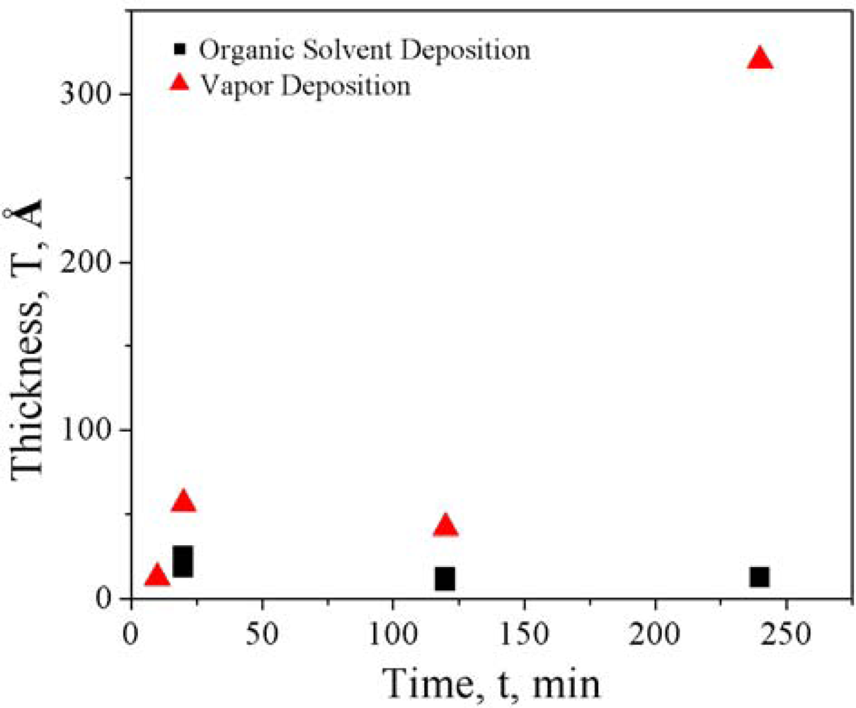
| O2 Plasma Etch | Piranha Etch | |
|---|---|---|
| Organic Solvent Deposition |
|
|
| Vapor Deposition |
|
|
© 2010 by the authors; licensee MDPI, Basel, Switzerland. This article is an open access article distributed under the terms and conditions of the Creative Commons Attribution license (http://creativecommons.org/licenses/by/3.0/).
Share and Cite
Hunt, H.K.; Soteropulos, C.; Armani, A.M. Bioconjugation Strategies for Microtoroidal Optical Resonators. Sensors 2010, 10, 9317-9336. https://doi.org/10.3390/s101009317
Hunt HK, Soteropulos C, Armani AM. Bioconjugation Strategies for Microtoroidal Optical Resonators. Sensors. 2010; 10(10):9317-9336. https://doi.org/10.3390/s101009317
Chicago/Turabian StyleHunt, Heather K., Carol Soteropulos, and Andrea M. Armani. 2010. "Bioconjugation Strategies for Microtoroidal Optical Resonators" Sensors 10, no. 10: 9317-9336. https://doi.org/10.3390/s101009317