Article

Development of an Efficient Electroporation Method for Iturin A-Producing Bacillus subtilis ZK

1
Key Laboratory of Environmental and Applied Microbiology, Chengdu Institute of Biology, the Chinese Academy of Sciences, No. 9 Section 4, Renmin Nan Road, Chengdu 610041, China
2
University of the Chinese Academy of Sciences, No. 19A Yuquan Road, Beijing 100049, China
*
Author to whom correspondence should be addressed.
Int. J. Mol. Sci. 2015, 16(4), 7334-7351; https://doi.org/10.3390/ijms16047334
Submission received: 19 January 2015 / Revised: 9 February 2015 / Accepted: 11 February 2015 / Published: 1 April 2015
(This article belongs to the Section Biochemistry)

Abstract

:
In order to efficiently introduce DNA into B. subtilis ZK, which produces iturin A at a high level, we optimized seven electroporation conditions and explored an efficient electroporation method. Using the optimal conditions, the electroporation efficiency was improved to 1.03 × 10⁷ transformants/μg of DNA, an approximately 10,000-fold increase in electroporation efficiency. This efficiency is the highest electroporation efficiency for B. subtilis and enables the construction of a directed evolution library or the knockout of a gene in B. subtilis ZK for molecular genetics studies. In the optimization process, the combined effects of three types of wall-weakening agents were evaluated using a response surface methodology (RSM) design, which led to a two orders of magnitude increase in electroporation efficiency. To the best of our limited knowledge, this study provides the first demonstration of using an RSM design for optimization of the electroporation conditions for B. subtilis. To validate the electroporation efficiency, a case study was performed and a gene (rapC) was inactivated in B. subtilis ZK using a suicide plasmid pMUTIN4. Moreover, we found that the rapC mutants exhibited a marked decrease in iturin A production, suggesting that the rapC gene was closely related to the iturin A production.

1. Introduction

B. subtilis, a type of Gram-positive bacterium, has been used as a cell factory for many enzymes, including proteases, amylases, and lipases [1,2,3]. Some subspecies produce antifungal lipopeptides (surfactin, iturin, and fengycin) that are widely applied for the control of plant pathogenic fungi in agriculture. B. subtilis ZK, a wild-type B. subtilis with high economic value, has been a workhorse for producing iturin A at a high level [4]. In order to study the high yield mechanism and further improve the iturin A production, it is desirable to perform molecular genetics studies in B. subtilis ZK. However, the fact that the genetic manipulation systems of B. subtilis ZK, such as the transformation system, gene deletion and mutation library, have not been established retards molecular genetic studies of this organism. The present work aimed at developing an efficient transformation method for the B. subtilis ZK in order to carry out molecular genetics studies.
There are five transformation systems available that have been developed to introduce exogenous DNA into B. subtilis, such as phage transduction [5], protoplast [6], natural transformation [7], recombinant method [8], and electroporation [9]. The first two methods are important methods for introducing DNA into Bacillus [10]. Unfortunately, these two methods have not been wildly used mainly due to heavy workload and time-consuming experimental processes [11]. Natural transformation and recombinant methods are usually used for introducing DNA into B. subtilis and lead to high transformation efficiency for some B. subtilis [7,8]. However, the transformation efficiency of these two methods is closely related to some genes, such as quorum sensing components, and competence master regulator comK. The divergent structures of those genes lead to a huge difference in the transformation efficiency [12,13]. Hence, the natural transformation and recombinant method are strain-specific. In contrast to other methods, electroporation is a common, time-saving transformation method and has been widely used for Bacillus [9,10,11,14,15]. Although the electroporation system for B. subtilis has been developed for many years, there is still no standard and universal protocol for all B. subtilis. We carried out an efficient protocol according to the reported literature [9]. Unfortunately, relatively low efficiency was obtained (about 1.0 × 10³ transformants/μg DNA) in wild-type B. subtilis ZK and the knockout of a gene was not to be performed, because the electroporation efficiency (1.0 × 10³ transformants/μg DNA) was insufficient and the frequency of a foreign plasmid integrating into genome was about 1.0 × 10⁴–1.0 × 10⁻⁶ per plasmid when the homologous region was less than 1 kb [16,17,18,19].
Many factors affect electroporation efficiency including growth medium [
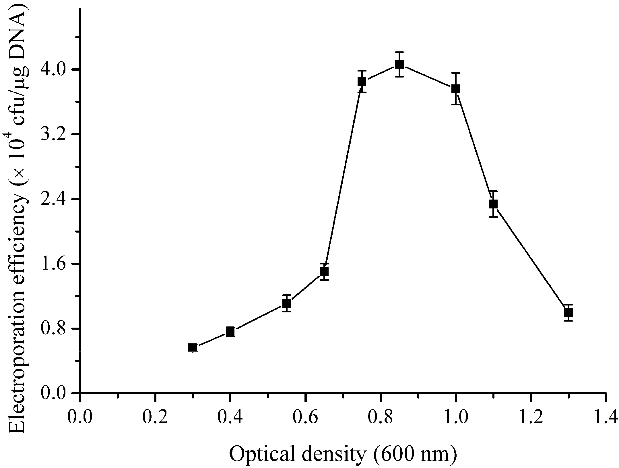
11], growth phase [14], electric field [20], weakening agent [
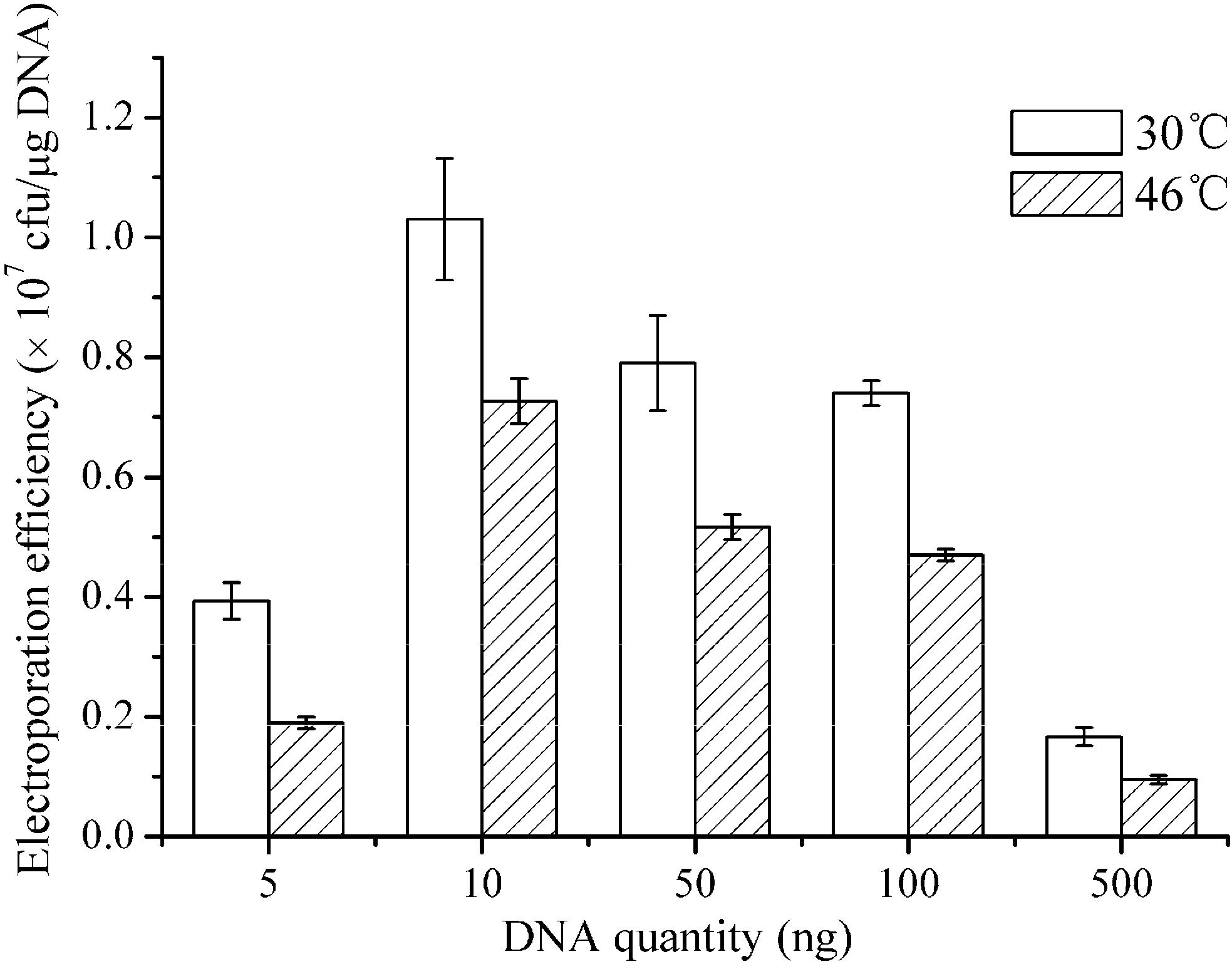
21], plasmid quantity [22], plasmid desalting [23], electroporation buffer [20], and heat treatment [24]. Some factors have combined effects on electroporation efficiency [19,25]. Hence, it is desirable to apply the multifactorial experimental design to investigate the combined effects of the conditions in order to improve electroporation efficiency. Response surface methodology (RSM) is a type of simple, commonly used multifactorial experimental design methodology used for the optimization of a small number of factors. It not only reflects the combined effects of the studied factors, but also allows completion of the optimization through a small number of experiments. To date, RSM design has been widely applied for the optimization of production conditions and media in industrial formulations [26,27] and has been rarely used to optimize electroporation conditions.
In the present study, the effects of seven factors on electroporation were investigated, including the growth media, growth phase, electric field, concentration of weakening agent, electroporation buffer, plasmid quantity, as well as heat treatment. In addition, RSM design was applied to investigate the combined effects of wall-weakening agents on the electroporation efficiency. We ultimately developed an efficient electroporation method for B. subtilis ZK that yielded a high efficiency (1.03 × 10⁷ transformants/μg of DNA), which would enable the construction of a large mutant library or the knockout of a gene in wild-type B. subtilis ZK. In order to validate the electroporation efficiency, a gene (rapC) was successfully inactivated using the suicide plasmid pMUTIN4 with the established electroporation method.

2. Results and Discussion

2.1. Screening of the Growth Medium for Electro-Competent Cells

An appropriate growth medium is an important factor for determining the electroporation efficiency of bacteria [11,28,29]. In the selected optimal growth medium, four types of growth medium (LBS, LBSP, NCM and BHIS) were investigated to prepare the electro-competent cells of B. subtilis ZK. These mediums have been used as the growth mediums for preparing electro-competent in Gram-positive strains [9,11,28]. The first three mediums are suitable for preparing electro-competent cells of B. amyloliquefaciens. Nutrient BHIS medium is regarded as the proper medium for the Gram-positve Corynebacterium glutamicum [28]. Table 1 shows that the LBSP medium yielded a higher efficiency (1.3 × 10⁴ transformants/μg of DNA) than the LBS, NCM, and BHIS media. Consequently, the LBSP medium was proved to be the appropriate medium for B. subtilis ZK and used to prepare the electro-competent cells in the subsequent experiments.
Table 1. Effect of growth media on the electroporation efficiency of B. subtilis ZK.
MediaEE aReference
LBS4 × 103 ± 0.62 × 10³[9]
LBSP1.3 × 104 ± 0.17 × 10⁴[29]
NCM7 × 102 ± 0.39 × 10²[30]
BHIS1.6 × 102 ± 0.82 × 10²[31]
When the OD600 value reached 0.65, the electro-transformed competent cells were prepared. Then, one hundred nanograms of pHT43 plasmid were used for each electroporation experiment. The field strength was 20 kV∙cm−1 and the electroporation buffer was TSM. a EE, electroporation efficiency (cfu/μg of DNA).
In the previous report, NCM medium yielded higher electroporation efficiency than LBSP medium in B. amyloliquefaciens [11]. However, the opposite result was found in the B. subtilis ZK and we found some lysis of B. subtilis ZK in the NCM medium, which indicated that the ion concentration in NCM may be too high for B. subtilis ZK.

2.2. Effect of Growth Phase on the Electroporation Efficiency

Growth phase markedly affects the electroporation efficiency [15,32]. To investigate the effect of growth phase on the electroporation efficiency, electro-competent cells were prepared at different OD600 values (0.3–1.3). Figure 1 presents the relationship between the electroporation efficiency and the growth phase. The results show that proper growth phase (OD600 0.7–1) yielded higher electroporation efficiency and an efficiency of 4 × 10⁴ transformants/μg of DNA was obtained when the OD600 value reached 0.85. In contrast with results of B. cereus [15] and B. thuringiensis [32], where the early phase of exponential growth (OD600 0.1–0.4) yielded the highest electroporation efficiency, our results indicated that the late phase of exponential growth (OD600 0.7–1) yielded the highest electroporation efficiency in B. subtilis ZK. Similar results have been observed in B. subtilis IH6140 and B. amyloliqueficence [9], as well as C. pseudotuberculosis [22] in which the late phase of exponential growth was required to obtain the highest electroporation efficiency.
Figure 1. Effects of growth phase of B. subtilis ZK on the electroporation efficiency. B. subtilis ZK was grown in LBSP medium and the electro-competent cells were prepared at different OD600 values (0.3–1.3). One hundred nanograms of the pHT43 plasmid were used for each electroporation experiment. The field strength was 20 kV·cm−1 and the electroporation buffer was TSM. The experiments were repeated three times. The error bars indicate the standard deviations from the average values.
Ijms 16 07334 g001

2.3. Effect of Electric Field on the Electroporation Efficiency

Applying high electric field is an approach to overcome the wall of B. subtilis, which improves the permeabilization of membrane and produces Bacillus cells accessible by exogenous plasmid [11]. However, an exorbitant electric field results in death of cells. To optimize the electric field for B. subtilis ZK, electroporation experiments were performed under a gradient of field strength (8–26 kV·cm−1). The relationship between the field strength and the electroporation efficiency is shown in Figure 2. An electric field of 20 kV·cm−1 led to the optimal efficiency (4 × 10⁴ transformants/μg of DNA), and this value was thus used in the follow-up experiments. The electric fields at around 5–10 kV·cm−1 are appropriate conditions for most bacterial cells and can produce the pores accessible by plasmid [10]. In the present study, we found that a high electric field (20 kV·cm−1) yielded the highest electroporation efficiency in B. subtilis ZK. Similarly high electric field (18–20 kV·cm−1) has been observed in B. amyloliqueficence [11], B. cereus [15], B. licheniformis [9], and B. thuringiensis [14]. These results indicated that the high electric field was required to achieve the high electroporation efficiency in Bacillus strains.
Figure 2. Effects of the field strength on the electroporation efficiency of B. subtilis ZK. B. subtilis ZK was grown in LBSP medium and the electro-competent cells were prepared when the OD600 value reached 0.85. The electroporation experiments were performed under a gradient of the field strength (8–26 kV·cm−1). One hundred nanograms of pHT43 plasmid were used for each electroporation experiment, and the electroporation buffer was TSM. The data shown are the averages of three independent experiments, and the error bars indicate the standard deviations from the average values.
Ijms 16 07334 g002

2.4. Investigation the Concentration of Wall-Weakening Agent for RSM Design

Application of weakening agents is an important means to improve the electroporation efficiency in Bacillus. Glycine, dl-threonine, Tween 80, and ampicillin are widely used to increase the electroporation efficiency in Bacillus strains. Glycine and dl-threonine can be integrated into the walls of cells and replace the alanine, deducing the peptidoglycan linkage and loosening the walls of cells [33]. The Tween 80 can increase the electroporation efficiency, likely because the Tween 80 loosens the cell walls and disturbs the cell–membrane fluidity [11].
To investigate the concentration of wall-weakening agent for RSM design, the four types of wall-weakening agents (glycine, dl-threonine, Tween 80, and ampicillin) were individually used to prepare the electro-competent cells. The results, which are exhibited in Figure 3, show the individual effect of the weakening agent on the electroporation efficiency. The four types of weakening agents all exhibited an enhancing effect on the electroporation efficiency. Specifically, 0.75% glycine, 1% dl-threonine, and 0.07% Tween 80 yielded efficiencies of 2.75 × 10⁵, 4.5 × 10⁵ and 3.4 × 10⁵ transformants/µg of DNA, respectively (Figure 3A–C). In addition, 10 µg/mL ampicillin showed best effect on the electroporation efficiency and improved the efficiency to 5 × 10⁵ transformants/µg of DNA, which is a 12.5-fold increase compared with the LBSP medium alone (Figure 3D). All these weakening agents yielded significant increases in electroporation efficiency (one order of magnitude) and the order in which the weakening agents contributed to electroporation efficiency was ampicillin > dl-threonine > Tween 80 > glycine. In contrast with the result of B. cereus [15], where the weakening agents (glycine, dl-threonine) had a negative effect on the electroporation efficiency, our results indicated that the wall-weakening agents played a crucial role in improving electroporation efficiency for B. subtilis ZK. A similar positive effect has been found in B. amyloliquefaciens [11], Gordonia [34], B. thuringiensis [14], and C. pseudotuberculosis [22], in which a certain ratio of weakening agents was required to obtain high electroporation efficiency.
Figure 3. Effects of wall-weakening agents on the electroporation efficiency of B. subtilis ZK. Various weakening agents at different concentration gradients (0.25%–0.85% glycine (A); 0.63%–1.56% dl-threonine (B); 15–110 mg/mL Tween 80 (C) and 1–20 μg/mL ampicillin (D)) were separately added to the LBSP medium when the OD600 value reached 0.5. After shaking for an additional 1 h, the electro-competent cells were prepared. One hundred nanograms of pHT43 were used for each electroporation experiment. The field strength was 20 kV·cm−1 and the electroporation buffer was TSM. The values shown are the averages from three independent experiments and the error bars indicate the standard deviations from the average values.
Ijms 16 07334 g003

2.5. RSM Design for Investigating the Combination of Wall-Weakening Agents

Operating parameters have combined effect on the electroporation efficiency [19,25]. To evaluate the combinatorial effects of wall-weakening agents on the electroporation efficiency, a Box-Behnken response surface design (BBD) including 17 trials was used. The BBD experimental design with the predicted and observed electroporation efficiencies are shown in Table 2. The observed electroporation efficiency was regressed to obtain a mathematical model shown in Equation (1). Although ampicillin yielded a 12.5-fold increase in above experiments, it did not exhibit a combined effect with other wall-weakening agents and reduced the electroporation efficiency in combinatorial experiments. Therefore, ampicillin was not used in the combinatorial experiments.
Y = 7.3 × 10⁶ − 6.505 × 10⁵·X1 + 5.633 × 10⁵·X2 + 7.764 × 10⁵·X3 + 5.75 × 10⁵·X1X2 + 8.141 × 10⁵·X2X3 + 8.154 × 10⁵·X1X3 − 2.668 × 10⁶·X12 − 1.144 × 10⁷·X2 − 3.976 × 10⁶·X32
Table 2. Box-Behnken response surface (BBD) design with the predicted and observed electroporation efficiencies.
No.VariablesElectroporation Efficiency (cfu/μg of DNA)
X1X2X3ObservedPredicted
1−1−102.1 × 1062.13 × 106
20007.28 × 1067.28 × 106
30007.2 × 1067.28 × 106
40−1−12.11 × 1062.15 × 106
50007.3 × 1067.28 × 106
61−107.1 × 1056.18 × 105
70007.28 × 1067.28 × 106
8−1106.35 × 1057.14 × 105
9−1011.32 × 1061.27 × 106
100−115.28 × 1055.51 × 105
11−10−13.6 × 1063.54 × 106
120115.3 × 1054.92 × 105
1301−15.8 × 1055.59 × 105
1410−11.11 × 1061.15 × 106
150007.31 × 1067.28 × 106
161011.53 × 1061.6 × 106
171105.14 × 1054.94 × 105
When the OD600 value reached 0.5, the combined weakening agents were added to the LBSP medium. After shaking for 1 h, the electro-competent competent cells were prepared. Then, one hundred nanograms of pHT43 plasmid were used for each electroporation experiment. The field strength was 20 kV∙cm−1 and the electroporation buffer was TSM. X1: code of glycine, X2: code of dl-threonine, X3: code of Tween 80.
A Pareto analysis of variance (ANOVA) was applied to evaluate the adjusted determination coefficient (R2Adj) and the coefficient of variance (CV), which represents the fitness of the quadratic polynomial and the accuracy of the quadratic polynomial, respectively. The R2Adj of the quadratic polynomial, which reached 0.997, showed good agreement between the predicted and the observed values, indicating that the mathematical model was reliable. The relatively low CV value for the quadratic polynomial (2.5%) exhibited the high precision and reliability of the conducted trials. The ANOVA of the quadratic polynomial is shown in Table 3. The Prob > F values of the coefficients are shown in Table 4 and all the coefficients were significant at the desired confidence level.
Table 3. ANOVA of the quadratic polynomial.
SourceSS aDF bMS cF Valuep-Value
Model1.37 × 101491.522 × 10132633.10<0.0001
Residual4.046 × 101075.78 × 109--
Lack of fit3.233 × 101031.078 × 10105.30.0705
Pure error8.133 × 10942.03 × 109--
Total1.37 × 101416---
a SS, sum of squares; b DF, degree of freedom; c MS, mean square.
Table 4. Significance of the regression coefficients.
FactorCE aSE bp-Value
Intercept7.3 × 1063.42 × 104<0.0001
X1−6.505 × 1052.84 × 1040.0003
X25.633 × 1055.35 × 1040.0016
X37.764 × 1053.62 × 1040.0044
X1X25.75 × 1056.74 × 1040.007
X1X38.154 × 1054.56 × 1040.042
X2X38.14 × 1058.08 × 1040.034
X12−2.668 × 1063.705 × 104<0.0001
X22−1.144 × 1071.19 × 105<0.0001
X32−3.98 × 1065.37 × 104<0.0001
a CE, coefficient estimate; b SE, standard error.
Using the optimal formulation (0.64% glycine, 1.02% dl-threonine, and 0.05% Tween 80) determined by the BBD design, an average efficiency of 6.3 × 10⁶ transformants/μg of DNA, which was close to the predicted value (7.38 × 10⁶ transformants/μg of DNA), was archived. These results indicated that RSM is a suitable tool for optimizing the electroporation conditions and provided the first demonstration of using a response surface design to improve the electroporation efficiency in B. subtilis.
In contrast with the many experimental designs for wall-weakening agents treatment [14,15,22,32], where the monofactorial experimental designs were used to optimize the concentration of weakening agent and small increases (less than one order of magnitude) in electroporation efficiency were obtained, our results indicated that weakening agents can yield higher increase (two orders of magnitude) with a multifactorial experimental design (RSM).

2.6. Effect of Electroporation Buffer on the Electroporation Efficiency

The composition of the electroporation buffer is closely related to the electroporation efficiency. Under the above-described optimized conditions, five types of buffers (MSG, MKK, SMKK, TSM, and TSMKK) were investigated. The first four buffers have been reported as the electroporation buffers for preparing electro-competent in Bacillus [9,14,35]. The results, which are listed in Table 5, show that the electroporation buffers supplemented with trehalose have positive effect on the electroporation efficiency and indicate that the TSMMKK buffer is a better electroporation buffer than other tested buffers for B. subtilis ZK. In contrast with results of other wild-type B. subtilis [20], where an electroporation buffer supplemented with trehalose (TSM) did not affect the electroporation efficiency, our results indicated that trehalose buffer is suitable for the wild-type B. subtilis ZK. The similar positive effects have been found in the B. subtilis DB104 and B. subtilis WB600 [20]. To the best of our knowledge, this is first study applying the TSMMKK buffer that found improved electroporation efficiency.
Table 5. Effects of electroporation buffers on the electroporation efficiency.
BufferEE aReference
MSG2.86 ± 0.21 × 10⁵[9]
MKK1.02 ± 0.19 × 10⁵[11]
SMKK4.73 ± 0.41 × 10⁵[14]
TSM6.3 ± 0.17 × 10⁶[35]
TSMMKK7.4 ± 0.31 × 10⁶This study
When the OD600 value reached 0.5, 0.64% Glycine, 1.02% dl-threonine, and 0.05% Tween 80 were added to the growth medium (LBSP). After shaking for 1 h, the electro-competent cells were prepared. Then, one hundred nanograms of pHT43 plasmid were used for each electroporation experiment. The field strength was 20 kV·cm−1. a EE, electroporation efficiency (cfu/μg of DNA).
In the present study, we found that freezing did not obviously decrease the electroporation efficiency of electro-competent cells within one month (data not shown). Since we did not add any glycerol or other cryoprotectants to store electro-competent cells at −80 °C, we suggest that the trehalose may act as cryoprotectants for electro-competent cells stored at −80 °C. Therefore, the TSMMKK buffer is a good option for improving electroporation efficiency and using the electro-competent cells within the following month.

2.7. The Effect of Heat Shock and Plasmid Quantity on the Electroporation Efficiency

Restriction-modification system is an obstacle of introducing the plasmid into Gram-positive strains. The heat shock is usually applied to reduce the activity of a host restriction-modification system and to improve transformation efficiency [24,36,37,38]. To determine whether the heat shock would improve the electroporation efficiency, a water bath suitable for Gram-positive strains was performed after electroporation according to previous reports [11,24]. In addition, different quantities of the plasmids (5–500 ng) were used to investigate the effect of plasmid quantity on the electroporation efficiency. The results exhibited in Figure 4 show that a water bath at 46 °C decreased the electroporation efficiency of B. subtilis ZK and the 10 nanograms yielded higher efficiency than other quantities. In contrast with results of C. pseudotuberculosis [22], C. glutamicum [24], and B. amyloliquefaciens [11], where a water bath at 46 °C after electroporation yielded a significant increase in transformation efficiency, our results indicated that heat treatment did not work for B. subtilis ZK and decreased the electroporation efficiency.
Figure 4. Plots of the electroporation efficiency as a function of the DNA quantity. When the OD600 value reached 0.5, the weakening agents (0.64% Glycine, 1.02% dl-threonine, and 0.05% Tween 80) were added to the LBSP medium and the electro-competent competent cells were prepared. B. subtilis ZK cells were transformed with various quantities of the plasmid DNA (5–500 ng). The field strength was 20 kV·cm−1 and the electroporation buffer was TSMMKK. After electroporation, the cells were incubated in a water bath at 30 or 46 °C. The electroporation experiments were repeated three times, and the data shown are the means of triplicate experiments. The error bars indicate the standard deviations from the average values.
Ijms 16 07334 g004

2.8. Validation of Electroporation Efficiency: A Case Study

Using the optimized conditions (Table 6), the electroporation efficiency was improved to 1.03 × 10⁷ transformants/μg of DNA. To validate the electroporation efficiency, we performed a case study.
Table 6. Optimal electroporation condition for B. subtilis ZK.
Optimized FactorsOptimal Condition
Growth mediumLBPS
Optical density (600 nm)0.85
Weakening-agent formulation0.64% Glycine, 1.02% dl-threonine, 0.05% Tween 80
Time for weakening treatment1 h
Electroporation bufferTSMMKK
Plasmid amount10 ng
Electric field20 kV·cm−1
Time constant4 ms
Recovery mediumLBMS
Water bathA water bath at 30 °C for 5 min
Recovery time 3 h
The suicide pMUTIN4 was used to obtain insertional mutation via a single crossing-event in B. subtilis [39]. We used the pMUTIN4 to inactivate a gene (rapC). The rapC regulates the transcription of srfA [40] to influence the expression of lipopeptide antibiotics gene (surfactin) [41]. The gene may be involved in regulation of iturin A biosynthesis. A middle fragment of phosphatase gene (rapC) was cloned to pMUTIN4 plasmid and the recombinant plasmid was used to inactivate the rapC. Seventy nine transformants were obtained, four transformants of which were randomly selected and verified by PCR. As shown in Figure 5, the mutants have 1069 bp fragment with the P3 and P4 as the primers, indicating that rapC genes were inserted by the pMUTIN4 and that the optimized method provided enough efficiency for further genetics studies.
When compared to the wild-type B. subtilis ZK, the rapC mutants exhibited markedly decrease (more than half) in iturin A production by shake flask fermentation in LB medium (Figure 6), suggesting that the rapC gene was closely related to the iturin A biosynthesis. This study provides the first demonstration of relationship between rapC and iturin A. However, further studies were required to confirm the relationship between rapC and iturin A production.
Figure 5. Inactivation of rapC gene by gene insertional mutation. (A) Construction of insertional mutation of the rapC gene; (B) Lane 1: middle encoding region of rapC (595 bp) with the P1 and P2 as the primers (C) Verification of the single crossover mutants by PCR. Lane 14: fragments (1069 bp) with the P3 and P4 as the primers; M: marker.
Ijms 16 07334 g005
Figure 6. HPLC analysis of the fermentation of wild-type strain and mutant. (A) Absorption peak of the iturin A of the wild-type B. subtilis ZK; (B) Absorption peaks of an iturin A standard sample; (C) Absorption peak of the iturin A of the mutant; and (D) The yields of iturin A of wild-type strain, mutant, and complementary mutant. The complementary mutant was grown in LB medium in presence of IPTG (0.3 mM).
Ijms 16 07334 g006aIjms 16 07334 g006b

3. Experimental Section

3.1. Chemicals

All of chemicals used in this study were purchased from Sangon Biotech (Shanghai, China), Tiangen (Beijing, China) or cdboruike (Chengdu, China).

3.2. Preparation of Desalted Plasmid for Electroporation

Escherichia coli JM110 carrying pHT43 (8057 bp; cat; MoBitech GmbH, Goettingen, Germany) was grown in LB (5 g of yeast extract, 5 g of NaCl, and 10 g of tryptone per litre) with 100 μg·mL−1 of ampicillin (Amp) at 37 °C, and 180 rpm for 18 h. The pHT43 plasmid was extracted from E. coli JM110 using the Plasmid Mini Kit (OMEGA Bio-tek, Norcross, GA, USA) and was desalted as described in the literatures [14,23]. Plasmid quantification was performed using a spectrophotometer (Nanodrop 2000C, Thermo, Wilmington, MA, USA). After purification, the plasmid was dissolved in ddH2O and stored at −20 °C.

3.3. Screening of the Growth Media for Electro-Competent Cells

A single clone of B. subtilis ZK was grown in 3 mL of LB at 30 °C and 150 rpm for 18 h. Then, 2.6 mL of the cultures were respectively added to 40 mL of the growth medium, including LBS (0.5 M sorbitol, 10 g of NaCl, 10 g of tryptone, and 5 g of yeast extract per litre of water), LBSP (0.5 M sorbitol, 0.05 M KH2PO4, 0.05 M K2HPO4, 10 g of NaCl, 10 g of tryptone, and 5 g of yeast extract per litre of water), NCM (0.2 M NaCl, 5 g of glucose, 0.1 M K2HPO4, 5 g of tryptone, 0.5 M sorbitol, 10 mM sodium citrate, 1 g of yeast extract, and 0.2 mM MgSO4∙7H2O per litre of water), and BHIS (91.1 g of sorbitol, 34 g of Brain Heart Infusion (Difco, Detroit, MI, USA) per litre of water).

3.4. The Individual Treatment of Wall-Weakening Agents

A 20% (w/v) glycine solution and a 5% Tween 80 (w/v) solution were individually sterilized at 115 °C for 20 min in advance, and 12.5% dl-threonine solution (w/v) and 50 mg/mL ampicillin were filtered for sterilization. B. subtilis ZK was grown in 40 mL of growth medium. When the optical density at 600 nm (OD600), as monitored using an U-1800 spectrophotometer (Hitachi, Tokyo, Japan) reached 0.5, the glycine, Tween 80, ampicillin, and dl-threonine solutions were added to the growth medium. After shaking at 37 °C and 200 rpm for 1 h, the electro-competent cells were prepared.

3.5. A Combinatorial Treatment of Wall-Weakening Agents

To optimize the concentrations of X1 (glycine), X2 (dl-threonine), and X3 (Tween 80) on the electroporation efficiency (Y), a Box-Behnken response surface design (BBD) were performed. A three-variable three-level design was employed, and the values of the variables were selected according to the independent wall-weakening treatment. The coded and un-coded values of the variables are listed in Table 7. The relationship between the three variables and the electroporation efficiency is expressed by the quadratic polynomial shown in Equation (2):
Y = β 0 + i = 1 m β i X i + i = 1 m β i X i 2 + i < j β i j X i X j
where Y is the predicted response (transformants/μg of DNA). Xi and Xj are variables. β0, βi, βj, and βij are constant, linear, square, and interaction coefficients respectively, and m is the number of variable; The BBD design, analysis of variance (ANOVA) and coefficients of the equation were calculated using the Design expert 8.05b software (Stat-Ease Inc., Minneapolis, MN, USA).
Table 7. Coded and un-coded values of the variables.
Variables (%)Un-CodedCoded
Gly concentration 0.55−1
0.650
0.751
dl-Thr concentration0.6−1
10
1.51
Tween 80 concentration0.02−1
0.060
0.11

3.6. Screening of the Electroporation Buffer and Preparation of Electro-Competent Cells

The cells in growth medium were chilled on ice for 10 min and harvested by centrifugation at 10,000× g and 4 °C for 5 min. The cells were purified four times using different buffers, including ice-cold MSG buffer (0.5 M sorbitol, 0.5 M mannitol and 10% glycerol), MKK buffer (0.5 mM MgCl2, 0.25 mM K2HPO4 and 0.25 mM KH2PO4 per litre of water, pH 7.2), SMKK buffer (272 mM sucrose, 0.5 mM MgCl2, 0.5 mM K2HPO4 and 0.5 mM KH2PO4 per litre of water, pH7.2), TSM buffer (0.5 M trehalose, 0.5 M sorbitol, and 0.5 M mannitol per litre of water), and TSMMKK buffer (0.5 M trehalose, 0.5 M sorbitol, 0.5 M mannitol, 0.5 mM MgCl2, 0.5 mM K2HPO4 and 0.5 mM KH2PO4 per litre of water, pH 7.2). After suspension in 1 mL of electroporation buffer, the cells were frozen in liquid nitrogen and stored at −80 °C for later use.

3.7. Electroporation

One aliquot (60 μL) of electro-competent cells was thawed on ice and then mixed with the plasmid. After incubation on ice for 3 min, the mixture was loaded into a prechilled electroporation cuvette (0.1-cm electrode gap) and exposed to a single pulse generated by a Gene Pulser System (Bio-Rad, Hercules, CA, USA). After the electrical pulse, 1 mL of LBMS (0.5 M sorbitol, 10 g of NaCl, 10 g of tryptone, 5 g of yeast extract, 0.5 M mannitol, and 0.38 M sorbitol per litre of water) was added immediately to the mixture. After shaking at 37 °C and 200 rpm for 3 h, the cells were harvested and plated on LB agar plates with 5 μg·mL−1 chloramphenicol (Cm). The transformants were counted following overnight incubation at 37 °C without agitation.

3.8. Treatment of Heat Inactivation

After electroporation, the cells were incubated at 30 or 46 °C for 6 min as previous report [24].

3.9. Construction of Plasmids and Analysis of Mutants

A 595 bp fragment encompassing middle encoding region of rapC was amplified with primers P-1 (5'-CCCCGGAATTCGGGCTTCTCGATTATTACGTCAACT-3') (with a EcoRI site, underlined) and P-2 (5'-CCCGCGGATCCACGGGATCGTCTGTTTCTTTAGCAT-3') (with a BamHI site, underlined). The fragment was digested with EcoRI and BamHI and then cloned to pMUTIN4 plasmid. Recombinant plasmid was extracted from E. coli and transferred into B. subtilis ZK. The mutants were selected on LB plates containing 0.3 μg·mL−1 erythromycin (Em). The total DNA of mutant was extracted using the bacterial DNA Kit (OMEGA Bio-tek, Norcross, GA, USA). The inactivate mutants were verified by PCR with the specific primers P-3 (5'-ATGAATCATCTTGAAACCGGCAGTC-3') within upstream of the rapC and P-4 (5'-ATCCACGGGATCGTCTGTTTCTTTA-3') within the downstream of the multiple clone site of pMUTIN4.
A 1146 bp full encoding region of rapC was amplified with primers P-5 (5'-ACATCAGCCGTAGGATCCATGAAGAGCGGGTTAATTCCTTCTT-3') (with a BamHI site, underlined) and P-6 (5'-TAGGCGGGCTGCCCCGGGTTAGATTTCAATTTCATACAAACCC-3') (with a SmaI site, underlined). The fragment was digested with BamHI and SmaI and then cloned to pHT01 plasmid. Recombinant plasmid was extracted from E. coli and transferred into rapC mutant. The transformants were selected on LB plates containing 5 μg·mL−1 chloramphenicol (Cm) and 0.3 μg·mL−1 erythromycin (Em).

3.10. Fermentation and Measurement of Iturin A

The ZK strains were activated in LB plates at 37 °C for 36 h. The activated strains were separately inoculated in 5 mL LB mediums (250 mL Erlenmeyer flask) in a shaker at 30 °C with 150 rpm for 22 h. The seed cultures were cultivated in 40 mL LB medium by 10% amount of inoculum at 30 °C, under 150 rpm for 2 days. The extraction and measurement of iturin A as previously reported [42].

4. Conclusions

In conclusion, a detailed and systematic protocol that results in improved electroporation efficiency for wild-type B. subtilis ZK is described in this manuscript. Using this new method, the electroporation efficiency for B. subtilis ZK is improved to 1 × 10⁷ transformants/μg of DNA, which is highest level found for wild-type B. subtilis. This efficiency allows the construction of a directed evolution library and allows the knockout of a gene in wild-type B. subtilis ZK. We hope that the proposed protocol can facilitate the molecular genetics study in this economically important strain and may shed light on improvements in other wild-type B. subtlis strains. In order to validate the electroporation efficiency, a gene (rapC) was successfully inactivated using a suicide plasmid pMUTIN4 with the established electroporation method. Moreover, we found that the rapC mutants exhibited markedly decrease in iturin A production, suggesting that the rapC gene was involved in the regulation of iturin A production.

Acknowledgments

We thank Yuanpeng, Zhang (Nanjing Agricultural University, Nanjing, China) for providing plasmids pHT01, pHT04, and pMUTIN4. This work is funded by the National Key Technologies R&D Program (No. 2011BAE06B04-18).

Author Contributions

Zhi Zhang: contributed to the laboratory work, data analyses and manuscript writing; Zhong-Tao Ding, Dan Shu and Di Luo: conceived the study and commented the manuscript; Hong Tan: conceived and designed the study. All authors read and approved the final manuscript.

Conflicts of Interest

The authors declare no conflict of interest.

References

  1. Rahman, M.S.; Ano, T.; Shoda, M. Biofilm fermentation of iturin A by a recombinant strain of Bacillus subtilis 168. J. Biotechnol. 2007, 127, 503–507. [Google Scholar] [CrossRef] [PubMed]
  2. Tsuge, K.; Ohata, Y.; Shoda, M. Gene yerP, involved in surfactin self-resistance in Bacillus subtilis. Antimicrob. Agents Chemother. 2001, 45, 3566–3573. [Google Scholar] [CrossRef] [PubMed]
  3. Tsuge, K.; Inoue, S.; Ano, T.; Itaya, M.; Shoda, M. Horizontal transfer of iturin A operon, itu, to Bacillus subtilis 168 and conversion into an iturin A producer. Antimicrob. Agents Chemother. 2005, 49, 4641–4648. [Google Scholar] [CrossRef] [PubMed]
  4. Peng, W.J.; Zhong, J.; Yang, J.; Ren, Y.L.; Xu, T.; Xiao, S.; Zhou, J.Y.; Tan, H. The artificial neural network approach based on uniform design to optimize the fed-batch fermentation condition: Application to the production of iturin A. Microb. Cell Fact. 2014, 13, 54. [Google Scholar] [CrossRef] [PubMed]
  5. Young, F.E.; Yasbin, R.E. Transduction in Bacillus subtilis by bacteriophage SPP1. J. Virol. 1974, 14, 1343–1348. [Google Scholar] [PubMed]
  6. Harwood, C.R.; Cutting, S.M. Molecular Biological Methods for Bacillus; Wiley: New York, NY, USA, 1990; p. 581. [Google Scholar]
  7. Anagnostopoulos, C.; Spizizen, J. Requirements for transformation in Bacillus subtilis. J. Bacteriol. 1961, 81, 741–746. [Google Scholar] [PubMed]
  8. Zhang, X.Z.; Zhang, Y.H.P. Simple, fast and high-efficiency transformation system for directed evolution of cellulase in Bacillus subtilis. Microb. Biotechnol. 2011, 4, 98–105. [Google Scholar] [CrossRef] [PubMed]
  9. Xue, G.P.; Johnson, J.S.; Dalrymple, B.P. High osmolarity improves the electro-transformation efficiency of the Gram-positive bacteria Bacillus subtilis and Bacillus licheniformis. J. Microbiol. Methods 1999, 34, 183–191. [Google Scholar] [CrossRef]
  10. Aune, T.E.; Aachmann, F.L. Methodologies to increase the transformation efficiencies and the range of bacteria that can be transformed. Appl. Microbiol. Biotechnol. 2010, 85, 1301–1313. [Google Scholar] [CrossRef] [PubMed]
  11. Zhang, G.Q.; Bao, P.; Zhang, Y.; Deng, A.H.; Chen, N.; Wen, T.Y. Enhancing electro-transformation competency of recalcitrant Bacillus amyloliquefaciens by combining cell-wall weakening and cell-membrane fluidity disturbing. Anal. Biochem. 2011, 409, 130–137. [Google Scholar] [CrossRef] [PubMed]
  12. Tran, L.S.P.; Nagai, T.; Itoh, Y. Divergent structure of the ComQXPA quorum-sensing components: Molecular basis of strain-specific communication mechanism in Bacillus subtilis. Mol. Microbiol. 2000, 37, 1159–1171. [Google Scholar] [CrossRef] [PubMed]
  13. Vojcic, L.; Despotovic, D.; Martinez, R.; Maurer, K.H.; Schwaneberg, U. An efficient transformation method for Bacillus subtilis DB104. Appl. Microbiol. Biotechnol. 2012, 94, 487–493. [Google Scholar] [CrossRef] [PubMed]
  14. Peng, D.; Luo, Y.; Guo, S.; Zeng, H.; Ju, S.; Yu, Z.; Sun, M. Elaboration of an electroporation protocol for large plasmids and wild-type strains of Bacillus thuringiensis. J. Appl. Microbiol. 2009, 106, 1849–1858. [Google Scholar] [CrossRef] [PubMed]
  15. Turgeon, N.; Laflamme, C.; Ho, J.; Duchaine, C. Elaboration of an electroporation protocol for Bacillus cereus ATCC 14579. J. Microbiol. Methods 2006, 67, 543–548. [Google Scholar] [CrossRef] [PubMed]
  16. Biswas, I.; Gruss, A.; Ehrlich, S.D.; Maguin, E. High-efficiency gene inactivation and replacement system for Gram-positive bacteria. J. Bacteriol. 1993, 175, 3628–3635. [Google Scholar] [PubMed]
  17. Leloup, L.; Ehrlich, S.D.; Zagorec, M.; MorelDeville, F. Single-crossover integration in the Lactobacillus sake chromosome and insertional inactivation of the ptsI and lacL genes. Appl. Environ. Microbiol. 1997, 63, 2117–2123. [Google Scholar] [PubMed]
  18. Schwarzer, A.; Puhler, A. Manipulation of Corynebacterium glutamicum by gene disruption and replacement. Biotechnol. N. Y. 1991, 9, 84–87. [Google Scholar] [CrossRef]
  19. Lu, S.L.; Nie, Y.; Tang, Y.Q.; Xiong, G.M.; Wu, X.L. A critical combination of operating parameters can significantly increase the electrotransformation efficiency of a Gram-positive Dietzia strain. J. Microbiol. Methods 2014, 103, 144–151. [Google Scholar] [CrossRef] [PubMed]
  20. Lu, Y.P.; Zhang, C.; Lv, F.X.; Bie, X.M.; Lu, Z.X. Study on the electro-transformation conditions of improving transformation efficiency for Bacillus subtilis. Lett. Appl. Microbiol. 2012, 55, 9–14. [Google Scholar] [CrossRef] [PubMed]
  21. Mcdonald, I.R.; Riley, P.W.; Sharp, R.J.; Mccarthy, A.J. Factors affecting the electroporation of Bacillus subtilis. J. Appl. Bacteriol. 1995, 79, 213–218. [Google Scholar] [CrossRef] [PubMed]
  22. Dorella, F.A.; Estevam, E.M.; Cardoso, P.G.; Savassi, B.M.; Oliveira, S.C.; Azevedo, V.; Miyoshi, A. An improved protocol for electrotransformation of Corynebacterium pseudotuberculosis. Vet. Microbiol. 2006, 114, 298–303. [Google Scholar] [CrossRef] [PubMed]
  23. Schlaak, C.; Hoffmann, P.; May, K.; Weimann, A. Desalting minimal amounts of DNA for electroporation in E. coli: A comparison of different physical methods. Biotechnol. Lett. 2005, 27, 1003–1005. [Google Scholar]
  24. Van der Rest, M.E.; Lange, C.; Molenaar, D. A heat shock following electroporation induces highly efficient transformation of Corynebacterium glutamicum with xenogeneic plasmid DNA. Appl. Microbiol. Biotechnol. 1999, 52, 541–545. [Google Scholar]
  25. Marciset, O.; Mollet, B. Multifactorial Experimental-designs for optimizing transformation: Electroporation of streptococcus-thermophilus. Biotechnol. Bioeng. 1994, 43, 490–496. [Google Scholar] [CrossRef] [PubMed]
  26. Azevedo, A.N.; Buarque, P.R.; Cruz, E.M.O.; Blank, A.F.; Alves, P.B.; Nunes, M.L.; Santana, L.C.L.D. Response surface methodology for optimisation of edible chitosan coating formulations incorporating essential oil against several foodborne pathogenic bacteria. Food Control 2014, 43, 1–9. [Google Scholar] [CrossRef]
  27. Maran, J.P.; Sivakumar, V.; Thirugnanasambandham, K.; Sridhar, R. Response surface modeling and analysis of barrier and optical properties of maize starch edible films. Int. J. Biol. Macromol. 2013, 60, 412–421. [Google Scholar] [CrossRef] [PubMed]
  28. Haynes, J.A.; Britz, M.L. The Effect of Growth-conditions of Corynebacterium glutamicum on the transformation frequency obtained by electroporation. J. Gen. Microbiol. 1990, 136, 255–263. [Google Scholar] [CrossRef]
  29. Vehmaanpera, J. Transformation of Bacillus amyloliquefaciens by electroporation. FEMS Microbiol. Lett. 1989, 61, 165–169. [Google Scholar] [CrossRef]
  30. Ito, M.; Nagane, M. Improvement of the electro-transformation efficiency of facultatively alkaliphilic Bacillus pseudofirmus OF4 by high osmolarity and glycine treatment. Biosci. Biotechnol. Biochem. 2001, 65, 2773–2775. [Google Scholar] [CrossRef] [PubMed]
  31. Hu, S.B.; Liu, P.; Ding, X.Z.; Yan, L.; Sun, Y.J.; Zhang, Y.M.; Li, W.P.; Xia, L.Q. Efficient constitutive expression of chitinase in the mother cell of Bacillus thuringiensis and its potential to enhance the toxicity of Cry1Ac protoxin. Appl. Microbiol. Biotechnol. 2009, 82, 1157–1167. [Google Scholar] [CrossRef] [PubMed]
  32. Zhang, H.H.; Li, Y.T.; Chen, X.M.; Sheng, H.M.; An, L.Z. Optimization of electroporation conditions for Arthrobacter with plasmid PART2. J. Microbiol. Methods 2011, 84, 114–120. [Google Scholar] [CrossRef] [PubMed]
  33. Hammes, W.; Schleifer, K.H.; Kandler, O. Mode of action of glycine on the biosynthesis of peptidoglycan. J. Bacteriol. 1973, 116, 1029–1053. [Google Scholar] [PubMed]
  34. Singh, P.; Srivastava, P. An improved protocol for electroporation in members of the genus Gordonia. J. Microbiol. Methods 2013, 95, 114–116. [Google Scholar] [CrossRef] [PubMed]
  35. Cao, G.Q.; Zhang, X.H.; Zhong, L.; Lu, Z.X. A modified electro-transformation method for Bacillus subtilis and its application in the production of antimicrobial lipopeptides. Biotechnol. Lett. 2011, 33, 1047–1051. [Google Scholar] [CrossRef] [PubMed]
  36. Tauch, A.; Kirchner, O.; Loffler, B.; Gotker, S.; Puhler, A.; Kalinowski, J. Efficient electrotransformation of Corynebacterium diphtheriae with a mini-replicon derived from the Corynebacterium glutamicum plasmid pGA1. Curr. Microbiol. 2002, 45, 362–367. [Google Scholar] [CrossRef] [PubMed]
  37. Zakataeva, N.P.; Nikitina, O.V.; Gronskiy, S.V.; Romanenkov, D.V.; Livshits, V.A. A simple method to introduce marker-free genetic modifications into the chromosome of naturally nontransformable Bacillus amyloliquefaciens strains. Appl. Microbiol. Biotechnol. 2010, 85, 1201–1209. [Google Scholar] [CrossRef] [PubMed]
  38. Sun, F.H.; Luo, D.; Shu, D.; Zhong, J.; Tan, H. Development of an intergeneric conjugal transfer system for Xinaomycins-producing Streptomyces noursei Xinao-4. Int. J. Mol. Sci. 2014, 15, 12217–12230. [Google Scholar] [CrossRef] [PubMed]
  39. Vagner, V.; Dervyn, E.; Ehrlich, S.D. A vector for systematic gene inactivation in Bacillus subtilis. Microbiology 1998, 144, 3097–3104. [Google Scholar] [CrossRef] [PubMed]
  40. Solomon, J.M.; Lazazzera, B.A.; Grossman, A.D. Purification and characterization of an extracellular peptide factor that affects two different developmental pathways in Bacillus subtilis. Genes Dev. 1996, 10, 2014–2024. [Google Scholar] [CrossRef] [PubMed]
  41. Jung, J.; Yu, K.O.; Ramzi, A.B.; Choe, S.H.; Kim, S.W.; Han, S.O. Improvement of surfactin production in Bacillus subtilis using synthetic wastewater by overexpression of specific extracellular signaling peptides, comX and phrC. Biotechnol. Bioeng. 2012, 109, 2349–2356. [Google Scholar] [CrossRef] [PubMed]
  42. Ren, Y.L.; Zhou, J.Y.; Zhang, X.Y.; Li, Z.D.; Zhong, J.; Yang, J.; Xu, T.; Tan, H. Analysis of Free amino acids during fermentation by Bacillus subtilis using capillary electrophoresis. Biotechnol. Bioprocess E 2012, 17, 1244–1251. [Google Scholar] [CrossRef]

