Article

Ecdysone Receptor (EcR) Is Involved in the Transcription of Cell Cycle Genes in the Silkworm

State Key Laboratory of Silkworm Genome Biology, Southwest University, Chongqing 400715, China
*
Authors to whom correspondence should be addressed.
Int. J. Mol. Sci. 2015, 16(2), 3335-3349; https://doi.org/10.3390/ijms16023335
Submission received: 17 October 2014 / Revised: 6 January 2015 / Accepted: 23 January 2015 / Published: 3 February 2015
(This article belongs to the Section Biochemistry)

Abstract

:
EcR (ecdysone receptor)-mediated ecdysone signaling pathway contributes to regulate the transcription of genes involved in various processes during insect development. In this work, we detected the expression of EcR gene in silkworm ovary-derived BmN4 cells and found that EcR RNAi result in an alteration of cell shape, indicating that EcR may orchestrate cell cycle progression. EcR RNAi and EcR overexpression analysis revealed that in the cultured BmN4 cells, EcR respectively promoted and suppressed the transcription of E2F-1 and CycE, two genes controlling cell cycle progression. Further examination demonstrated that ecdysone application in BmN4 cells not only changed the transcription of these two cell cycle genes like that under EcR overexpression, but also induced cell cycle arrest at G2/M phase. In vivo analysis confirmed that E2F-1 expression was elevated in silk gland of silkworm larvae after ecdysone application, which is same as its response to ecdysone in BmN4 cells. However, ecdysone also promotes CycE transcription in silk gland, and this is converse with the observation in BmN4 cells. These results provide new insights into understanding the roles of EcR-mediated ecdysone signaling in the regulation of cell cycle.

1. Introduction

Ecdysone receptor (EcR), as a member of nuclear receptor family, was identified and well-characterized in insects and primarily contributes to meditate the signaling of steroid hormone ecdysone [1,2,3]. Generally, ecdysone directly binds to the heterodimer comprising of EcR and Ultraspiracle (USP) and subsequently activates a transcriptional response of its primary and secondary response genes [4,5]. More evidence supports that EcR-mediated ecdysone pathway is required for all aspects of insect growth and development, including developmental timing [6], embryogenesis [7], larval molting [8], degeneration, remodeling or specification of organs during metamorphosis [9], and reproduction [10].
Cell cycle-modulated cell growth and cell proliferation have been shown to connect to the growth and development of multicellular organisms including insects [11,12,13,14]. Extensive studies provided a comprehensive understanding that cell cycle is principally and directly governed by the paired members from several protein families of cyclins, cyclin-dependent kinases (CDK), and cdk inhibitors [15,16]. It is conservation among multicellular organisms that cyclin E (CycE) and its CDK partner Cdk2 are responsible for initiating the G1/S transition by promoting DNA replication [17], and CycB interacts with Cdk1 to promote the G2/M transition by triggering mitosis [18,19]. In addition, both CycE and CycB are transcriptionally regulated by E2F-1 protein [20,21].
Recently increasing reports have shown that ecdysone signal also plays roles in the regulation of cell cycle progression in insects [22,23,24,25,26]. In the fruit fly (Drosophila melanogaster), ecdysone and its receptor EcR modulate cell cycle progression in wing disc by repressing the expression of Wingless (Wg) gene [27,28]. Wingless acts to down-regulate the key cell cycle genes, such as E2F-1 and cycE [29,30]. However, to date, the mechanism of ecdysone regulation of cell cycle progression in insects is poorly understood.
From our previously obtained microarray data of gene expression in cultured silkworm (Bombyx mori) ovary-derived BmN4-SID1 cells [31], we found that EcR gene revealed a weak expression in BmN4-SID1 cells, indicating that EcR may be also involved in the regulation of the transcription of cell cycle genes in silkworm cells. Here, we performed RNA interference (RNAi)-mediated knockdown of EcR gene and ecdysone treatment in the silkworm at cellular and individual scales, and found that EcR-mediated ecdysone signaling can regulate the transcription of two cell cycle genes, E2F-1 and CycE.

2. Results

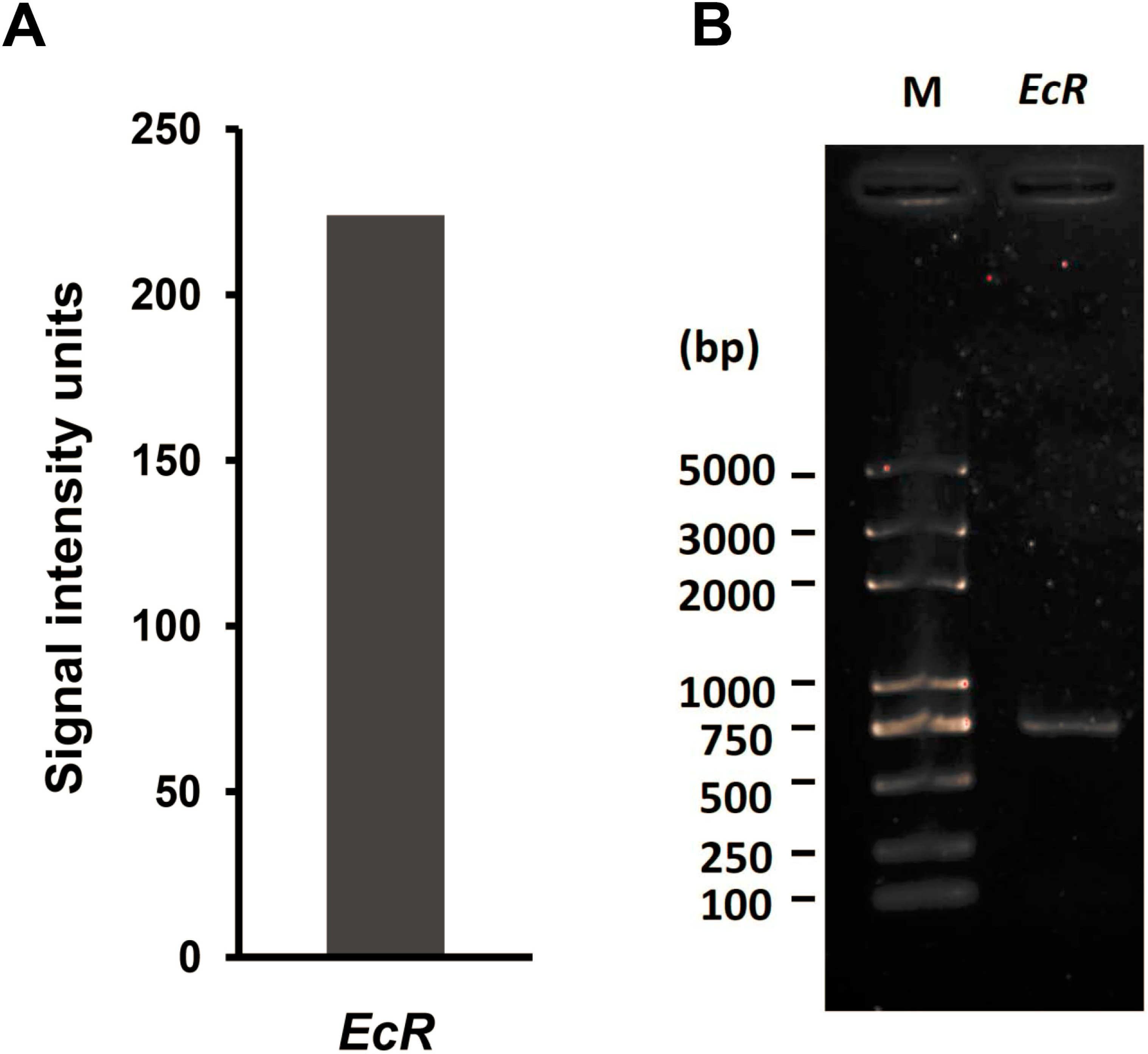
2.1. EcR Was Expressed in Silkworm BmN4 Cells

We previously profiled the genome-wide expression in cultured silkworm ovary-derived BmN4 cells by using microarray approach [31]. From the related microarray data (GSE no.: GSE34246), we observed that the expression signal intensity of two probes (sw11842 and sw11434) for silkworm EcR gene exceeded a value of 200 units (Figure 1A), suggesting that EcR gene is likely expressed in cultured BmN4 cells. Quantitative RT-PCR examination confirmed an obvious expression of EcR gene in BmN4 cells (Figure 1B). Together with the observation that EcR expression could be detected in cultured mosquito (Aedes albopictus) cells [32], we speculated that EcR may be involved in cell cycle or cell growth in cultured cells without the existence of ecdysone.
Figure 1. EcR expression in BmN4 cells. (A) Microarray data of mRNA expression of silkworm EcR gene in BmN4 cells; (B) Quantitative RT-PCR detection of mRNA expression of silkworm EcR gene in BmN4 cells. M: Molecular weight marker.
Ijms 16 03335 g001

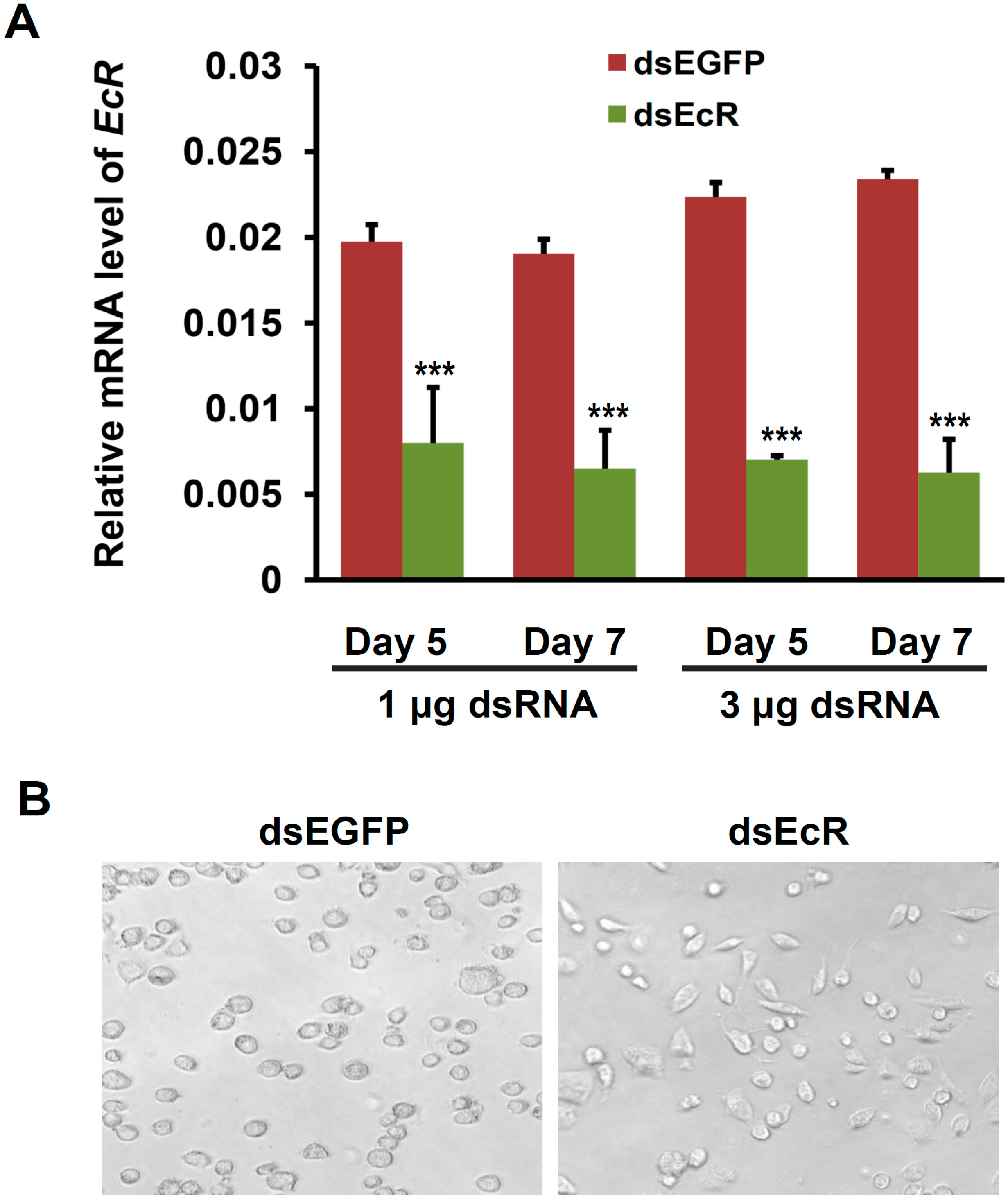
2.2. EcR RNAi Alters the Shape of Silkworm BmN4-SID1 Cells

In order to ascertain the roles of EcR in BmN4 cells, we performed a RNAi experiment of EcR gene in cultured BmN4-SID1 cells, which is established by overexpressing the SID1 gene, a gene with high efficiency in the uptake of exogenous double strand RNA (dsRNA) into host cells, in BmN4 cells [33]. The dsRNAs targeting the EcR gene and EGFP (enhanced green fluorescent protein) gene as control were separately transfected into BmN4-SID1 cells in a dosage of either 1 or 3 μg per plate well. Quantitative RT-PCR analysis showed that compared with the control of EGFP dsRNA treatment, EcR expression was remarkably silenced at both the fifth and seventh day after the treatment with EcR dsRNAs (Figure 2A). Further microscopy analysis found that the morphology of the BmN4-SID1 cells was transformed into fusiform from roundness (Figure 2B). This observation is similar to the morphological response of the fruit fly Kc Cells to ecdysone [25], indicating that cell cycle progression of the BmN4-SID1 cells was changed after EcR RNAi.
Figure 2. EcR RNAi changes the shape of BmN4-SID1 cells. (A) Quantitative RT-PCR assay of RNAi-based knockdown efficiency of EcR expression in silkworm BmN4-SID1 cells. EGFP RNAi was used as control. Error bars represents mean and S.D., *** p < 0.001, compared with the control; (B) Effects of EcR RNAi on the shape of BmN4-SID1 cells. The BmN4-SID1 cells were checked on the seventh day after the treatment with different dsRNA (dsEcR for EcR gene or dsEGFP for EGFP gene) using microscope.
Ijms 16 03335 g002

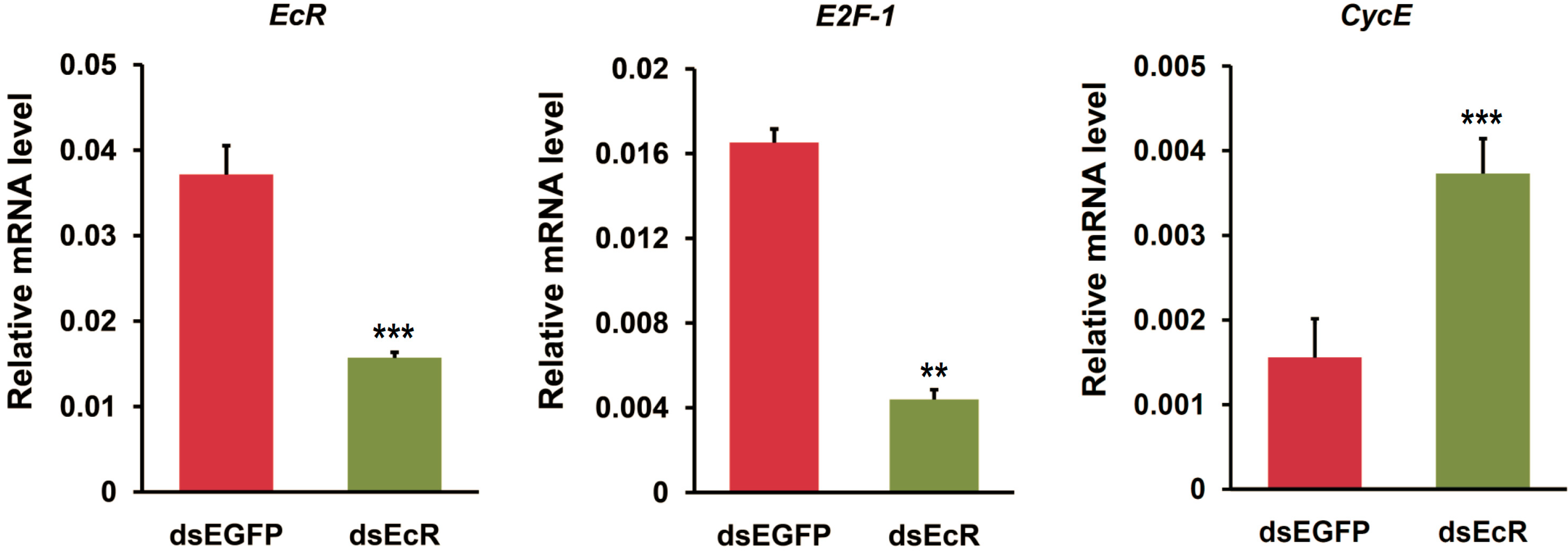
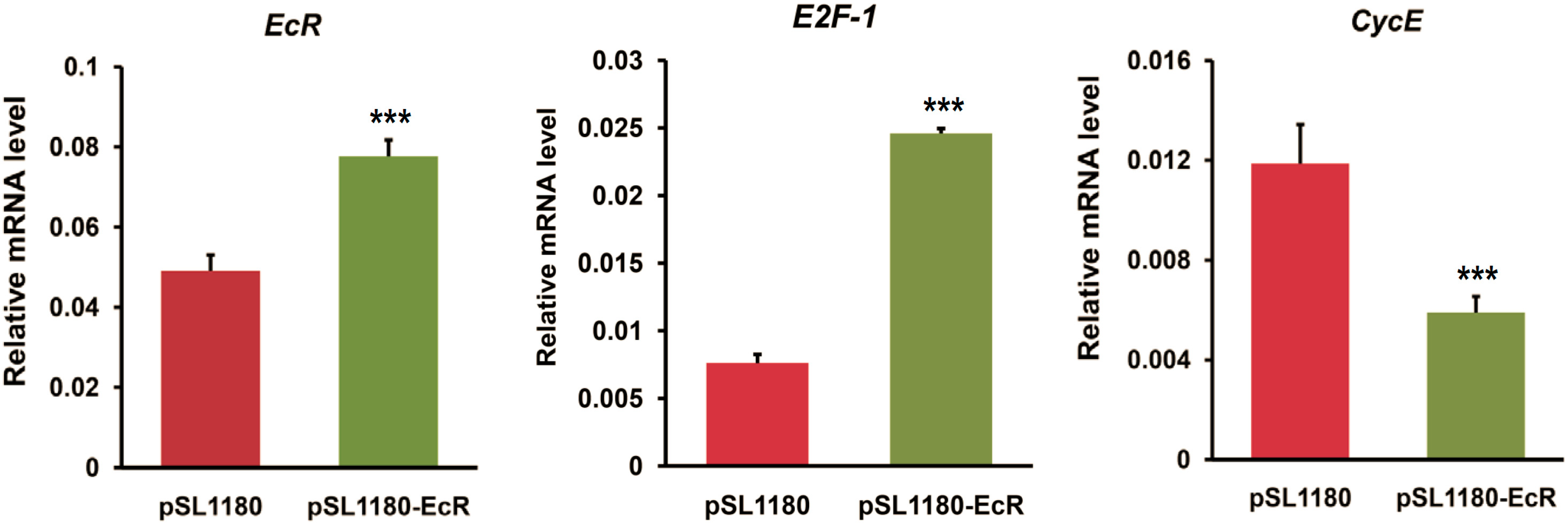
2.3. RNAi or Overexpression of EcR Gene Disrupts the Expression of Cell Cycle Genes in BmN4 Cells

Given that EcR RNAi changed the shape of BmN4 cells and may alter cell cycle progression, we proposed that EcR may be involved in regulating the expression of cell cycle genes. Here, we focused on two DNA replication-related genes, E2F-1 and CycE, and checked whether their expressions could be changed after EcR RNAi. As expected, quantitative RT-PCR examination showed that in BmN4-SID1 cells after EcR RNAi, E2F-1 and CycE were down- and up-regulated, respectively (Figure 3). This is obviously different with the previous observation in the fruit fly wing that CycE expression is positively regulated by E2F-1 [20,21].
Figure 3. EcR RNAi changes the expression of cell cycle genes in BmN4-SID1 cells. The expression changes of cell cycle genes (E2F-1 and CycE) after the RNAi of EcR and EGFP in silkworm BmN4-SID1 cells were examined using quantitative RT-PCR. Error bars represents mean and S.D., ** p < 0.01, *** p < 0.001, compared with the control.
Ijms 16 03335 g003
To further confirm the roles of EcR in the transcription of E2F-1 and CycE, we performed an overexpression of EcR gene in BmN4 cells. As shown in Figure 4, E2F-1 expression was promoted following EcR overexpression. Conversely, CycE expression was inhibited after EcR overexpression. This is contrary to the effects of EcR RNAi on the transcription of both E2F-1 and CycE.
Figure 4. EcR overexpression disrupts the expression of cell cycle genes in BmN4 cells. The expression changes of cell cycle genes (E2F-1 and CycE) after the overexpression of EcR gene in silkworm BmN4 cells were examined using quantitative RT-PCR. Expression vector excluding EcR gene was used as control. Error bars represents mean and S.D., *** p < 0.001, compared with the control.
Ijms 16 03335 g004

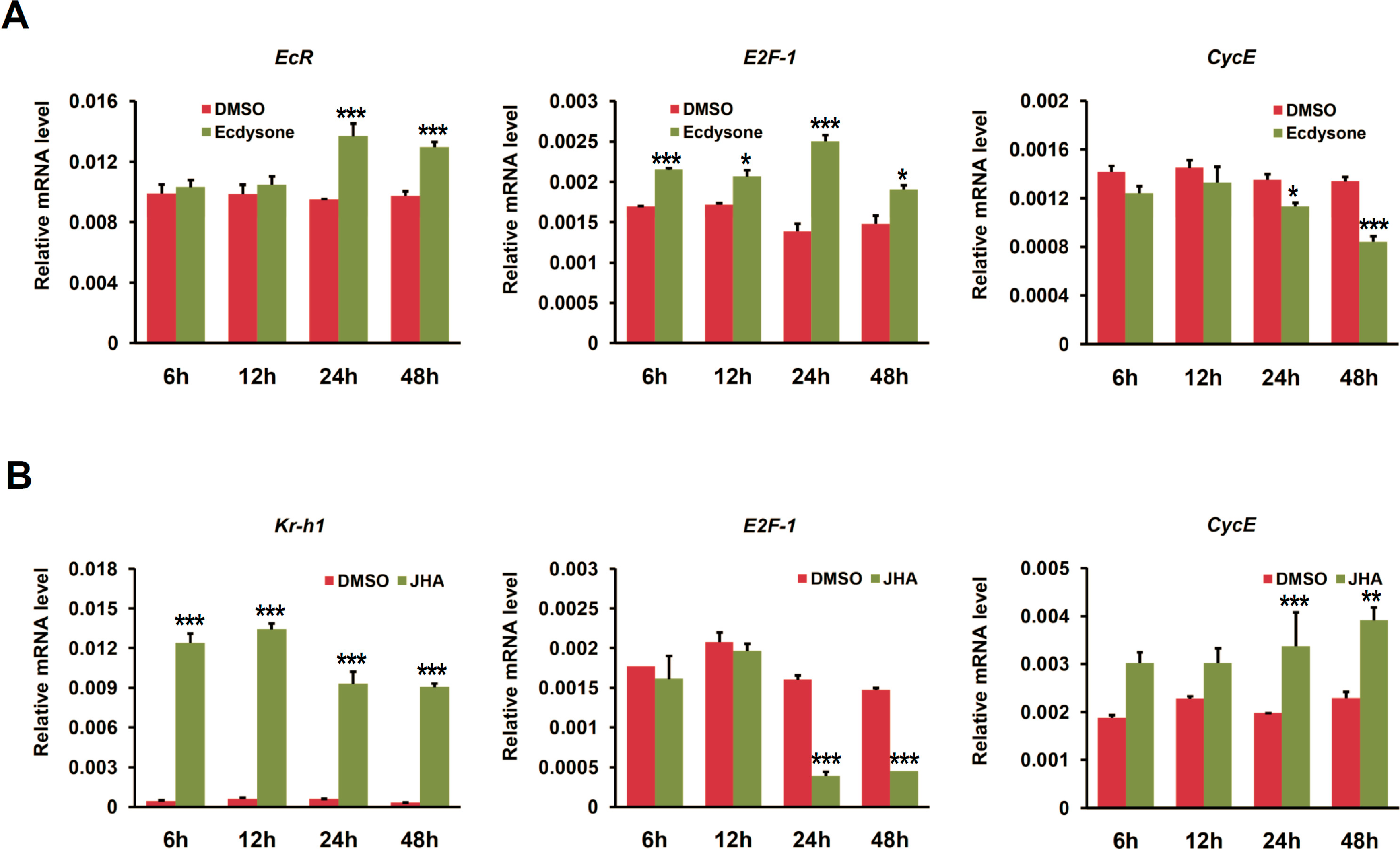
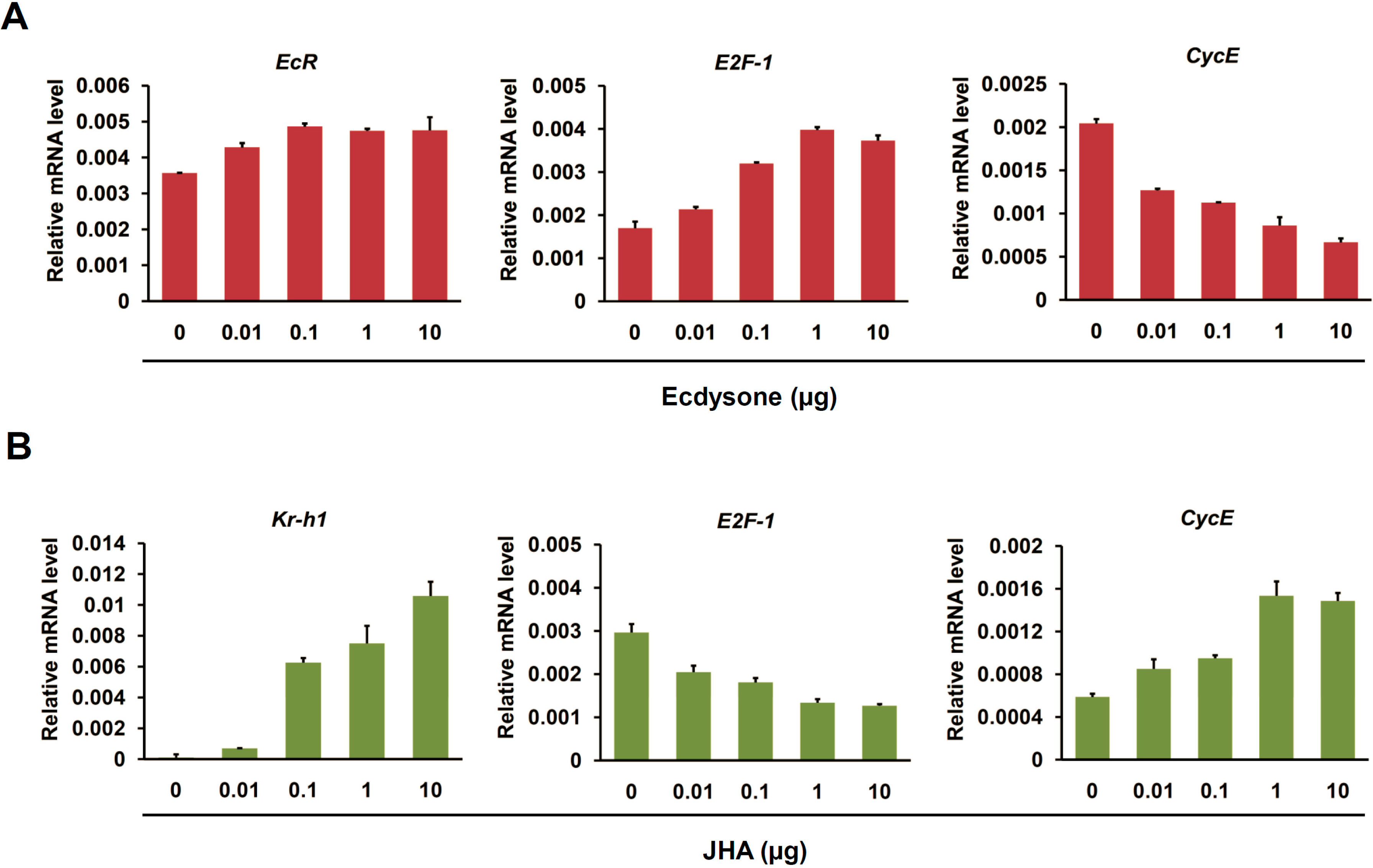
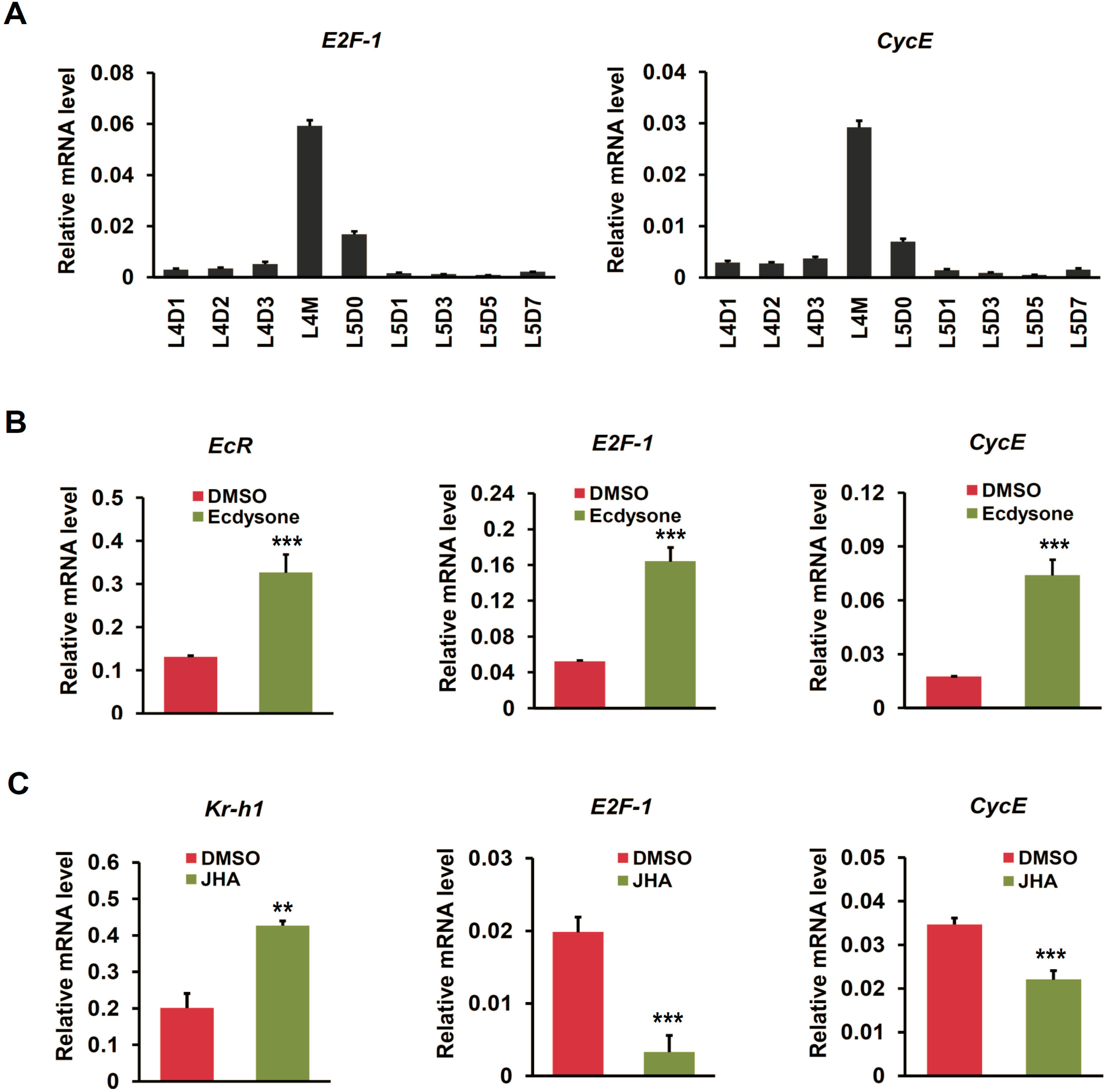
2.4. Ecdysone Treatment Changes the Expression of Cell Cycle Genes in BmN4 Cells

Given that EcR is a key receptor in mediating ecdysone signaling in insects [1], we further examined the effects of ecdysone application on the expression of cell cycle genes in BmN4 cells. We firstly checked time-dependent effects of ecdysone treatment with a dosage of 1 μg per mL per plate well on the transcription of cell cycle genes. As shown in Figure 5A, EcR expression was significantly up-regulated after 24 h with ecdysone treatment. Further examination revealed that E2F-1 expression was remarkably elevated at 6 h after ecdysone treatment and CycE was inhibited at 24 h (Figure 5A). In addition, given that the role of juvenile hormone (JH) is antagonistic to that of ecdysone in controlling developmental processes in insects [34,35], we also checked the effects of JH analog (JHA) on the expression of cell cycle genes in BmN4 cells. In our experiment, the dosage of JHA is 1 μg per plate well. Obviously, Kr-h1 gene, a key transcription factor that is activated by JH and is involved in JH signaling [36,37], was up-regulated in BmN4 cells at 6 h after JHA treatment (Figure 5B). E2F-1 and CycE were significantly down- and up-regulated after 24 h with JHA treatment, respectively, which is consistent with the observation in EcR RNAi, but converse with the effects of either EcR overexpression or ecdysone application.
Figure 5. Time-dependent expression changes of cell cycle genes induced by ecdysone and JH Analog (JHA) in BmN4 cells. (A) The expression changes of cell cycle genes (E2F-1 and CycE) at different time points, including 6, 12, 24, and 48 h, after ecdysone treatment in silkworm BmN4 cells were examined using quantitative RT-PCR. DMSO treatment was used as control. Error bars represents mean and S.D., * p < 0.05, ** p < 0.01, *** p < 0.001, compared with the control; (B) The expression changes of cell cycle genes (E2F-1 and CycE) at different time points, including 6, 12, 24, and 48 h, after JHA treatment in silkworm BmN4 cells were examined.
Ijms 16 03335 g005
We further investigated the effects of ecdysone and JHA treatment with different dosages on the transcription of cell cycle genes in BmN4 cells. As shown in Figure 6A, EcR expression was up-regulated at 24 h after ecdysone treatment with a dosage of 0.01 μg per plate well. E2F-1 and CycE was elevated and inhibited after ecdysone treatment in a dosage-dependent manner, respectively (Figure 6A). Similarly, JHA treatment resulted in an up-regulation of Kr-h1 and CycE but a down-regulation of E2F-1 at transcriptional level in a dosage-dependent manner (Figure 6B). Taken together, we proposed that EcR-mediated ecdysone signaling is transcriptionally involved in the promotion of E2F-1 and suppression of CycE in the cultured silkworm BmN4 cells.
Figure 6. Dosage-dependent expression changes of cell cycle genes induced by ecdysone in BmN4 cells. (A) Dosage-dependent expression changes of cell cycle genes (E2F-1 and CycE) after ecdysone treatment in silkworm BmN4 cells were examined using quantitative RT-PCR. Ecdysone treatment was initiated with different dosage, including 0, 0.01, 0.1, 1, and 10 µg per plate well. The cells were collected at 24 h after ecdysone treatment for further analysis; (B) Dosage-dependent expression changes of cell cycle genes (E2F-1 and CycE) after the treatment of JHA as ecdysone antagonists were tested by quantitative RT-PCR. The analyzing approach for JHA treatment was same as ecdysone treatment.
Ijms 16 03335 g006

2.5. Cell Cycle Genes Respond to Ecdysone Treatment in Silkworm Silk Gland

Our results revealed that ecdysone-mediated change of CycE expression in silkworm BmN4 cells was opposite to that of E2F-1. This is inconsistent with the observation in the fruit fly wing that CycE expression is positively regulated by E2F-1 [20,21]. To test whether ecdysone signaling in regulating the transcription of E2F-1 and CycE may be different at cellular and individual scales, we examined the expression changes of these two cell cycle genes in silk gland of silkworm larvae with ecdysone treatment. Firstly, we noted that E2F-1 and CycE were highly expressed during larval molting (Figure 7A), further indicating these two cell cycle genes were also transcriptionally regulated by ecdysone in silk gland. This is because ecdysone pulse is highly present during larval molting [38]. Furthermore, after ecdysone treatment, EcR was up-regulated, and both E2F-1 and CycE were synchronously induced to be up-regulated (Figure 7B). Obviously, ecdysone-mediated up-regulation of CycE transcription in silkworm silk gland is consistent with the findings in the fruit fly wing, but opposite with that in the cultured BmN4 cells.
Figure 7. Ecdysone-induced changes in the expression of cell cycle genes in silkworm silk gland. (A) Expression patterns of cell cycle genes (E2F-1 and CycE) in silk gland during silkworm larval molting. Nine developmental time points, including L4D1, L4D2, L4D3, L4M, L5D0, L5D1, L5D3, L5D5, and L5D7, were selected; (B) The expression changes of cell cycle genes (E2F-1 and CycE) at 12 h after ecdysone treatment on 1-day-old fourth instar silkworm larvae were examined using quantitative RT-PCR. Error bars represents mean and S.D., ** p < 0.01, *** p < 0.001, compared with the control; (C) The expression changes of cell cycle genes (E2F-1 and CycE) at 12 h after JHA treatment on 1-day-old fourth instar silkworm larvae were examined.
Ijms 16 03335 g007
We also examined the transcriptional response of cell cycle genes in silk gland to JHA treatment. As shown in Figure 7C, JH responsive gene Kr-h1 was up-regulated in silk gland of silkworm larvae with JHA treatment. Remarkably, in response to JHA, the transcriptions of both E2F-1 and CycE were suppressed, which is completely converse with the effects of ecdysone. Taken together, we suggested that both the transcriptions of both E2F-1 and CycE were positively regulated by ecdysone at individual scale.

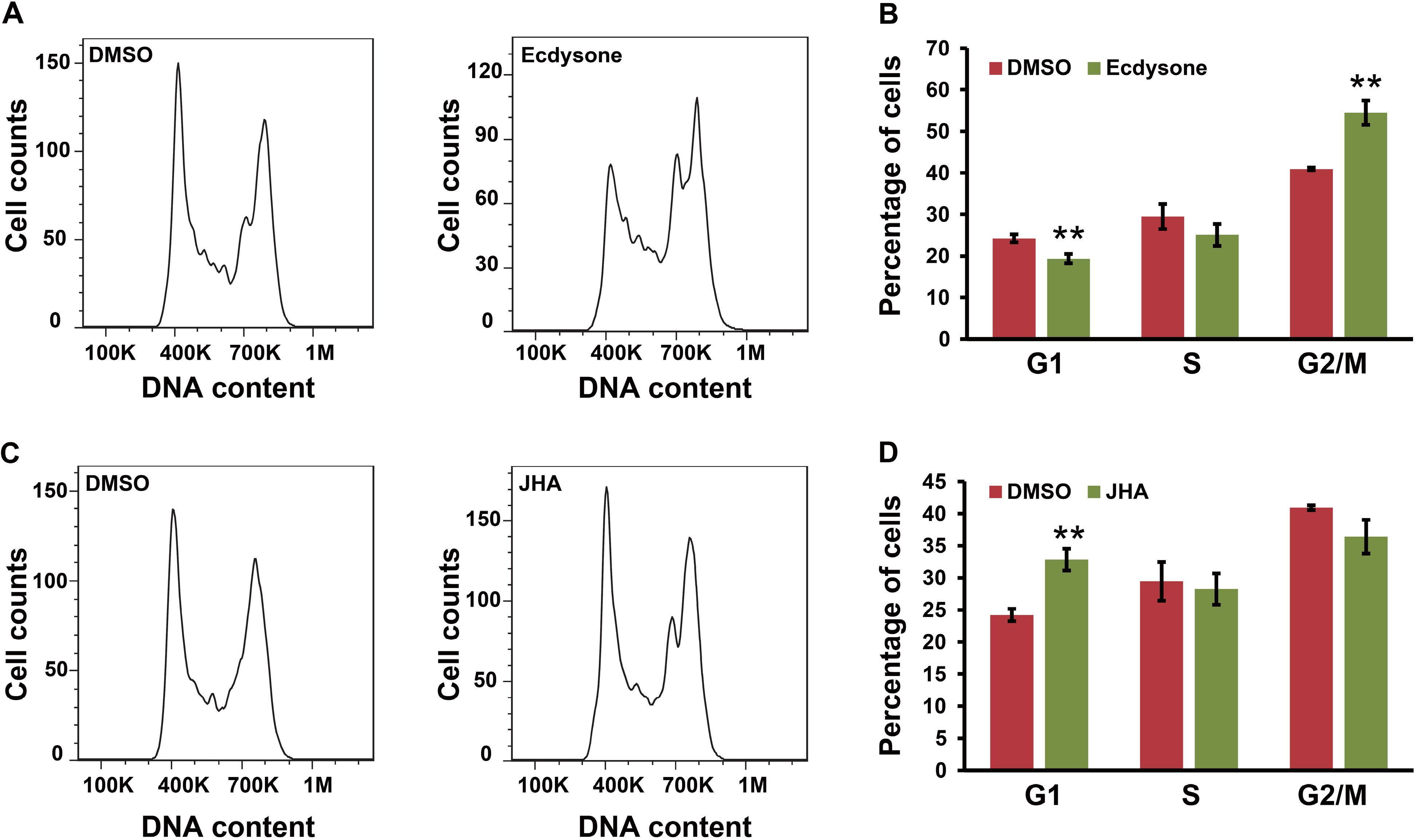
2.6. Ecdysone Treatment Induces Cell Cycle Arrest in BmN4 Cells

Undoubtedly, our findings raised a question why ecdysone regulation on CycE transcription was not positively correlated with its induction on E2F-1 transcription in BmN4 cells. Previous reports have demonstrated that during normal cell cycle progression, CycE expression is periodically elevated during G1/S transition but is declined during G2/M transition [39,40], and E2F-1 is expressed throughout the cell cycle and is dramatically increased in late G1 [41,42]. Therefore, we wonder whether the disruption of ecdysone signaling could induce cell cycle arrest, which in turn perturb the transcription of CycE transcription in BmN4 cells. To test this hypothesis, we further examined the effect of ecdysone treatment on cell cycle progression in BmN4 cells via flow cytometry analysis. As shown in Figure 8A,B, ecdysone treatment induced cell cycle arrest at G2/M phase, a time when CycE expression should be reduced as expected. The proportion of cells at G2/M phase was significantly increased from 40.9% in the control (DMSO treatment) to 54.5% after ecdysone treatment.
Figure 8. Ecdysone and JHA treatment induce cell cycle arrest. (A,B) Effect of ecdysone on cell cycle progression in BmN4 cells. At 24 h after ecdysone treatment, BmN4 cells were stained with Cell Cycle and Apoptosis Analysis Kit and subsequently detected by flow cytometry; (C,D) Effect of JHA on cell cycle progression in BmN4 cells. Similarly, at 24 h after JHA treatment, BmN4 cells were stained with Cell Cycle and Apoptosis Analysis Kit and subsequently detected by flow cytometry. Error bars represents mean and S.D., ** p < 0.01, compared with the control.
Ijms 16 03335 g008
Furthermore, we analyzed the effect of JHA treatment on cell cycle progression in BmN4 cells. As shown in Figure 8C,D, JHA treatment induced cell cycle arrest at G1 phase, a time when CycE expression should be increased. A significant accumulation of cells at G1 phase could be observed. Taken together with the cell cycle-dependent changes of CycE expression, we speculated that although E2F-1 transcription could be increased and decreased by ecdysone and JHA, respectively, the changes of CycE transcription after the treatment with either ecdysone or JHA is most possibly caused by cell cycle arrest induced by ecdysone or JHA, rather than E2F-1-dependent regulation.

3. Discussion

An increasing number of studies have reported that the ecdysone signaling pathway regulates cell cycle progression in insect. For example, the analysis in insect cell lines demonstrated that ecdysone application results in an accumulation of cells in the G1 phase in mosquito cells [43], and G2 arrest in the indianmeal moth (Plodia interpunctella) imaginal disc cells and fall armyworm (Spodoptera frugiperda) Sf9 cells [44,45,46]. In vivo analysis revealed that the ecdysone signaling pathway is required for cell cycle progression in the wing and eye disc of the fruit fly larvae [23,27,28,47]. Intriguingly, we found that EcR as ecdysone receptor is expressed in silkworm ovary-derived BmN4 cells at mRNA level. Although it is unclear how EcR transcription is activated in BmN4 cells, our investigation revealed that EcR-mediated ecdysone signaling is involved in cell cycle progression.
E2F-1 plays a crucial role in the regulation of cell cycle progression [48,49]. Previous studies in the fruit fly indicated that E2F-1 is likely a candidate target of EcR and is positively regulated by ecdysone at transcriptional level [23,50]. Intriguingly, based on cellular and individual analysis in silkworm, we found that E2F-1 expression was up-regulated by EcR overexpression and ecdysone treatment in both cultured BmN4 cells and silk gland of larvae, but reduced by EcR RNAi and the treatment of JHA as an antagonist of ecdysone. This strongly supported that E2F-1 transcription is positively regulated by EcR-mediated ecdysone signaling pathway.
As well known, CycE functions in controlling the G1/S transition during cell cycle progression [17], and CycE expression is positively regulated by E2F-1 in the fruit fly wing and salivary gland [20,21,51]. Our results showed that CycE expression was promoted after ecdysone treatment but suppressed after JHA application in silkworm silk gland. This is consistent with the changing tendency of E2F-1 expression under the same condition, indicating that E2F-1 similarly contributes to regulate positively the transcription of CycE gene in silkworm silk gland. Curiously, in silkworm BmN4 cells, CycE expression was suppressed by ecdysone treatment but promoted by either JHA application or EcR RNAi, which was negatively associated with ecdysone or JHA-induced changes of E2F-1 transcription.
It has been previously shown that CycE is highly expressed during G1/S transition and decreased during G2/M transition [39,40], whereas E2F-1 transcription continues throughout the cell cycle [41,42]. Our further examination demonstrated that ecdysone and JHA could induce cell cycle arrest at G2/M and G1 phases in BmN4 cells, respectively. This appears to be well synchronized with decreased transcription of CycE after ecdysone treatment and increased transcription of CycE after JHA treatment. Taken together with the fact of cell cycle-dependent changes of CycE transcription, we proposed that unlike E2F-1, ecdysone-mediated changes of CycE transcription in BmN4 cells is due most possibly to the response to ecdysone-induced cell cycle arrest, rather than E2F-1 regulation from ecdysone signaling cascades. Undoubtedly, the detailed mechanism underlying the regulation of EcR-mediated ecdysone signaling on CycE transcription needs to be further elucidated.

4. Experimental Section

4.1. Cell Lines

The silkworm cell lines of BmN4 and BmN4-SID1 were used in our experiment. The BmN4 cell line was derived from the silkworm ovary and the BmN4-SID1 cell line was established by introducing the nematode (Caenorhabditis elegans) SID1 gene into BmN4 cells, which can greatly enhance the uptake of exogenous dsRNA [33]. The BmN4-SID1 and BmN4 cell line were used to check the effect of EcR RNAi and hormone treatment on the transcription of cell cycle genes, respectively. The BmN4 and BmN4-SID1 cell lines were cultured at 27 °C in IPL-41 medium.

4.2. RNAi Knockdown of EcR Gene in BmN4-SID1 Cells

RNAi knockdown of EcR gene in BmN4-SID1 cells was performed according to the previously described approach [31]. In summary, the dsRNAs against silkworm EcR gene and EGFP (enhanced green fluorescent protein; dsEGFP) were separately synthesized and transfected into BmN4-SID1 cells. We collected the BmN4-SID1 cells at the 7th day after dsRNA treatment for twice to check cell shape using microscope and examined expression change of the selected genes.

4.3. RT-PCR Assay

Semi-quantitative and quantitative RT-PCR experiments were performed to determine whether the selected genes were expressed in BmN4 cells. RNA extraction, cDNA synthesis, and RT-PCR reaction were carried out following previous reports [52]. The silkworm ribosomal protein L3 (RpL3) gene was selected as a control. The primers used here are listed in Table S1.

4.4. Overexpression of EcR Gene in BmN4 Cells

For the overexpression of silkworm EcR in BmN4 cells, the opening reading frame sequence of silkworm EcR gene was cloned into the expression vector pSL1180. The constructs was transfected into BmN4 cells in 12-well plates. At 72 h after transfection, the BmN4 cells were collected to extract RNA for RT-PCR-based expression pattern analysis.

4.5. Hormone Treatment in BmN4 Cells

The BmN4 cells were seeded into 12-well plates with approximately 106 cells per well and cultured for 12 h. After a replacement with 1 mL fresh medium, the cells were subsequently treated with different dosage of either ecdysone or JHA. The treated BmN4 cells were cultured for 24 h at 27 °C and were collected for further RT-PCR analysis. In addition, for a time-dependent analysis, the BmN4 cells were treated with 1 μg per well and were collected at 6, 12, 24 and 48 h after hormone treatment.

4.6. Gene Expression Assay in Silkworm Silk Gland

The silkworm strain Dazao were reared with fresh mulberry leaves under the conditions of 25 °C and 12 h light/12 h dark photoperiod. The silk gland was isolated at nine developmental time points, including L4D1 (Day 1 of the fourth larval instar), L4D2, L4D3, L4M (fourth larval molting), L5D0 (0 h after fourth molts), L5D1 (day 1 of the fifth larval instar), L5D3, L5D5, and L5D7, and used for further RT-PCR analysis. Moreover, ecdysone and JHA with the dosage of 1 μg per individual were separately injected into silkworm larvae on Day 1 of the fourth instar. DMSO treatment was used as a control. At 12 h after hormone treatment, silk gland was isolated for further RT-PCR analysis.

4.7. Cell Cycle Analysis by Flow Cytometry

The BmN4 cells that were seeded into 12-well plate with 106 cells per well in 1 mL medium were used for cell cycle analysis. After culturing for 12 h, the cells were treated by ecdysone or JHA with a dosage of 1 μg per well, respectively. DMSO treatment was used as a control. At 24 h after treatment, the cells were stained with Cell Cycle and Apoptosis Analysis Kit (Beyotime Biotechnology, Nantong, China) and subsequently analyzed on flow cytometer (Becton, Dickinson and Company, Franklin Lakes, NJ, USA). Data was analyzed using Flow Jo 7.6 software (Flowjo, Ashland, OR, USA).

5. Conclusions

In this study, we demonstrated at cellular and individual scales that the EcR-mediated ecdysone signaling pathway is involved in regulating the transcription of two cell cycle genes, E2F-1 and CycE, in the silkworm. It is expected to facilitate the future work about deciphering the roles of EcR-mediated ecdysone signaling in cell cycle progression in insect.

Supplementary Materials

Supplementary materials can be found at https://www.mdpi.com/1422-0067/16/02/3335/s1.

Acknowledgments

We thank Takahiro Kusakabe (Kyushu University) for kindly providing silkworm ovary-derived BmN4-SID1 cell line. This work was supported by grants from the National Basic Research Program of China (no. 2012CB114600), the National Natural Science Foundation of China (no. 31172267 and no. 31272503), the National Hi-Tech Research and Development Program of China (no. 2011AA100306), and the Municipal Natural Science Foundation of Chongqing (no. cstc2012jjA80023).

Author Contributions

Daojun Cheng and Qingyou Xia conceived and designed experiments; Wenliang Qian, Lixia Kang, Tianlei Zhang, Meng Meng, and Yonghu Wang performed all experiments. Daojun Cheng, Wenliang Qian, Lixia Kang and Zhiqing Li analyzed the data. Daojun Cheng and Wenliang Qian wrote the paper. All authors read and approved the final manuscript.

Conflicts of Interest

The authors declare no conflict of interest.

References

  1. Koelle, M.R.; Talbot, W.S.; Segraves, W.A.; Bender, M.T.; Cherbas, P.; Hogness, D.S. The Drosophila EcR gene encodes an ecdysone receptor, a new member of the steroid receptor superfamily. Cell 1991, 67, 59–77. [Google Scholar] [CrossRef] [PubMed]
  2. Fahrbach, S.E.; Smagghe, G.; Velarde, R.A. Insect nuclear receptors. Annu. Rev. Entomol. 2012, 57, 83–106. [Google Scholar] [CrossRef] [PubMed]
  3. King-Jones, K.; Thummel, C.S. Nuclear receptors—A perspective from Drosophila. Nat. Rev. Genet. 2005, 6, 311–323. [Google Scholar] [CrossRef] [PubMed]
  4. Andres, A.J.; Thummel, C.S. Hormones, puffs and flies: The molecular control of metamorphosis by ecdysone. Trends Genet. 1992, 8, 132–138. [Google Scholar] [CrossRef] [PubMed]
  5. Thummel, C.S. Flies on steroids—Drosophila metamorphosis and the mechanisms of steroid hormone action. Trends Genet. 1996, 12, 306–310. [Google Scholar] [CrossRef] [PubMed]
  6. Yamanaka, N.; Rewitz, K.F.; O’Connor, M.B. Ecdysone control of developmental transitions: Lessons from Drosophila research. Annu. Rev. Entomol. 2013, 58, 497–516. [Google Scholar] [CrossRef] [PubMed]
  7. Carney, G.E.; Wade, A.A.; Sapra, R.; Goldstein, E.S.; Bender, M. DHR3, an ecdysone-inducible early-late gene encoding a Drosophila nuclear receptor, is required for embryogenesis. Proc. Natl. Acad. Sci. USA 1997, 94, 12024–12029. [Google Scholar] [CrossRef] [PubMed]
  8. Li, T.; Bender, M. A conditional rescue system reveals essential functions for the ecdysone receptor (EcR) gene during molting and metamorphosis in Drosophila. Development 2000, 127, 2897–2905. [Google Scholar] [PubMed]
  9. Baehrecke, E.H. Ecdysone signaling cascade and regulation of Drosophila metamorphosis. Arch. Insect Biochem. Physiol. 1996, 33, 231–244. [Google Scholar] [CrossRef] [PubMed]
  10. Schwedes, C.C.; Carney, G.E. Ecdysone signaling in adult Drosophila melanogaster. J. Insect Physiol. 2012, 58, 293–302. [Google Scholar] [CrossRef] [PubMed]
  11. Tapon, N.; Moberg, K.H.; Hariharan, I.K. The coupling of cell growth to the cell cycle. Curr. Opin. Cell Biol. 2001, 13, 731–737. [Google Scholar] [CrossRef] [PubMed]
  12. Levine, E. Cell cycling through development. Development 2004, 131, 2241–2246. [Google Scholar] [CrossRef] [PubMed]
  13. Neufeld, T.P.; Edgar, B.A. Connections between growth and the cell cycle. Curr. Opin. Cell Biol. 1998, 10, 784–790. [Google Scholar] [CrossRef] [PubMed]
  14. Swanhart, L.; Kupsco, J.; Duronio, R.J. Developmental control of growth and cell cycle progression in Drosophila. Methods Mol. Biol. 2005, 296, 69–94. [Google Scholar] [PubMed]
  15. Sanchez, I.; Dynlacht, B.D. New insights into cyclins, CDKs, and cell cycle control. Semin. Cell Dev. Biol. 2005, 16, 311–321. [Google Scholar] [CrossRef] [PubMed]
  16. Lim, S.; Kaldis, P. Cdks, cyclins and CKIs: Roles beyond cell cycle regulation. Development 2013, 140, 3079–3093. [Google Scholar] [CrossRef] [PubMed]
  17. Knoblich, J.A.; Sauer, K.; Jones, L.; Richardson, H.; Saint, R.; Lehner, C.F. Cyclin E controls S phase progression and its down-regulation during Drosophila embryogenesis is required for the arrest of cell proliferation. Cell 1994, 77, 107–120. [Google Scholar] [CrossRef] [PubMed]
  18. Edgar, B.A.; Lehman, D.A.; O’Farrell, P.H. Transcriptional regulation of string (cdc25): A link between developmental programming and the cell cycle. Development 1994, 120, 3131–3143. [Google Scholar] [PubMed]
  19. Nigg, E.A. Cyclin-dependent protein kinases: Key regulators of the eukaryotic cell cycle. BioEssays 1995, 17, 471–480. [Google Scholar] [CrossRef] [PubMed]
  20. Neufeld, T.P.; de la Cruz, A.F.; Johnston, L.A.; Edgar, B.A. Coordination of growth and cell division in the Drosophila wing. Cell 1998, 93, 1183–1193. [Google Scholar] [CrossRef] [PubMed]
  21. Reis, T.; Edgar, B.A. Negative regulation of dE2F1 by cyclin-dependent kinases controls cell cycle timing. Cell 2004, 117, 253–264. [Google Scholar] [CrossRef] [PubMed]
  22. Edgar, B.A.; Lehner, C.F. Developmental control of cell cycle regulators: A fly’s perspective. Science 1996, 274, 1646–1652. [Google Scholar] [CrossRef] [PubMed]
  23. Cranna, N.; Quinn, L. Impact of steroid hormone signals on Drosophila cell cycle during development. Cell Div. 2009, 4. [Google Scholar] [CrossRef]
  24. Ninov, N.; Manjon, C.; Martin-Blanco, E. Dynamic control of cell cycle and growth coupling by ecdysone, EGFR, and PI3K signaling in Drosophila histoblasts. PLoS Biol. 2009, 7, e1000079. [Google Scholar] [CrossRef] [PubMed]
  25. Stevens, B.; Alvarez, C.M.; Bohman, R.; O’Connor, J.D. An ecdysteroid-induced alteration in the cell cycle of cultured Drosophila cells. Cell 1980, 22, 675–682. [Google Scholar] [CrossRef] [PubMed]
  26. Fallon, A.M.; Gerenday, A. Ecdysone and the cell cycle: Investigations in a mosquito cell line. J. Insect Physiol. 2010, 56, 1396–1401. [Google Scholar] [CrossRef] [PubMed]
  27. Mitchell, N.C.; Lin, J.I.; Zaytseva, O.; Cranna, N.; Lee, A.; Quinn, L.M. The Ecdysone receptor constrains wingless expression to pattern cell cycle across the Drosophila wing margin in a Cyclin B-dependent manner. BMC Dev. Biol. 2013, 13. [Google Scholar] [CrossRef]
  28. Mitchell, N.; Cranna, N.; Richardson, H.; Quinn, L. The Ecdysone-inducible zinc-finger transcription factor Crol regulates Wg transcription and cell cycle progression in Drosophila. Development 2008, 135, 2707–2716. [Google Scholar] [CrossRef] [PubMed]
  29. Johnston, L.A.; Edgar, B.A. Wingless and Notch regulate cell-cycle arrest in the developing Drosophila wing. Nature 1998, 394, 82–84. [Google Scholar] [CrossRef] [PubMed]
  30. Johnston, L.A.; Sanders, A.L. Wingless promotes cell survival but constrains growth during Drosophila wing development. Nat. Cell Biol. 2003, 5, 827–833. [Google Scholar] [CrossRef] [PubMed]
  31. Li, Z.; Cheng, D.; Mon, H.; Tatsuke, T.; Zhu, L.; Xu, J.; Lee, J.M.; Xia, Q.; Kusakabe, T. Genome-wide identification of polycomb target genes reveals a functional association of Pho with Scm in Bombyx mori. PLoS One 2012, 7, e34330. [Google Scholar] [CrossRef] [PubMed]
  32. Jayachandran, G.; Fallon, A.M. Evidence for expression of EcR and USP components of the 20-hydroxyecdysone receptor by a mosquito cell line. Arch. Insect Biochem. Physiol. 2000, 43, 87–96. [Google Scholar] [CrossRef] [PubMed]
  33. Mon, H.; Kobayashi, I.; Ohkubo, S.; Tomita, S.; Lee, J.; Sezutsu, H.; Tamura, T.; Kusakabe, T. Effective RNA interference in cultured silkworm cells mediated by overexpression of Caenorhabditis elegans SID-1. RNA Biol. 2012, 9, 40–46. [Google Scholar] [CrossRef] [PubMed]
  34. Truman, J.W.; Riddiford, L.M. Endocrine insights into the evolution of metamorphosis in insects. Annu. Rev. Entomol. 2002, 47, 467–500. [Google Scholar] [CrossRef] [PubMed]
  35. Dubrovsky, E.B. Hormonal cross talk in insect development. Trends Endocrinol. Metab. 2005, 16, 6–11. [Google Scholar] [CrossRef] [PubMed]
  36. Minakuchi, C.; Zhou, X.; Riddiford, L.M. Kruppel homolog 1 (Kr-h1) mediates juvenile hormone action during metamorphosis of Drosophila melanogaster. Mech. Dev. 2008, 125, 91–105. [Google Scholar] [CrossRef] [PubMed]
  37. Kayukawa, T.; Minakuchi, C.; Namiki, T.; Togawa, T.; Yoshiyama, M.; Kamimura, M.; Mita, K.; Imanishi, S.; Kiuchi, M.; Ishikawa, Y.; et al. Transcriptional regulation of juvenile hormone-mediated induction of Kruppel homolog 1, a repressor of insect metamorphosis. Proc. Natl. Acad. Sci. USA 2012, 109, 11729–11734. [Google Scholar] [CrossRef] [PubMed]
  38. Mizoguchi, A.; Ohashi, Y.; Hosoda, K.; Ishibashi, J.; Kataoka, H. Developmental profile of the changes in the prothoracicotropic hormone titer in hemolymph of the silkworm Bombyx mori: Correlation with ecdysteroid secretion. Insect Biochem. Mol. Biol. 2001, 31, 349–358. [Google Scholar] [CrossRef] [PubMed]
  39. Siu, K.T.; Rosner, M.R.; Minella, A.C. An integrated view of cyclin E function and regulation. Cell Cycle 2012, 11, 57–64. [Google Scholar] [CrossRef] [PubMed]
  40. Keyomarsi, K.; Herliczek, T.W. The role of cyclin E in cell proliferation, development and cancer. Prog. Cell Cycle Res. 1997, 3, 171–191. [Google Scholar] [PubMed]
  41. Johnson, D.G.; Ohtani, K.; Nevins, J.R. Autoregulatory control of E2F1 expression in response to positive and negative regulators of cell cycle progression. Genes Dev. 1994, 8, 1514–1525. [Google Scholar] [CrossRef] [PubMed]
  42. Adams, M.R.; Sears, R.; Nuckolls, F.; Leone, G.; Nevins, J.R. Complex transcriptional regulatory mechanisms control expression of the E2F3 locus. Mol. Cell. Biol. 2000, 20, 3633–3639. [Google Scholar] [CrossRef] [PubMed]
  43. Gerenday, A.; Fallon, A.M. Ecdysone-induced accumulation of mosquito cells in the G1 phase of the cell cycle. J. Insect Physiol. 2004, 50, 831–838. [Google Scholar] [CrossRef] [PubMed]
  44. Siaussat, D.; Bozzolan, F.; Porcheron, P.; Debernard, S. The 20-hydroxyecdysone-induced signalling pathway in G2/M arrest of Plodia interpunctella imaginal wing cells. Insect Biochem. Mol. Biol. 2008, 38, 529–539. [Google Scholar] [CrossRef] [PubMed]
  45. Giraudo, M.; Califano, J.; Hilliou, F.; Tran, T.; Taquet, N.; Feyereisen, R.; le Goff, G. Effects of hormone agonists on Sf9 cells, proliferation and cell cycle arrest. PLoS One 2011, 6, e25708. [Google Scholar] [CrossRef] [PubMed]
  46. Siaussat, D.; Bozzolan, F.; Queguiner, I.; Porcheron, P.; Debernard, S. Cell cycle profiles of EcR, USP, HR3 and B cyclin mRNAs associated to 20E-induced G2 arrest of Plodia interpunctella imaginal wing cells. Insect Mol. Biol. 2005, 14, 151–161. [Google Scholar] [CrossRef] [PubMed]
  47. Zelhof, A.C.; Ghbeish, N.; Tsai, C.C.; Evans, R.M.; McKeown, M. A role for ultraspiracle, the Drosophila RXR, in morphogenetic furrow movement and photoreceptor cluster formation. Development 1997, 124, 2499–2506. [Google Scholar] [PubMed]
  48. Sala, A.; Nicolaides, N.C.; Engelhard, A.; Bellon, T.; Lawe, D.C.; Arnold, A.; Grana, X.; Giordano, A.; Calabretta, B. Correlation between E2f-1 requirement in the S-Phase and E2f-1 transactivation of cell cycle-related genes in human cells. Cancer Res. 1994, 54, 1402–1406. [Google Scholar] [PubMed]
  49. Duronio, R.J.; Ofarrell, P.H.; Xie, J.E.; Brook, A.; Dyson, N. The transcription factor E2f is required for S-Phase during Drosophila embryogenesis. Gene Dev. 1995, 9, 1445–1455. [Google Scholar] [CrossRef] [PubMed]
  50. Gauhar, Z.; Sun, L.V.; Hua, S.; Mason, C.E.; Fuchs, F.; Li, T.R.; Boutros, M.; White, K.P. Genomic mapping of binding regions for the Ecdysone receptor protein complex. Genome Res. 2009, 19, 1006–1013. [Google Scholar] [CrossRef] [PubMed]
  51. Duronio, R.J.; Ofarrell, P.H. Developmental control of the G(1) to S-transition in Drosophila—Cyclin-E Is a limiting downstream target of E2f. Gene Dev. 1995, 9, 1456–1468. [Google Scholar] [CrossRef] [PubMed]
  52. Li, Z.; Tatsuke, T.; Sakashita, K.; Zhu, L.; Xu, J.; Mon, H.; Lee, J.M.; Kusakabe, T. Identification and characterization of Polycomb group genes in the silkworm, Bombyx mori. Mol. Biol. Rep. 2012, 39, 5575–5588. [Google Scholar] [CrossRef] [PubMed]

