Review

Autophagy in DNA Damage Response

1
Department of Molecular Genetics, University of Lodz, Pomorska 141/143, 90-236 Lodz, Poland
2
Department of Orthodontics, Medical University of Lodz, Pomorska 251, 92-216 Lodz, Poland
3
Department of Infectious and Liver Diseases, Medical University of Lodz, Kniaziewicza 1/5, 92-347 Lodz, Poland
4
Department of Ophthalmology, Institute of Clinical Medicine, University of Eastern Finland, Kuopio FI-70211, Finland
5
Department of Ophthalmology, Kuopio University Hospital, Kuopio FI-70211, Finland
*
Author to whom correspondence should be addressed.
Int. J. Mol. Sci. 2015, 16(2), 2641-2662; https://doi.org/10.3390/ijms16022641
Submission received: 15 December 2014 / Accepted: 12 January 2015 / Published: 23 January 2015
(This article belongs to the Special Issue DNA Damage and Repair in Degenerative Diseases 2014)

Abstract

:
DNA damage response (DDR) involves DNA repair, cell cycle regulation and apoptosis, but autophagy is also suggested to play a role in DDR. Autophagy can be activated in response to DNA-damaging agents, but the exact mechanism underlying this activation is not fully understood, although it is suggested that it involves the inhibition of mammalian target of rapamycin complex 1 (mTORC1). mTORC1 represses autophagy via phosphorylation of the ULK1/2–Atg13–FIP200 complex thus preventing maturation of pre-autophagosomal structures. When DNA damage occurs, it is recognized by some proteins or their complexes, such as poly(ADP)ribose polymerase 1 (PARP-1), Mre11–Rad50–Nbs1 (MRN) complex or FOXO3, which activate repressors of mTORC1. SQSTM1/p62 is one of the proteins whose levels are regulated via autophagic degradation. Inhibition of autophagy by knockout of FIP200 results in upregulation of SQSTM1/p62, enhanced DNA damage and less efficient damage repair. Mitophagy, one form of autophagy involved in the selective degradation of mitochondria, may also play role in DDR. It degrades abnormal mitochondria and can either repress or activate apoptosis, but the exact mechanism remains unknown. There is a need to clarify the role of autophagy in DDR, as this process may possess several important biomedical applications, involving also cancer therapy.

Graphical Abstract

1. Introduction

Cells respond to different stress stimuli in order to survive, duplicate and avoid cancer transformation. The DNA damage response (DDR) plays an important role against detrimental effects of stress. It coordinates many processes, including DNA repair, regulation of cell cycle checkpoints, transcription of DDR genes, and ultimately induction of a programmed cell death, most often apoptosis, when DNA damage cannot be repaired (Figure 1). A growing body of evidence suggests, that autophagy, a catabolic process considered to be a cellular survival mechanism, may also play a role in DDR.
Figure 1. Cellular response to DNA damage. DNA damage can be induced by exogenous chemical and physical factors, or by endogenous influences following from cellular and DNA metabolism. The induction of DNA damage triggers the DNA damage response (DDR). Three proteins from the phosphatidylinositol 3-kinase-like protein kinases (PIKKs) family plays a major role in DDR: ataxia telangiectasia mutated (ATM), DNA protein kinase (DNA-PK) and ataxia telangiectasia and Rad3 related (ATR), two proteins of the poly(ADP-ribose) polymerase (PARP) family: PARP1 and PARP2, and heterotrimeric complex of Rad9, Rad1 and Hus1 (9–1–1 complex). These proteins are activated by either DNA damage itself or by other proteins. After ATM, ATR, DNA-PK, PARP1/2 or 9–1–1 complexes are activated, they transfer signals via signal mediators to regulate many cellular processes, including DNA repair, cell checkpoint activation or deactivation, activation or silencing of transcription, apoptosis and autophagy.
Ijms 16 02641 g001

2. DNA Damage and Its Cellular Response

2.1. DNA Damage and Repair

DNA damage can be induced by a variety of physical and chemical factors, generated exogenously or endogenously, by ultraviolet (UV) light and ionic radiation (IR) as well as metabolic reactions, producing reactive oxygen and nitrogen species (ROS and RNS, respectively) [1,2,3].
If a DNA damage is left unrepaired or is misrepaired, it can be changed into a mutation, which may play a role in pathogenesis of diseases, including cancer [3,4,5]. Therefore, an accurate DNA repair system is important for normal life of the cell. Some DNA damages can be repaired in a simple one step chemical reaction. This kind of repair pathway is known as DNA damage direct reversal. In this pathway methylated bases can be demethylated using the “suicide” enzyme O(6)-methylguanine-DNA methyltransferase (MGMT) [6,7]. Another two pathways, base excision repair (BER) and nucleotide excision repair (NER) can restore single-strand DNA damage. BER removes small chemical modifications, such as those caused by oxidative bases, while NER can repair damage affecting more than one DNA base, e.g., pyrimidine dimers (Figure 1). Mismatch repair (MMR) replaces a wrongly incorporated nucleotide with the correct one [8,9]. DNA double-strand breaks (DSBs), belonging to the most serious DNA damage, can be repaired by three pathways: homologous recombination repair (HRR), non-homologous end joining (NHEJ) and single-strand annealing (SSA), along with their several variants [10].
Mitochondrial DNA (mtDNA) is more prone to damage than its nuclear counterpart (nDNA) [11,12,13]. First, due to its close proximity to the electron transport chain (ETC), mtDNA is exposed to a relatively high level of ROS and RNS. Second, mtDNA lacks histones and non-histone proteins associated with DNA, forming a “molecular shield” protecting nDNA from damage. Third, mitochondrial mechanisms of DNA repair are less efficient and limited when compared to their nuclear analogues. Moreover, the repair of mtDNA is usually performed at sites of ROS generation. In addition, many reports suggest the lack of efficient NER in mitochondria [14]. Since mtDNA is especially prone to ROS- and RNS-induced lesions, BER is the main pathway activated in response to mtDNA damage [15,16]. Other pathways are also present in mitochondria, but their mechanisms are less known and may differ from their nuclear counterparts [17,18,19,20].

2.2. DNA Damage Signaling

Fast and precise transduction of the DNA damage signal is crucial for the efficiency of its repair. This signal is transduced mainly by a cascade of phosphorylation/dephosphorylation reactions [21].
Proteins from the phosphatidylinositol 3-kinase-like protein kinases (PIKKs) and poly(ADP)ribose polymerase (PARP) families play the major role in DDR signaling (Figure 1) [22,23]. ATM (Ataxia telangiectasia mutated) and DNA-PK (DNA-dependent protein kinase) are recruited to the site of DSBs [24,25]. Unlike DNA-PK, which only coordinates proteins responsible for DSB end joining, ATM controls more processes, including DNA replication, transcription, metabolic signaling and DNA splicing [1,26,27]. After creating a complex with ATR (Ataxia telangiectasia mutated and Rad3-related protein)-interacting protein (ATRIP) ATR recognizes persistent single-strand DNA (ssDNA) coated with replication protein A (RPA), which is present at stalled replication forks and DSBs [28]. Similarly to ATM, ATR regulates also other important cellular processes [26,27]. The 9–1–1 complex (heterotrimeric complex of Rad9, Rad1 and Hus1) and Rad17 are also involved in the detection of ssDNA coated with RPA. This complex creates a ring structure resembling the proliferating cell nuclear antigen (PCNA), and Rad17 shares a high homology with replication factor C (RFC) [29,30,31,32,33,34,35]. Rad17 recruits 9–1–1 to the DNA damage in a similar way that RFC engages PCNA. Two members of PARP (poly(ADP-ribose) polymerase) protein family involved in the DDR build chains of poly(ADP-ribose) (PAR) in the regionof single-strand breaks (SSBs) and DSBs occurrence to recruit other DDR proteins [36].
The involvement of ATM and ATR in DDR has been described (Figure 1). ATM, in its inactive form, creates a homodimer, which is recruited to the site of DSB by the Mre11–Rad50–Nbs1 (MRN) complex, then undergoes autophosphorylation and separates into two active monomers [37]. After recruitment and activation, ATM and ATR interact with many mediator and executing proteins, including Checkpoint kinase 1 and 2 (CHK1 and CHK2) involved in the cell cycle control, p53, a multifunctional protein essential for cell survival, breast cancer type 1 susceptibility protein (BRCA1)-associated genome surveillance complex (BASC) containing DNA damage repair proteins, histone deacetylases 1 and 2 (HDAC1 and HDAC2) responsible for remodeling the structure of chromatin, and transcription factor FOXO3, regulating genes involved in DNA repair [38,39,40,41,42].

2.3. Programmed Cell Death and DNA Damage

When DNA damage is left non-repaired, it may induce cell transformation or death. Morphologically, two different types of cell death: necrosis and programmed cell death, most often apoptosis, can be considered (Figure 1) [43]. Necrosis is characterized by an enlargement of cell volume (oncosis) and swelling of organelles. When oncosis reaches a critical point, the cell membrane breaks and the entire content of the cell flow into the extracellular space, often leading to inflammation. Although it is assumed that necrosis is an uncontrolled process occurring after overwhelming stress, there are some data suggesting that it can be regulated to some extent [44]. Apoptosis involves chromatin condensation, fragmentation of the nucleus, plasma membrane blebbing and creation of apoptotic bodies [45]. Triggering of apoptosis may occur by extrinsic or intrinsic pathways. Briefly, apoptosis via the extrinsic pathway is activated by death receptors belonging to the tumor necrosis factor receptor (TNFR) gene superfamily containing the evolutionary conserved death domain (DD). These receptors, such as TNFR-1/TNF-α or Fas/CD95, become activated when they bind specific ligands, form trimmers, and transduce signals via cytoplasmic death receptors. The signal from FAS receptors is transduced via Fas-Associated protein with death domain (FADD) and the signal from TNF receptors via TNFR-1-associated death domain protein (TRADD) with additional recruitment of FADD and receptor-interacting protein kinase (RIP) [46,47]. Similarly to DD, FADD contains a conserved motif called the Death Effector Domain (DED), which is also present in procaspase-8. Due to dimerization of these domains, FADD recruits procaspase-8 and this creates a death-inducing signaling complex (DISC). After DISC is formed, procaspase-8 undergoes auto-catalytic activation and, as caspase-8, triggers the implementation phase of apoptosis [48]. The intrinsic pathway is also called a mitochondrial pathway, because its crucial step is the release of pro-apoptotic proteins from the mitochondrial intermembrane space into the cytosol, (mitochondrial outer membrane permeabilization (MOMP) [49]. Thus, MOMP liberates the Smac/DIABLO (direct IAP (the inhibitor of apoptosis protein)-binding protein with low pl) complex, which promotes apoptosis by suppressing the inhibitors of apoptosis (IAP) and cytochrome c, which together with Apaf-1 and procaspase-9 creates the apoptosome, leading to caspase-9 activation [50]. There are also other pro-apoptotic proteins, including AIF (apoptosis-inducing factor), endonuclease G and CAD, which are responsible for DNA fragmentation [51]. The process of MOMP is regulated by members of the Bcl-2 family, containing pro- and anti-apoptotic proteins [52].
Severe DNA damage can induce both the extrinsic and intrinsic apoptosis pathways. As mentioned above, in response to DNA damage, ATM and ATR are activated and transduce signals via phosphorylation to other proteins, including p53. This chemical modification increases stability of p53, which can trigger apoptosis via mitochondria either as a transcription activator of pro-apoptotic proteins BAX (Bcl-2-assciated X protein 1), BID (BH3 interacting-domain death agonist), NOXA (Phorbol-12-myristate-13-acetate-induced protein), PUMA (p53 upregulated modulator of apoptosis) and FAS (tumor necrosis factor receptor superfamily 6) or by binding to anti-apoptotic proteins of the Bcl-2 family [53,54]. Moreover, ATR participates in phosphorylation of BRCA1 in response to UV light [55,56,57,58]. BRCA1, a protein involved in homologous recombination repair (HRR) and non-homologous end joining (NHEJ) DNA repair pathways, was found to stimulate apoptosis in a p53-independend manner [59,60]. DNA-PK can also induce apoptosis via phosphorylation of p53 [61]. This induction occurs in response to severe DNA damage or due to critically shortened telomeres [62,63]. Another protein involved in DDR and apoptosis is PARP1 [64]. It was shown that inhibition of PARP1 in combination with inhibition of epidermal growth factor receptor (EGFR) induces intrinsic apoptosis [65]. This approach has been recently translated pre-clinically as EGFR inhibition reduced HRR and NHEJ pathways and PARP1 inhibition thus augments the effect of chemotherapy as well as targeted radionuclide therapy [66]. In addition, PARP1 inhibition triggers caspase-independent cell death by mitochondrial release of AIF, which causes nDNA fragmentation [67,68].
It should be noted that programmed cell death in animals most often, but not exclusively, occurs by apoptosis and several other programmed pathways can be considered, including autophagy, anoikis excitotoxicity, ferroptosis, cornification and Wallerian degeneration. Therefore, there are many effector pathways exist downstream of DDR, including autophagy. However, autophagy can also contribute to survival mechanism of the cell after genotoxic insult, along with other mechanisms, including cell cycle arrest and mitotic arrest as well as reversible senescence (see [69] for review).

3. Autophagy

Autophagy, from Greek literally meaning “self-eating”, is a tightly regulated, evolutionary conserved, catabolic process, in which damaged proteins and organelles are degraded in lysosomes [70]. Three basic types of autophagy can be considered: microautophagy, chaperon-mediated autophagy (CMA) and macroautophagy [69,71,72]. Microautophagy is a nonselective process sequestering cytosolic proteins via invagination of the lysosomal membrane [73,74]. In CMA, proteins are selectively delivered to the lysosomes after recognition of their consensus sequence by a molecular chaperones, for example by HCS70 (heta-shock cognate) [75,76]. Macroautophagy which will be subsequently referred to as “autophagy” starts with the formation of autophagosome, a double-membrane vacuole, which encloses bulk proteins and organelles in the cytosol and delivers them to lysosome for degradation, resulting in the release of amino acids and fatty acids that can be reused by the cell [77,78,79]. Autophagy is triggered in response to the various stress stimuli, including nutrient and energy stresses, endoplasmic reticulum (ER) stress, hormone stimulation, hypoxia, redox stress, mitochondrial damage and DNA damage [80]. Although autophagy is considered to be a major protective mechanism against stress stimuli and plays an important role in many physiological processes, extensive autophagy may lead to cell death [43,81]. Autophagic cell death is characterized by the presence of many autophagic structures and, in the contrast to apoptosis, chromatin condensation occurs in the later steps, there is no DNA fragmentation or formation of apoptotic bodies and debris of cell are phagocytized later or there may be no remnants to be phagocytized at all [81].
One of the best described pathways leading to autophagy is activated during starvation. In this pathway mammalian target of rapamycin (mTOR) plays a central role as a negative regulator of autophagy. mTOR complex 1 (mTORC1) is associated with another complex of proteins consisting of ULK1 (Unc-51-like kinase 1), which is a mammalian analog of the yeast master regulator of autophagy, Atg1, the mammalian analog of Atg13 and the scaffold protein FIP200 (FAK (focal adhesion kinase)-family interacting protein of 200 kDa) [81,82]. Inhibition of autophagy is achieved by the phosphorylation of ULK1 and Atg13 by mTOR [83,84]. During starvation, mTORC1 dissociates from the Atg13–FIP200–ULK1 complex leading to dephosphorylation of ULK1 and Atg13, activation of ULK1, which phosphorylates Atg13 and FIP200 [85,86,87]. After activation of autophagy, a structure called the pre-autophagosomal membrane, which later will become the autophagosome, is formed. The pre-autophagosome is created by the complex of Atg5–Atg12–Atg16L. Atg5 is an E3 ubiquitin ligase, which is conjugated to the Atg12 protein [88]. The conjugate interacts with Atg16L to form a multimeric complex by homo-oligomerization of Atg16 [89]. The process of autophagosome maturation is controlled by the class III PI3 kinase (PI3KIII) Vps34, which forms complex with Beclin-1 and Vps15/p150, and its kinase activity is enhanced by binding with UVRAG (UV radiation resistance-associated gene) and Bif-1 (BAX-interacting factor-1) [90,91,92,93,94]. Vps34 interacts with Atg14 and targets it to the pre-autophagosome membrane, where Atg14 recruits Atg16 and LC3 [95]. LC3 is the mammalian analog of Atg8 and its cytoplasmic form, LC3-I is lipidated upon conjugation with phosphatidyl-ethanolamine (PE) to form LC3-II, which is responsible for expanding the autophagosome membrane [94,95]. This process is promoted by the Atg5–Atg12–Atg16L complex. After the autophagosome is assembled, it merges with the lysosome, where proteins and organelles are degraded into amino acids and fatty acids. The process of fusion is regulated by the pH of organelles’ interior: low pH promotes fusion, whereas an alkaline pH inhibits this process [95].

4. DNA Damage Response and Autophagy

An emerging body of evidence suggest that autophagy can play an important role in DDR (Figure 1). It was observed that after exposure to DNA-damaging agents, including ionizing radiation (IR), etoposide, camptothecin, tomozolomide or p-anilioaniline, not only the cell cycle was stopped, but also autophagy was induced [96,97,98]. Autophagy plays a cytoprotective role during anticancer therapy with DNA-damaging agents and its inhibition can sensitize cancer cells to these agents [66,99,100,101,102,103]. Additionally, the suppression of autophagy results in chromosomal instability, especially after metabolic stress, which may result in oncogene activation and tumor progression [102,103,104].

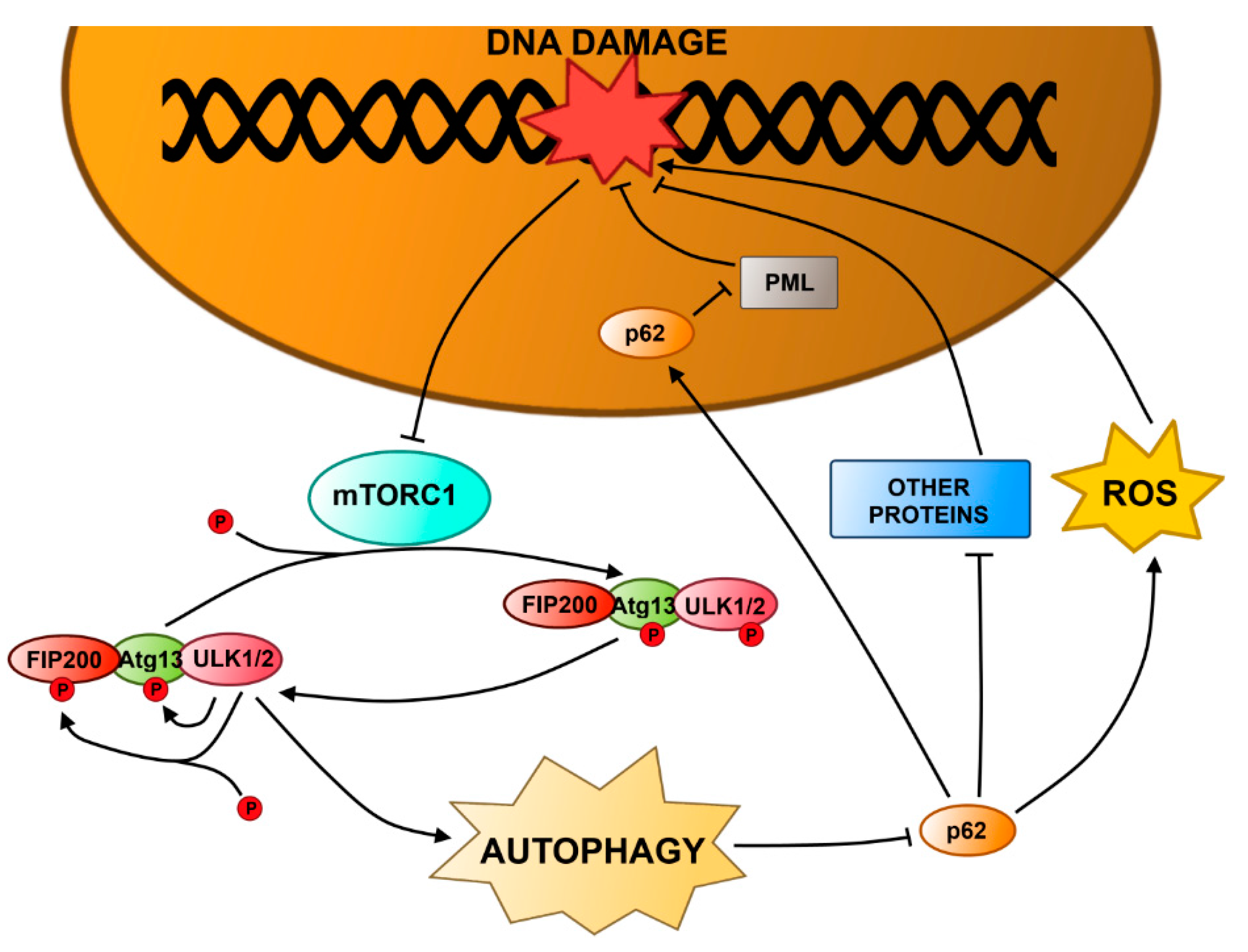
4.1. Autophagy and DNA Damage Response in the Nucleus

ATM is activated in response to DNA damage by the MRN complex and it plays a key role in DDR (Figure 1 and Figure 2). Activation of ATM after exposure to genotoxic and oxidative agents causes repression of mTORC1 and induction of autophagy [105,106]. The transduction of the signal from ATM to mTORC1 is mediated via the AMPK (5' adenosine-monophosphate-activated protein kinase) pathway, which involves tuberous sclerosis complex 1 and 2 (TSC1/2) and phosphorylation of ULK1 [107,108,109,110]. ATM can be also activated by the transcriptional factor FOXO3a (forkhead box O3) via phosphorylation [111]. Under normal conditions, FOXO3a is attached to DNA, but upon DNA damage, it dissociates from DNA and interacts with ATM promoting DNA repair. Additionally, FOXO3a regulates transcription of autophagy-related genes, including LC3 or Bnip3 [112,113,114].
Figure 2. Autophagy in the DNA damage response. Activation of autophagy in response to DNA damage is mainly achieved by inhibition of the mTOR complex 1 (mTORC1), which is a negative regulator of autophagy. p53 acts as a transcription factor, and if there is DNA damage it can upregulate expression of proteins inducing autophagy. One of them, DRAM (damage-regulated autophagy modulator), is responsible for autophagy degradation of VRK1 (vaccina-related kinase 1), a protein involved in cell cycle checkpoint activation, thus it arrests the cell cycle.
Ijms 16 02641 g002
The p53 protein is crucial for DDR and plays an important role in the regulation of autophagy [115,116,117,118,119,120,121,122,123,124].
PARP-1 is another protein of DDR involved in the regulation of autophagy. As described above, after a DNA lesion, PARP-1 synthesizes poly(ADP-ribose) chains that recruit the DNA damage repair proteins (Figure 2). On the other hand, when PARP-1 is hyperactivated in response to DNA damage, it causes NAD+ depletion leading to cellular energy failure and necrotic cell death [125,126]. Recently, it was shown that during starvation conditions, ROS-induced DNA damage activated PARP-1, which caused a depletion of ATP and activation of AMP-activated protein kinase (AMPK) [127]. Then AMPK sensed the energy depletion by measuring the ratio of AMP to ATP and it inhibited mTOR via TSC1/2, thus inducing autophagy [128]. In addition, H2O2- and doxorubicin-induced DNA damage activates a pathway involving PARP-1, which can induce either necrosis or autophagy [129,130]. Similarly, PARP-1 together with the catalytic subunit of DNA-PK (DNA-PKcs) was found to be activated by ATM during the autophagy induced by a chemopreventive agent, capsaicin, leading to DNA repair and the survival of breast cancer MCF-7 cells [130].
Results of recent research suggest that sirtuins, a family of protein deacetylases dependent on NAD+, may play an important role in autophagy and DDR [131]. The mammalian sirtuins family consists of 7 members, SIRT1–7, but in the light of recent studies, SIRT1 seems to play the most important role in autophagy/DDR, first of all due to its involvement in cellular reaction to oxidative stress and programmed death [132]. SIRT1 can induce the formation of autophagosome by interaction and deacetylation of the Atg5, Atg7 and Atg8 proteins in a NAD+-dependent fashion [133]. In addition, mediators of autophagy, mTOR1 and FOXO may be targeted by SIRT1 [134,135]. SIRT1 interacts with many protein which can be, directly or indirectly, involved in DDR, but its interaction with p53 seems to be crucial for its role in DDR, because it affects transcriptional activity of p53 regulating expression of p53 downstream effectors important for cell cycle regulation and programmed death under DNA-damaging conditions [136]. Therefore, SIRT1 may be involved in the regulation of autophagy in nontoxic stress, but precise mechanism underlying this involvement is not known and requires further studies.
FIP200, a 200 kDa FAK-family interacting protein, is a multifunctional protein regulating many cellular processes, including proliferation, cell migration and apoptosis, by interacting with FAK, Pyk2 (proline-rich tyrosine kinase 2), TSC1, p53, ASK1 (apoptosis signal-regulating kinase 1) or TRAF2 (TNF receptor-associated factor 2) [137,138,139,140] In addition, FIP200 is a component of the ULK1/2–Atg13–FIP200 complex and is essential for activation of autophagy (Figure 3). This complex is directly regulated by mTORC1 [141,142,143]. Under normal conditions, mTORC1 interacts with the complex by phosphorylation of ULK1/2 and Atg13, but when mTORC1 is inhibited, the level of phosphorylation of ULK1/2 and Atg13 decreases. This results in an increased kinase activity of ULK1/2, subsequent phosphorylation of Atg13 and FIP200, and translocation of the ULK1/2–Atg13–FIP200 complex to pre-autophagosomal structures [144,145]. Recently, it was shown that mouse embryonic fibroblasts (MEFs) with FIP200 knockout (KO) displayed a less efficient repair of the DNA damage induced by IR and two anticancer agents, camptothecin and etoposide, compared to the wild-type cells [146]. Moreover, KO of FIP200 caused up-regulation of SQSTM1 (sequestome 1)/p62 expression and formation of aggregates containing SQSTM1/p62. Re-expression of FIP200 restored the wild phenotype in FIP200 KO MEFs and suppressed SQSTM1/p62 expression. This indicates, that FIP200 regulates DNA damage response by autophagy and regulation of SQSTM1/p62 expression [147,148,149].
SQSTM1/p62 is an ubiquitin binding and scaffolding protein [141]. Its multiple domain structure enables controlling many processes, including osteoclastogenesis, inflammation, differentiation, neurotrophin properties and obesity [150]. SQSTM1/p62 is also involved in selective degradation via autophagy [151]. Damaged or unfolded proteins are polyubiquitinated, which recruits SQSTM1/p62 and induce binding to Atg8/LC3 presented on autophagosome membrane that finally leads to autophagic degradation of the aggregates [151]. It is not known how SQSTM1/p62 regulates the efficiency of DDR. SQSTM1/p62 is localized mainly in the cytoplasm, but it has a nuclear export signal (Figure 3). In the nucleus, it interacts with promyelocytic leukemia (PML) nuclear bodies that contain DDR proteins: BLM (Bloom syndrome)/WRN (Werner syndrome) DNA helicases, MRN or TopBP1 (DNA topoisomerase II binding protein), and are involved in DDR [152,153]. On the other hand, no significant relocalization of SQSTM1/p62 was observed and its influence on DNA repair efficiency may be indirectly mediated by an interaction with other proteins in the cytoplasm via its scaffold function. Additionally, up-regulation of SQSTM1/p62 expression causes increased ROS production, which may also contribute to increased DNA damage and create an amplification loop [154].
Figure 3. FIP200 (FAK (focal adhesion kinase)-family interacting protein of 200 kDa) and p62 in autophagy and DNA damage response. The mTOR1 (mammalian target of rapamycin) complex (mTORC1) interacts with ULK1/2 (UNC-51-like kinase 1/2)–Atg13 autophagy-related protein 13)–FIP200 complex and phosphorylates Atg13 and ULK1/2. If there is DNA damage, mTORC1 is inhibited, causing a slow decrease in phosphorylation of Atg13 and ULK1/2. Unphosphorylated ULK1/2 exhibits its kinase activity triggering phosphorylation of FIP200 and Atg13 and activating the ULK1/2–Atg13–FIP200 complex. The complex translocates to the pre-autophagosomal structure promoting autophagy. p62, a multifunctional ubiquitin-binding protein, is degraded by autosphagy. Inhibition of autophagy up-regulates p62, which causes an increase in the amount of DNA damage. The increase is caused either by generation of reactive oxygen species (ROS) or inhibition DNA repair via a direct interaction of p62 with promyelocytic leukemia (PML) nuclear bodies containing DDR proteins or an indirect interaction with other proteins in the cytosol.
Ijms 16 02641 g003

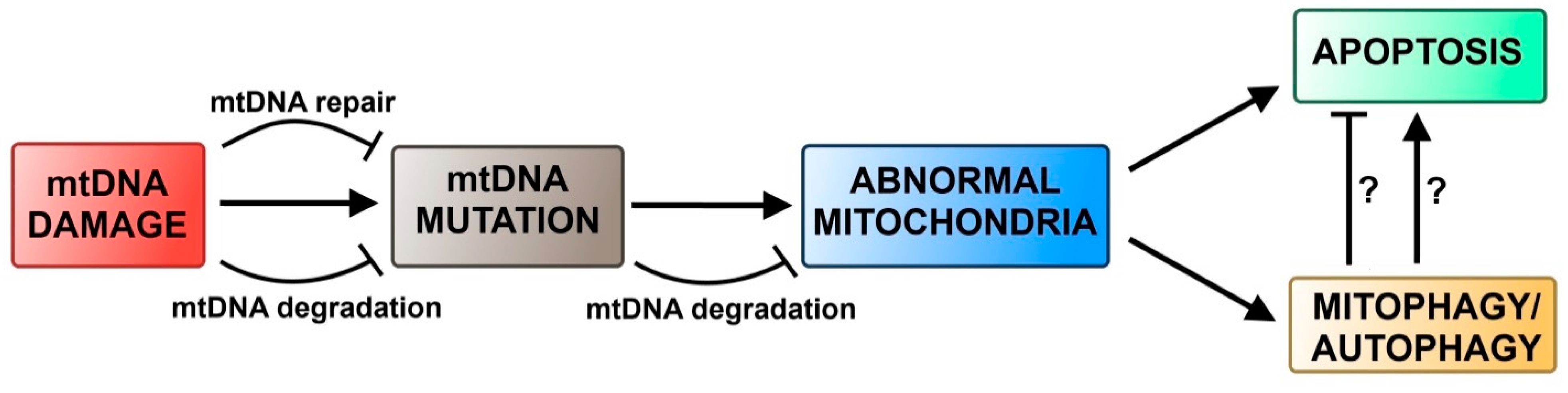
4.2. Autophagy and DNA Damage Response in Mitochondria

As mentioned above, DNA repair systems in mitochondria are less efficient, when compared to their nuclear counterparts. Although the mitochondrial genome contains only 37 genes, mutations and deletions of mtDNA are responsible for a significant number of inherited mitochondrial diseases, indicating the importance of mtDNA integrity for human health [155,156]. Damaged mitochondria may produce elevated levels of ROS, thus inducing even more DNA damage [43]. Since there are more than one mitochondrion in a cell and each mitochondrion has several copies of mtDNA, the damaged molecules can be degraded in live cells. It has been shown that degradation of mitochondria and mtDNA can be executed by a selective autophagic pathway, called mitophagy (Figure 4). The receptor to ensure the selectivity of mitophagy is the Nix protein [157]. After recruitment of Nix in response to mitochondrial depolarization, these mitochondria are marked for degradation by ubiquitination of this mitochondrial protein by the E3 ligase Parkin [158,159,160]. Recently, it was shown that SQSTM1/p62 binds to ubiquitinated mitochondrial membrane through its ubiquitin-binding domain and recruits the pre-autophagosome by LC3 binding domain [161,162,163,164]. The importance of SQSTM1/p62 in mitophagy remains to be elucidated. On the one hand, knockout of SQSTM1/p62 disabled elimination of mitochondria with compromised membrane potential, but other studies showed that SQSTM1/p62 is only involved in mitochondria aggregation, not in mitophagy itself. Nevertheless, it has been reported that the inductions of autophagy and mitophagy were triggered by toxic exposure, mtDNA mutations, ROS and UV [165,166,167]. In addition, blockage of autophagy and mitophagy can result in the accumulation of dysfunctional mitochondria, damaged mtDNA and an increased rate of apoptotic cell death [168,169,170]. In yeast mutations causing mitochondrial dysfunctions, especially these impairing the mitochondrial electrochemical transmembrane potential, have induced mitophagy even during nonstarvation conditions [171]. A similar observation was made in mammalian cells, when either mutations in mtDNA or drug-induced loss of mitochondrial membrane potential caused mitochondrial elimination by autophagy [172,173]. Autophagy may play a protective role against apoptosis, because it eliminates damaged mitochondria, thus preventing them from releasing proapoptotic proteins [173]. On the other hand, in the presence of caspase inhibitors, elimination of mitochondria by autophagy is crucial in triggering cell death [174]. Overall, these findings indicate that mtDNA damage can trigger mitophagy in an indirect way by the induction of mutations and changes in mitochondrial physiology, rather than by any direct signal.
Figure 4. Autophagy in mitochondrial DNA damage response. Damaged mitochondrial DNA (mtDNA) can be either repaired or degraded. This prevents transition of the damage to a mutation. When a mutation occurs, it can cause degradation of mutated mtDNA. Nevertheless, replication of mutated mtDNA molecules can cause a decrease in the mutation threshold and result in abnormalities in mitochondrial physiology. On the one hand, such abnormalities can cause apoptosis via the intrinsic pathway but on the other hand, these damaged mitochondria can be degraded via mitophagy. Question marks denote hypothetical pathways.
Ijms 16 02641 g004

5. Conclusions and Perspectives

Autophagy is a central player in the regulation of DDR. Impairments in this process have been connected to increased susceptibility of the cells to genotoxic agents, which may be important in anticancer therapy. It was shown that a DNA topisomerase II and tyrosine kinase 3 inhibitor induced autophagy in cancer cell lines and this process was associated with acquiring of senescent phenotype, which might be essential for a cytostatic action of this drug [175]. Senscence, associated with stable cell arrest, does not inhibit cellular apoptosis, as in apoptosis [176]. Genotoxic stress, leading to activation of DDR, may evoke autophagy as an early adaptative response, which can be compared with the DDR mechanisms of DNA damage tolerance, but this issue needs further research and explanation. Regulation of mechanisms of cross-talk between autophagy and apoptosis and senescence may be important for the regulation of DDR and cell fate and should be further studied as some controversial results were obtained so far. As mentioned, autophagy in DDR may be determined by the involvement of p53, a multifunction tumor suppressor, which is essential for determining the cell fate after a stress stimulus, but which mechanism of anticancer action is not fully known. Recent studies revealed several novel elements of p53 and a large autophagy network regulated by p53 and its family members, first of all p63 and p73 [176]. These studies revealed that when activated by p53, autophagy did not promote survival, but induced p53-dependent apoptosis. Therefore, further studies on the role of p53 in autophagy in the context of DDR may bring some important information on tumor suppressor role of p53 with potential relevance to anticancer therapy. Although many lines of evidence suggest the feasibility of autophagy as a target in anticancer therapy, it should be taken into account that this process may play a context-dependent role in cancer development. On one hand, its involvement in DDR may induce apoptosis and prevent genomic instability, which is a hallmark of cancer transformation, but on the other hand, it may promote survival of cancer cells in unfavorable, stress conditions, including those following from anticancer therapy. Autophagy is seen as a pro-survival mechanism due to its critical role in maintaining cellular protostasis and in the regulation of inflammation and cell death in conditions of metabolic stress in tumor cells [177]. In contrast, inhibition of autophagy has been shown to sensitize tumor cells to the cytotoxic effects of both chemotherapy and irradiation and thus it can improve the results of these kinds of cancer treatment [178]. Autophagy cell death is one of the standard cell death mechanisms. Therefore, a powerful promotion of autophagy by drugs would be predicted to achieve a better therapeutic efficacy. Induction of autophagic cell death might be a therapeutic aim if the apoptotic signaling is defective in tumor cells. Thus, the dual role of autophagy in tumor cells is not only of clinical interest but it also provides opportunities for the development of novel chemotherapeutic strategies. Various autophagy-regulating drugs could function as many different ways e.g., photosensitizors, lysosomotrophic agents, apoptosis inducers, proton pump inhibitors, toll-like receptor agonists, microtubule depolymerizators, cell cycle controllers, ROS generators, mTOR kinase inhibitors, tyrosine kinase inhibitors, AMP-kinase regulators and histamine receptor antagonists; all of these kinds of agents have been evaluated a potential cancer therapy alternatives [178,179,180]. However, the question about exact role of autophagy in DDR and its implications for cancer therapy is still waiting for the answer.

Acknowledgments

The authors thank Anna Luczynska for her editorial help.

Author Contributions

Janusz Blasiak and Kai Kaarniranta created the idea and synopsis of the manuscript and Piotr Czarny wrote the first draft version of it, which was then corrected and developed by Janusz Blasiak, Kai Kaarniranta, Elzbieta Pawlowska and Jolanta Bialkowska-Warzecha. Janusz Blasiak revised the manuscript to its final form.

Conflicts of Interest

The authors declare no conflict of interest.

References

  1. Ciccia, A.; Elledge, S.J. The DNA damage response: Making it safe to play with knives. Mol. Cell 2010, 40, 179–204. [Google Scholar]
  2. Ferguson, L.R. Chronic inflammation and mutagenesis. Mutat. Res. 2010, 690, 3–11. [Google Scholar]
  3. Jackson, S.P.; Bartek, J. The DNA-damage response in human biology and disease. Nature 2009, 461, 1071–1078. [Google Scholar]
  4. Kaarniranta, K.; Sinha, D.; Blasiak, J.; Kauppinen, A.; Veréb, Z.; Salminen, A.; Boulton, M.E.; Petrovski, G. Autophagy and heterophagy dysregulation leads to retinal pigment epithelium dysfunction and development of age-related macular degeneration. Autophagy 2013, 9, 973–984. [Google Scholar]
  5. Simpson, P.T.; Vargas, A.C.; Al-Ejeh, F.; Khanna, K.K.; Chenevix-Trench, G.; Lakhani, S.R. Application of molecular findings to the diagnosis and management of breast disease: Recent advances and challenges. Hum. Pathol. 2011, 42, 153–165. [Google Scholar]
  6. Eker, A.P.; Quayle, C.; Chaves, I.; van der Horst, G.T. DNA repair in mammalian cells. Cell. Mol. Life Sci. 2009, 66, 968–980. [Google Scholar]
  7. Lindahl, T.; Barnes, D.E. Repair of endogenous DNA damage. Cold Spring Harb. Symp. Quant. Biol. 2000, 65, 127–133. [Google Scholar]
  8. Hoeijmakers, J.H. DNA damage, aging, and cancer. N. Engl. J. Med. 2009, 361, 1475–1485. [Google Scholar]
  9. Jiricny, J. The multifaceted mismatch-repair system. Nat. Rev. Mol. Cell Biol. 2006, 7, 335–346. [Google Scholar]
  10. Chapman, J.R.; Taylor., M.R.; Boulton, S.J. Playing the end game: DNA double-strand break repair pathway choice. Mol. Cell 2012, 47, 497–510. [Google Scholar]
  11. Yakes, F.M.; van Houten, B. Mitochondrial DNA damage is more extensive and persists longer than nuclear DNA damage in human cells following oxidative stress. Proc. Natl. Acad. Sci. USA 1997, 94, 514–519. [Google Scholar]
  12. Reddy, V.N.; Kasahara, E.; Hiraoka, M.; Lin, L.R.; Ho, Y.S. Effects of variation in superoxide dismutases (SOD) on oxidative stress and apoptosis in lens epithelium. Exp. Eye Res. 2004, 79, 859–868. [Google Scholar]
  13. Banmeyer, I.C.; Clippe, A.; Knoops, B. Human mitochondrial peroxiredoxin 5 protects from mitochondrial DNA damages induced by hydrogen peroxide. FEBS Lett. 2005, 579, 2327–2333. [Google Scholar]
  14. Pascucci, B.; Versteegh, A.; van Hoffen, A.; van Zeeland, A.A.; Mullenders, L.H.; Dogliotti, E. DNA repair of UV photoproducts and mutagenesis in human mitochondrial DNA. J. Mol. Biol. 1997, 273, 417–427. [Google Scholar]
  15. Boesch, P.; Weber-Lotfi, F.; Ibrahim, N.; Tarasenko, V.; Cosset, A.; Paulus, F.; Lightowlers, R.N.; Dietrich, A. DNA repair in organelles: Pathways, organization, regulation, relevance in disease and aging. Biochim. Biophys. Acta 2011, 1813, 186–200. [Google Scholar]
  16. Le Doux, S.P.; Wilson, G.L. Base excision repair of mitochondrial DNA damage in mammalian cells. Prog. Nucleic Acid Res. Mol. Biol. 2001, 68, 273–284. [Google Scholar]
  17. De Souza-Pinto, N.C.; Mason, P.A.; Hashiguchi, K.; Weissman, L.; Tian, J.; Guay, D.; Lebel, M.; Stevnsner, T.V.; Rasmussen, L.J.; Bohr, V.A.; et al. Novel DNA mismatch-repair activity involving YB-1 in human mitochondria. DNA Repair 2009, 8, 704–719. [Google Scholar]
  18. Kraytsberg, Y.; Schwartz, M.; Brown, T.A.; Ebralidse, K.; Kunz, W.S.; Clayton, D.A.; Vissing, J.; Khrapko, K. Recombination of human mitochondrial DNA. Science 2004, 304, 981. [Google Scholar]
  19. Sage, J.M.; Gildemeister, O.S.; Knight, K.L. Discovery of a novel function for human Rad51: Maintenance of the mitochondrial genome. J. Biol. Chem. 2010, 285, 18984–18990. [Google Scholar]
  20. Lakshmipathy, U.; Campbell, C. Double strand break rejoining by mammalian mitochondrial extracts. Nucleic Acids Res. 1999, 27, 11198–11204. [Google Scholar]
  21. Cui, R.; Widlund, H.R.; Feige, E.; Lin, J.Y.; Wilensky, D.L.; Igras, V.E.; D’Orazio, J.; Fung, C.Y.; Schanbacher, C.F.; Granter, S.R.; et al. Central role of p53 in the suntan response and pathologic hyperpigmentation. Cell 2007, 128, 853–864. [Google Scholar]
  22. Schreiber, V.; Dantzer, F.; Ame, J.C.; de Murcia, G. Poly(ADP-ribose): Novel functions for an old molecule. Nat. Rev. Mol. Cell Biol. 2006, 7, 517–528. [Google Scholar]
  23. Sancar, A.; Lindsey-Boltz, L.A.; Unsal-Kacmaz, K.; Linn, S. Molecular mechanisms of mammalian DNA repair and the DNA damage checkpoints. Annu. Rev. Biochem. 2004, 73, 39–85. [Google Scholar]
  24. Meek, K.; Dang, V.; Lees-Miller, S.P. DNA-PK: The means to justify the ends? Adv. Immunol. 2008, 99, 33–58. [Google Scholar]
  25. Harpe, J.W.; Elledge, S.J. The DNA damage response: Ten years after. Mol. Cell 2007, 28, 739–745. [Google Scholar]
  26. Matsuoka, S.; Ballif, B.A.; Smogorzewska, A.; McDonald, E.R., 3rd; Hurov, K.E.; Luo, J.; Bakalarski, C.E.; Zhao, Z.; Solimini, N.; Lerenthal, Y.; et al. ATM and ATR substrate analysis reveals extensive protein networks responsive to DNA damage. Science 2007, 316, 1160–1166. [Google Scholar]
  27. Paulsen, R.D.; Soni, D.V.; Wollman, R.; Hahn, A.T.; Yee, M.C.; Guan, A.; Hesley, J.A.; Miller, S.C.; Cromwell, E.F.; Solow-Cordero, D.E.; et al. A genome-wide siRNA screen reveals diverse cellular processes and pathways that mediate genome stability. Mol. Cell 2009, 35, 228–239. [Google Scholar]
  28. Cimprich, K.A.; Cortez, D. ATR: An essential regulator of genome integrity. Nat. Rev. Mol. Cell Biol. 2008, 9, 616–627. [Google Scholar]
  29. Paulovich, A.G.; Margulies, R.U.; Garvik, B.M.; Hartwell, L.H. RAD9, RAD17, and RAD24 are required for S phase regulation in Saccharomyces cerevisiae in response to DNA damage. Genetics 1997, 145, 45–62. [Google Scholar]
  30. Volkmer, E.; Karnitz, L.M. Human homologs of Schizosaccharomyces pombe Rad1, Hus1, and Rad9 form a DNA damage-responsive protein complex. J. Biol. Chem. 1999, 274, 567–570. [Google Scholar]
  31. Thelen, M.P.; Venclovas, C.; Fidelis, K. A sliding clamp model for the Rad1 family of cell cycle checkpoint proteins. Cell 1999, 96, 769–770. [Google Scholar]
  32. Griffiths, D.J.; Barbet, N.C.; McCready, S.; Lehmann, A.R.; Carr, A.M. Fission yeast rad17: A homologue of budding yeast RAD24 that shares regions of sequence similarity with DNA polymerase accessory proteins. EMBO J. 1995, 14, 5812–5823. [Google Scholar]
  33. Green, C.M.; Erdjument-Bromage, H.; Tempst, P.; Lowndes, N.F. A novel Rad24 checkpoint protein complex closely related to replication factor C. Curr. Biol. 2000, 10, 39–42. [Google Scholar]
  34. Naiki, T.; Shimomura, T.; Kondo, T.; Matsumoto, K.; Sugimoto, K. Rfc5, in cooperation with Rad24, controls DNA damage checkpoints throughout the cell cycle in Saccharomyces cerevisiae. Mol. Cell. Biol. 2000, 20, 5888–5896. [Google Scholar]
  35. Lindsey-Boltz, L.A.; Bermudez, V.P.; Hurwitz, J; Sancar, A. Purification and characterization of human DNA damage checkpoint Rad complexes. Proc. Natl. Acad. Sci. USA 2001, 98, 11236–11241. [Google Scholar]
  36. Golia, B.; Singh, H.R.; Timinszki, G. Poly-ADP-ribosylation signaling during DNA damage repair. Front. Biosci. 2015, 20, 440–457. [Google Scholar]
  37. Bakkenist, C.J.; Kastan, M.B. DNA damage activates ATM through intermolecular autophosphorylation and dimer dissociation. Nature 2003, 421, 499–506. [Google Scholar]
  38. Wang, Y.; Cortez, D.; Yazdi, P.; Neff, N.; Elledge, S.J.; Qin, J. BASC, a super complex “of BRCA1-associated proteins involved in the recognition and repair of aberrant DNA structures. Genes Dev. 2000, 14, 927–939. [Google Scholar]
  39. Kim, G.D.; Choi, Y.H.; Dimtchev, A.; Jeong, S.J.; Dritschilo, A.; Jung, M. Sensing of ionizing radiation-induced DNA damage by ATM through interaction with histone deacetylase. J. Biol. Chem. 1999, 274, 31127–33130. [Google Scholar]
  40. Schmidt, D.R.; Schreiber, S.L. Molecular association between ATR and two components of the nucleosome remodeling and deacetylating complex, HDAC2 and CHD4. Biochemistry 1999, 38, 14711–14717. [Google Scholar]
  41. Tran, H.; Brunet, A.; Grenier, J.M.; Datta, S.R.; Fornace, A.J., Jr.; DiStefano, P.S.; Chiang, L.W.; Greenberg, M.E. DNA repair pathway stimulated by the forkhead transcription factor FOXO3a through the Gadd45 protein. Science 2002, 296, 530–534. [Google Scholar]
  42. Thurn, K.T.; Thomas, S.; Raha, P.; Qureshi, I.; Munster, P.N. Histone deacetylase regulation of ATM-mediated DNA damage signaling. Mol. Cancer Ther. 2013, 12, 2078–2087. [Google Scholar]
  43. Rodriguez-Rochaa, H.; Garcia-Garciaa, A.; Panayiotidisb, M.I.; Francoa, R. DNA damage and autophagy. Mutat. Res. 2011, 711, 158–166. [Google Scholar]
  44. Festjens, N.; VandenBerghe, T.; Vandenabeele, P. Necrosis, a well-orchestrated form of cell demise: Signaling cascades, important mediators and concomitant immune response. Biochim. Biophys. Acta 2006, 1757, 1371–1387. [Google Scholar]
  45. Huang, C.; Freter, C. Lipid metabolism, apoptosis and cancer therapy. Int. J. Mol. Sci. 2015, 16, 924–949. [Google Scholar]
  46. Hsu, H.; Xiong, J.; Goeddel, D.V. The TNF receptor 1-associated protein TRADD signals cell death and NFκB activation. Cell 1995, 81, 495–504. [Google Scholar]
  47. Wajant, H. The Fas signaling pathway: More than a paradigm. Science 2002, 29, 1635–1636. [Google Scholar]
  48. Kischkel, F.C.; Hellbardt, S.; Behrmann, I.; Germer, M.; Pawlita, M.; Krammer, P.H.; Peter, M.E. Cytotoxicity dependent APO-1 (Fas/CD95)-associated proteins form a death-inducing signaling complex (DISC) with the receptor. EMBO J. 1995, 14, 5579–5588. [Google Scholar]
  49. Saelens, X.; Festjens, N.; vande Walle, L.; van Gurp, M.; van Loo, G.; Vandenabeele, P. Toxic proteins released from mitochondria in cell death. Oncogene 2004, 23, 2861–2874. [Google Scholar]
  50. Parsons, M.J.; Green, D.R. Mitochondria in cell death. Essays Biochem. 2010, 47, 99–114. [Google Scholar]
  51. Elmore, S. Apoptosis: A review of programmed cell death. Toxicol. Pathol. 2007, 35, 495–516. [Google Scholar]
  52. Schuler, M.; Green, D.R. Mechanisms of p53-dependent apoptosis. Biochem. Soc. Trans. 2001, 29, 684–688. [Google Scholar]
  53. Roos, W.P.; Kaina, B. DNA damage-induced cell death by apoptosis. Trends Mol. Med. 2006, 12, 440–450. [Google Scholar]
  54. Chipuk, J.E.; Green, D.R. Dissecting p53-dependent apoptosis. Cell Death Differ. 2006, 13, 994–1002. [Google Scholar]
  55. Kung, C.P.; Budina, A.; Balaburski, G.; Bergenstock, M.K.; Murphy, M. Autophagy in tumor suppression and cancer therapy. Crit. Rev. Eukaryot. Gene Expr. 2011, 21, 71–100. [Google Scholar]
  56. Levine, B.; Klionsky, D.J. Development by self-digestion: Molecular mechanisms and biological functions of autophagy. Cell 2004, 6, 463–477. [Google Scholar]
  57. Cuervo, A.M. Autophagy: Many paths to the same end. Mol. Cell. Biochem. 2004, 263, 55–72. [Google Scholar]
  58. Marzella, L.; Ahlberg, J.; Glaumann, H. Autophagy, heterophagy, microautophagy and crinophagy as the means for intracellular degradation. Virchows Arch. B 1981, 36, 219–234. [Google Scholar]
  59. Shao, N.; Chai, Y.L.; Shyam, E.; Reddy, P.; Rao, V.N. Induction of apoptosis by the tumor suppressor protein BRCA1. Oncogene 1996, 13, 1–7. [Google Scholar]
  60. Martin, S.A.; Ouchi, T. BRCA1 phosphorylation regulates caspase-3 activation in UV-induced apoptosis. Cancer Res. 2005, 65, 10657–10662. [Google Scholar]
  61. Burma, S.; Chen, D.J. Role of DNA-PK in the cellular response to DNA double-strand breaks. DNA Repair 2004, 3, 909–918. [Google Scholar]
  62. Espejel, S.; Franco, S.; Sgura, A.; Gae, D.; Bailey, S.M.; Taccioli, G.E.; Blasco, M.A. Functional interaction between DNA-PKcs and telomerase in telomere length maintenance. EMBO J. 2002, 21, 6275–6287. [Google Scholar]
  63. Espejel, S.; Martín, M.; Klatt, P.; Martín-Caballero, J.; Flores, J.M.; Blasco, M.A. Shorter telomeres, accelerated ageing and increased lymphoma in DNA-PKcs deficient mice. EMBO Rep. 2004, 5, 503–509. [Google Scholar]
  64. Luo, X.; Kraus, W.L. On PAR with PARP: Cellular stress signaling through poly (ADP-ribose) and PARP-1. Genes Dev. 2012, 26, 417–432. [Google Scholar]
  65. Nowsheen, S.; Bonner, J.A.; Lo Buglio, A.F.; Trummell, H.; Whitley, A.C.; Dobelbower, M.C.; Yang, E.S. Cetuximab augments cytotoxicity with poly (ADP-ribose) polymerase inhibition in head and neck cancer. PLoS One 2011, 6, e24148. [Google Scholar]
  66. Al-Ejeh, F.; Shi, W.; Miranda, M.; Simpson, P.T.; Vargas, A.C.; Song, S.; Wiegmans, A.P.; Swarbrick, A.; Welm, A.L.; Brown, M.P.; et al. Treatment of triple-negative breast cancer using anti-EGFR-directed radioimmunotherapy combined with radiosensitizing chemotherapy and PARP inhibitor. J. Nucl. Med. 2013, 54, 913–921. [Google Scholar]
  67. Yu, S.W.; Wang, H.; Poitras, M.F.; Coombs, C.; Bowers, W.J.; Federoff, H.J.; Poirier, G.G.; Dawson, T.M.; Dawson, V.L. Mediation of poly(ADP-ribose) polymerase-1-dependent cell death by apoptosis-inducing factor. Science 2002, 297, 259–263. [Google Scholar]
  68. Cregan, S.P.; Dawson, V.L.; Slack, R.S. Role of AIF in caspase-dependent and caspase-independent cell death. Oncogene 2004, 23, 2785–2796. [Google Scholar]
  69. Al-Ejeh, F.; Kumar, R.; Wiegmans, A.; Lakhani, S.R.; Brown, M.P.; Khanna, K.K. Harnessing the complexity of DNA-damage response pathways to improve cancer treatment outcomes. Oncogene 2010, 29, 6085–6098. [Google Scholar]
  70. Mortimore, G.E.; Lardeux, B.R.; Adams, C.E. Regulation of microautophagy and basal protein turnover in rat liver. Effects of short-term starvation. J. Biol. Chem. 1988, 263, 2506–2512. [Google Scholar]
  71. Agarraberes, F.; Dice, J.F. A molecular chaperone complex at the lysosomal membrane is required for protein translocation. J. Cell Sci. 2001, 114, 2491–2499. [Google Scholar]
  72. Majeski, A.E.; Dice, J.F. Mechanisms of chaperone-mediated autophagy. Int. J. Biochem. Cell Biol. 2004, 36, 2435–2444. [Google Scholar]
  73. Massey, A.C.; Zhang, C.; Cuervo, A.M. Chaperone-mediated autophagy in aging and disease. Curr. Top. Dev. Biol. 2006, 73, 205–235. [Google Scholar]
  74. Klionsky, D.J.; Emr, S.D. Autophagy as a regulated pathway of cellular degradation. Science 2000, 290, 1717–1721. [Google Scholar]
  75. Baehrecke, E.H. Autophagy: Dual roles in life and death? Nat. Rev. Mol. Cell Biol 2005, 6, 505–510. [Google Scholar]
  76. Klionsky, D.J.; Abdalla, F.C.; Abeliovich, H.; Abraham, R.T.; Acevedo-Arozena, A.; Adeli, K.; Agholme, L.; Agnello, M.; Agostinis, P.; Aguirre-Ghiso, J.A.; et al. Guidelines for the use and interpretation of assays for monitoring autophagy. Autophagy 2012, 8, 445–544. [Google Scholar]
  77. Filomeni, G.; de Zio, D.; Cecconi, F. Oxidative stress and autophagy: the clash between damage and metabolic needs. Cell Death Differ. 2014. [Google Scholar] [CrossRef]
  78. Kroemer, G.; Mariño, G.; Levine, B. Autophagy and the integrated stress response. Mol. Cell 2010, 40, 280–293. [Google Scholar]
  79. Gozuacik, D.; Kimchi, A. Autophagy and cell death. Curr. Top. Dev. Biol. 2007, 78, 217–245. [Google Scholar]
  80. Wei, H.; Wang, C.; Croce, C.M.; Guan, J.L. p62/SQSTM1 synergizes with autophagy for tumor growth in vivo. Genes Dev. 2014, 28, 1204–1216. [Google Scholar]
  81. Kamada, Y.; Funakoshi, T.; Shintani, T.; Nagano, K.; Ohsumi, M.; Ohsumi, Y. Tor-mediated induction of autophagy via an Apg1 protein kinase complex. J. Cell Biol. 2000, 150, 1507–1513. [Google Scholar]
  82. Mizushima, N. The role of the Atg1/ULK1 complex in autophagy regulation. Curr. Opin. Cell Biol. 2010, 22, 132–139. [Google Scholar]
  83. Wei, H.; Guan, J.L. Pro-tumorigenic function of autophagy in mammalian oncogenesis. Autophagy 2012, 8, 129–131. [Google Scholar]
  84. Dunlop, E.A.; Tee, A.R. mTOR and autophagy: A dynamic relationship governed by nutrients and energy. Semin. Cell Dev. Biol. 2014, 36, 121–129. [Google Scholar]
  85. Roberts, D.J.; Miyamoto, S. Hexokinase II integrates energy metabolism and cellular protection: Akting on mitochondria and TORcing to autophagy. Cell Death Differ. 2015, 22, 248–257. [Google Scholar]
  86. Mizushima, N.; Noda, T.; Ohsumi, Y. Apg16p is required for the function of the Apg12p–Apg5p conjugate in the yeast autophagy pathway. EMBO J. 1999, 18, 3888–3896. [Google Scholar]
  87. Kuma, A.; Mizuchima, N.; Ishihara, N.; Ohsumi, Y. Formation of the approximately 350 kDa Apg12–Apg5–Apg16 multimeric complex, mediated by Apg16 oligomerization, is essential for autophagy in yeast. J. Biol. Chem. 2002, 277, 18619–18625. [Google Scholar]
  88. Scott, S.V.; Nice, D.C., 3rd; Nau, J.J.; Weisman, L.S.; Kamada, Y.; Keizer-Gunnink, I.; Funakoshi, T.; Veenhuis, M.; Ohsumi, Y.; Klionsky, D.J.; et al. Apg13p and Vac8p are part of a complex of phosphoproteins that are required for cytoplasm to vacuole targeting. J. Biol. Chem. 2000, 275, 25840–25849. [Google Scholar]
  89. Itakura, E.; Kishi, C.; Inoue, K.; Mizushima, N. Beclin-1 forms two distinct phosphatidylinositol 3-kinase complexes with mammalian Atg14 and UVRAG. Mol. Biol. Cell 2008, 19, 5360–5372. [Google Scholar]
  90. Liang, C.; Feng, P.; Ku, B.; Dotan, I.; Canaani, D.; Oh, B.H.; Jung, J.U. Autophagic and tumour suppressor activity of a novel Beclin1-binding protein UVRAG. Nat. Cell Biol. 2006, 8, 688–699. [Google Scholar]
  91. Sun, Q.; Fan, W.; Chen, K.; Ding, X.; Chen, S.; Zhong, Q. Identification of Barkor as a mammalian autophagy-specific factor for Beclin 1 and class III phosphatidylinositol 3-kinase. Proc. Natl. Acad. Sci. USA 2008, 105, 19211–19216. [Google Scholar]
  92. Takahashi, Y.; Coppola, D.; Matsushita, N.; Cualing, H.D.; Sun, M.; Sato, Y.; Liang, C.; Jung, J.U.; Cheng, J.Q.; Mulé, J.J.; et al. Bif-1 interacts with Beclin 1 through UVRAG and regulates autophagy and tumorigenesis. Nat. Cell Biol. 2007, 9, 1142–1151. [Google Scholar]
  93. Kihara, A.; Noda, T.; Ishihara, N.; Ohsumi, Y. Two distinct Vps34 phosphatidylinositol 3-kinase complexes function in autophagy and carboxypeptidase Y sorting in Saccharomyces cerevisiae. J. Cell Biol. 2001, 152, 519–530. [Google Scholar]
  94. Ichimura, Y.; Kirisako, T.; Takao, T.; Satomi, Y.; Shimonishi, Y.; Ishihara, N.; Mizushima, N.; Tanida, I.; Kominami, E.; Ohsumi, M.; et al. An ubiquitin-like system mediates protein lipidation. Nature 2000, 408, 488–492. [Google Scholar]
  95. Kawai, A.; Uchiyama, H.; Takano, S.; Nakamura, N.; Ohkuma, S. Autophagosome-lysosome fusion depends on the pH in acidic compartments in CHO cells. Autophagy 2007, 3, 154–157. [Google Scholar]
  96. Rieber, M.; Rieber, M.S. Sensitization to radiation-induced DNA damage accelerates loss of Bcl-2 and increases apoptosis and autophagy. Cancer Biol. Ther. 2008, 7, 1561–1566. [Google Scholar]
  97. Katayama, M.; Kawaguchi, T.; Berger, M.S.; Pieper, R.O. DNA damaging agent induced autophagy produces a cytoprotective adenosine triphosphate surge in malignant glioma cells. Cell Death Differ. 2007, 14, 548–558. [Google Scholar]
  98. Abedin, M.J.; Wang, D.; McDonnell, M.A.; Lehmann, U.; Kelekar, A. Autophagy delays apoptotic death in breast cancer cells following DNA damage. Cell Death Differ. 2007, 14, 500–510. [Google Scholar]
  99. Elliott, A.; Reiners, J.J., Jr. Suppression of autophagy enhances the cytotoxicity of the DNA-damaging aromatic amine p-anilinoaniline. Toxicol. Appl. Pharmacol. 2008, 232, 169–179. [Google Scholar]
  100. Apel, A.; Herr, I.; Schwarz, H.; Rodemann, H.P.; Mayer, A. Blocked autophagy sensitizes resistant carcinoma cells to radiation therapy. Cancer Res. 2008, 68, 1485–1494. [Google Scholar]
  101. Qadir, M.A.; Kwok, B.; Dragowska, W.H.; To, K.H.; Le, D.; Bally, M.B.; Gorski, S.M. Macroautophagy inhibition sensitizes tamoxifen-resistant breast cancer cells and enhances mitochondrial depolarization. Breast Cancer Res. Treat. 2008, 112, 389–403. [Google Scholar]
  102. Amaravadi, R.K.; Yu, D.; Lum, J.J.; Bui, T.; Christophorou, M.A.; Evan, G.I.; Thomas-Tikhonenko, A.; Thompson, C.B. Autophagy inhibition enhances therapy-induced apoptosis in a Myc-induced model of lymphoma. J. Clin. Investig. 2007, 117, 326–336. [Google Scholar]
  103. Zhao, Z.; Ni, D.; Ghozalli, I.; Piroz, S.D.; Ma, B.; Liang, C. UVARG at the crossroad of autophagy and genomic stability. Autophagy 2012, 8, 1392–1393. [Google Scholar]
  104. Karantza-Wadsworth, V.; Patel, S.; Kravchuk, O.; Chen, G.; Mathew, R.; Jin, S.; White, E. Autophagy mitigates metabolic stress and genome damage in mammary tumorigenesis. Genes Dev. 2007, 21, 1621–1635. [Google Scholar]
  105. Alexander, A.; Kim, J.; Walker, C.L. ATM engages the TSC2/mTORC1 signaling node to regulate autophagy. Autophagy 2010, 6, 672–673. [Google Scholar]
  106. Alexander, A.; Cai, S.L.; Kim, J.; Nanez, A.; Sahin, M.; MacLean, K.H.; Inoki, K.; Guan, K.L.; Shen, J.; Person, M.D.; et al. ATM signals to TSC2 in the cytoplasm to regulate mTORC1 in response to ROS. Proc. Natl. Acad. Sci. USA 2010, 107, 4153–4158. [Google Scholar]
  107. Inoki, K.; Zhu, T.; Guan, K.L. TSC2 mediates cellular energy response to control cell growth and survival. Cell 2003, 115, 577–590. [Google Scholar]
  108. Zhao, M.; Klionsky, D.J. AMPK-dependent phosphorylation of ULK1 induces autophagy. Cell Metab. 2011, 13, 119–120. [Google Scholar]
  109. Tsai, W.B.; Chung, Y.M.; Takahashi, Y.; Xu, Z.; Hu, M.C. Functional interaction between FOXO3a and ATM regulates DNA damage response. Nat. Cell Biol. 2008, 10, 460–467. [Google Scholar]
  110. Mammucari, C.; Milan, G.; Romanello, V.; Masiero, E.; Rudolf, R.; del Piccolo, P.; Burden, S.J.; di Lisi, R.; Sandri, C.; Zhao, J.; et al. FoxO3 controls autophagy in skeletal muscle in vivo. Cell Metab. 2007, 6, 458–471. [Google Scholar]
  111. Salminen, A.; Kaarniranta, K. Regulation of the aging process by autophagy. Trends Mol. Med. 2009, 15, 217–224. [Google Scholar]
  112. Chiacchiera, F.; Simone, C. The AMPK-FoxO3A axis as a target for cancer treatment. Cell Cycle 2010, 9, 1091–1096. [Google Scholar]
  113. Tasdemir, E.; Chiara Maiuri, M.; Morselli, E.; Criollo, A.; D’Amelio, M.; Djavaheri-Mergny, M.; Cecconi, F.; Tavernarakis, N.; Kroemer, G. A dual role of p53 in the control of autophagy. Autophagy 2008, 4, 810–814. [Google Scholar]
  114. Zong, W.X.; Moll, U. p53 in autophagy control. Cell Cycle 2008, 7, 2947–2948. [Google Scholar]
  115. Kang, K.B.; Zhu, C.; Yong, S.K.; Gao, Q.; Wong, M.C. Enhanced sensitivity of celecoxib in human glioblastoma cells: Induction of DNA damage leading to p53-dependent G1 cell cycle arrest and autophagy. Mol. Cancer 2009, 8, 66. [Google Scholar]
  116. Feng, Z.; Zhang, H.; Levine, A.J.; Jin, S. The coordinate regulation of the p53 and mTOR pathways in cells. Proc. Natl. Acad. Sci. USA 2005, 102, 8204–8209. [Google Scholar]
  117. Fortini, P.; Dogliotti, E. Mechanisms of dealing with DNA damage in terminally differentiated cells. Mutat. Res. 2010, 685, 38–44. [Google Scholar]
  118. Jin, S. p53, Autophagy and tumor suppression. Autophagy 2005, 1, 171–173. [Google Scholar]
  119. Crighton, D.; Wilkinson, S.; O’Prey, J.; Syed, N.; Smith, P.; Harrison, P.R.M.; Garrone, O.; Crook, T.; Ryan, K.M. DRAM, a p53-induced modulator of autophagy, is critical for apoptosis. Cell 2006, 126, 121–134. [Google Scholar]
  120. Crighton, D.; Wilkinson, S.; Ryan, K.M. DRAM links autophagy to p53 and programmed cell death. Autophagy 2007, 3, 72–74. [Google Scholar]
  121. Valbuena, A.; Castro-Obregón, S.; Lazo, P.A. Down-regulation of VRK1 by p53 in response to DNA damage is mediated by the autophagic pathway. PLoS One 2011, 6, e17320. [Google Scholar]
  122. Klerkx, E.P.; Lazo, P.A.; Askjaer, P. Emerging biological functions of the vaccinia-related kinase (VRK) family. Histol. Histopathol. 2009, 24, 749–759. [Google Scholar]
  123. Sanz-Garcia, M.; Valbuena González, M.; López-Sánchez, A.; Blanco, I.; Fernández, S.; Vázquez Cedeira, I.F.; Lazo, M.; Pedro, A. Vaccinia-related kinase (VRK) signaling in cell and tumor biology. In Emerging Signaling Pathways in Tumor Biology; Lazo, P.A., Ed.; Transworld Research Networks: Kerala, India, 2010; pp. 135–156. [Google Scholar]
  124. Dyavaiah, M.; Rooney, J.P.; Chittur, S.V.; Lin, Q.; Begley, T.J. Autophagy-dependent regulation of the DNA damage response protein ribonucleotide reductase 1. Mol. Cancer Res. 2011, 9, 462–475. [Google Scholar]
  125. Ha, H.C.; Snyder, S.H. Poly(ADP-ribose) polymerase is a mediator of necrotic cell death by ATP depletion. Proc. Natl. Acad. Sci. USA 1999, 96, 13978–13982. [Google Scholar]
  126. Rodríguez-Vargas, J.M.; Ruiz-Magaña, M.J.; Ruiz-Ruiz, C.; Majuelos-Melguizo, J.; Peralta-Leal, A.; Rodríguez, M.I.; Muñoz-Gámez, J.A.; de Almodóvar, M.R.; Siles, E.; Rivas, A.L.; et al. ROS-induced DNA damage and PARP-1 are required for optimal induction of starvation-induced autophagy. Cell Res. 2012, 22, 1181–1198. [Google Scholar]
  127. Hoyer-Hansen, M.; Jaattela, M. AMP-activated protein kinase: A universal regulator of autophagy? Autophagy 2007, 3, 381–383. [Google Scholar]
  128. Munoz-Gamez, J.A.; Rodríguez-Vargas, J.M.; Quiles-Pérez, R.; Aguilar-Quesada, R.; Martín-Oliva, D.; de Murcia, G.; Menissier de Murcia, J.; Almendros, A.; Ruiz de Almodóvar, M.; Oliver, F.J.; et al. PARP-1 is involved in autophagy induced by DNA damage. Autophagy 2009, 5, 61–74. [Google Scholar]
  129. Huang, Q.; Shen, H.M. To die or to live: The dual role of poly(ADP-ribose) polymerase-1 in autophagy and necrosis under oxidative stress and DNA damage. Autophagy 2009, 5, 273–276. [Google Scholar]
  130. Yoon, J.H.; Ahn, S.G.; Lee, B.H.; Jung, S.H.; Oh, S.H. Role of autophagy in chemoresistance: Regulation of the ATM-mediated DNA-damage signaling pathway through activation of DNA-PKcs and PARP-1. Biochem. Pharmacol. 2012, 83, 747–757. [Google Scholar]
  131. Abbi, S.; Ueda, H.; Zheng, C.; Cooper, L.A.; Zhao, J.; Christopher, R.; Guan, J.L. Regulation of focal adhesion kinase by a novel protein inhibitor FIP200. Mol. Biol. Cell 2002, 13, 3178–3191. [Google Scholar]
  132. Rajendran, R.; Garva, R.; Krstic-Demonacos, M.; Demonacos, C. Sirtuins: Molecular traffic lights in the crossroad of oxidative stress, chromatin remodeling, and transcription. J. Biomed. Biotechnol. 2011, 2011, 368276. [Google Scholar]
  133. Kitada, M.; Kume, S.; Takeda-Watanabe, A.; Kanasaki, K.; Koya, D. Sirtuins and renal diseases: Relationship with aging and diabetic nephropathy. Clin. Sci. 2013, 124, 153–164. [Google Scholar]
  134. Hariharan, N.; Maejima, Y.; Nakae, J.; Paik, J.; Depinho, R.A.; Sadoshima, J. Deacetylation of FoxO by Sirt1 plays an essential role in mediating starvation-induced autophagy in cardiac myocytes. Circ. Res. 2010, 107, 1470–1482. [Google Scholar]
  135. Hands, S.L.; Proud, C.G.; Wyttenbach, A. mTOR’s role in ageing: Protein synthesis or autophagy? Aging 2009, 1, 586–597. [Google Scholar]
  136. Yi, J.; Luo, J. SIRT1 and p53, effect on cancer, senescence and beyond. Biochim. Biophys. Acta 2010, 1804, 1684–1689. [Google Scholar]
  137. Ueda, H.; Abbi, S.; Zheng, C.; Guan, J.L. Suppression of Pyk2 kinase and cellular activities by FIP200. J. Cell Biol. 2000, 149, 423–430. [Google Scholar]
  138. Gan, B.; Melkoumian, Z.K.; Wu, X.; Guan, K.L.; Guan, J.L. Identification of FIP200 interaction with the TSC1–TSC2 complex and its role in regulation of cell size control. J. Cell Biol. 2005, 170, 379–389. [Google Scholar]
  139. Melkoumian, Z.K.; Peng, X.; Gan, B.; Wu, X.; Guan, J.L. Mechanism of cell cycle regulation by FIP200 in human breast cancer cells. Cancer Res. 2005, 65, 6676–6684. [Google Scholar]
  140. Gan, B.; Peng, X.; Nagy, T.; Alcaraz, A.; Gu, H.; Guan, J.L. Role of FIP200 in cardiac and liver development and its regulation of TNFα and TSC–mTOR signaling pathways. J. Cell Biol. 2006, 175, 121–133. [Google Scholar]
  141. Ganley, I.G.; du Lam, H.; Wang, J.; Ding, X.; Chen, S.; Jiang, X. ULK1–ATG13–FIP200 complex mediates mTOR signaling and is essential for autophagy. J. Biol. Chem. 2009, 284, 12297–12305. [Google Scholar]
  142. Hosokawa, N.; Hara, T.; Kaizuka, T.; Kishi, C.; Takamura, A.; Miura, Y.; Iemura, S.; Natsume, T.; Takehana, K.; Yamada, N.; et al. Nutrient-dependent mTORC1 association with the ULK1–Atg13–FIP200 complex required for autophagy. Mol. Biol. Cell 2009, 20, 1981–1991. [Google Scholar]
  143. Jung, C.H.; Jun, C.B.; Ro, S.H.; Kim, Y.M.; Otto, N.M.; Cao, J.; Kundu, M.; Kim, D.H. ULK–Atg13–FIP200 complexes mediate mTOR signaling to the autophagy machinery. Mol. Biol. Cell 2009, 20, 1992–2003. [Google Scholar]
  144. Alers, S.; Löffler, A.S.; Wesselborg, S.; Stork, B. The incredible ULKs. Cell Commun. Signal. 2012, 10, 7. [Google Scholar]
  145. Bae, H.; Guan, J.L. Suppression of autophagy by FIP200 deletion impairs DNA damage repair and increases cell death upon treatments with anticancer agents. Mol. Cancer Res. 2011, 9, 1232–1241. [Google Scholar]
  146. Moscat, J.; Diaz-Meco, M.T.; Wooten, M.W. Signal integration and diversification through the p62 scaffold protein. Trends Biochem. Sci. 2007, 32, 95–100. [Google Scholar]
  147. Vadlamudi, R.K.; Joung, I.; Strominger, J.L.; Shin, J. p62, a phosphotyrosine-independent ligand of the SH2 domain of p56lck, belongs to a new class of ubiquitin-binding proteins. J. Biol. Chem. 1996, 271, 20235–20237. [Google Scholar]
  148. Pohl, C.; Jentsch, S. Midbody ring disposal by autophagy is a post-abscission event of cytokinesis. Nat. Cell Biol. 2009, 11, 65–70. [Google Scholar]
  149. Pankiv, S.; Clausen, T.H.; Lamark, T.; Brech, A.; Bruun, J.A.; Outzen, H.; Øvervatn, A.; Bjørkøy, G.; Johansen, T. p62/SQSTM1 binds directly to Atg8/LC3 to facilitate degradation of ubiquitinated protein aggregates by autophagy. J. Biol. Chem. 2007, 282, 24131–24145. [Google Scholar]
  150. Wooten, M.W.; Geetha, T.; Babu, J.R.; Seibenhener, M.L.; Peng, J.; Cox, N.; Diaz-Meco, M.T.; Moscat, J. Essential role of sequestosome 1/p62 in regulating accumulation of Lys63-ubiquitinated proteins. J. Biol. Chem. 2008, 283, 6783–6789. [Google Scholar]
  151. Pankiv, S.; Lamark, T.; Bruun, J.A.; Øvervatn, A.; Bjørkøy, G.; Johansen, T. Nucleocytoplasmic shuttling of p62/SQSTM1 and its role in recruitment of nuclear polyubiquitinated proteins to promyelocytic leukemia bodies. J. Biol. Chem. 2010, 285, 5941–5953. [Google Scholar]
  152. Lallemand-Breitenbach, V.; de The, H. PML nuclear bodies. Cold Spring Harb. Perspect. Biol. 2010, 2, a000661. [Google Scholar]
  153. Mathew, R.; Kongara, S.; Beaudoin, B.; Karp, C.M.; Bray, K.; Degenhardt, K.; Chen, G.; Jin, S.; White, E. Autophagy suppresses tumor progression by limiting chromosomal instability. Genes Dev. 2007, 21, 1367–1381. [Google Scholar]
  154. Scheffler, I.E. Mitochondria,, 2nd ed.; Wiley: Hoboken, NJ, USA, 2008. [Google Scholar]
  155. Greaves, L.C.; Reeve, A.K.; Taylor, R.W.; Turnbull, D.M. Mitochondrial DNA and disease. J. Pathol. 2012, 226, 274–286. [Google Scholar]
  156. Kim, I.; Rodriguez-Enriquez, S.; Lemasters, J.J. Selective degradation of mitochondria by mitophagy. Arch. Biochem. Biophys. 2007, 462, 245–253. [Google Scholar]
  157. Ashford, T.P.; Porter, K.R. Cytoplasmic components in hepatic cell lysosomes. J. Cell Biol. 1962, 12, 198–202. [Google Scholar]
  158. Novak, I.; Kirkin, V.; McEwan, D.G.; Zhang, J.; Wild, P.; Rozenknop, A.; Rogov, V.; Löhr, F.; Popovic, D.; Occhipinti, A.; et al. Nix is a selective autophagy receptor for mitochondrial clearance. EMBO Rep. 2010, 11, 45–51. [Google Scholar]
  159. Ding, W.X.; Ni, H.M.; Li, M.; Liao, Y.; Chen, X.; Stolz, D.B.; Dorn, G.W., 2nd.; Yin, X.M. Nix is critical to two distinct phases of mitophagy, reactive oxygen species-mediated autophagy induction and Parkin-ubiquitin-p62-mediated mitochondrial priming. J. Biol. Chem. 2010, 285, 27879–27890. [Google Scholar]
  160. Matsuda, S.; nakanishi, A.; Minami, A.; Wada, Y.; Kitagishi, Y. Functions and characteristics of PINK1 and Parkin in cancer. Front. Biosci. 2015, 20, 491–501. [Google Scholar]
  161. Geisler, S.; Holmström, K.M.; Treis, A.; Skujat, D.; Weber, S.S.; Fiesel., F.C.; Kahle, P.J.; Springer, W. The PINK1/Parkin-mediated mitophagy is compromised by PD-associated mutations. Autophagy 2010, 6, 871–878. [Google Scholar]
  162. Narendra, D.; Tanaka, A.; Suen, D.F.; Youle, R.J. Parkin is recruited selectively to impaired mitochondria and promotes their autophagy. J. Cell Biol. 2008, 183, 795–803. [Google Scholar]
  163. Vives-Bauza, C.; Zhou, C.; Huang, Y.; Cui, M.; de Vries, R.L.; Kim, J.; May, J.; Tocilescu, M.A.; Liu, W.; Ko, H.S.; et al. PINK1-dependent recruitment of Parkin to mitochondria in mitophagy. Proc. Natl. Acad. Sci. USA 2010, 107, 378–383. [Google Scholar]
  164. Vila, M.; Ramonet, D.; Perier, C. Mitochondrial alterations in Parkinson’s disease: New clues. J. Neurochem. 2008, 107, 317–328. [Google Scholar]
  165. Gil, J.M.; Rego, A.C. Mechanisms of neurodegeneration in Huntington’s disease. Eur. J. Neurosci. 2008, 27, 2803–2820. [Google Scholar]
  166. Bess, A.S.; Ryde, I.T.; Hinton, D.E.; Meyer, J.N. UVC-induced mitochondrial degradation via autophagy correlates with mtDNA damage removal in primary human fibroblasts. J. Biochem. Mol. Toxicol. 2013, 27, 28–41. [Google Scholar]
  167. Scheibye-Knudsen, M.; Ramamoorthy, M.; Sykora, P.; Maynard, S.; Lin, P.C.; Minor, R.K.; Wilson, D.M., 3rd.; Cooper, M.; Spencer, R.; de Cabo, R.; et al. Cockayne syndrome group B protein prevents the accumulation of damaged mitochondria by promoting mitochondrial autophagy. J. Exp. Med. 2012, 209, 855–869. [Google Scholar]
  168. Monick, M.M.; Powers, L.S.; Walters, K.; Lovan, N.; Zhang, M.; Gerke, A.; Hansdottir, S.; Hunninghake, G.W. Identification of an autophagy defect in smokers’ alveolar macrophages. J. Immunol. 2010, 185, 5425–5435. [Google Scholar]
  169. Chen, L.H.; Chu, P.M.; Lee, Y.J.; Tu, P.H.; Chi, C.W.; Lee, H.C.; Chiou, S.H. Targeting protective autophagy exacerbates UV-triggered apoptotic cell death. Int. J. Mol. Sci. 2012, 13, 1209–1224. [Google Scholar]
  170. Rodriguez-Hernandez, A.; Cordero, M.D.; Salviati, L.; Artuch, R.; Pineda, M.; Briones, P.; Gómez Izquierdo, L.; Cotán, D.; Navas, P.; Sánchez-Alcázar, J.A.; et al. Coenzyme Q deficiency triggers mitochondria degradation by mitophagy. Autophagy 2009, 5, 19–32. [Google Scholar] [Green Version]
  171. Cotan, D.; Cordero, M.D.; Garrido-Maraver, J.; Oropesa-Ávila, M.; Rodríguez-Hernández, A.; Gómez Izquierdo, L.; de la Mata, M.; de Miguel, M.; Lorite, J.B.; Infante, E.R.; et al. Secondary coenzyme Q10 dficiency triggers mitochondria degradation by mitophagy in MELAS fibroblasts. FASEB J. 2011, 25, 2669–2687. [Google Scholar]
  172. Priault, M.; Salin, B.; Schaeffer, J.; Vallette, F.M.; di Rago, J.P.; Martinou, J.C. Impairing the bioenergetic status and the biogenesis of mitochondria triggers mitophagy in yeast. Cell Death Differ. 2005, 12, 1613–1621. [Google Scholar]
  173. Elmore, S.P.; Qian, T.; Grissom, S.F.; Lemasters, J.J. The mitochondrial permeability transition initiates autophagy in rat hepatocytes. FASEB J. 2001, 15, 2286–2287. [Google Scholar]
  174. Gu, Y.; Wang, C.; Cohen, A. Effect of IGF-1 on the balance between autophagy of dysfunctional mitochondria and apoptosis. FEBS Lett. 2004, 577, 357–360. [Google Scholar]
  175. Amaravadi, R.K.; Lippincott-Schwartz, J.; Yin, X.M.; Weiss, W.A.; Takebe, N.; Timmer, W.; di Paola, R.S.; Lotze, M.T.; White, E. Principles and current strategies for targeting autophagy for cancer treatment. Clin. Cancer Res. 2011, 17, 654–666. [Google Scholar]
  176. Edinger, A.L.; Thompson, C.B. Death by design: Apoptosis, necrosis and autophagy. Curr. Opin. Cell Biol. 2004, 16, 663–669. [Google Scholar]
  177. Maycotte, P.; Thorburn, A. Autophagy and cancer therapy. Cancer Biol. Ther. 2011, 11, 127–137. [Google Scholar]
  178. Kenzelmann Broz, D.; Spano Mello, S.; Bieging, K.T.; Jiang, D.; Dusek, R.L.; Brady, C.A.; Sidow, A.; Attardi, L.D. Global genomic profiling reveals an extensive p53-regulated autophagy program contributing to key p53 responses. Genes Dev. 2013, 27, 1016–1031. [Google Scholar]
  179. Liu, H.; He, Z.; Simon, H.U. Targeting autophagy as a potential therapeutic approach for melanoma therapy. Semin. Cancer Biol. 2013, 23, 352–360. [Google Scholar]
  180. Cui, J.; Gong, Z.; Shen, H.M. The role of autophagy in liver cancer: Molecular mechanisms and potential therapeutic targets. Biochim. Biophys. Acta 2013, 1836, 15–26. [Google Scholar]

