Article

The Voltage-Dependent Anion Channel 1 (AtVDAC1) Negatively Regulates Plant Cold Responses during Germination and Seedling Development in Arabidopsis and Interacts with Calcium Sensor CBL1

1
The Genetic Engineering International Cooperation Base of Chinese Ministry of Science and Technology, Chinese National Center of Plant Gene Research (Wuhan) HUST Part, College of Life Science and Technology, Huazhong University of Science & Technology (HUST), Wuhan 430074, China
2
Institute of Crop Science, Chinese Academy of Agricultural Sciences (CAAS)/National Key Facility for Crop Gene Resources and Genetic Improvement, Key Laboratory of Biology and Genetic Improvement of Triticeae Crops, Ministry of Agriculture, Beijing 100081, China
*
Authors to whom correspondence should be addressed.
Int. J. Mol. Sci. 2013, 14(1), 701-713; https://doi.org/10.3390/ijms14010701
Submission received: 26 October 2012 / Revised: 12 December 2012 / Accepted: 12 December 2012 / Published: 4 January 2013
(This article belongs to the Special Issue Abiotic and Biotic Stress Tolerance Mechanisms in Plants)

Abstract

:
The voltage-dependent anion channel (VDAC), a highly conserved major mitochondrial outer membrane protein, plays crucial roles in energy metabolism and metabolite transport. However, knowledge about the roles of the VDAC family in plants is limited. In this study, we investigated the expression pattern of VDAC1 in Arabidopsis and found that cold stress promoted the accumulation of VDAC1 transcripts in imbibed seeds and mature plants. Overexpression of VDAC1 reduced tolerance to cold stress in Arabidopsis. Phenotype analysis of VDAC1 T-DNA insertion mutant plants indicated that a vdac1 mutant line had faster germination kinetics under cold treatment and showed enhanced tolerance to freezing. The yeast two-hybrid system revealed that VDAC1 interacts with CBL1, a calcium sensor in plants. Like the vdac1, a cbl1 mutant also exhibited a higher seed germination rate. We conclude that both VDAC1 and CBL1 regulate cold stress responses during seed germination and plant development.

1. Introduction

The voltage-dependent anion channel (VDAC) encoded by a small gene family is the major transport protein in the outer membrane of mitochondria, which are present in all organisms from fungi to animals and plants. In Saccharomyces cerevisiae, there are two VDAC genes and mammals, including mice and human, have three isoforms [1,2]. Genomic sequence analysis revealed that there are five VDAC isoforms in Arabidopsis [3]. At least three isoforms are present in Oryza sativa (rice) and Nicotiana tabacum (tobacco) [46]. Five different VDACs have been identified in Lotus japonicas and Medicago truncatula[3,7]. Up to now, VDAC isoforms have been identified in maize, wheat and rape [8,9]. Plants appear to contain more VDAC isoforms than yeast and mammals.
VDACs are thought to function in the regulation of metabolite transport between mitochondria and the cytoplasm [10,11]. In mammals VDAC, also known as a component of the permeability transition pore (PTP) complex formed in the junction site of mitochondrial outer and inner membrane, is a key player in mitochondria-mediated cell death [12,13]. The general importance of VDAC in plant has only recently emerged. VDAC might be the important component of the tRNA import machinery in plant mitochondria [1416]. Besides their role in metabolite transport, plant VDACs are also involved in programmed cell death [17]. The expression of VDACs in plants can be affected by different abiotic and biotic stresses, including cold, drought, salinity and pathogen defense. Differential expression of VDAC genes in response to such stresses has been reported. Four VDAC isoforms from Arabidopsis were upregulated in response to a bacterial pathogen, but were not affected by salt, cold or drought stresses during 24 h [18]. A pearl millet VDAC, identified as a salinity-inducible gene, was also upregulated by drought, cold and salicylic acid, but not by abscisic acid [19]. Three isoforms of tobacco VDACs were upregulated by inoculation with a non-host bacterial pathogen, but not by the host pathogen [6]. The above data indicate that the expression of plant VDAC members is often modulated by certain abiotic and biotic stimuli. Characterization of the biochemical and physiological functions of VDAC isoforms in plants have also been characterized. Three of the VDACs in Arabidopsis, viz. VDAC1, VDAC2 and VDAC4, may play important roles in plant growth [20,21]. Transgenic Arabidopsis plants suppressing AtVDAC2 expression showed an abscisic acid (ABA)-insensitive phenotype, suggesting the role of VDAC in ABA signaling [22]. Transgenic rice plants overexpressing millet VDAC became tolerant to salinity stress [19]. Thus the fine-tuned enhancement of VDAC expression may improve tolerance to environmental stresses.
Nevertheless, information regarding plant VDACs is fragmented, and individual functions and the physiological significance of VDAC isoforms are still needed to be well studied. This prompted further investigation of VDAC members from plant species. The VDAC family in Arabidopsis was selected here for examination, because Arabidopsis is a model plant. In this study, we found that VDAC1 was involved in cold response during germination and plant development. By using the yeast two-hybrid system, a potential interactor of VDAC1 was identified as the unique plant Ca2+ sensor (calcineurin B-like) CBL1. The biological relevance of VDAC1 and CBL1 is further discussed.

2. Results

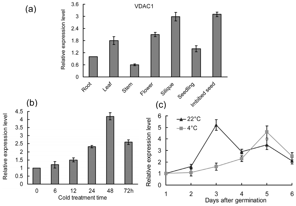
2.1. Expression Patterns of the VDAC1 in Arabidopsis

To better understand the physiological function of VDAC1 in Arabidopsis, we designed real-time PCR experiments to examine the genetic temporal and spatial expression changes in detail. As shown in Figure 1a, VDAC1 transcripts were abundant in imbibed seeds and siliques. AtVDAC1 expression was previously shown to be not affected by several abiotic stresses [18]. However, VDAC1 was promoted after 48 h under cold stress in mature plants in this study (Figure 1b). VDAC1 perhaps plays a role in cold response at the mature stage. In addition, VDAC1 was up-regulated at either 22 or 4 °C during seed germination (Figure 1c). The mRNA level of VDAC1 exhibited a sudden increase at 3 days or 5 days, when wild-type seeds were germinated at 22 or 4 °C, respectively, indicating VDAC1 involvement in seed germination.

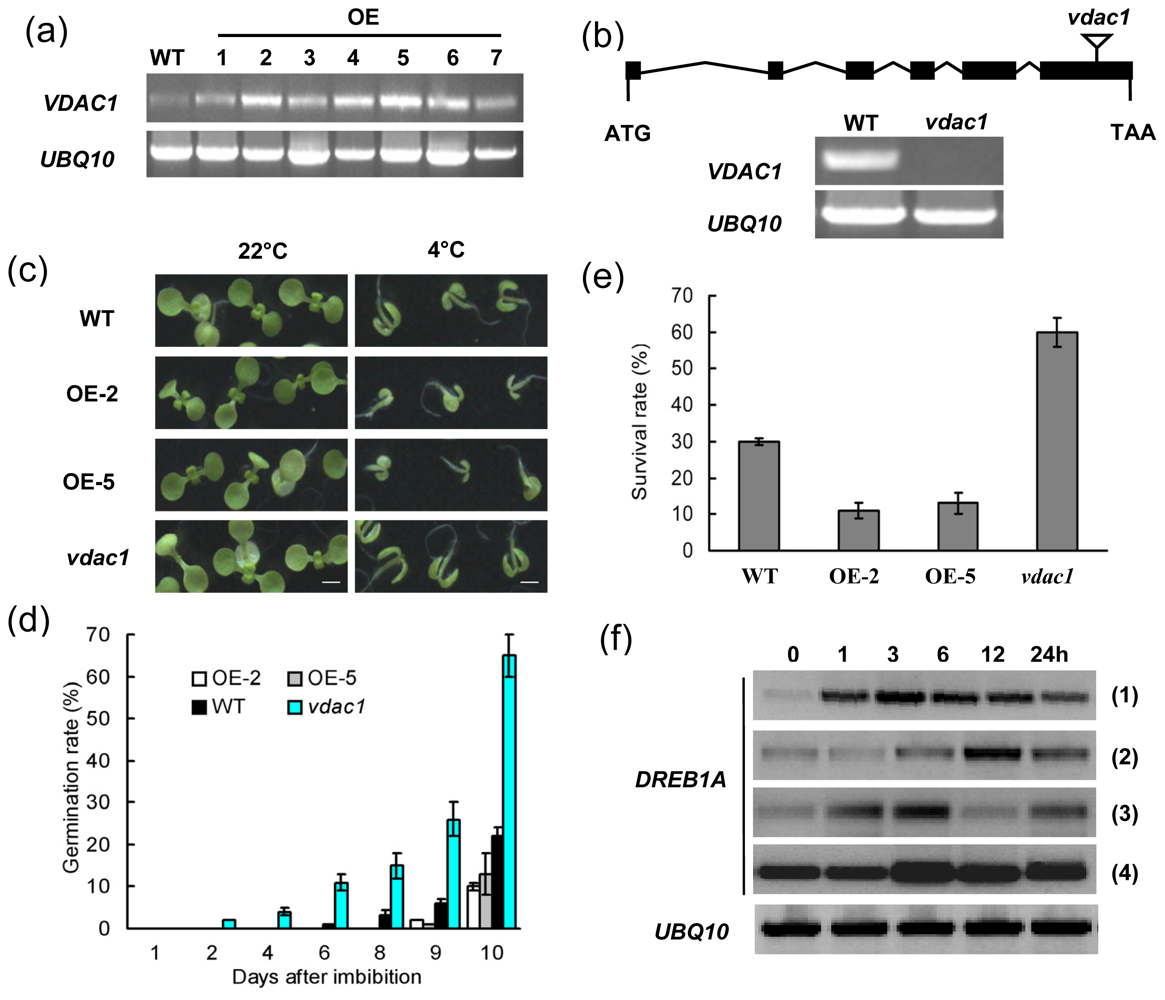
2.2. VDAC1 Overexpressing Arabidopsis Plants is Sensitive to Cold Stress

A comparative analysis was done to investigate the physiological roles of VDAC1 on germination and response to cold stress. To examine germination and seedling growth of VDAC1 overexpressing plants, a vdac1 mutant, and wild-type (WT) (Columbia) were examined under designated conditions.
Semi-quantitative analysis showed that VDAC1 mRNA levels in transgenic Arabidopsis lines were much higher level than in WT plants (Figure 2a). The vdac1 mutant with a T-DNA insertion site in the last exon of the genome sequence had no detectable VDAC1 transcript (Figure 2b). As shown in Figure 2c,d, there was no visible difference when seeds of the vdac1 mutant, transgenics and WT were germinated on MS medium under normal conditions (22 °C). However, seeds of WT and VDAC1 overexpressing lines (OE-2 and OE-5) germinated more slowly than the vdac1 mutant at 4 °C, and the root growth and cotyledon expansion of transgenic seedlings were severely inhibited compared with either WT or mutant plants. The germination rates of the VDAC1 overexpressing lines were even lower than the WT. These results demonstrated that VDAC1 functions during seed germination and seedling growth at low temperatures.
We also compared the survival rates of the VDAC1 overexpressing lines with those of WT and vdac1 mutant plants following cold stress. As shown in Figure 2e, the VDAC1 overexpressing lines were less tolerant of the freezing treatment than WT plants, whereas with the same treatment, 60% of vdac1 mutant plants survived compared with a 30% of WT plants. In addition, cold induction of DREB1A was less in the VDAC1 overexpressing lines compared with WT control plants (Figure 2f). Moreover, expression of DREB1A was obviously enhanced in vdac1 mutant plants relative to in WT. The results from the germination and freezing tolerance assays support the idea that VDAC1 functions cold stress response in plants.

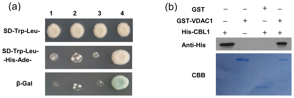
2.3. Interaction of VDAC1-CBL1

Interactions of proteins with other proteins or nucleic acids are important for most biological functions. A search for interacting partners is necessary to understand the functions of VDAC1. A positive interactor of VDAC1 was identified, and sequence analysis revealed that the VDAC1-interacting protein was CBL1. In yeast two-hybrid screening, strong growth on SD-Trp-Leu-Ade-His medium and activity of the reporter gene were observed only in yeast cells co-transformed with pGBKT7-VDAC1 and pGADT7-CBL1 (Figure 3a), indicating interaction of VDAC1 and CBL1 in yeast. His-CBL1 also physically interacted with GST-VDAC1, as shown by anti-His antibody on the resulting western blot (Figure 3b). These results demonstrated that VDAC1 interacts with CBL1.

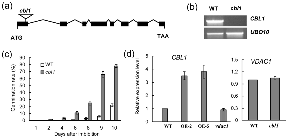
2.4. cbl1 Was Insensitive to Low Temperature during Seed Germination

Previous study showed that disruption of CBL1 promoted freezing stress tolerance in Arabidopsis [23]. Here, seed germination under cold treatment of a CBL1 T-DNA insertion mutant was evaluated. The cbl1 mutant had a T-DNA insertion site in the first exon of CBL1 and did not produce a detectable CBL1 transcript in a reverse transcription experiment (Figure 4a,b). There were no obvious morphological or developmental differences between WT and cbl1 mutant plants grown on MS medium at 22 °C. However, seeds of WT germinated more slowly than the cbl1 mutant on MS medium at 4 °C (Figure 4c). This suggests that CBL1 mutation alters cold stress response during seed germination.

3. Discussion

Plant research to date has mainly focused on identification and expression patterns of VDAC isoforms. There is limited data on the functions of VDAC genes. Specific roles could be determined through VDAC knock-down or knock-out experiments. Previous studies revealed that VDAC1 is associated with mitochondria, and its disruption affects pollen germination and pollen tube growth in Arabidopsis [20,21]. In this study, we gained insight into the novel roles of VDAC1 in Arabidopsis based on comparisons of overexpressing transgenic lines, wild-type and T-DNA insertion mutants.
The time, location and levels of gene transcripts are finely regulated in all organisms, ensuring normal development and optimal survival of plants under both normal or stress conditions. Spatiotemporal expression profiles of VDAC1 in Arabidopsis were described in transgenic Arabidopsis plants carrying VDAC1 promoter-GUS constructs [20]. The result showed that the VDAC1 promoter was active in shoot meristems and surrounding tissues. Here, VDAC1 transcripts were detected in all tested tissues and organs, which is consistent with the analysis of the microarray expression database [24]. However, it was strongly expressed in flowers and siliques (Figure 1a); not exactly the same as in the previous study, which showed the level of transcripts of VDAC1 decreases with the stage of development in Arabidopsis [11]. More research is needed to elucidate this significant difference. In addition, VDAC1 was obviously upregulated during seed germination (Figure 1c), which agrees with the data given by the Genvestigator microarray expression database [24], hence indicating an important role in seed germination. Previous studies showed that down-regulated VDAC expression, or expression based on mutants, could lead to changes in channel activities, such as cytosolic ATP levels and mitochondrial ATP-synthesis rates [2527]. For instance, reduction in the expression of VDAC1 in human cells led to a decrease in the ATP level, suggesting a demand on VDAC proteins for control of ATP flux [26]. Plant VDAC proteins may share similar functions in control of ATP levels, which are important for degradative metabolism. Germination of seeds following a dormant stage requires an efficient energy source that comes from degradative metabolism [28]. Thus, the high VDAC1 gene expression level observed in imbibed seeds could be explained by the energy demand for seed germination. Sequent study showed that VDAC1 overproduction increased cold stress sensitivity during both seed germination and seedling growth, whereas vdac1 T-DNA insertion lines exhibited a higher seed germination frequency than VDAC1 overexpressing and wild-type lines at 4 °C (Figure 2d). In addition, a loss-of-function mutant vdac1 in Arabidopsis showed enhanced tolerance to freezing (Figure 2e). These results suggest that VDAC1 plays a negative role in cold stress response.
In animal cells, several proteins were suggested to interact with VDAC [29]. These included mitochondrial creatinekinase, microtubule-associated protein 2, mitochondrial heat shock protein 70 and several glycolytic enzymes, such as hexokinase (Hxk), aldolase, and glyceraldehyde 3-phosphate dehydrogenase (GAPDH). In Arabidopsis plants, Hexokinase 1 (HXK1) is an interaction protein of VDAC, and AtHXK1 overexpressing transgenic plants have faster germination kinetics, which coincides with observations on seed germination of AtVDAC2 antisense transgenic lines in the presence of ABA [22,30]. To gain insights into how VDAC1 affects plant response to cold stress, we performed a yeast two-hybrid screen using VDAC1 as bait. A few polypeptides were identified as potential interaction partners, among which we focused on the Ca2+ sensor, CBL1.
In Arabidopsis, CBL1 is activated by various abiotic stresses and functions as a positive regulator in salt or drought stress responses [23]. It was also described as a negative regulator of cold response [23]. By contrast, VDAC1 was stable under salt, cold or drought stresses in Arabidopsis during 24 h detected by semi-quantitative RT-PCR [18], whereas its expression was enhanced in mature plants after 48 h under cold stress examined by real-time PCR method (Figure 1b), indicating a possible role in cold stress response. Overexpression of VDAC1 reduced freezing tolerance, whereas vdac1 mutant plants showed superior performance to WT under freezing stress, suggesting that VDAC1 functions as a negative regulator in cold stress response. This is consistent with the role of CBL1 in cold stress response. CBL1 was shown to alter the expression of a series of cold-induced genes when subjected to cold stress [23]. To test if VDAC1 and CBL1 could be involved in the regulation of a common set of genes, the expression of genes found to be altered by CBL1 under cold treatment were quantified in VDAC1 overexpressing and vdac1 mutant lines. Among these genes, the expression of DREB1A, a gene specifically responsive to cold, was inhibited in VDAC1-overexpressing plants, suggesting that VDAC1 overexpression exerted negative regulation on DREB1A. On the contrary, DREB1A was highly induced in vdac1 mutant plants (Figure 2f). These results indicate that VDAC1 and CBL1 have similar effects on the expression of DREB1A[23]. The vdac1 mutant also showed superior performance during seed germination under low temperature, and the phenotypes observed for cbl1 resembled those of vdac1 (Figure 4c). To determine if the visual phenotypes of the mutants at 4 °C were due to quicker germination, but not faster growth, seeds were germinated at 22 °C for two days before being transferred to 4 °C. The mutants did not present any obvious morphological differences (data not shown). These results demonstrated that both VDAC1 and CBL1 function during seed germination at low temperature, suggesting a biological relevance of the two genes.
To analyze a possible cross-regulation between VDAC1 and CBL1, their mRNA levels in cbl1 and VDAC1 overexpressing plants were quantified. As shown in Figure 4d, no significant differences in mRNA levels of VDAC1 were detected between the wild-type and cbl1 mutant plants. The mRNA level of CBL1 was also found to be stable in vdac1 mutant plants. However, CBL1 was induced more than three-fold in VDAC1 overexpressing plants. Previous studies showed that Ca2+ flux across the mitochondria outer membrane occurs through VDAC and VDAC has a role in regulation of the Ca2+ homoeostasis [31]. Perhaps, overproduction of VDAC1 promotes calcium flux from mitochondria and then triggers expression of CBL1, which functions as a unique Ca2+ sensor in plants [32]. VDAC proteins could regulate Ca2+ permeation to sense changes in the physiological state of the cell [31,33]. Deletion of VDAC1 may modify the perception of calcium influx by cold stress to result in altered cold-induced signaling and adaptation. However, whether and how VDAC proteins influence calcium flux in plant cells remain to be elucidated.
It was found that the VDAC (At5g15090) protein was improved during heat- and senescence-associated cell death, but its corresponding transcripts did not show any change [34,35]. And, three rice VDACs (OsVDAC1-3) were found to be up-regulated in roots during the recovery period from osmotic stress, whereas the overall VDAC protein levels stayed constant [5]. There might be no obvious linear relationship between the level of transcripts and that of the corresponding active protein products, which are the functional elements of cells, tissues and organisms [11]. These data suggest that there is a post-transcriptional regulation in VDAC protein level. In this study, expression of VDAC1 was promoted in normal conditions (22 °C) during germination. However, it seems that VDAC1 is not essential for germination in normal conditions (22 °C), as shown in Figure 2. In addition, VDAC1 were found to be promoted slowly, which displays some inconsistencies with the previous results. It perhaps is due to the post-transcriptional regulation of VDAC1 or other unexpected mechanisms. More research is needed to answer such questions.

4. Experimental Section

4.1. Plant Material and Growth Conditions

Seeds of Arabidopsis (Columbia strain), mutant lines vdac1 (SALK_034648) and cbl1 (SALK_110426) were surface sterilized with bleach and thoroughly washed five times with sterile water before incubation in a growth chamber following three days of cold treatment. For the mutant screen, seedlings were transferred from plates to soil and grown at 22 °C in a growth room with a 16 h photoperiod. All plant materials were harvested and stored at −80 °C for DNA and RNA isolation.

4.2. Screening of T-DNA Insertion Mutants

Plant DNA isolation was performed as described [36]. The locations of the T-DNA insertion sites in the vdac1 and cbl1 homozygous mutants were determined by direct sequencing of PCR products amplified using DNA as a template by the T-DNA border primer LBb1.3 [37] for SALK lines and gene-specific primers as follows: SALK_034648LP: 5′-TTATTACAGGCCAACAATGCC-3′ and SALK_034648RP: 5′-GTGATTGGCTCCAATGTCTTG-3′ for vdac1; SALK_110426LP: 5′-GGGCTACGATACATTGAATCG-3′ and SALK_110426RP: 5′-TTGATCGTCTGGTTTCGAATC-3′ for cbl1.
Homozygous mutant plants were further confirmed by RT-PCR with gene-specific primers of VDAC1 (GenBank accession number: AT3G01280): 5′-CTTGCCCTTGGTACTGATGTTT-3′ (forward) and 5′-GAGGGTGTTCTCATTGGTTGAG-3′ (reverse) and of CBL1 (GenBank accession number: AT4G17615): 5′-AATGAAACTGGCTGATGAAACC-3′ (forward) and 5′-CCTCCGAATGGAAGACAAAACT-3′ (reverse).

4.3. RNA Isolation and Real Time PCR

Total RNA was isolated from different plant materials using Trizol reagent (Takara, city, country), and reverse transcription were performed with 2 μg of total RNA for the first strand cDNA synthesis with a PrimeScript 1st Strand cDNA Synthesis kit (Takara, Dalian, China), according to the manufacturer’s instructions. Real-time PCR was performed with the following gene-specific primers: VDAC1, 5′-CAGAAGCTACGAGACCAGAAGTTG-3′ (forward) and 5′-CTATGGTACCATGAACCTTGCATG-3′ (reverse); and CBL1, 5′-TTTTTCCAGAGGCAATCATG-3′ (forward) and 5′-GCCCATTTGGTGGTATCTTC-3′ (reverse). Real-time PCR analyses were performed on an ABI7300 system with SYBR Premix Ex Taq II (Takara, Dalian, China), as previously described [38]. Relative quantitative results were calculated by normalization to UBQ10 (GenBank accession number: AT4G05320).

4.4. Construction of Expression Vector and Isolation of Transgenic Plants

The open reading frame of VDAC1 was amplified by PCR from the cDNA with the specific primers: 5′-AACCATGAGCAAAGGTCCAGGACTC-3′ (forward) and 5′-CTCAAGGTTTGAGAGAAGAGAGAGACC-3′ (reverse). PCR fragments were cloned into the pMD18-T vector (Takara, Dalian, China), and the resulting plasmid was sequenced. To construct the eukaryotic expression vector, the VDAC1 fragment was cloned into the pBI121 previously digested with BamHI and SacI. After sequencing, the transgenic Arabidopsis (Columbia accession) lines were obtained by Agrobacterium-mediated dip flora using 50 mg/L kanamycin as a selective agent as, previously described [39].

4.5. Seed Germination Assay

For seed germination assays, seeds from the different genotypes were harvested from plants grown simultaneously in a glasshouse and then stored for four weeks. For the seed germination assay at 4 °C, about 80 sterile seeds for each genotype were kept in an incubator at 4 °C with a 16 h photoperiod. Germination was scored by radicle emergence. For the cold tolerance assay, four-week-old plants were treated at 4 °C for two days and then −6 °C for 2 h before recovery under normal growing condition. Survival rates were determined after re-watering for two days.

4.6. Yeast Two-Hybrid Screening

An Arabidopsis seedling cDNA library was constructed in a pGADT7-Rec2 vector containing a GAL4 activation domain using Matchmaker Library Construction (Clontech) and then transformed into yeast strain AH109 (Clontech). For yeast two-hybrid assays, the PCR product of VDAC1 was cloned into the BamHI and SalI restriction sites of pGBKT7 vector (Clontech) to generate the pGBKT-VDAC1 bait vector. Screening of the cDNA library for candidate interaction partners of VDAC1 was performed using the MATCHMAKER two-hybrid system (Clontech). Transformants were selected by growth on a synthetic dropout (SD) medium that lacked Trp, Leu, Ade and His (SD-Trp-Leu-Ade-His), but supplemented with an optimal concentration of 3-amino-1,2,4-triazole (3-AT) to reduce any artificial interaction. Surviving clones were retransferred to SD-Trp-Leu-His-Ade-medium and assayed for β-galactosidase activity according to the manufacturer’s instructions (Clontech). For the yeast two-hybrid assay, the full coding sequence of CBL1 was amplified with the specific primers: 5′-CGTCGCCGCACTCACTTT-3′ (forward) and 5′-TCCCACCAATCACATAACC TAAA-3′ (reverse) and cloned into the BamHI and SacI restriction sites of the pGADT7-Rec2 vector.

4.7. GST Pull-Down Assay

For the GST pull-down assay, the full coding fragment of Arabidopsis VDAC1 was cloned into the BamHI and SalI restriction sites of the pGEX-4T-1 vector (Promega) to generate GST-VDAC1. The full length CBL1 was cloned into the BamHI and SacI restriction sites of the pET28a (+) vector (Novagen) to generate His-CBL1. The recombinant proteins GST-VDAC1 and His-CBL1 were expressed in Escherichia coli strain BL21 (DE3) by induction with 0.5 mM isopropyl-1-thio-b-d-thiogalactoside (IPTG) and purified with glutathione sepharose or Ni-activated His-binding resin (GE Healthcare) according to the manufacturers’ instructions. Aliquots of GST and GST-VDAC1 beads (100 mL beads containing 15 mg of protein) were incubated overnight at 4 °C with the purified His-CBL1 protein on a rotary incubator. After being washed with ice-cold phosphate-buffered saline for five times, they were re-suspended in SDS gel-loading buffer and then loaded on SDS-PAGE (12%, w/v) gels. Protein bound to GST-VDAC1 was detected by western blotting using an anti-His antibody and visualized using chemiluminescence following the manufacturer’s recommendations (Amersham plc., Amersham, UK).

5. Conclusions

In this study, we investigated the expression pattern of VDAC1 in Arabidopsis. The results showed that VDAC1 was redundant in imbibed seeds and induced by cold stress in mature plants. Further phenotype analysis confirmed that VDAC1 is involved in cold stress response during the seed germination and plant development. Using yeast two-hybrid system, a novel interactor of VDAC1 was identified as plant Ca2+ sensor CBL1. In summary, new insights were gained into the possible role of VDAC1 in Arabidopsis, particularly in showing a diversity of roles of the VDAC family in plants.

Acknowledgments

This research was financially supported by the National Natural Science Foundation of China (31171546) and International S & T Cooperation Key Projects of MoST (Grant No. 2009DFB30340). We are grateful to Qi Xie (Institute of Genetics and Developmental Biology, Chinese Academy of Sciences) for kindly providing seeds of the vdac1 mutant and Weihua Wu (China Agricultural University) for generously providing the cbl1. We also thank RA McIntosh (Plant Breeding Institute, University of Sydney) for critically reading the manuscript.

References

  1. Lee, A.C.; Xu, X.; Blachly-Dyson, E.; Forte, M.; Colombini, M. The role of yeast VDAC genes on the permeability of the mitochondrial outer membrane. J. Membr. Biol 1998, 161, 173–181. [Google Scholar]
  2. Sampson, M.J.; Lovell, R.S.; Craigen, W.J. The murine voltagedependent anion channel gene family. Conserved structure and function. J. Biol. Chem 1997, 272, 18966–18973. [Google Scholar]
  3. Clausen, C.; Ilkavets, I.; Thompson, R.; Philippar, K.; Vojta, A.; Möhlmann, T.; Neuhaus, E.; Fulgosi, H.; Soll, J. Intracellular localization of VDAC proteins in plants. Planta 2004, 220, 30–37. [Google Scholar]
  4. Roosens, N.; Al Bitar, F.; Jacobs, M.; Homble, F. Characterization of a cDNA encoding a rice mitochondrial voltage-dependent anion channel and its gene expression studied upon plant development and osmotic stress. BBA-Biomembranes 2000, 1463, 470–476. [Google Scholar]
  5. Al Bitar, F.; Roosens, N.; Smeyers, M.; Vauterin, M.; van, B.J.; Jacobs, M.; Homble, F. Sequence analysis, transcriptional and posttranscriptional regulation of the rice vdac family. BBA-Gene Struct. Expr 2003, 1625, 43–51. [Google Scholar]
  6. Tateda, C.; Yamashita, K.; Takahashi, F.; Kusano, T.; Takahashi, Y. Plant voltage-dependent anion channels are involved in host defense against Pseudomonas cichorii and in Bax-induced cell death. Plant Cell Rep 2009, 28, 41–51. [Google Scholar]
  7. Wandrey, M.; Trevaskis, B.; Brewin, N.; Udvardi, M.K. Molecular and cell biology of a family of voltage-dependent anion channel porins in Lotus japonicus. Plant Physiol 2004, 134, 182–193. [Google Scholar]
  8. Elkeles, A.; Breiman, A.; Zizi, M. Functional differences among wheat voltage-dependent anion channel (VDAC) isoforms expressed in yeast. Indication for the presence of a novel VDAC-modulating protein? J. Biol. Chem 1997, 272, 6252–6260. [Google Scholar]
  9. Wang, J.; Zhang, L.D.; Zuo, K.J.; Oian, H.M.; Cao, Y.F.; Tang, K.X. Cloning and expressional studies of the voltage-dependent anion channel gene from Brassica rapa L. J. Integr. Plant Biol 2006, 48, 197–203. [Google Scholar]
  10. Colombini, M. VDAC: The channel at the interface between mitochondria and the cytosol. Mol. Cell Biochem 2004, 256, 107–115. [Google Scholar]
  11. Homblé, F.; Krammer, E.M.; Prévost, M. Plant VDAC: Facts and speculations. Biochim. Biophys. Acta 2012, 1818, 1486–1501. [Google Scholar]
  12. Xu, X.; Decker, W.; Sampson, M.J.; Craigen, W.J.; Colombini, M. Mouse VDAC isoforms expressed in yeast: Channel properties and their roles in mitochondrial outer membrane permeability. J. Membr. Biol 1999, 170, 89–102. [Google Scholar]
  13. Shoshan-Barmatz, V.; Keinan, N.; Zaid, H. Uncovering the role of VDAC in the regulation of cell life and death. J. Bioenerg. Biomembr 2008, 40, 183–191. [Google Scholar]
  14. Salinas, T.; Duchêne, A.M.; Delage, L.; Nilsson, S.; Glaser, E.; Zaepfel, M.; Maréchal-Drouard, L. The voltage-dependent anion channel, a major component of the tRNA import machinery in plant mitochondria. Proc. Natl. Acad. Sci. USA 2006, 103, 18362–18367. [Google Scholar]
  15. Salinas, T.; Duchêne, A.M.; Maréchal-Drouard, L. Recent advances in tRNA mitochondrial import. Trends Biochem. Sci 2008, 33, 320–329. [Google Scholar]
  16. Sieber, F.; Placido, A.; El Farouk-Ameqrane, S.; Duchêne, A.M.; Maréchal-Drouard, L. A protein shuttle system to target RNA into mitochondria. Nucleic Acids Res 2011, 39, e96. [Google Scholar]
  17. Godbole, A.; Varghese, J.; Sarin, A.; Mathew, M.K. VDAC is a conserved element of death pathways in plant and animal systems. Biochim. Biophys. Acta 2003, 1642, 87–96. [Google Scholar]
  18. Lee, S.M.; Hoang, M.H.; Han, H.J.; Kim, H.S.; Lee, K.; Kim, K.E.; Kim, D.H.; Lee, S.Y.; Chung, W.S. Pathogen inducible voltage-dependent anion channel (AtVDAC) isoforms are localized to mitochondria membrane in Arabidopsis. Mol. Cells 2009, 27, 321–327. [Google Scholar]
  19. Desai, M.K.; Mishra, R.N.; Verma, D.; Nair, S.; Sopory, S.K.; Reddy, M.K. Structural and functional analysis of a salt stress inducible gene encoding voltage dependent anion channel (VDAC) from pearl millet (Pennisetum glaucum). Plant Physiol. Biochem 2006, 44, 483–493. [Google Scholar]
  20. Tateda, C.; Watanabe, K.; Kusano, T.; Takahashi, Y. Molecular and genetic characterization of the gene family encoding the voltage-dependent anion channel in Arabidopsis. J. Exp. Bot 2011, 62, 4773–4785. [Google Scholar]
  21. Robert, N.; d’Erfurth, I.; Marmagne, A.; Erhardt, M.; Allot, M.; Boivin, K.; Gissot, L.; Monachello, D.; Michaud, M.; Duchêne, A.M.; et al. Voltage-dependent-anion-channels (VDACs) in Arabidopsis have a dual localization in the cell but show a distinct role in mitochondria. Plant Mol. Biol 2012, 78, 431–446. [Google Scholar]
  22. Yan, J.P.; He, H.; Tong, S.B.; Zhang, W.R.; Wang, J.M.; Li, X.F.; Yang, Y. Voltage-dependent anion channel 2 of Arabidopsis thaliana (AtVDAC2) is involved in ABA-mediated early seedling development. Int. J. Mol. Sci 2009, 10, 2476–2486. [Google Scholar]
  23. Cheong, Y.H.; Kim, K.N.; Pandey, G.K.; Gupta, R.; Grant, J.J.; Luan, S. CBL1, a calcium sensor that differentially regulates salt, drought and cold responses in Arabidopsis. Plant Cell 2003, 15, 1833–1845. [Google Scholar]
  24. Zimmermann, P.; Hirsch-Hoffmann, M.; Hennig, L.; Gruissem, W. GENEVESTIGATOR. Arabidopsis microarray database and analysis toolbox. Plant Physiol 2004, 136, 2621–2632. [Google Scholar]
  25. Kmita, H.; Budzińska, M.; Stobienia, O. Modulation of the voltagedependent anion-selective channel by cytoplasmic proteins from wild type and the channel depleted cells of Saccharomyces cerevisiae. Acta Biochim. Polonica 2003, 50, 415–424. [Google Scholar]
  26. Abu-Hamad, S.; Sivan, S.; Shoshan-Barmatz, V. The expression level of the voltage-dependent anion channel controls life and death of the cell. Proc. Natl. Acad. Sci. USA 2006, 103, 5787–5792. [Google Scholar]
  27. Krauskopf, A.; Eriksson, O.; Craigen, W.J.; Forte, M.A.; Bernardi, P. Properties of the permeability transition in VDAC1(−/−) mitochondria. Biochim. Biophys. Acta 2006, 1757, 590–595. [Google Scholar]
  28. Perl, M. ATP synthesis and utilization in the early stage of seed germination in relation to seed dormancy and quality. Physiol. Plantarum 1986, 66, 177–182. [Google Scholar]
  29. Shoshan-Barmatz, V.; Israelson, A. The voltage-dependent anion channel in endoplasmic/sarcoplasmic reticulum: Characterization, modulation and possible function. J. Membr. Biol 2005, 204, 57–66. [Google Scholar]
  30. Balasubramanian, R.; Karve, A.; Kandasamy, M.; Meagher, R.B.; Moore, B. A role for F-actin in hexokinase-mediated glucose signaling. Plant Physiol 2007, 145, 1423–1434. [Google Scholar]
  31. Gincel, D.; Zaid, H.; Shoshan-Barmatz, V. Calcium binding and translocation by the voltage dependent anion channel: A possible regulatory mechanism in mitochondrial function. Biochem. J 2001, 358, 147–155. [Google Scholar]
  32. Luan, S.; Kudla, J.; Rodriguez-Concepcion, M.; Yalovsky, S.; Gruissem, W. Calmodulins and calcineurin B-like proteins: Calcium sensors for specific signal response coupling in plants. Plant Cell 2002, 14, S389–S400. [Google Scholar]
  33. Trewavas, A.J.; Malho, R. Ca2+ signaling in plant cells: The big network! Curr. Opin. Plant Biol 1998, 1, 428–433. [Google Scholar]
  34. Swidzinski, J.A.; Sweetlove, L.J.; Leaver, C.J. A custom microarray analysis of gene expression during programmed cell death in Arabidopsis thaliana. Plant J 2002, 30, 431–446. [Google Scholar]
  35. Swidzinski, J.A.; Leaver, C.J.; Sweetlove, L.J. A proteomic analysis of plant programmed cell death. Phytochemistry 2004, 65, 1829–1838. [Google Scholar]
  36. Murray, M.G.; Thompson, W.F. Rapid isolation of high molecular weight plant DNA. Nucleic Acids Res 1980, 8, 4321–4325. [Google Scholar]
  37. Li, Z.Y.; Xu, Z.S.; He, G.Y.; Yang, G.X.; Chen, M.; Li, L.C.; Ma, Y.Z. A mutation in Arabidopsis BSK5 encoding a brassinosteroid-signaling kinase protein affects responses to salinity and abscisic acid. Biochem. Biophys. Res. Commun 2012, 426, 522–527. [Google Scholar]
  38. Zhang, H.Y.; Mao, X.G.; Jing, R.L.; Chang, X.P.; Xie, H. Characterization of a common wheat (Triticum aestivum L.) TaSnRK2.7 gene involved in abiotic stress responses. J. Exp. Bot 2011, 62, 975–988. [Google Scholar]
  39. Xu, Z.S.; Xia, L.Q.; Chen, M.; Cheng, X.G.; Zhang, R.Y.; Li, L.C.; Zhao, Y.X.; Lu, Y.; Ni, Z.Y.; Liu, L.; et al. Isolation and molecular characterization of the Triticum aestivum L. ethylene-responsive factor 1 (TaERF1) that increases multiple stress tolerance. Plant Mol. Biol 2007, 65, 719–732. [Google Scholar]
Figure 1. Expression patterns of VDAC1 in Arabidopsis. (a) Expression of VDAC1 in various tissues or organs. The transcript level in roots was used as the control, where the VDAC1 mRNA level is given as 1. (b) Expression analysis of VDAC1 under cold treatment. (c) Real-time PCR analyses of VDAC1 expression in wild-type during seed germination under normal conditions (22 °C) or low temperature (4 °C). The expression of VDAC1 was normalized to the expression of UBQ10 in each PCR assay. Means and SD were calculated from three independent experiments.
Ijms 14 00701f1
Figure 2. Involvement of VDAC1 in seed germination and cold stress response. (a) RT-PCR expression analysis of VDAC1 in different lines. Transcripts of VDAC1 in wild-type (WT) and transgenic Arabidopsis lines (OE-1 to 7) were detected using semi-quantitative RT-PCR with 23 and 26 reaction cycles for VDAC1 and UBQ10, respectively. (b) VDAC1 gene structure and RT-PCR analysis of the WT and vdac1 mutant. Introns are shown as lines, and exons are shown as black boxes. The triangle indicates the location of the T-DNA insertion site in vdac1. The numbers of semi-quantitative RT-PCR reaction cycles for VDAC1 and UBQ10 were 26. (c) Representative images of different phenotypes at day 15 after imbibition at (22 °C) or 4 °C. Bars = 2 mm. (d) Seed germination frequencies in WT, vdac1 mutant and transgenic plants (OE-2 and OE-5) at 4 °C. Data points represent the mean ± SE of three independent biological determinations. (e) Survival rates after cold stress. Survival rates were calculated 48 h after re-watering. Data represent means ± SD (n = 20). (f) Semi-quantitative RT-PCR expression analysis of cold-responsive DREB1A gene with gene specific primers: 5′-TCTCTGAACCAGAGTCGTTT-3′ (forward) and 5′-CTTCTTCTCACCGTCTTCAC-3′ (reverse) in WT (1), transgenics (2 and 3) and vdac1 mutant (4) plants induced by cold stress. Similar results were obtained in at least three biological replicates. The UBQ10 gene was amplified as an internal control.
Ijms 14 00701f2
Figure 3. Interaction between VDAC1 and CBL1. (a) Yeast two-hybrid interactions. Vectors were co-introduced into yeast AH109 strain in different combinations: 1) pGADT7 and pGBKT7; 2) pGADT7-CBL1 and pGBKT7; 3) pGADT7 and pGBKT7-VDAC1; and 4) pGADT7-CBL1 and pGBKT7-VDAC1. Transformants were placed on the selection medium and grown for four days before the β-galactosidase (β-Gal) assay. (b) In vitro GST pull-down assay. GST-VDAC1 and His-CBL1 were expressed in E. coli and used for analysis. The presence or absence of each protein in the reaction mixture is shown as + or −, respectively. After transformed from the SDS-PAGE gel, the Polyvinylidene Fluoride (PVDF) membrane was stained with Coomassie Brilliant Blue (CBB). Unrelated His-tagged recombinant CBL1 protein at the left channel was used as a control. Experiments were performed three times, and a representative result is shown.
Ijms 14 00701f3
Figure 4. CBL1 is involved in seed germination at 4 °C. (a) The gene structure of CBL1 and T-DNA insertion sits in the cbl1 mutants. Introns are shown as lines and exons as black boxes. Triangle indicates the location of the T-DNA insertion site. (b) RT-PCR analysis of the WT and cbl1 mutant. The CBL1 transcript was absent in the cbl1 mutant. The UBQ10 transcript was amplified as an internal control. (c) Seed germination frequency in WT and cbl1 mutant at 4 °C. Data points represent the mean ± SE of three independent biological determinations. (d) Relative expression levels of VDAC1 and CBL1 are quantified in cbl1 and VDAC1 overexpressing plants (OE-2 and OE-5). Mean values were normalized to the transcript level of the internal control, UBQ10.
Ijms 14 00701f4

