<?xml version="1.0" encoding="UTF-8"?>
<!DOCTYPE article PUBLIC "-//NLM//DTD Journal Publishing DTD v2.3 20070202//EN" "journalpublishing.dtd">
<article xmlns:xlink="http://www.w3.org/1999/xlink" xml:lang="en" article-type="research-article">
<front>
<journal-meta>
<journal-id journal-id-type="publisher-id">ijms</journal-id>
<journal-title>International Journal of Molecular Sciences</journal-title>
<abbrev-journal-title>Int. J. Mol. Sci.</abbrev-journal-title>
<issn pub-type="epub">1422-0067</issn>
<publisher>
<publisher-name>Molecular Diversity Preservation International (MDPI)</publisher-name></publisher></journal-meta>
<article-meta>
<article-id pub-id-type="doi">10.3390/ijms14010532</article-id>
<article-id pub-id-type="publisher-id">ijms-14-00532</article-id>
<article-categories>
<subj-group>
<subject>Articles</subject></subj-group></article-categories>
<title-group>
<article-title>Ultraviolet B (UVB) Irradiation-Induced Apoptosis in Various Cell Lineages <italic>in Vitro</italic></article-title></title-group>
<contrib-group>
<contrib contrib-type="author">
<name><surname>Salucci</surname><given-names>Sara</given-names></name><xref ref-type="aff" rid="af1-ijms-14-00532">1</xref></contrib>
<contrib contrib-type="author">
<name><surname>Burattini</surname><given-names>Sabrina</given-names></name><xref ref-type="aff" rid="af1-ijms-14-00532">1</xref></contrib>
<contrib contrib-type="author">
<name><surname>Battistelli</surname><given-names>Michela</given-names></name><xref ref-type="aff" rid="af1-ijms-14-00532">1</xref></contrib>
<contrib contrib-type="author">
<name><surname>Baldassarri</surname><given-names>Valentina</given-names></name><xref ref-type="aff" rid="af1-ijms-14-00532">1</xref></contrib>
<contrib contrib-type="author">
<name><surname>Maltarello</surname><given-names>Maria Cristina</given-names></name><xref ref-type="aff" rid="af2-ijms-14-00532">2</xref></contrib>
<contrib contrib-type="author">
<name><surname>Falcieri</surname><given-names>Elisabetta</given-names></name><xref ref-type="aff" rid="af1-ijms-14-00532">1</xref><xref ref-type="aff" rid="af3-ijms-14-00532">3</xref><xref ref-type="corresp" rid="c1-ijms-14-00532">*</xref></contrib></contrib-group>
<aff id="af1-ijms-14-00532">
<label>1</label>DiSTeVA, University of Urbino “Carlo Bo”, Urbino 61029, Italy; E-Mails: <email>sara.salucci@uniurb.it</email> (S.S.); <email>sabrina.burattini@uniurb.it</email> (S.B.); <email>michela.battistelli@uniurb.it</email> (M.B.); <email>valentina.baldassarri@uniurb.it</email> (V.B.)</aff>
<aff id="af2-ijms-14-00532">
<label>2</label>Laboratory of Musculoskeletal Cell Biology, Rizzoli Orthopaedic Institute, Bologna 40136, Italy; E-Mail: <email>mariacristina.maltarello@ior.it</email></aff>
<aff id="af3-ijms-14-00532">
<label>3</label>IGM, CNR, Rizzoli Orthopaedic Institute, Bologna 40136, Italy</aff>
<author-notes>
<corresp id="c1-ijms-14-00532">
<label>*</label>Author to whom correspondence should be addressed; E-Mail: <email>elisabetta.falcieri@uniurb.it</email>; Tel.: +39-0722304284; Fax: +39-0722304244.</corresp></author-notes>
<pub-date pub-type="collection">
<year>2013</year></pub-date>
<pub-date pub-type="epub">
<day>27</day>
<month>12</month>
<year>2012</year></pub-date>
<volume>14</volume>
<issue>1</issue>
<fpage>532</fpage>
<lpage>546</lpage>
<history>
<date date-type="received">
<day>31</day>
<month>10</month>
<year>2012</year></date>
<date date-type="rev-recd">
<day>19</day>
<month>12</month>
<year>2012</year></date>
<date date-type="accepted">
<day>21</day>
<month>12</month>
<year>2012</year></date></history>
<permissions>
<copyright-statement>© 2013 by the authors; licensee Molecular Diversity Preservation International, Basel, Switzerland.</copyright-statement>
<copyright-year>2013</copyright-year>
<license license-type="open-access" xlink:href="http://creativecommons.org/licenses/by/3.0">
<p>This article is an open-access article distributed under the terms and conditions of the Creative Commons Attribution license (http://creativecommons.org/licenses/by/3.0/).</p></license></permissions>
<abstract>
<p>Ultraviolet B (UVB) radiation acts as a strong apoptotic trigger in many cell types, in tumor and normal cells. Several studies have demonstrated that UVB-induced cell death occurs through the generation of reactive oxygen species. The consequent oxidative stress includes the impairment of cellular antioxidants, the induction of DNA damage and the occurrence of apoptosis. In this review, we investigated UVB apoptotic action in various cell models by using ultrastructural, molecular and cytofluorimetric techniques. Myeloid leukemia HL-60, T-lymphoblastoid Molt-4 and myelomonocytic U937 human cells, generally affected by apoptotic stimuli, were studied. Human chondrocytes and C2C12 skeletal muscle cells, known to be more resistant to damage, were also considered. All of them, when exposed to UVB radiation, revealed a number of characteristic apoptotic markers. Membrane blebbing, cytoplasm shrinkage and chromatin condensation were detected by means of electron microscopy. DNA cleavage, investigated by using agarose gel electrophoresis and TUNEL reaction, was observed in suspended cells. Differently, in chondrocytes and in skeletal muscle cells, oligonucleosomic DNA fragmentation did not appear, even if a certain TUNEL positivity was detected. These findings demonstrate that UVB radiation appears to be an ideal tool to study the apoptotic behavior.</p></abstract>
<kwd-group>
<kwd>UVB</kwd>
<kwd>apoptosis</kwd>
<kwd>HL-60</kwd>
<kwd>Molt-4</kwd>
<kwd>U937</kwd>
<kwd>chondrocytes</kwd>
<kwd>skeletal muscle cells</kwd></kwd-group></article-meta></front>
<body>
<sec sec-type="intro">
<title>1. Introduction</title>
<p>UVB radiation-induced apoptosis has been extensively studied in human keratinocytes, which represent the major cellular target for solar UVB radiation [<xref ref-type="bibr" rid="b1-ijms-14-00532">1</xref>]. Skin cells undergo apoptosis because of irreversible DNA damage [<xref ref-type="bibr" rid="b2-ijms-14-00532">2</xref>], and it can prevent the accumulation of abnormal cells, which could lead to cutaneous malignancies [<xref ref-type="bibr" rid="b3-ijms-14-00532">3</xref>].</p>
<p>The molecular pathways leading to UVB radiation-induced apoptosis include the formation of cyclobutane pyrimidine dimers (CPDs) and photoproducts [<xref ref-type="bibr" rid="b4-ijms-14-00532">4</xref>,<xref ref-type="bibr" rid="b5-ijms-14-00532">5</xref>], the activation of death receptors, including CD95 (Fas/APO-1) [<xref ref-type="bibr" rid="b4-ijms-14-00532">4</xref>,<xref ref-type="bibr" rid="b6-ijms-14-00532">6</xref>], and the formation of reactive oxygen species (ROS) [<xref ref-type="bibr" rid="b7-ijms-14-00532">7</xref>].</p>
<p>Recently, several studies have demonstrated that many harmful effects of short-wavelength UVB rays (290–320 nm) occur through the generation of ROS [<xref ref-type="bibr" rid="b8-ijms-14-00532">8</xref>–<xref ref-type="bibr" rid="b10-ijms-14-00532">10</xref>]. Oxidative stress response of cells and tissues includes the induction of oxidative damage to various cellular components (e.g., membrane lipids, proteins and DNA) and the occurrence of inflammation, immunosuppression and apoptosis [<xref ref-type="bibr" rid="b11-ijms-14-00532">11</xref>–<xref ref-type="bibr" rid="b15-ijms-14-00532">15</xref>]. After UVB exposure, DNA double-strand breaks occur; they are particularly toxic and may not be correctly repaired. This could lead to chromosomal translocations and to the formation of highly unstable dicentric chromosomes or acentric chromosomal fragments [<xref ref-type="bibr" rid="b16-ijms-14-00532">16</xref>], which are correlated to the induction of apoptosis [<xref ref-type="bibr" rid="b17-ijms-14-00532">17</xref>].</p>
<p>The purpose of this review is to report and discuss the UVB radiation role in apoptotic cell death, induced <italic>in vitro</italic> in different cell lines.</p>
<p>In fact, UVB radiation is a known inducer of apoptosis in cultured cells [<xref ref-type="bibr" rid="b18-ijms-14-00532">18</xref>–<xref ref-type="bibr" rid="b21-ijms-14-00532">21</xref>]. It can trigger both the extrinsic and the intrinsic apoptotic pathways, but it remains unclear how these pathways are interrelated [<xref ref-type="bibr" rid="b22-ijms-14-00532">22</xref>]. Recent studies demonstrated that UVB-induced cell death mostly occurs through the intrinsic apoptotic pathway [<xref ref-type="bibr" rid="b23-ijms-14-00532">23</xref>,<xref ref-type="bibr" rid="b24-ijms-14-00532">24</xref>], even if the presence of caspase-independent mechanisms cannot be excluded. Anyhow, a mitochondrial involvement in UVB-induced apoptosis is certain. In fact, it is well known that UVB radiation alters the structure of the outer mitochondrial membrane, causing its permeabilization and the cytochrome c release [<xref ref-type="bibr" rid="b24-ijms-14-00532">24</xref>–<xref ref-type="bibr" rid="b26-ijms-14-00532">26</xref>].</p>
<p>Cell exposure to UVB is one of the best experimental systems to study apoptosis in response to DNA damage [<xref ref-type="bibr" rid="b27-ijms-14-00532">27</xref>,<xref ref-type="bibr" rid="b28-ijms-14-00532">28</xref>]. Morphological observations showed that low doses of UV induced apoptosis [<xref ref-type="bibr" rid="b27-ijms-14-00532">27</xref>], whereas higher doses triggered both apoptosis and necrosis [<xref ref-type="bibr" rid="b29-ijms-14-00532">29</xref>]. UVB, which is an oxidant and pro-apoptotic agent widely demonstrated in keratinocytes, melanocytes and epidermal cells [<xref ref-type="bibr" rid="b30-ijms-14-00532">30</xref>–<xref ref-type="bibr" rid="b32-ijms-14-00532">32</xref>], appeared also useful to study apoptotic behavior in other cell cultures <italic>in vitro.</italic></p>
<p>In the early years of our research, the UVB apoptotic effect was analyzed in different models: myeloid leukemia HL-60, T-lymphoblastoid Molt-4 and myelomonocytic U937 human tumor cell lines that grow in suspension. They have a high proliferative rate and, for this reason, they are usually more sensitive to cell death stimuli. As described in the literature, UVB radiation induces apoptosis rapidly and essentially in the entire cell population (about 90%). This aspect has been discussed in several reports in which apoptosis of human myeloblastic cells, induced by UV radiation, has been investigated [<xref ref-type="bibr" rid="b18-ijms-14-00532">18</xref>,<xref ref-type="bibr" rid="b33-ijms-14-00532">33</xref>].</p>
<p>Several studies, in the last decade, showed that apoptosis, necessary for maintaining tissue homeostasis, plays an active role not only in carcinogenesis [<xref ref-type="bibr" rid="b7-ijms-14-00532">7</xref>,<xref ref-type="bibr" rid="b34-ijms-14-00532">34</xref>]. It contributes to articular cartilage damage in osteoarthritis, and it is correlated to a number of cartilage disorders [<xref ref-type="bibr" rid="b35-ijms-14-00532">35</xref>]. It is also necessary for the regulation of skeletal muscle homeostasis, and it has been described in several myopathies, as well as after denervation or disuse [<xref ref-type="bibr" rid="b36-ijms-14-00532">36</xref>,<xref ref-type="bibr" rid="b37-ijms-14-00532">37</xref>].</p>
<p>Thus, the UVB-induced DNA damage was recently investigated by our group in human chondrocytes and in C2C12 murine skeletal muscle cells, both known to be somehow apoptosis-resistant. Differently from suspended cell models, routinely used to study apoptosis, primary cell lines (chondrocytes) and murine skeletal myoblasts originally derived from satellite stem cells appeared as complex cell systems in which several molecules and genes are involved in promoting cell survival or driving the apoptotic process. For these reasons, they can be considered more resistant to apoptotic induction if compared to leukemic cells.</p>
<p>In this review, to obtain a massive apoptosis, cells were exposed to UVB (dose 72 J/cm<sup>2</sup>, wavelength 312 nm, Transilluminator 2000, Bio-Rad Laboratories, Hercules, CA, USA) for 30 min at room temperature, at a distance of 1 cm, and then post-incubated for 4 h at 37 °C in humidified air with 5% CO<sub>2</sub>. Different UVB exposure times were considered for chondrocytes and C2C12 cells, but after 1 h of treatment, a massive necrotic cell death appeared. Thus, 30 min of UVB exposure has been chosen for our experiments with all cell types [<xref ref-type="bibr" rid="b18-ijms-14-00532">18</xref>,<xref ref-type="bibr" rid="b25-ijms-14-00532">25</xref>].</p>
<p>This review demonstrates that UVB radiation induces apoptosis in various cell lines using different approaches: Scanning (SEM) and Transmission (TEM) Electron Microscopy used to evidence morphological features, such as surface blebbing, cytoplasm shrinkage and chromatin condensation [<xref ref-type="bibr" rid="b38-ijms-14-00532">38</xref>–<xref ref-type="bibr" rid="b41-ijms-14-00532">41</xref>]; agarose gel electrophoresis and TUNEL reaction, the latter both at fluorescence and electron microscopy, used to investigate DNA [<xref ref-type="bibr" rid="b42-ijms-14-00532">42</xref>–<xref ref-type="bibr" rid="b44-ijms-14-00532">44</xref>]; and flow cytometry used to analyze cell cycle patterns and mitochondrial activity [<xref ref-type="bibr" rid="b45-ijms-14-00532">45</xref>,<xref ref-type="bibr" rid="b46-ijms-14-00532">46</xref>].</p>
<sec>
<title>
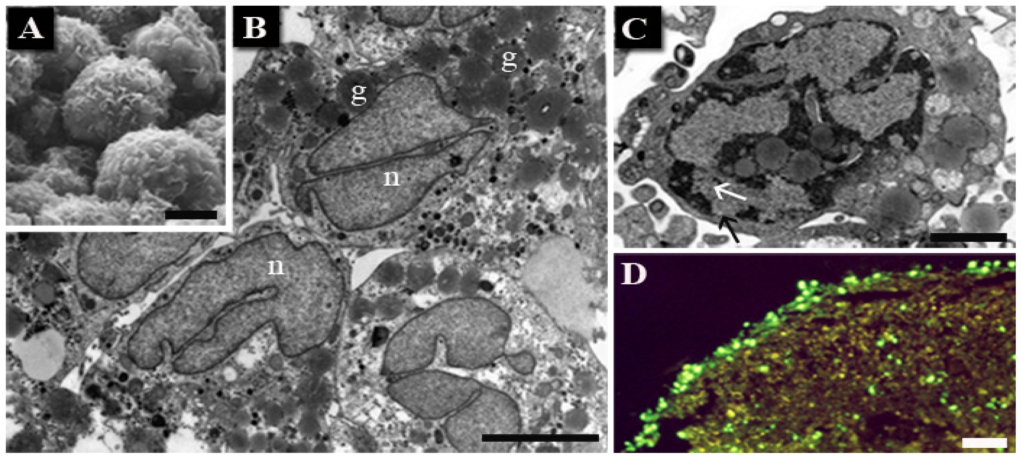
1.1. Human Hemopoietic Cells
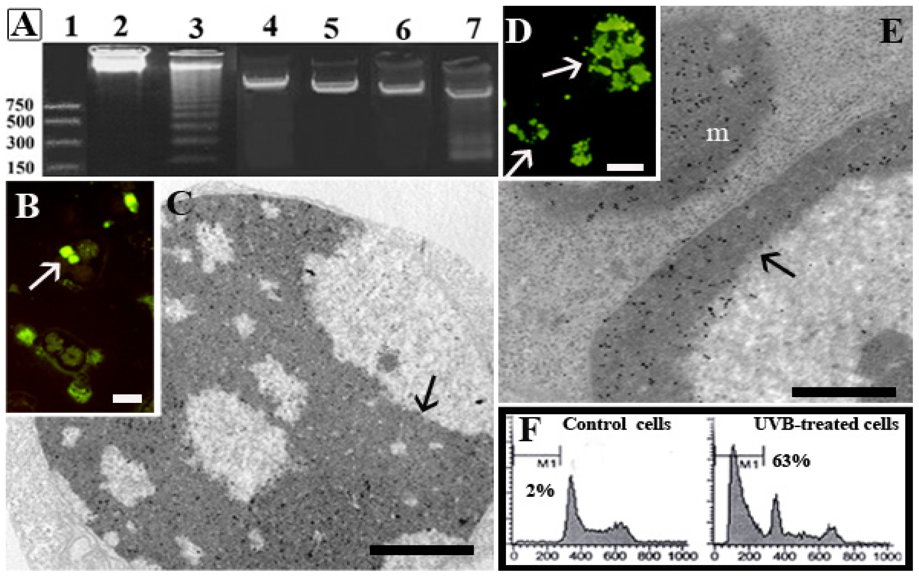
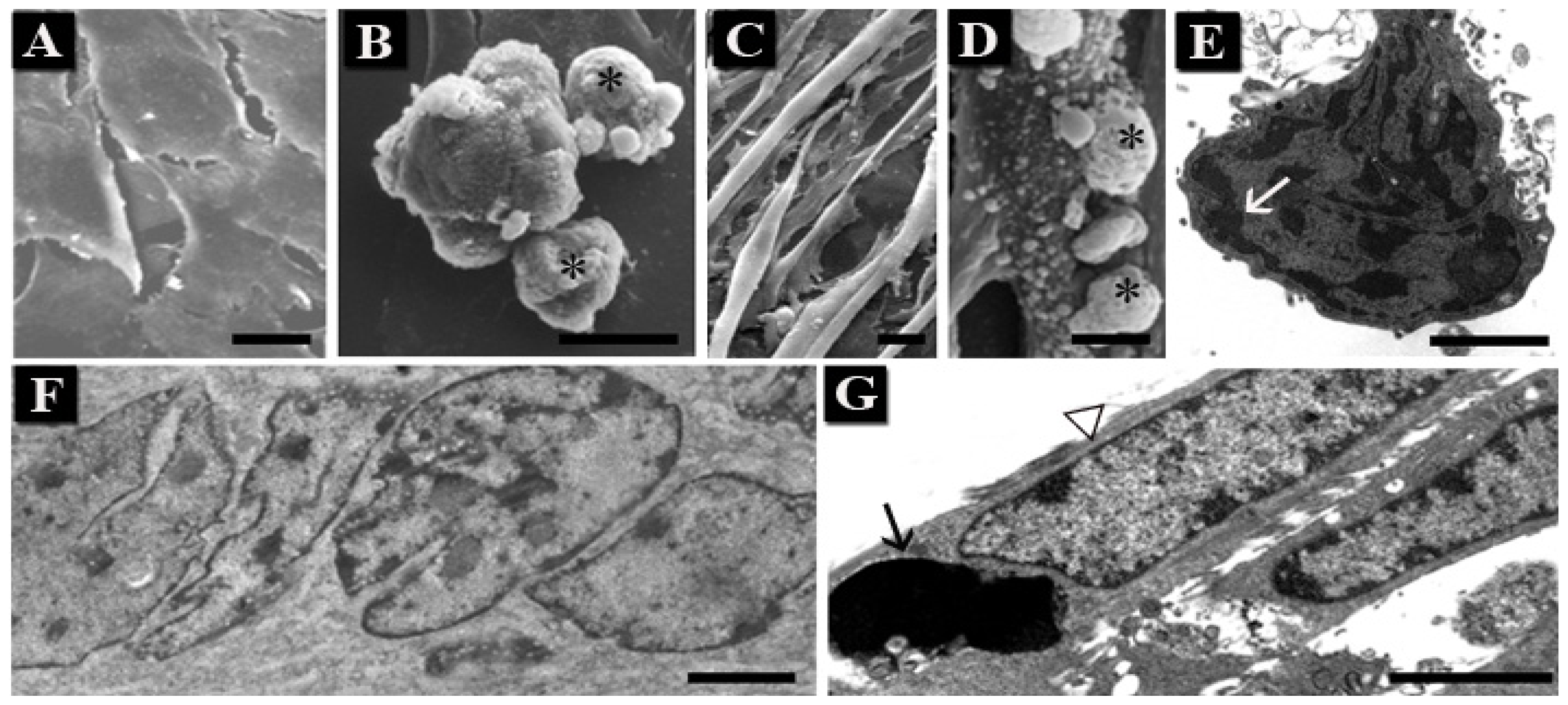
</title>
<p>HL-60 (human myeloid leukemia cells), Molt-4 (T-lymphoblastoid cells) and U937 (myelomonocytic human leukemia cells) are round mononuclear cells, with microvilli. It is known that HL-60 and other human cell lines were found to rapidly undergo apoptosis after short periods of UV radiation, whereas prolonged exposure induced necrotic cell death [<xref ref-type="bibr" rid="b47-ijms-14-00532">47</xref>].</p>
<p>Thus, cells were exposed to 30 min UVB, post-incubated for 4 h in growing media and then processed for ultrastructural, molecular and cytofluorimetric investigations. After UVB treatment, all cell types showed apoptotic patterns (<xref ref-type="fig" rid="f1-ijms-14-00532">Figure 1</xref>). Ultrastructural observations evidenced surface blebbing (<xref ref-type="fig" rid="f1-ijms-14-00532">Figure 1B</xref>), cytoplasm shrinkage and chromatin condensation (<xref ref-type="fig" rid="f1-ijms-14-00532">Figure 1D,E</xref>) [<xref ref-type="bibr" rid="b43-ijms-14-00532">43</xref>,<xref ref-type="bibr" rid="b46-ijms-14-00532">46</xref>]. Control and UVB-treated specimens were processed for SEM and TEM, as previously reported [<xref ref-type="bibr" rid="b39-ijms-14-00532">39</xref>].</p>
<p>Renò <italic>et al.</italic> 1998 [<xref ref-type="bibr" rid="b48-ijms-14-00532">48</xref>] studied the plasma membrane behavior in HL-60 and Molt-4 cells after UVB exposure, to investigate its involvement in apoptosis. The results showed that during the early stages of apoptosis, a membrane lipid rearrangement occurs and involves phosphatidylserine translocation from the inner to the outer leaflet, independently from nuclear activity. Moreover, in <xref ref-type="fig" rid="f2-ijms-14-00532">Figure 2</xref>, DNA behavior has been also investigated showing that in HL60, a widely studied leukemia cell line, the oligonucleosomic DNA cleavage occurred (<xref ref-type="fig" rid="f2-ijms-14-00532">Figure 2A</xref>, lane 3). On the other hand, in Molt-4, oligonucleosomic DNA fragmentation was not observed (<xref ref-type="fig" rid="f2-ijms-14-00532">Figure 2A</xref>, lane 5), even in the presence of typical apoptotic features: chromatin condensation, cell shrinkage with preservation of the plasma membrane structure, nuclear splitting and micronuclei formation. Molt-4 cell response to UVB was investigated not only at the standard post-incubation time (<italic>i.e.</italic>, 4 h), but also at 6, 8, 10 and 24 h and DNA cleavage never occurred [<xref ref-type="bibr" rid="b44-ijms-14-00532">44</xref>]. The lack of endonuclease activation can be postulated in this apoptotic model, despite the existence of a metabolic pathway responsible for chromatin rearrangement and other apoptotic patterns [<xref ref-type="bibr" rid="b38-ijms-14-00532">38</xref>]. In addition, after conventional agarose gel electrophoresis, a typical DNA ladder appeared in U937 cells, too (<xref ref-type="fig" rid="f2-ijms-14-00532">Figure 2A</xref>, lane 7) [<xref ref-type="bibr" rid="b44-ijms-14-00532">44</xref>], as also described in another work [<xref ref-type="bibr" rid="b49-ijms-14-00532">49</xref>].</p>
<p>Cells were processed for TUNEL reaction at fluorescence microscope (TUNEL/FM). Moreover, to better highlight DNA fragmentation, the same technique was adapted for electron microscopy (TUNEL/TEM) [<xref ref-type="bibr" rid="b50-ijms-14-00532">50</xref>,<xref ref-type="bibr" rid="b51-ijms-14-00532">51</xref>]. In this way, a precise localization of DNA break points within the different chromatin domains was obtained (<xref ref-type="fig" rid="f2-ijms-14-00532">Figure 2</xref>). In UVB-treated Molt-4 cells, TUNEL/FM showed several positive cells (<xref ref-type="fig" rid="f2-ijms-14-00532">Figure 2B</xref>), also observed by TUNEL/TEM. The latter revealed a number of nuclear gold particles in the dense chromatin (<xref ref-type="fig" rid="f2-ijms-14-00532">Figure 2C</xref>). A peculiar chromatin behavior, reported to as “moth-eaten” [<xref ref-type="bibr" rid="b45-ijms-14-00532">45</xref>], was observed in this cell lineage exposed to UVB, where small negative areas of diffuse chromatin scattered throughout the positive dense chromatin.</p>
<p>Numerous U937 cells with homogeneously fluorescent micronuclei (<xref ref-type="fig" rid="f2-ijms-14-00532">Figure 2D</xref>) appeared at TUNEL/FM. An intense gold labeling (TUNEL/TEM) was observed in dense apoptotic chromatin and micronuclei (<xref ref-type="fig" rid="f2-ijms-14-00532">Figure 2E</xref>) [<xref ref-type="bibr" rid="b52-ijms-14-00532">52</xref>].</p>
<p>Moreover, Luchetti <italic>et al.</italic> 2006 [<xref ref-type="bibr" rid="b46-ijms-14-00532">46</xref>] studied melatonin antiapoptic activity in UVB-treated U937 cells, analyzing the cell cycle profile by means of flow cytometry. A conspicuous hypodiploid peak appeared after UVB treatment (<xref ref-type="fig" rid="f2-ijms-14-00532">Figure 2F</xref>), revealing an apoptotic cell population with DNA cleavage also evidenced by Liu <italic>et al.</italic> 2005 [<xref ref-type="bibr" rid="b53-ijms-14-00532">53</xref>] in the study on oridonin role in enhancing phagocytosis of UV-irradiated apoptotic U937 cells. Moreover, in this cell line, mitochondrial activity was investigated using mitochondrial fluorescent probes, such as Mito Tracker and JC-1, that revealed an alteration of mitochondrial membrane potential. This event has been evidenced using the cardiolipin-sensitive probe 10-nonyl acridine orange (NAO), to monitor changes in mitochondrial lipids. A decrease in cardiolipin content, induced by ROS increase, occurred in concomitance with mitochondrial permeability alteration and, successively, with the release of cytochrome c into the cytosol [<xref ref-type="bibr" rid="b54-ijms-14-00532">54</xref>].</p></sec>
<sec>
<title>1.2. Chondrocytes</title>
<p>After local Ethics Committee approval, fragments of articular cartilage were obtained from 16 patients (mean age 67 years, range 41–81 years) who were undergoing knee replacement. The tissue was finely minced and subject to enzymatic digestion; primary chondrocytes were cultured in micromass [<xref ref-type="bibr" rid="b35-ijms-14-00532">35</xref>], which represent a convenient model to study chondrocyte biology [<xref ref-type="bibr" rid="b55-ijms-14-00532">55</xref>] and, in particular, their death, in the context of a tridimensional culture model.</p>
<p>Chondrocyte morphology (<xref ref-type="fig" rid="f3-ijms-14-00532">Figure 3</xref>) in control condition appears very similar to that of human articular cartilage. Cells are round or slightly elongated with a plurilobated nucleus and dispersed chromatin. Large amount of glycogen masses and lipid granules can be observed scattered throughout the cytoplasm. Proteoglycans and collagen fibers are present in the intercellular space, indicating a good extra-cellular matrix production (<xref ref-type="fig" rid="f3-ijms-14-00532">Figure 3A,B</xref>).</p>
<p>After UVB treatment, chromatin condensation appears, even if dense cup-shaped masses, comparable to those of more classic apoptotic models, could not be found. The evidenced nuclear features, when analyzed in detail, suggest apoptosis [<xref ref-type="bibr" rid="b56-ijms-14-00532">56</xref>].</p>
<p>Gel electrophoresis did not show oligonucleosomic DNA cleavage (data not shown). Nevertheless, after UVB exposure, TUNEL evidenced the presence of positive nuclei, in particular in chondrocytes at the micromass periphery (<xref ref-type="fig" rid="f3-ijms-14-00532">Figure 3D</xref>).</p></sec>
<sec>
<title>
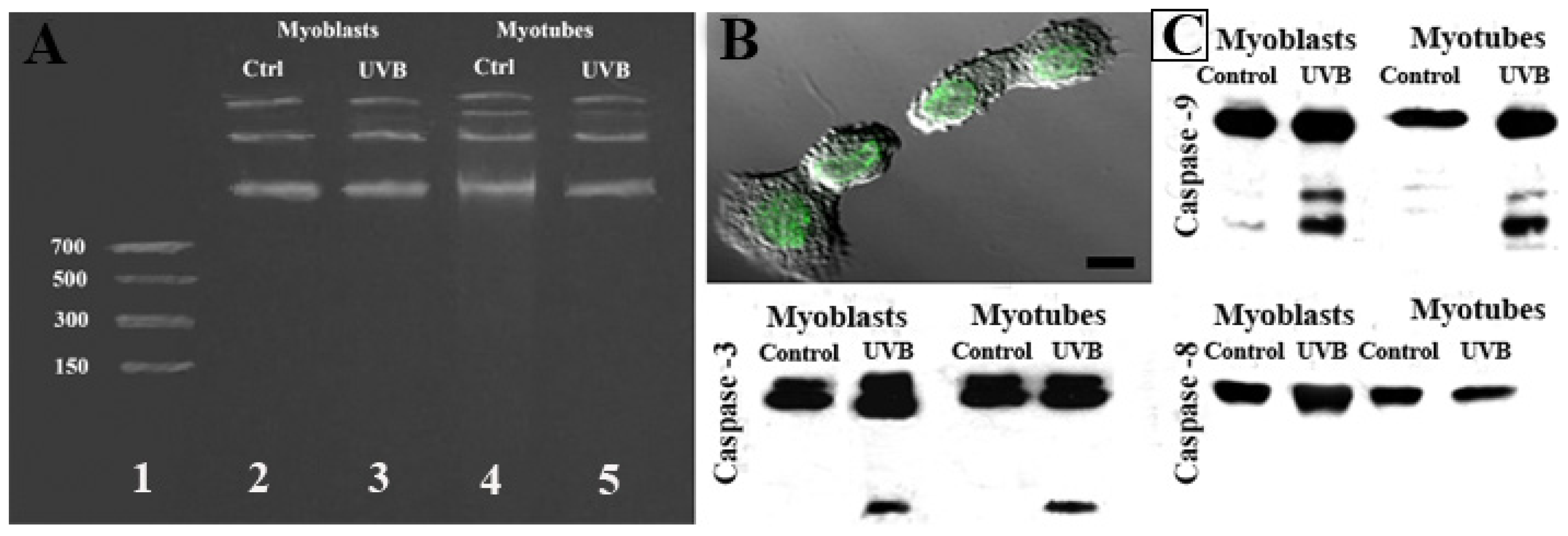
1.3. Skeletal Muscle Cells</title>
<p>Murine C2C12 myoblasts have been widely used as a model to study apoptosis in developing muscle, because extensive cell death occurs during myogenic differentiation [<xref ref-type="bibr" rid="b57-ijms-14-00532">57</xref>].</p>
<p>C2C12 is an adherent cell line of murine myoblasts that can be induced to differentiate <italic>in vitro</italic> into multinucleated myotubes, which progressively become muscle fibers [<xref ref-type="bibr" rid="b58-ijms-14-00532">58</xref>]. These cells are considered apoptosis-resistant, in particular myotubes [<xref ref-type="bibr" rid="b59-ijms-14-00532">59</xref>], but when exposed to UVB, undifferentiated and differentiated C2C12 showed a certain number of apoptotic features (<xref ref-type="fig" rid="f4-ijms-14-00532">Figure 4</xref>). Most cells began to detach from the substrate and membrane blebbing could be observed at SEM (<xref ref-type="fig" rid="f4-ijms-14-00532">Figure 4B,D</xref>).</p>
<p>Apoptotic nuclei showed condensed chromatin, which marginated beneath the nuclear membrane, occasionally forming micronuclei, even if the classical cup-shaped masses were not visible (<xref ref-type="fig" rid="f4-ijms-14-00532">Figure 4E</xref>). In addition, the simultaneous presence of normal and apoptotic nuclei inside the same myotube (<xref ref-type="fig" rid="f4-ijms-14-00532">Figure 4G</xref>) has been observed at TEM, suggesting a compartmentalization in nuclear domains. In the report by D’Emilio <italic>et al.</italic>[<xref ref-type="bibr" rid="b25-ijms-14-00532">25</xref>] we hypothesized, indeed, the presence of territorial areas, which maintain along the syncytium, a certain specificity and the capability to differently respond to external stimuli.</p>
<p>Furthermore, after UVB radiation of C2C12 muscle cells, autophagy appeared (<xref ref-type="fig" rid="f5-ijms-14-00532">Figure 5</xref>). This process, recognizable by the presence of cytoplasmic vacuoles filled with membranes, organelles and mitochondria remnants, is clearly evident in treated myotubes, while absent in control condition.</p>
<p>In <xref ref-type="fig" rid="f6-ijms-14-00532">Figure 6</xref>, agarose gel electrophoresis did not show oligonucleosomic DNA cleavage in myoblasts nor in myotubes (<xref ref-type="fig" rid="f6-ijms-14-00532">Figure 6A</xref>), but the TUNEL technique revealed a diffuse positivity (<xref ref-type="fig" rid="f6-ijms-14-00532">Figure 6B</xref>) and confirmed the presence of positive and negative nuclei inside the same myotube (data not shown). Moreover, molecular analyses evidenced, in both myoblasts and myotubes, the activation of caspase-9 and -3, but not caspase-8 (<xref ref-type="fig" rid="f6-ijms-14-00532">Figure 6C</xref>). D’Emilio <italic>et al.</italic> 2010 [<xref ref-type="bibr" rid="b25-ijms-14-00532">25</xref>], demonstrated that caspase-9 and -3 inhibitors only partially reduced the apoptotic rate, suggesting that apoptosis occurred by following both caspase-dependent and -independent pathways.</p></sec></sec>
<sec sec-type="conclusions">
<title>2. Conclusions</title>
<p>In this review, the apoptotic behavior of different cell lineages after UVB exposure has been investigated by means of several techniques, evidencing that UVB radiation plays a central role in the induction of apoptotic cell death. UVB is a powerful apoptotic stimulus in hemopoietic tumor cells. In these models, classical morphological apoptotic features were indeed observed, together with oligonucleosomic DNA cleavage evidenced after electrophoresis, TUNEL-FM and -TEM and cell cycle analysis. Cytofluorimetric analyses, used to test mitochondrial activity, showed that UVB induced cardiolipin decrease with alteration of mitochondrial membrane permeabilization and, as a consequence, cytochrome c release [<xref ref-type="bibr" rid="b54-ijms-14-00532">54</xref>]. In suspended cells, UVB induced apoptosis mostly follows the mitochondrial pathway. Luchetti <italic>et al.</italic> 2009 [<xref ref-type="bibr" rid="b18-ijms-14-00532">18</xref>] evidenced that melatonin was able to downregulate superoxide anion production, mitochondrial damage and caspase-dependent apoptosis in U937 cells exposed to UVB. In keratinocytes, UV-induced apoptosis is a complex event and involves different pathways. Apoptosis can be triggered by direct DNA damage, ROS production and death receptor activation and mitochondrial damage [<xref ref-type="bibr" rid="b12-ijms-14-00532">12</xref>,<xref ref-type="bibr" rid="b27-ijms-14-00532">27</xref>,<xref ref-type="bibr" rid="b60-ijms-14-00532">60</xref>–<xref ref-type="bibr" rid="b62-ijms-14-00532">62</xref>]. Several studies [<xref ref-type="bibr" rid="b26-ijms-14-00532">26</xref>,<xref ref-type="bibr" rid="b63-ijms-14-00532">63</xref>] discussed the molecular mechanisms of UV-induced apoptosis in keratinocytes, demonstrating that mitochondrial changes and cytochrome c release are involved and can be considered the predominant mechanisms in UV-mediated apoptosis.</p>
<p>UVB application to other cell models has been considered; chondrocytes and skeletal muscle cells, even if known to be resistant to apoptosis, were studied. Intriguingly, chondrocytes appeared sensitive to UVB treatment, at ultrastructural and molecular analyses, showing chromatin condensation, pore clustering and DNA fragmentation only revealed by TUNEL reaction, whereas DNA fragmentation was absent.</p>
<p>Moreover, as also reported by D’Emilio <italic>et al.</italic> 2010 [<xref ref-type="bibr" rid="b25-ijms-14-00532">25</xref>], skeletal muscle cells, as undifferentiated myoblasts and in the form of highly differentiate myotubes, appeared sensitive to UVB-induced apoptosis.</p>
<p>Chromatin changes, with progressive compaction and formation of micronuclei, were identified after ultrastructural observations. In this cell line, oligonucleosomic DNA cleavage does not seem to occur [<xref ref-type="bibr" rid="b15-ijms-14-00532">15</xref>], but double-strand DNA breaks are shown by TUNEL reaction. The presence of normal and apoptotic nuclei inside the same syncytium seems to indicate that each myonucleus regulates a certain fiber volume, suggesting a cytoplasmic compartmentalization in nuclear domains. Thus, individual myonuclear apoptosis, as well as complete cell death, can occur, as previously described by other authors [<xref ref-type="bibr" rid="b64-ijms-14-00532">64</xref>,<xref ref-type="bibr" rid="b65-ijms-14-00532">65</xref>]. The partial inhibition of apoptosis by caspase inhibitors (both -9 and -3) indicates that UVB radiation is able to trigger apoptosis through the activation of the mitochondrial pathway, as occurs in epidermal cells [<xref ref-type="bibr" rid="b32-ijms-14-00532">32</xref>]. Moreover, as reported by Sitailo <italic>et al.</italic> 2002 [<xref ref-type="bibr" rid="b63-ijms-14-00532">63</xref>], in keratinocytes, UV radiation triggers activation of caspase-3, -9 and -8. UVB radiation activates primarily the mitochondrial or intrinsic apoptotic pathway, resulting in activation of procaspase-9, whereas activation of procaspase-8 via death receptors is a relatively minor pathway. As a consequence, UVB is able to induce apoptosis through the mitochondrial pathway in all cell systems considered in this review, as well as in epidermal cells.</p>
<p>Furthermore, molecular analyses demonstrated that DNA cleavage on agarose gel electrophoresis appeared only in U937 and HL-60 cell lines. Molt-4 cells exposed to UVB showed the lack of DNA fragmentation even if morphological apoptotic features appeared in the presence of TUNEL reaction positivity, detected both by fluorescence and electron microscopy. The lack of DNA cleavage can be linked to endonuclease absence [<xref ref-type="bibr" rid="b66-ijms-14-00532">66</xref>] or, as suggested by Yanagisawa-Shiota <italic>et al.</italic> 1995 [<xref ref-type="bibr" rid="b67-ijms-14-00532">67</xref>], endonuclease activity might be differently regulated in myelogenous (U937, HL-60) and non-myelogenous leukemic cell lines (Molt-4).</p>
<p>In our experiments, oligonucleosomic DNA fragmentation did not appear after UVB exposure in chondrocytes and skeletal muscle cells. The DNase responsible for the apoptotic DNA laddering and the precise molecular mechanisms are poorly understood in the case of these cell lines. Shiokawa <italic>et al.</italic> 2002 [<xref ref-type="bibr" rid="b57-ijms-14-00532">57</xref>] demonstrated that an olinucleosomic DNA fragmentation occurred in myoblasts only when apoptosis appeared during induction of differentiation.</p>
<p>Moreover, in muscle cells, autophagy was observed after UVB exposure. Recent papers, evaluating the induction of LC3 lipidation and the increase of beclin-1 expression, demonstrated that UVB radiation induces autophagy in epidermal cells [<xref ref-type="bibr" rid="b68-ijms-14-00532">68</xref>], where it appeared to be a protective response to damage. Inhibition of autophagy exacerbated UVB-induced cell death, whereas its stimulation provided protection [<xref ref-type="bibr" rid="b69-ijms-14-00532">69</xref>,<xref ref-type="bibr" rid="b70-ijms-14-00532">70</xref>]. It is known that autophagy is an important catabolic program fundamental for tissue homeostasis; in fact, it promotes physiological protein turnover and removes damaged proteins and organelles [<xref ref-type="bibr" rid="b69-ijms-14-00532">69</xref>]. However, the role of autophagy as a mechanism of cell survival or death is still controversial, even if some evidences demonstrated its protective action against cell death after UV radiation [<xref ref-type="bibr" rid="b71-ijms-14-00532">71</xref>].</p>
<p>In C2C12 control cells, as demonstrated by our images, autophagic vacuoles were not observed. After UVB-induced cell death, both myoblasts and myotubes evidenced a certain number of autophagic vacuoles, which appeared to be more diffuse in cells that do not show apoptotic changes.</p>
<p>Finally, these data demonstrated that UVB radiation is ideal to trigger apoptosis in different cell models, where they mainly act through the mitochondrial pathway. Thus, in all cell types considered in this review, as well as in skin cells, which are the major target of solar radiations, UVB seems to induce apoptosis through the same pathway. Unexpectedly, in skeletal muscle cells, as well as in keratynocytes, as recently evidenced by other works, UVB radiation induced autophagy; further studies are required to investigate its role through molecular and biochemical approaches.</p></sec></body>
<back>
<ack>
<title>Acknowledgements</title>
<p>This work was supported by Urbino University and the Ministry of Education, University and Research (PRIN 2009). We would like to thank A.M. Martelli, C. Evangelisti, R.M. Borzì, E. Olivotto, B. Canonico, F. Luchetti and P. Ferri for collaborating in various projects.</p></ack>
<fn-group><fn id="fn1-ijms-14-00532">
<p><bold>Conflict of Interest</bold></p>
<p>The authors declare no conflict of interest.</p></fn></fn-group>
<ref-list>
<title>References</title>
<ref id="b1-ijms-14-00532"><label>1</label><citation citation-type="journal"><person-group person-group-type="author"><name><surname>Narayanan</surname><given-names>D.L.</given-names></name><name><surname>Saladi</surname><given-names>R.N.</given-names></name><name><surname>Fox</surname><given-names>J.L.</given-names></name></person-group><article-title>Ultraviolet radiation and skin cancer</article-title><source>Int. J. Dermatol</source><year>2010</year><volume>49</volume><fpage>978</fpage><lpage>986</lpage><pub-id pub-id-type="doi">10.1111/j.1365-4632.2010.04474.x</pub-id><pub-id pub-id-type="pmid">20883261</pub-id></citation></ref>
<ref id="b2-ijms-14-00532"><label>2</label><citation citation-type="journal"><person-group person-group-type="author"><name><surname>Jackson</surname><given-names>S.</given-names></name><name><surname>Harwood</surname><given-names>C.</given-names></name><name><surname>Thomas</surname><given-names>M.</given-names></name><name><surname>Banks</surname><given-names>L.</given-names></name><name><surname>Storey</surname><given-names>A.</given-names></name></person-group><article-title>Role of Bak in UV-induced apoptosis in skin cancer and abrogation by HPV E6 proteins</article-title><source>Genes Dev</source><year>2000</year><volume>14</volume><fpage>3065</fpage><lpage>3073</lpage><pub-id pub-id-type="doi">10.1101/gad.182100</pub-id><pub-id pub-id-type="pmid">11114894</pub-id></citation></ref>
<ref id="b3-ijms-14-00532"><label>3</label><citation citation-type="journal"><person-group person-group-type="author"><name><surname>Katiyar</surname><given-names>S.K.</given-names></name><name><surname>Mantena1</surname><given-names>S.K.</given-names></name><name><surname>Meeran1</surname><given-names>S.M.</given-names></name></person-group><article-title>Silymarin protects epidermal keratinocytes from ultraviolet radiation-induced apoptosis and DNA damage by nucleotide excision repair mechanism</article-title><source>PLoS One</source><year>2011</year><volume>6</volume><fpage>e21410</fpage><pub-id pub-id-type="doi">10.1371/journal.pone.0021410</pub-id><pub-id pub-id-type="pmid">21731736</pub-id></citation></ref>
<ref id="b4-ijms-14-00532"><label>4</label><citation citation-type="journal"><person-group person-group-type="author"><name><surname>Kulms</surname><given-names>D.</given-names></name><name><surname>Pöppelmann</surname><given-names>B.</given-names></name><name><surname>Yarosh</surname><given-names>D.</given-names></name><name><surname>Luger</surname><given-names>T.A.</given-names></name><name><surname>Krutmann</surname><given-names>J.</given-names></name><name><surname>Schwarz</surname><given-names>T.</given-names></name></person-group><article-title>Nuclear and cell membrane effects contribute independently to the induction of apoptosis in human cells exposed to UVB radiation</article-title><source>Proc. Natl. Acad. Sci. USA</source><year>1999</year><volume>96</volume><fpage>7974</fpage><lpage>7979</lpage><pub-id pub-id-type="doi">10.1073/pnas.96.14.7974</pub-id><pub-id pub-id-type="pmid">10393932</pub-id></citation></ref>
<ref id="b5-ijms-14-00532"><label>5</label><citation citation-type="journal"><person-group person-group-type="author"><name><surname>Stege</surname><given-names>H.</given-names></name><name><surname>Roza</surname><given-names>L.</given-names></name><name><surname>Vink</surname><given-names>A.A.</given-names></name><name><surname>Grewe</surname><given-names>M.</given-names></name><name><surname>Ruzicka</surname><given-names>T.</given-names></name><name><surname>Grether-Beck</surname><given-names>S.</given-names></name><name><surname>Krutmann</surname><given-names>J.</given-names></name></person-group><article-title>Enzyme plus light therapy to repair DNA damage in ultraviolet-B-irradiated human skin</article-title><source>Proc. Natl. Acad. Sci. USA</source><year>2000</year><volume>97</volume><fpage>1790</fpage><lpage>1795</lpage><pub-id pub-id-type="doi">10.1073/pnas.030528897</pub-id><pub-id pub-id-type="pmid">10660687</pub-id></citation></ref>
<ref id="b6-ijms-14-00532"><label>6</label><citation citation-type="journal"><person-group person-group-type="author"><name><surname>Aragane</surname><given-names>Y.</given-names></name><name><surname>Kulms</surname><given-names>D.</given-names></name><name><surname>Metze</surname><given-names>D.</given-names></name><name><surname>Wilkes</surname><given-names>G.</given-names></name><name><surname>Pöppelmann</surname><given-names>B.</given-names></name><name><surname>Luger</surname><given-names>T.A.</given-names></name><name><surname>Schwarz</surname><given-names>T.</given-names></name></person-group><article-title>Ultraviolet light induces apoptosis via direct activation of CD95 (Fas/APO-1) independently of its ligand CD95L</article-title><source>J. Cell Biol</source><year>1998</year><volume>140</volume><fpage>171</fpage><lpage>182</lpage><pub-id pub-id-type="doi">10.1083/jcb.140.1.171</pub-id><pub-id pub-id-type="pmid">9425165</pub-id></citation></ref>
<ref id="b7-ijms-14-00532"><label>7</label><citation citation-type="journal"><person-group person-group-type="author"><name><surname>Kulms</surname><given-names>D.</given-names></name><name><surname>Zeise</surname><given-names>E.</given-names></name><name><surname>Pöppelmann</surname><given-names>B.</given-names></name><name><surname>Schwarz</surname><given-names>T.</given-names></name></person-group><article-title>DNA damage, death receptor activation and reactive oxygen species contribute to ultraviolet radiation-induced apoptosis in an essential and independent way</article-title><source>Oncogene</source><year>2002</year><volume>21</volume><fpage>5844</fpage><lpage>5851</lpage><pub-id pub-id-type="doi">10.1038/sj.onc.1205743</pub-id><pub-id pub-id-type="pmid">12185583</pub-id></citation></ref>
<ref id="b8-ijms-14-00532"><label>8</label><citation citation-type="journal"><person-group person-group-type="author"><name><surname>Wölfle</surname><given-names>U.</given-names></name><name><surname>Esser</surname><given-names>P.R.</given-names></name><name><surname>Simon-Haarhaus</surname><given-names>B.</given-names></name><name><surname>Martin</surname><given-names>S.F.</given-names></name><name><surname>Lademann</surname><given-names>J.</given-names></name><name><surname>Schempp</surname><given-names>C.M.</given-names></name></person-group><article-title>UVB-induced DNA damage, generation of reactive oxygen species and inflammation are effectively attenuated by the flavonoid luteolin <italic>in vitro</italic> and <italic>in vivo</italic></article-title><source>Free Radic. Biol. Med</source><year>2011</year><volume>50</volume><fpage>1081</fpage><lpage>1093</lpage><pub-id pub-id-type="doi">10.1016/j.freeradbiomed.2011.01.027</pub-id><pub-id pub-id-type="pmid">21281711</pub-id></citation></ref>
<ref id="b9-ijms-14-00532"><label>9</label><citation citation-type="journal"><person-group person-group-type="author"><name><surname>Yasui</surname><given-names>H.</given-names></name><name><surname>Hakozaki</surname><given-names>T.</given-names></name><name><surname>Date</surname><given-names>A.</given-names></name><name><surname>Yoshii</surname><given-names>T.</given-names></name><name><surname>Sakurai</surname><given-names>H.</given-names></name></person-group><article-title>Real-time chemiluminescent imaging and detection of reactive oxygen species generated in the UVB-exposed human skin equivalent model</article-title><source>Biochem. Biophys. Res. Commun</source><year>2006</year><volume>347</volume><fpage>83</fpage><lpage>88</lpage><pub-id pub-id-type="doi">10.1016/j.bbrc.2006.06.046</pub-id><pub-id pub-id-type="pmid">16808904</pub-id></citation></ref>
<ref id="b10-ijms-14-00532"><label>10</label><citation citation-type="journal"><person-group person-group-type="author"><name><surname>Perluigi</surname><given-names>M.</given-names></name><name><surname>Di Domenico</surname><given-names>F.</given-names></name><name><surname>Blarzino</surname><given-names>C.</given-names></name><name><surname>Foppoli</surname><given-names>C.</given-names></name><name><surname>Cini</surname><given-names>C.</given-names></name><name><surname>Giorgi</surname><given-names>A.</given-names></name><name><surname>Grillo</surname><given-names>C.</given-names></name><name><surname>De Marco</surname><given-names>F.</given-names></name><name><surname>Butterfield</surname><given-names>D.A.</given-names></name><name><surname>Schininà</surname><given-names>M.E.</given-names></name><etal/></person-group><article-title>Effects of UVB-induced oxidative stress on protein expression and specific protein oxidation in normal human epithelial keratinocytes: A proteomic approach</article-title><source>Proteome Sci</source><year>2010</year><volume>8</volume><fpage>13</fpage><lpage>27</lpage><pub-id pub-id-type="doi">10.1186/1477-5956-8-13</pub-id><pub-id pub-id-type="pmid">20298559</pub-id></citation></ref>
<ref id="b11-ijms-14-00532"><label>11</label><citation citation-type="journal"><person-group person-group-type="author"><name><surname>Hanson</surname><given-names>C.J.</given-names></name><name><surname>Bootman</surname><given-names>M.D.</given-names></name><name><surname>Distelhorst</surname><given-names>C.W.</given-names></name><name><surname>Maraldi</surname><given-names>T.</given-names></name><name><surname>Roderick</surname><given-names>H.L.</given-names></name></person-group><article-title>The cellular concentration of Bcl-2 determines its pro- or anti-apoptotic effect</article-title><source>Cell Calcium</source><year>2008</year><volume>44</volume><fpage>243</fpage><lpage>258</lpage><pub-id pub-id-type="doi">10.1016/j.ceca.2007.11.014</pub-id><pub-id pub-id-type="pmid">18215418</pub-id></citation></ref>
<ref id="b12-ijms-14-00532"><label>12</label><citation citation-type="journal"><person-group person-group-type="author"><name><surname>Muthusamy</surname><given-names>V.</given-names></name><name><surname>Piva</surname><given-names>T.J.</given-names></name></person-group><article-title>The UV response of the skin: A review of the MAPK, NFkappaB and TNFalpha signal transduction pathways</article-title><source>Arch. Dermatol. Res</source><year>2010</year><volume>302</volume><fpage>5</fpage><lpage>17</lpage><pub-id pub-id-type="doi">10.1007/s00403-009-0994-y</pub-id><pub-id pub-id-type="pmid">19756672</pub-id></citation></ref>
<ref id="b13-ijms-14-00532"><label>13</label><citation citation-type="journal"><person-group person-group-type="author"><name><surname>Maverakis</surname><given-names>E.</given-names></name><name><surname>Miyamura</surname><given-names>Y.</given-names></name><name><surname>Bowen</surname><given-names>M.P.</given-names></name><name><surname>Correa</surname><given-names>G.</given-names></name><name><surname>Ono</surname><given-names>Y.</given-names></name><name><surname>Goodarzi</surname><given-names>H.</given-names></name></person-group><article-title>Light, including ultraviolet</article-title><source>J. Autoimmun</source><year>2010</year><volume>34</volume><fpage>J247</fpage><lpage>J257</lpage><pub-id pub-id-type="doi">10.1016/j.jaut.2009.11.011</pub-id><pub-id pub-id-type="pmid">20018479</pub-id></citation></ref>
<ref id="b14-ijms-14-00532"><label>14</label><citation citation-type="journal"><person-group person-group-type="author"><name><surname>Filip</surname><given-names>A.</given-names></name><name><surname>Daicoviciu</surname><given-names>D.</given-names></name><name><surname>Clichici</surname><given-names>S.</given-names></name><name><surname>Mocan</surname><given-names>T.</given-names></name><name><surname>Muresan</surname><given-names>A.</given-names></name><name><surname>Postescu</surname><given-names>I.D.</given-names></name></person-group><article-title>Photoprotective effects of two naturals products on ultraviolet B-induced oxidative stress and apoptosis in SKH-1 mouse skin</article-title><source>J. Med. Food</source><year>2011</year><volume>14</volume><fpage>761</fpage><lpage>766</lpage><pub-id pub-id-type="doi">10.1089/jmf.2010.0142</pub-id><pub-id pub-id-type="pmid">21470043</pub-id></citation></ref>
<ref id="b15-ijms-14-00532"><label>15</label><citation citation-type="journal"><person-group person-group-type="author"><name><surname>Halliday</surname><given-names>G.M.</given-names></name></person-group><article-title>Common links among the pathways leading to UV-induced immunosuppression</article-title><source>J. Invest. Dermatol</source><year>2010</year><volume>130</volume><fpage>1209</fpage><lpage>1212</lpage><pub-id pub-id-type="doi">10.1038/jid.2009.374</pub-id><pub-id pub-id-type="pmid">20393477</pub-id></citation></ref>
<ref id="b16-ijms-14-00532"><label>16</label><citation citation-type="journal"><person-group person-group-type="author"><name><surname>Su</surname><given-names>J.</given-names></name><name><surname>Pearce</surname><given-names>D.J.</given-names></name><name><surname>Feldman</surname><given-names>S.R.</given-names></name></person-group><article-title>The role of commercial tanning beds and ultraviolet A light in the treatment of psoriasis</article-title><source>J. Dermatol. Treat</source><year>2005</year><volume>16</volume><fpage>324</fpage><lpage>326</lpage><pub-id pub-id-type="doi">10.1080/09546630500336009</pub-id></citation></ref>
<ref id="b17-ijms-14-00532"><label>17</label><citation citation-type="journal"><person-group person-group-type="author"><name><surname>Bao</surname><given-names>Y.</given-names></name><name><surname>Shen</surname><given-names>X.</given-names></name></person-group><article-title>Chromatin remodeling in DNA double-strand break repair</article-title><source>Curr. Opin. Genet. Dev</source><year>2007</year><volume>17</volume><fpage>126</fpage><lpage>131</lpage><pub-id pub-id-type="doi">10.1016/j.gde.2007.02.010</pub-id><pub-id pub-id-type="pmid">17320375</pub-id></citation></ref>
<ref id="b18-ijms-14-00532"><label>18</label><citation citation-type="journal"><person-group person-group-type="author"><name><surname>Luchetti</surname><given-names>F.</given-names></name><name><surname>Betti</surname><given-names>M.</given-names></name><name><surname>Canonico</surname><given-names>B.</given-names></name><name><surname>Arcangeletti</surname><given-names>M.</given-names></name><name><surname>Ferri</surname><given-names>P.</given-names></name><name><surname>Galli</surname><given-names>F.</given-names></name><name><surname>Papa</surname><given-names>S.</given-names></name></person-group><article-title>ERK MAPK activation mediates the antiapoptotic signaling of melatonin in UVB-stressed U937 cells</article-title><source>Free Radic. Biol. Med</source><year>2009</year><volume>46</volume><fpage>339</fpage><lpage>351</lpage><pub-id pub-id-type="doi">10.1016/j.freeradbiomed.2008.09.017</pub-id><pub-id pub-id-type="pmid">18930812</pub-id></citation></ref>
<ref id="b19-ijms-14-00532"><label>19</label><citation citation-type="journal"><person-group person-group-type="author"><name><surname>Liu</surname><given-names>S.</given-names></name><name><surname>Mizu</surname><given-names>H.</given-names></name><name><surname>Yamauchi</surname><given-names>H.</given-names></name></person-group><article-title>Molecular response to phototoxic stress of UVB-irradiated ketoprofen through arresting cell cycle in G2/M phase and inducing apoptosis</article-title><source>Biochem. Biophys. Res. Commun</source><year>2007</year><volume>364</volume><fpage>650</fpage><lpage>655</lpage><pub-id pub-id-type="doi">10.1016/j.bbrc.2007.10.046</pub-id><pub-id pub-id-type="pmid">17964538</pub-id></citation></ref>
<ref id="b20-ijms-14-00532"><label>20</label><citation citation-type="journal"><person-group person-group-type="author"><name><surname>Pozzi</surname><given-names>D.</given-names></name><name><surname>Grimaldi</surname><given-names>P.</given-names></name><name><surname>Gaudenzi</surname><given-names>S.</given-names></name><name><surname>Di Giambattista</surname><given-names>L.</given-names></name><name><surname>Silvestri</surname><given-names>I.</given-names></name><name><surname>Morrone</surname><given-names>S.</given-names></name><name><surname>Congiu Castellano</surname><given-names>A.</given-names></name></person-group><article-title>UVB-radiation-induced apoptosis in Jurkat cells: A coordinated fourier transform infrared spectroscopy-flow cytometry study</article-title><source>Radiat. Res</source><year>2007</year><volume>168</volume><fpage>698</fpage><lpage>705</lpage><pub-id pub-id-type="doi">10.1667/RR0991.1</pub-id><pub-id pub-id-type="pmid">18088183</pub-id></citation></ref>
<ref id="b21-ijms-14-00532"><label>21</label><citation citation-type="journal"><person-group person-group-type="author"><name><surname>Paz</surname><given-names>M.L.</given-names></name><name><surname>González Maglio</surname><given-names>D.H.</given-names></name><name><surname>Weill</surname><given-names>F.S.</given-names></name><name><surname>Bustamante</surname><given-names>J.</given-names></name><name><surname>Leoni</surname><given-names>J.</given-names></name></person-group><article-title>Mitochondrial dysfunction and cellular stress progression after ultraviolet B irradiation in human keratinocytes</article-title><source>Photodermatol. Photoimmunol. Photomed</source><year>2008</year><volume>24</volume><fpage>115</fpage><lpage>122</lpage><pub-id pub-id-type="doi">10.1111/j.1600-0781.2008.00348.x</pub-id><pub-id pub-id-type="pmid">18477129</pub-id></citation></ref>
<ref id="b22-ijms-14-00532"><label>22</label><citation citation-type="journal"><person-group person-group-type="author"><name><surname>Sandri</surname><given-names>M.</given-names></name><name><surname>El Meslemani</surname><given-names>A.H.</given-names></name><name><surname>Sandri</surname><given-names>C.</given-names></name><name><surname>Schjerling</surname><given-names>P.</given-names></name><name><surname>Vissing</surname><given-names>K.</given-names></name><name><surname>Andersen</surname><given-names>J.L.</given-names></name><name><surname>Rossini</surname><given-names>K.</given-names></name><name><surname>Carraro</surname><given-names>U.</given-names></name><name><surname>Angelini</surname><given-names>C.</given-names></name></person-group><article-title>Caspase 3 expression correlates with skeletal muscle apoptosis in Duchenne and facioscapulo human muscular dystrophy. A potential target for pharmacological treatment?</article-title><source>J. Neuropathol. Exp. Neurol</source><year>2001</year><volume>60</volume><fpage>302</fpage><lpage>312</lpage><pub-id pub-id-type="pmid">11245214</pub-id></citation></ref>
<ref id="b23-ijms-14-00532"><label>23</label><citation citation-type="journal"><person-group person-group-type="author"><name><surname>Ji</surname><given-names>C.</given-names></name><name><surname>Yang</surname><given-names>B.</given-names></name><name><surname>Yang</surname><given-names>Z.</given-names></name><name><surname>Tu</surname><given-names>Y.</given-names></name><name><surname>Yang</surname><given-names>Y.L.</given-names></name><name><surname>He</surname><given-names>L.</given-names></name><name><surname>Bi</surname><given-names>Z.G.</given-names></name></person-group><article-title>Ultra-violet B (UVB)-induced skin cell death occurs through a cyclophilin D intrinsic signaling pathway</article-title><source>Biochem. Biophys. Res. Commun</source><year>2012</year><volume>425</volume><fpage>825</fpage><lpage>829</lpage><pub-id pub-id-type="doi">10.1016/j.bbrc.2012.07.160</pub-id><pub-id pub-id-type="pmid">22892127</pub-id></citation></ref>
<ref id="b24-ijms-14-00532"><label>24</label><citation citation-type="journal"><person-group person-group-type="author"><name><surname>Hilder</surname><given-names>T.L.</given-names></name><name><surname>Carlson</surname><given-names>G.M.</given-names></name><name><surname>Haystead</surname><given-names>T.A.</given-names></name><name><surname>Krebs</surname><given-names>E.G.</given-names></name><name><surname>Graves</surname><given-names>L.M.</given-names></name></person-group><article-title>Caspase-3 dependent cleavage and activation of skeletal muscle phosphorylase b kinase</article-title><source>Mol. Cell. Biochem</source><year>2005</year><volume>275</volume><fpage>233</fpage><lpage>242</lpage><pub-id pub-id-type="doi">10.1007/s11010-005-2411-y</pub-id><pub-id pub-id-type="pmid">16335803</pub-id></citation></ref>
<ref id="b25-ijms-14-00532"><label>25</label><citation citation-type="journal"><person-group person-group-type="author"><name><surname>D’Emilio</surname><given-names>A.</given-names></name><name><surname>Biagiotti</surname><given-names>L.</given-names></name><name><surname>Burattini</surname><given-names>S.</given-names></name><name><surname>Battistelli</surname><given-names>M.</given-names></name><name><surname>Canonico</surname><given-names>B.</given-names></name><name><surname>Evangelisti</surname><given-names>C.</given-names></name><name><surname>Ferri</surname><given-names>P.</given-names></name><name><surname>Papa</surname><given-names>S.</given-names></name><name><surname>Martelli</surname><given-names>A.M.</given-names></name><name><surname>Falcieri</surname><given-names>E.</given-names></name></person-group><article-title>Morphological and biochemical patterns in skeletal muscle apoptosis</article-title><source>Histol. Histopathol</source><year>2010</year><volume>25</volume><fpage>21</fpage><lpage>32</lpage><pub-id pub-id-type="pmid">19924638</pub-id></citation></ref>
<ref id="b26-ijms-14-00532"><label>26</label><citation citation-type="journal"><person-group person-group-type="author"><name><surname>Nys</surname><given-names>K.</given-names></name><name><surname>van Laethem</surname><given-names>A.</given-names></name><name><surname>Michiels</surname><given-names>C.</given-names></name><name><surname>Rubio</surname><given-names>N.</given-names></name><name><surname>Piette</surname><given-names>J.G.</given-names></name><name><surname>Garmyn</surname><given-names>M.</given-names></name><name><surname>Agostinis</surname><given-names>P.A.</given-names></name></person-group><article-title>p38(MAPK)/HIF-1 pathway initiated by UVB irradiation is required to induce Noxa and apoptosis of human keratinocytes</article-title><source>J. Invest. Dermatol</source><year>2010</year><volume>130</volume><fpage>2269</fpage><lpage>2276</lpage><pub-id pub-id-type="doi">10.1038/jid.2010.93</pub-id><pub-id pub-id-type="pmid">20393480</pub-id></citation></ref>
<ref id="b27-ijms-14-00532"><label>27</label><citation citation-type="journal"><person-group person-group-type="author"><name><surname>Kulms</surname><given-names>D.</given-names></name><name><surname>Schwarz</surname><given-names>T.</given-names></name></person-group><article-title>Molecular mechanisms of UV-induced apoptosis</article-title><source>Photodermatol. Photoimmunol. Photomed</source><year>2000</year><volume>16</volume><fpage>195</fpage><lpage>201</lpage><pub-id pub-id-type="doi">10.1034/j.1600-0781.2000.160501.x</pub-id><pub-id pub-id-type="pmid">11068857</pub-id></citation></ref>
<ref id="b28-ijms-14-00532"><label>28</label><citation citation-type="journal"><person-group person-group-type="author"><name><surname>Svobodová</surname><given-names>A.R.</given-names></name><name><surname>Galandáková</surname><given-names>A.</given-names></name><name><surname>Sianská</surname><given-names>J.</given-names></name><name><surname>Doležal</surname><given-names>D.</given-names></name><name><surname>Lichnovská</surname><given-names>R.</given-names></name><name><surname>Ulrichová</surname><given-names>J.</given-names></name><name><surname>Vostálová</surname><given-names>J.</given-names></name></person-group><article-title>DNA damage after acute exposure of mice skin to physiological doses of UVB and UVA light</article-title><source>Arch. Dermatol. Res</source><year>2012</year><volume>304</volume><fpage>407</fpage><lpage>412</lpage><pub-id pub-id-type="doi">10.1007/s00403-012-1212-x</pub-id><pub-id pub-id-type="pmid">22271212</pub-id></citation></ref>
<ref id="b29-ijms-14-00532"><label>29</label><citation citation-type="journal"><person-group person-group-type="author"><name><surname>Caricchio</surname><given-names>R.</given-names></name><name><surname>McPhie</surname><given-names>L.</given-names></name><name><surname>Cohen</surname><given-names>P.L.</given-names></name></person-group><article-title>Ultraviolet B radiation-induced cell death: Critical role of ultraviolet dose in inflammation and lupus autoantigen redistribution</article-title><source>J. Immunol</source><year>2003</year><volume>171</volume><fpage>5778</fpage><lpage>5786</lpage><pub-id pub-id-type="pmid">14634086</pub-id></citation></ref>
<ref id="b30-ijms-14-00532"><label>30</label><citation citation-type="journal"><person-group person-group-type="author"><name><surname>Abu-Yousif</surname><given-names>A.O.</given-names></name><name><surname>Smith</surname><given-names>K.A.</given-names></name><name><surname>Getsios</surname><given-names>S.</given-names></name><name><surname>Green</surname><given-names>K.J.</given-names></name><name><surname>van Dross</surname><given-names>R.T.</given-names></name><name><surname>Pelling</surname><given-names>J.C.</given-names></name></person-group><article-title>Enhancement of UVB-induced apoptosis by apigenin in human keratinocytes and organotypic keratinocyte cultures</article-title><source>Cancer Res</source><year>2008</year><volume>68</volume><fpage>3057</fpage><lpage>3065</lpage><pub-id pub-id-type="doi">10.1158/0008-5472.CAN-07-2763</pub-id><pub-id pub-id-type="pmid">18413777</pub-id></citation></ref>
<ref id="b31-ijms-14-00532"><label>31</label><citation citation-type="journal"><person-group person-group-type="author"><name><surname>Huynh</surname><given-names>T.T.</given-names></name><name><surname>Chan</surname><given-names>K.S.</given-names></name><name><surname>Piva</surname><given-names>T.J.</given-names></name></person-group><article-title>Effect of ultraviolet radiation on the expression of pp38MAPK and furin in human keratinocyte-derived cell lines</article-title><source>Photodermatol. Photoimmunol. Photomed</source><year>2009</year><volume>25</volume><fpage>20</fpage><lpage>29</lpage><pub-id pub-id-type="doi">10.1111/j.1600-0781.2009.00395.x</pub-id><pub-id pub-id-type="pmid">19152512</pub-id></citation></ref>
<ref id="b32-ijms-14-00532"><label>32</label><citation citation-type="journal"><person-group person-group-type="author"><name><surname>Bivik</surname><given-names>C.A.</given-names></name><name><surname>Larsson</surname><given-names>P.K.</given-names></name><name><surname>Kågedal</surname><given-names>K.M.</given-names></name><name><surname>Rosdahl</surname><given-names>I.K.</given-names></name><name><surname>Ollinger</surname><given-names>K.M.</given-names></name></person-group><article-title>UVA/B-induced apoptosis in human melanocytes involves translocation of cathepsins and Bcl-2 family members</article-title><source>J. Invest. Dermatol</source><year>2006</year><volume>126</volume><fpage>1119</fpage><lpage>1127</lpage><pub-id pub-id-type="doi">10.1038/sj.jid.5700124</pub-id><pub-id pub-id-type="pmid">16528366</pub-id></citation></ref>
<ref id="b33-ijms-14-00532"><label>33</label><citation citation-type="journal"><person-group person-group-type="author"><name><surname>Arrebola</surname><given-names>F.</given-names></name><name><surname>Fernández-Segura</surname><given-names>E.</given-names></name><name><surname>Campos</surname><given-names>A.</given-names></name><name><surname>Crespo</surname><given-names>P.V.</given-names></name><name><surname>Skepper</surname><given-names>J.N.</given-names></name><name><surname>Warley</surname><given-names>A.</given-names></name></person-group><article-title>Changes in intracellular electrolyte concentrations during apoptosis induced by UV irradiation of human myeloblastic cells</article-title><source>Am. J. Physiol. Cell Physiol</source><year>2006</year><volume>290</volume><fpage>C638</fpage><lpage>C649</lpage><pub-id pub-id-type="pmid">16162654</pub-id></citation></ref>
<ref id="b34-ijms-14-00532"><label>34</label><citation citation-type="journal"><person-group person-group-type="author"><name><surname>Elkholi</surname><given-names>R.</given-names></name><name><surname>Floros</surname><given-names>K.V.</given-names></name><name><surname>Chipuk</surname><given-names>J.E.</given-names></name></person-group><article-title>The role of BH3-only proteins in tumor cell development, signaling and treatment</article-title><source>Genes Cancer</source><year>2011</year><volume>2</volume><fpage>523</fpage><lpage>537</lpage><pub-id pub-id-type="doi">10.1177/1947601911417177</pub-id><pub-id pub-id-type="pmid">21901166</pub-id></citation></ref>
<ref id="b35-ijms-14-00532"><label>35</label><citation citation-type="journal"><person-group person-group-type="author"><name><surname>Olivotto</surname><given-names>E.</given-names></name><name><surname>Vitellozzi</surname><given-names>R.</given-names></name><name><surname>Fernandez</surname><given-names>P.</given-names></name><name><surname>Falcieri</surname><given-names>E.</given-names></name><name><surname>Battistelli</surname><given-names>M.</given-names></name><name><surname>Burattini</surname><given-names>S.</given-names></name><name><surname>Facchini</surname><given-names>A.</given-names></name><name><surname>Flamigni</surname><given-names>F.</given-names></name><name><surname>Santi</surname><given-names>S.</given-names></name><name><surname>Facchini</surname><given-names>A.</given-names></name><etal/></person-group><article-title>Chondrocyte hypertrophy and apoptosis induced by GROalpha require three-dimensional interaction with the extracellular matrix and a co-receptor role of chondroitin sulfate and are associated with the mitochondrial splicing variant of cathepsin B</article-title><source>J. Cell. Physiol</source><year>2007</year><volume>210</volume><fpage>417</fpage><lpage>427</lpage><pub-id pub-id-type="doi">10.1002/jcp.20864</pub-id><pub-id pub-id-type="pmid">17096385</pub-id></citation></ref>
<ref id="b36-ijms-14-00532"><label>36</label><citation citation-type="journal"><person-group person-group-type="author"><name><surname>Ferreira</surname><given-names>R.</given-names></name><name><surname>Neuparth</surname><given-names>M.J.</given-names></name><name><surname>Vitorino</surname><given-names>R.</given-names></name><name><surname>Appell</surname><given-names>H.J.</given-names></name><name><surname>Amado</surname><given-names>F.</given-names></name><name><surname>Duarte</surname><given-names>J.A.</given-names></name></person-group><article-title>Evidences of apoptosis during the early phases of soleus muscle atrophy in hindlimb suspended mice</article-title><source>Physiol. Res</source><year>2008</year><volume>57</volume><fpage>601</fpage><lpage>611</lpage><pub-id pub-id-type="pmid">17705678</pub-id></citation></ref>
<ref id="b37-ijms-14-00532"><label>37</label><citation citation-type="journal"><person-group person-group-type="author"><name><surname>Adhihetty</surname><given-names>P.J.</given-names></name><name><surname>O’Leary</surname><given-names>M.F.N.</given-names></name><name><surname>Hood</surname><given-names>D.A.</given-names></name></person-group><article-title>Mitochondria in skeletal muscle: Adaptable rheostats of apoptotic susceptibility</article-title><source>Exerc. Sport Sci. Rev.</source><year>2009</year><volume>36</volume><fpage>116</fpage><lpage>121</lpage></citation></ref>
<ref id="b38-ijms-14-00532"><label>38</label><citation citation-type="journal"><person-group person-group-type="author"><name><surname>Falcieri</surname><given-names>E.</given-names></name><name><surname>Martelli</surname><given-names>A.M.</given-names></name><name><surname>Bareggi</surname><given-names>R.</given-names></name><name><surname>Cataldi</surname><given-names>A.</given-names></name><name><surname>Cocco</surname><given-names>L.</given-names></name></person-group><article-title>The protein kinase inhibitor staurosporine induces morphological changes typical of apoptosis in MOLT-4 cells without concomitant DNA fragmentation</article-title><source>Biochem. Biophys. Res. Commun</source><year>1993</year><volume>193</volume><fpage>19</fpage><lpage>25</lpage><pub-id pub-id-type="doi">10.1006/bbrc.1993.1584</pub-id><pub-id pub-id-type="pmid">8503906</pub-id></citation></ref>
<ref id="b39-ijms-14-00532"><label>39</label><citation citation-type="journal"><person-group person-group-type="author"><name><surname>Luchetti</surname><given-names>F.</given-names></name><name><surname>Burattini</surname><given-names>S.</given-names></name><name><surname>Ferri</surname><given-names>P.</given-names></name><name><surname>Papa</surname><given-names>S.</given-names></name><name><surname>Falcieri</surname><given-names>E.</given-names></name></person-group><article-title>Actin involvement in apoptotic chromatin changes of hemopoietic cells undergoing hyperthermia</article-title><source>Apoptosis</source><year>2002</year><volume>7</volume><fpage>143</fpage><lpage>152</lpage><pub-id pub-id-type="doi">10.1023/A:1014362415047</pub-id><pub-id pub-id-type="pmid">11865198</pub-id></citation></ref>
<ref id="b40-ijms-14-00532"><label>40</label><citation citation-type="journal"><person-group person-group-type="author"><name><surname>Salucci</surname><given-names>S.</given-names></name><name><surname>Battistelli</surname><given-names>M.</given-names></name><name><surname>Burattini</surname><given-names>S.</given-names></name><name><surname>Squillace</surname><given-names>C.</given-names></name><name><surname>Canonico</surname><given-names>B.</given-names></name><name><surname>Gobbi</surname><given-names>P.</given-names></name><name><surname>Papa</surname><given-names>S.</given-names></name><name><surname>Falcieri</surname><given-names>E.</given-names></name></person-group><article-title>C2C12 myoblast sensitivity to different apoptotic chemical triggers</article-title><source>Micron</source><year>2010</year><volume>41</volume><fpage>966</fpage><lpage>973</lpage><pub-id pub-id-type="doi">10.1016/j.micron.2010.07.002</pub-id><pub-id pub-id-type="pmid">20674376</pub-id></citation></ref>
<ref id="b41-ijms-14-00532"><label>41</label><citation citation-type="journal"><person-group person-group-type="author"><name><surname>Maraldi</surname><given-names>T.</given-names></name><name><surname>Prata</surname><given-names>C.</given-names></name><name><surname>Caliceti</surname><given-names>C.</given-names></name><name><surname>Dalla Sega</surname><given-names>F.V.</given-names></name><name><surname>Zambonin</surname><given-names>L.</given-names></name><name><surname>Fiorentini</surname><given-names>D.</given-names></name><name><surname>Hakim</surname><given-names>G.</given-names></name></person-group><article-title>VEGF-induced ROS generation from NAD(P)H oxidase protects human leukemic cells from apoptosis</article-title><source>Int. J. Oncol</source><year>2010</year><volume>36</volume><fpage>1581</fpage><lpage>1589</lpage><pub-id pub-id-type="pmid">20428783</pub-id></citation></ref>
<ref id="b42-ijms-14-00532"><label>42</label><citation citation-type="journal"><person-group person-group-type="author"><name><surname>Falcieri</surname><given-names>E.</given-names></name><name><surname>Burattini</surname><given-names>S.</given-names></name><name><surname>Bortul</surname><given-names>R.</given-names></name><name><surname>Luchetti</surname><given-names>F.</given-names></name><name><surname>Tabellini</surname><given-names>G.</given-names></name><name><surname>Tazzari</surname><given-names>P.L.</given-names></name><name><surname>Cappellini</surname><given-names>A.</given-names></name><name><surname>Cocco</surname><given-names>L.</given-names></name><name><surname>Martelli</surname><given-names>A.M.</given-names></name></person-group><article-title>Intranucleolar localization of DNA topoisomerase IIalpha is a distinctive feature of necrotic, but not of apoptotic, Jurkat T-cells</article-title><source>Microsc. Res. Tech</source><year>2003</year><volume>62</volume><fpage>192</fpage><lpage>200</lpage><pub-id pub-id-type="doi">10.1002/jemt.10386</pub-id><pub-id pub-id-type="pmid">14506684</pub-id></citation></ref>
<ref id="b43-ijms-14-00532"><label>43</label><citation citation-type="journal"><person-group person-group-type="author"><name><surname>Renò</surname><given-names>F.</given-names></name><name><surname>Tontini</surname><given-names>A.</given-names></name><name><surname>Burattini</surname><given-names>S.</given-names></name><name><surname>Papa</surname><given-names>S.</given-names></name><name><surname>Falcieri</surname><given-names>E.</given-names></name><name><surname>Tarzia</surname><given-names>G.</given-names></name></person-group><article-title>Mimosine induces apoptosis in the HL60 human tumor cell line</article-title><source>Apoptosis</source><year>1999</year><volume>4</volume><fpage>469</fpage><lpage>477</lpage><pub-id pub-id-type="doi">10.1023/A:1009608628076</pub-id><pub-id pub-id-type="pmid">14634332</pub-id></citation></ref>
<ref id="b44-ijms-14-00532"><label>44</label><citation citation-type="journal"><person-group person-group-type="author"><name><surname>Burattini</surname><given-names>S.</given-names></name><name><surname>Ferri</surname><given-names>P.</given-names></name><name><surname>Battistelli</surname><given-names>M.</given-names></name><name><surname>D’Emilio</surname><given-names>A.</given-names></name><name><surname>Biagiotti</surname><given-names>L.</given-names></name><name><surname>Sestili</surname><given-names>P.</given-names></name><name><surname>Rocchi</surname><given-names>M.B.</given-names></name><name><surname>Falcieri</surname><given-names>E.</given-names></name></person-group><article-title>Apoptotic DNA fragmentation can be revealed <italic>in situ</italic>: An ultrastructural approach</article-title><source>Microsc. Res. Tech</source><year>2009</year><volume>72</volume><fpage>913</fpage><lpage>923</lpage><pub-id pub-id-type="doi">10.1002/jemt.20735</pub-id><pub-id pub-id-type="pmid">19484747</pub-id></citation></ref>
<ref id="b45-ijms-14-00532"><label>45</label><citation citation-type="journal"><person-group person-group-type="author"><name><surname>Zamai</surname><given-names>L.</given-names></name><name><surname>Burattini</surname><given-names>S.</given-names></name><name><surname>Luchetti</surname><given-names>F.</given-names></name><name><surname>Canonico</surname><given-names>B.</given-names></name><name><surname>Ferri</surname><given-names>P.</given-names></name><name><surname>Melloni</surname><given-names>E.</given-names></name><name><surname>Gonelli</surname><given-names>A.</given-names></name><name><surname>Guidotti</surname><given-names>L.</given-names></name><name><surname>Papa</surname><given-names>S.</given-names></name><name><surname>Falcieri</surname><given-names>E.</given-names></name></person-group><article-title><italic>In vitro</italic> apoptotic cell death during erythroid differentiation</article-title><source>Apoptosis</source><year>2004</year><volume>9</volume><fpage>235</fpage><lpage>246</lpage><pub-id pub-id-type="doi">10.1023/B:APPT.0000018805.63663.a5</pub-id><pub-id pub-id-type="pmid">15004520</pub-id></citation></ref>
<ref id="b46-ijms-14-00532"><label>46</label><citation citation-type="journal"><person-group person-group-type="author"><name><surname>Luchetti</surname><given-names>F.</given-names></name><name><surname>Canonico</surname><given-names>B.</given-names></name><name><surname>Curci</surname><given-names>R.</given-names></name><name><surname>Battistelli</surname><given-names>M.</given-names></name><name><surname>Mannello</surname><given-names>F.</given-names></name><name><surname>Papa</surname><given-names>S.</given-names></name><name><surname>Tarzia</surname><given-names>G.</given-names></name><name><surname>Falcieri</surname><given-names>E.</given-names></name></person-group><article-title>Melatonin prevents apoptosis induced by UV-B treatment in U937 cell line</article-title><source>J. Pineal Res</source><year>2006</year><volume>40</volume><fpage>158</fpage><lpage>167</lpage><pub-id pub-id-type="doi">10.1111/j.1600-079X.2005.00293.x</pub-id><pub-id pub-id-type="pmid">16441553</pub-id></citation></ref>
<ref id="b47-ijms-14-00532"><label>47</label><citation citation-type="journal"><person-group person-group-type="author"><name><surname>Martin</surname><given-names>S.J.</given-names></name><name><surname>Cotter</surname><given-names>T.G.</given-names></name></person-group><article-title>Ultraviolet B irradiation of human leukemia HL-60 cells <italic>in vitro</italic> induces apoptosis</article-title><source>Int. J. Radiat. Biol</source><year>1991</year><volume>59</volume><fpage>1001</fpage><lpage>1016</lpage><pub-id pub-id-type="doi">10.1080/09553009114550891</pub-id><pub-id pub-id-type="pmid">1674267</pub-id></citation></ref>
<ref id="b48-ijms-14-00532"><label>48</label><citation citation-type="journal"><person-group person-group-type="author"><name><surname>Renò</surname><given-names>F.</given-names></name><name><surname>Burattini</surname><given-names>S.</given-names></name><name><surname>Rossi</surname><given-names>S.</given-names></name><name><surname>Luchetti</surname><given-names>F.</given-names></name><name><surname>Columbaro</surname><given-names>M.</given-names></name><name><surname>Santi</surname><given-names>S.</given-names></name><name><surname>Papa</surname><given-names>S.</given-names></name><name><surname>Falcieri</surname><given-names>E.</given-names></name></person-group><article-title>Phospholipid rearrangement of apoptotic membrane does not depend on nuclear activity</article-title><source>Histochem. Cell Biol.</source><year>1998</year><volume>110</volume><fpage>467</fpage><lpage>476</lpage><pub-id pub-id-type="doi">10.1007/s004180050308</pub-id><pub-id pub-id-type="pmid">9826126</pub-id></citation></ref>
<ref id="b49-ijms-14-00532"><label>49</label><citation citation-type="journal"><person-group person-group-type="author"><name><surname>Shin</surname><given-names>S.W.</given-names></name><name><surname>Park</surname><given-names>C.I.</given-names></name><name><surname>Yang</surname><given-names>C.H.</given-names></name><name><surname>Park</surname><given-names>J.W.</given-names></name></person-group><article-title>Protective effect of <italic>Rehmannia glutinosa</italic> on the UV induced apoptosis in U937 cells</article-title><source>Am. J. Chin. Med</source><year>2008</year><volume>36</volume><fpage>1159</fpage><lpage>1170</lpage><pub-id pub-id-type="doi">10.1142/S0192415X08006491</pub-id><pub-id pub-id-type="pmid">19051343</pub-id></citation></ref>
<ref id="b50-ijms-14-00532"><label>50</label><citation citation-type="journal"><person-group person-group-type="author"><name><surname>Kawashima</surname><given-names>K.</given-names></name><name><surname>Doi</surname><given-names>H.</given-names></name><name><surname>Ito</surname><given-names>Y.</given-names></name><name><surname>Shibata</surname><given-names>M.A.</given-names></name><name><surname>Yoshinaka</surname><given-names>R.</given-names></name><name><surname>Otsuki</surname><given-names>Y.</given-names></name></person-group><article-title>Evaluation of cell death and proliferation in psoriatic epidermis</article-title><source>J. Dermatol. Sci</source><year>2004</year><volume>35</volume><fpage>207</fpage><lpage>214</lpage><pub-id pub-id-type="doi">10.1016/j.jdermsci.2004.05.008</pub-id><pub-id pub-id-type="pmid">15381242</pub-id></citation></ref>
<ref id="b51-ijms-14-00532"><label>51</label><citation citation-type="journal"><person-group person-group-type="author"><name><surname>Ito</surname><given-names>Y.</given-names></name><name><surname>Shibata</surname><given-names>M.A.</given-names></name><name><surname>Kusakabe</surname><given-names>K.</given-names></name><name><surname>Otsuki</surname><given-names>Y.</given-names></name></person-group><article-title>Method of specific detection of apoptosis using formamide-induced DNA denaturation assay</article-title><source>J. Histochem. Cytochem.</source><year>2006</year><volume>54</volume><fpage>683</fpage><lpage>692</lpage><pub-id pub-id-type="doi">10.1369/jhc.5A6799.2006</pub-id><pub-id pub-id-type="pmid">16714424</pub-id></citation></ref>
<ref id="b52-ijms-14-00532"><label>52</label><citation citation-type="journal"><person-group person-group-type="author"><name><surname>Biagiotti</surname><given-names>L.</given-names></name><name><surname>Ferri</surname><given-names>P.</given-names></name><name><surname>D’Emilio</surname><given-names>A.</given-names></name><name><surname>Rocchi</surname><given-names>M.B.L.</given-names></name><name><surname>Falcieri</surname><given-names>E.</given-names></name><name><surname>Burattini</surname><given-names>S.</given-names></name></person-group><article-title>Light and electron microscopy of apoptotic DNA fragmentation</article-title><source>Microscopie</source><year>2008</year><volume>1</volume><fpage>45</fpage><lpage>52</lpage></citation></ref>
<ref id="b53-ijms-14-00532"><label>53</label><citation citation-type="journal"><person-group person-group-type="author"><name><surname>Liu</surname><given-names>Y.Q.</given-names></name><name><surname>You</surname><given-names>S.</given-names></name><name><surname>Zhang</surname><given-names>C.L.</given-names></name><name><surname>Tashiro</surname><given-names>S.</given-names></name><name><surname>Onodera</surname><given-names>S.</given-names></name><name><surname>Ikejima</surname><given-names>T.</given-names></name></person-group><article-title>Oridonin enhances phagocytosis of UV-irradiated apoptotic U937 cells</article-title><source>Biol. Pharm. Bull</source><year>2005</year><volume>28</volume><fpage>461</fpage><lpage>467</lpage><pub-id pub-id-type="doi">10.1248/bpb.28.461</pub-id><pub-id pub-id-type="pmid">15744069</pub-id></citation></ref>
<ref id="b54-ijms-14-00532"><label>54</label><citation citation-type="journal"><person-group person-group-type="author"><name><surname>Luchetti</surname><given-names>F.</given-names></name><name><surname>Canonico</surname><given-names>B.</given-names></name><name><surname>Mannello</surname><given-names>F.</given-names></name><name><surname>Masoni</surname><given-names>C.</given-names></name><name><surname>D’Emilio</surname><given-names>A.</given-names></name><name><surname>Battistelli</surname><given-names>M.</given-names></name><name><surname>Papa</surname><given-names>S.</given-names></name><name><surname>Falcieri</surname><given-names>E.</given-names></name></person-group><article-title>Melatonin reduces early changes in intramitochondrial cardiolipin during apoptosis in U937 cell line</article-title><source>Toxicol In Vitro</source><year>2007</year><volume>21</volume><fpage>293</fpage><lpage>301</lpage><pub-id pub-id-type="doi">10.1016/j.tiv.2006.08.003</pub-id><pub-id pub-id-type="pmid">17045454</pub-id></citation></ref>
<ref id="b55-ijms-14-00532"><label>55</label><citation citation-type="journal"><person-group person-group-type="author"><name><surname>Battistelli</surname><given-names>M.</given-names></name><name><surname>Borzì</surname><given-names>R.M.</given-names></name><name><surname>Olivotto</surname><given-names>E.</given-names></name><name><surname>Vitellozzi</surname><given-names>R.</given-names></name><name><surname>Burattini</surname><given-names>S.</given-names></name><name><surname>Facchini</surname><given-names>A.</given-names></name><name><surname>Falcieri</surname><given-names>E.</given-names></name></person-group><article-title>Cell and matrix morpho-functional analysis in chondrocyte micromasses</article-title><source>Microsc. Res. Tech.</source><year>2005</year><volume>67</volume><fpage>286</fpage><lpage>295</lpage><pub-id pub-id-type="doi">10.1002/jemt.20210</pub-id><pub-id pub-id-type="pmid">16173090</pub-id></citation></ref>
<ref id="b56-ijms-14-00532"><label>56</label><citation citation-type="journal"><person-group person-group-type="author"><name><surname>Falcieri</surname><given-names>E.</given-names></name><name><surname>Zamai</surname><given-names>L.</given-names></name><name><surname>Santi</surname><given-names>S.</given-names></name><name><surname>Cinti</surname><given-names>C.</given-names></name><name><surname>Gobbi</surname><given-names>P.</given-names></name><name><surname>Bosco</surname><given-names>D.</given-names></name><name><surname>Cataldi</surname><given-names>A.</given-names></name><name><surname>Betts</surname><given-names>C.</given-names></name><name><surname>Vitale</surname><given-names>M.</given-names></name></person-group><article-title>The behavior of nuclear domains in the course of apoptosis</article-title><source>Histochemistry</source><year>1994</year><volume>102</volume><fpage>221</fpage><lpage>231</lpage><pub-id pub-id-type="doi">10.1007/BF00268899</pub-id><pub-id pub-id-type="pmid">7868364</pub-id></citation></ref>
<ref id="b57-ijms-14-00532"><label>57</label><citation citation-type="journal"><person-group person-group-type="author"><name><surname>Shiokawa</surname><given-names>D.</given-names></name><name><surname>Kobayashi</surname><given-names>T.</given-names></name><name><surname>Tanuma</surname><given-names>S.</given-names></name></person-group><article-title>Involvement of DNase gamma in apoptosis associated with myogenic differentiation of C2C12 cells</article-title><source>J. Biol. Chem</source><year>2002</year><volume>277</volume><fpage>31031</fpage><lpage>31037</lpage><pub-id pub-id-type="doi">10.1074/jbc.M204038200</pub-id><pub-id pub-id-type="pmid">12050166</pub-id></citation></ref>
<ref id="b58-ijms-14-00532"><label>58</label><citation citation-type="journal"><person-group person-group-type="author"><name><surname>Burattini</surname><given-names>S.</given-names></name><name><surname>Ferri</surname><given-names>P.</given-names></name><name><surname>Battistelli</surname><given-names>M.</given-names></name><name><surname>Curci</surname><given-names>R.</given-names></name><name><surname>Luchetti</surname><given-names>F.</given-names></name><name><surname>Falcieri</surname><given-names>E.</given-names></name></person-group><article-title>C2C12 murine myoblasts as a model of skeletal muscle development: Morpho-functional characterization</article-title><source>Eur. J. Histochem</source><year>2004</year><volume>48</volume><fpage>223</fpage><lpage>233</lpage><pub-id pub-id-type="pmid">15596414</pub-id></citation></ref>
<ref id="b59-ijms-14-00532"><label>59</label><citation citation-type="journal"><person-group person-group-type="author"><name><surname>Primeau</surname><given-names>A.J.</given-names></name><name><surname>Adhihetty</surname><given-names>P.J.</given-names></name><name><surname>Hood</surname><given-names>D.A.</given-names></name></person-group><article-title>Apoptosis in heart and skeletal muscle</article-title><source>Can. J. Appl. Physiol</source><year>2002</year><volume>27</volume><fpage>349</fpage><lpage>395</lpage><pub-id pub-id-type="doi">10.1139/h02-020</pub-id><pub-id pub-id-type="pmid">12442352</pub-id></citation></ref>
<ref id="b60-ijms-14-00532"><label>60</label><citation citation-type="journal"><person-group person-group-type="author"><name><surname>Pustisek</surname><given-names>N.</given-names></name><name><surname>Situm</surname><given-names>M.</given-names></name></person-group><article-title>UV-radiation, apoptosis and skin</article-title><source>Coll Antropol</source><year>2011</year><volume>2</volume><fpage>339</fpage><lpage>241</lpage></citation></ref>
<ref id="b61-ijms-14-00532"><label>61</label><citation citation-type="journal"><person-group person-group-type="author"><name><surname>Assefa</surname><given-names>Z.</given-names></name><name><surname>van Laethem</surname><given-names>A.</given-names></name><name><surname>Garmyn</surname><given-names>M.</given-names></name><name><surname>Agostinis</surname><given-names>P.</given-names></name></person-group><article-title>Ultraviolet radiation-induced apoptosis in keratinocytes: On the role of cytosolic factors</article-title><source>Biochim. Biophys. Acta</source><year>2005</year><volume>1755</volume><fpage>90</fpage><lpage>106</lpage><pub-id pub-id-type="pmid">15964692</pub-id></citation></ref>
<ref id="b62-ijms-14-00532"><label>62</label><citation citation-type="journal"><person-group person-group-type="author"><name><surname>Herrlich</surname><given-names>P.</given-names></name><name><surname>Karin</surname><given-names>M.</given-names></name><name><surname>Weiss</surname><given-names>C.</given-names></name></person-group><article-title>Supreme EnLIGHTenment: Damage recognition and signaling in the mammalian UV response</article-title><source>Mol. Cell</source><year>2008</year><volume>29</volume><fpage>279</fpage><lpage>290</lpage><pub-id pub-id-type="doi">10.1016/j.molcel.2008.01.001</pub-id><pub-id pub-id-type="pmid">18280234</pub-id></citation></ref>
<ref id="b63-ijms-14-00532"><label>63</label><citation citation-type="journal"><person-group person-group-type="author"><name><surname>Sitailo</surname><given-names>L.A.</given-names></name><name><surname>Tibudan</surname><given-names>S.S.</given-names></name><name><surname>Denning</surname><given-names>M.F.</given-names></name></person-group><article-title>Activation of caspase-9 is required for UV-induced apoptosis of human keratinocytes</article-title><source>J. Biol. Chem</source><year>2002</year><volume>277</volume><fpage>19346</fpage><lpage>19352</lpage><pub-id pub-id-type="doi">10.1074/jbc.M200401200</pub-id><pub-id pub-id-type="pmid">11919192</pub-id></citation></ref>
<ref id="b64-ijms-14-00532"><label>64</label><citation citation-type="journal"><person-group person-group-type="author"><name><surname>Aravamudan</surname><given-names>B.</given-names></name><name><surname>Mantilla</surname><given-names>C.B.</given-names></name><name><surname>Zhan</surname><given-names>W.Z.</given-names></name><name><surname>Sieck</surname><given-names>G.C.</given-names></name></person-group><article-title>Denervation effects on myonuclear domain size of rat diaphragm fibers</article-title><source>J. Appl. Physiol</source><year>2006</year><volume>100</volume><fpage>1617</fpage><lpage>1622</lpage><pub-id pub-id-type="doi">10.1152/japplphysiol.01277.2005</pub-id><pub-id pub-id-type="pmid">16410375</pub-id></citation></ref>
<ref id="b65-ijms-14-00532"><label>65</label><citation citation-type="journal"><person-group person-group-type="author"><name><surname>Marzetti</surname><given-names>E.</given-names></name><name><surname>Privitera</surname><given-names>G.</given-names></name><name><surname>Simili</surname><given-names>V.</given-names></name><name><surname>Wohlgemuth</surname><given-names>S.E.</given-names></name><name><surname>Aulisa</surname><given-names>L.</given-names></name><name><surname>Pahor</surname><given-names>M.</given-names></name><name><surname>Leeuwenburgh</surname><given-names>C.</given-names></name></person-group><article-title>Multiple pathways to the same end: Mechanisms of myonuclear apoptosis in sarcopenia of aging</article-title><source>Scientific World J.</source><year>2010</year><volume>10</volume><fpage>340</fpage><lpage>349</lpage><pub-id pub-id-type="doi">10.1100/tsw.2010.27</pub-id></citation></ref>
<ref id="b66-ijms-14-00532"><label>66</label><citation citation-type="journal"><person-group person-group-type="author"><name><surname>Walker</surname><given-names>P.R.</given-names></name><name><surname>Leblanc</surname><given-names>J.</given-names></name><name><surname>Smith</surname><given-names>B.</given-names></name><name><surname>Pandey</surname><given-names>S.</given-names></name><name><surname>Sikorska</surname><given-names>M.</given-names></name></person-group><article-title>Detection of DNA fragmentation and endonucleases in apoptosis</article-title><source>Methods</source><year>1999</year><volume>17</volume><fpage>329</fpage><lpage>338</lpage><pub-id pub-id-type="doi">10.1006/meth.1999.0747</pub-id><pub-id pub-id-type="pmid">10196104</pub-id></citation></ref>
<ref id="b67-ijms-14-00532"><label>67</label><citation citation-type="journal"><person-group person-group-type="author"><name><surname>Yanagisawa-Shiota</surname><given-names>F.</given-names></name><name><surname>Sakagami</surname><given-names>H.</given-names></name><name><surname>Kuribayashi</surname><given-names>N.</given-names></name><name><surname>Iida</surname><given-names>M.</given-names></name><name><surname>Sakagami</surname><given-names>T.</given-names></name><name><surname>Takeda</surname><given-names>M.</given-names></name></person-group><article-title>Endonuclease activity and induction of DNA fragmentation in human myelogenous leukemic cell lines</article-title><source>Anticancer Res</source><year>1995</year><volume>15</volume><fpage>259</fpage><lpage>265</lpage><pub-id pub-id-type="pmid">7762992</pub-id></citation></ref>
<ref id="b68-ijms-14-00532"><label>68</label><citation citation-type="journal"><person-group person-group-type="author"><name><surname>Yang</surname><given-names>Y.</given-names></name><name><surname>Wang</surname><given-names>H.</given-names></name><name><surname>Wang</surname><given-names>S.</given-names></name><name><surname>Xu</surname><given-names>M.</given-names></name><name><surname>Liu</surname><given-names>M.</given-names></name><name><surname>Liao</surname><given-names>M.</given-names></name><name><surname>Frank</surname><given-names>J.A.</given-names></name><name><surname>Adhikari</surname><given-names>S.</given-names></name><name><surname>Bower</surname><given-names>K.A.</given-names></name><name><surname>Shi</surname><given-names>X.</given-names></name><etal/></person-group><article-title>GSK3β signaling is involved in ultraviolet B-induced activation of autophagy in epidermal cells</article-title><source>Int. J. Oncol</source><year>2012</year><volume>41</volume><fpage>1782</fpage><lpage>1788</lpage><pub-id pub-id-type="pmid">22961228</pub-id></citation></ref>
<ref id="b69-ijms-14-00532"><label>69</label><citation citation-type="journal"><person-group person-group-type="author"><name><surname>Wang</surname><given-names>Q.</given-names></name><name><surname>Ye</surname><given-names>Y.</given-names></name><name><surname>Liu</surname><given-names>W.</given-names></name><name><surname>Jiang</surname><given-names>S.</given-names></name><name><surname>Tashiro</surname><given-names>S.</given-names></name><name><surname>Onodera</surname><given-names>S.</given-names></name><name><surname>Gu</surname><given-names>F.</given-names></name><name><surname>Wang</surname><given-names>Y.</given-names></name><name><surname>Ikejima</surname><given-names>T.</given-names></name></person-group><article-title>Dual effects of silibinin treatment on autophagy-regulated dermal apoptosis retardation and epidermal apoptosis up-regulation in UVB-induced skin inflammation</article-title><source>J. Asian Nat. Prod. Res</source><year>2012</year><volume>14</volume><fpage>688</fpage><lpage>699</lpage><pub-id pub-id-type="doi">10.1080/10286020.2012.685725</pub-id><pub-id pub-id-type="pmid">22574995</pub-id></citation></ref>
<ref id="b70-ijms-14-00532"><label>70</label><citation citation-type="journal"><person-group person-group-type="author"><name><surname>Levine</surname><given-names>B.</given-names></name><name><surname>Klionsky</surname><given-names>D.J.</given-names></name></person-group><article-title>Development by self-digestion: Molecular mechanisms and biological functions of autophagy</article-title><source>Dev. Cell</source><year>2004</year><volume>6</volume><fpage>463</fpage><lpage>477</lpage><pub-id pub-id-type="doi">10.1016/S1534-5807(04)00099-1</pub-id><pub-id pub-id-type="pmid">15068787</pub-id></citation></ref>
<ref id="b71-ijms-14-00532"><label>71</label><citation citation-type="journal"><person-group person-group-type="author"><name><surname>Wang</surname><given-names>Y.</given-names></name><name><surname>Singh</surname><given-names>R.</given-names></name><name><surname>Massey</surname><given-names>A.C.</given-names></name><name><surname>Kane</surname><given-names>S.S.</given-names></name><name><surname>Kaushik</surname><given-names>S.</given-names></name><name><surname>Grant</surname><given-names>T.</given-names></name><name><surname>Xiang</surname><given-names>Y.</given-names></name><name><surname>Cuervo</surname><given-names>A.M.</given-names></name><name><surname>Czaja</surname><given-names>M.J.</given-names></name></person-group><article-title>Loss of macroautophagy promotes or prevents fibroblast apoptosis depending on the death stimulus</article-title><source>J. Biol. Chem.</source><year>2008</year><volume>283</volume><fpage>4766</fpage><lpage>4777</lpage><pub-id pub-id-type="pmid">18073215</pub-id></citation></ref></ref-list>
<sec sec-type="display-objects">
<title>Figures</title>
<fig id="f1-ijms-14-00532" position="float">
<label>Figure 1</label>
<caption>
<p>Control (<bold>A</bold>) and Ultraviolet B (UVB)-treated (<bold>B</bold>) HL-60 cells at Scanning Electron Microscopy (SEM). Surface blebbing (<bold>B</bold>, asterisks), absent in the control (<bold>A</bold>), appears. Control (<bold>C</bold>) <italic>vs</italic>. treated U937 (<bold>D</bold>) cells and treated Molt-4 (<bold>E</bold>) cells at Transmission Electron Microscopy (TEM). Condensed chromatin, organized in cup-shaped masses (arrows) under the nuclear envelope, can be observed in both cases (<bold>D</bold>,<bold>E</bold>). Scale bars: <bold>A</bold>–<bold>C</bold>, 2 μm; D,E, 1 μm.</p></caption>
<graphic xlink:href="ijms-14-00532f1.gif"/></fig>
<fig id="f2-ijms-14-00532" position="float">
<label>Figure 2</label>
<caption>
<p>In HL-60 (lane 2), Molt-4 (lane 4) and U937 (lane 6) control cells, as well as in UVB-treated Molt-4 (lane 5), DNA cleavage is absent (<bold>A</bold>). In treated HL-60 (lane 3) and U937 (lane 7) cells, the ladder, due to oligonucleosomic fragmentation, appeared. Terminal deoxynucleotidyl transferase-mediated dUTP nick-end labeling reaction at fluorescence microscope (TUNEL/FM) (<bold>B</bold>,<bold>D</bold>) and TUNEL/TEM (<bold>C</bold>,<bold>E</bold>) of Molt-4 (<bold>B</bold>,<bold>C</bold>) and U937 (<bold>D</bold>,<bold>E</bold>) cells. Fluorescent micronuclei (arrows) are evident in both cases (<bold>B</bold>,<bold>D</bold>). Colloidal gold particles can be detected in dense chromatin (<bold>C</bold>,<bold>E</bold>, arrows) and micronuclei (m) appear labeled (<bold>E</bold>). U937 cell DNA content, evaluated by flow cytometry is shown in (<bold>F</bold>). A sub-G1 peak (63%) is revealed in treated cells, whereas it is absent in the control ones. Scale bars: (<bold>B</bold>,<bold>D</bold>) 20 μm; (<bold>C</bold>) 0.5 μm; (<bold>E</bold>) 0.1 μm.</p></caption>
<graphic xlink:href="ijms-14-00532f2.gif"/></fig>
<fig id="f3-ijms-14-00532" position="float">
<label>Figure 3</label>
<caption>
<p>Untreated chondrocytes at SEM (<bold>A</bold>) and TEM (<bold>B</bold>). Condensed chromatin can be observed after UVB treatment (<bold>C</bold>), where pore translocation appears at diffuse chromatin level (arrows). Nuclear TUNEL-positivity is shown at fluorescence microscope (<bold>D</bold>). n: nuclei; g: lipid granules. Scale bars: (<bold>A</bold>–<bold>C</bold>) 5 μm; (<bold>D</bold>) 40 μm.</p></caption>
<graphic xlink:href="ijms-14-00532f3.gif"/></fig>
<fig id="f4-ijms-14-00532" position="float">
<label>Figure 4</label>
<caption>
<p>Surface blebs (asterisks), absent in control myoblasts (<bold>A</bold>) and myotubes (<bold>C</bold>), appear after UVB treatment (<bold>B</bold> and <bold>D</bold>) at SEM. Myoblast chromatin condensation (arrow) can be also revealed (<bold>E</bold>). TEM of control (<bold>F</bold>) and treated myotubes (<bold>G</bold>) shows nuclear peculiarities. In the latter normal (arrowhead) and apoptotic (arrow) nuclei coexist (<bold>G</bold>). Scale bars: (<bold>A</bold>–<bold>D</bold>) 10 μm; (<bold>E</bold>,<bold>G</bold>) 2 μm; (<bold>F</bold>) 2.5 μm.</p></caption>
<graphic xlink:href="ijms-14-00532f4.gif"/></fig>
<fig id="f5-ijms-14-00532" position="float">
<label>Figure 5</label>
<caption>
<p>Autophagy occurs in UVB-radiation response of differentiated C2C12 cells. Autophagic vacuoles (arrows), absent in C2C12 control myotubes (<bold>A</bold>), appeared in UVB-treated myotubes (<bold>B</bold>). n: nuclei. Scale bars: (<bold>A</bold>) 2.5 μm; (<bold>B</bold>) 1 μm.</p></caption>
<graphic xlink:href="ijms-14-00532f5.gif"/></fig>
<fig id="f6-ijms-14-00532" position="float">
<label>Figure 6</label>
<caption>
<p>DNA cleavage (<bold>A</bold>) is absent in control myoblasts (lane 2) and myotubes (lane 4) and in UVB-treated myoblasts (lane 3) and myotubes (lane 5). Nevertheless, TUNEL positive nuclei were observed in treated myoblasts at confocal microscopy (<bold>B</bold>). Caspase-9 and -3 activation and caspase-8 absence were demonstrated by Western blotting, both in myoblasts and myotubes (<bold>C</bold>). Scale bar: (<bold>B</bold>) 20 μm.</p></caption>
<graphic xlink:href="ijms-14-00532f6.gif"/></fig></sec></back></article>
