Article

The Combination of Catechin and Epicatechin Gallate from Fructus Crataegi Potentiates β-Lactam Antibiotics Against Methicillin-Resistant Staphylococcus aureus (MRSA) in Vitro and in Vivo

1
Department of Pharmacology, College of Pharmacy, The Third Military Medical University, Chongqing 400038, China
2
Medical Research Center, Southwestern Hospital, The Third Military Medical University, Chongqing 400038, China
*
Authors to whom correspondence should be addressed.
These authors contributed equally to this work.
Int. J. Mol. Sci. 2013, 14(1), 1802-1821; https://doi.org/10.3390/ijms14011802
Submission received: 8 October 2012 / Revised: 29 November 2012 / Accepted: 8 January 2013 / Published: 16 January 2013
(This article belongs to the Special Issue Plant-Derived Pharmaceuticals by Molecular Farming 2012)

Abstract

:
Fructus crataegi (hawthorn) is the common name of all plant species in the genus Crataegus of the Rosaceae family. In the present study, three monomers of (+)-catechin (C), (−)-epicatechin gallate (ECg) and (−)-epigallocatechin (EGC) were isolated from the hawthorn under the guide of antibacterial sensitization activity. The bioactivity of the composite fraction in enhancing the antibacterial effect of oxacillin against methicillin-resistant Staphylococcus aureus (MRSA) was greater than that of the individual monomer of the hawthorn extract in vitro. Two-fold dilution and checkerboard methods were used to analyze antibacterial activity and screen for the combination and proportion of monomers with the best bioactivity. The result showed that C (128 mg/L) combined with ECg (16 mg/L) had the greatest effect and the combination also reduced the bacterial load in blood of septic mice challenged with a sublethal dose of MRSA, increased daunomycin accumulation within MRSA and down-regulated the mRNA expression of norA, norC and abcA, three important efflux pumps of MRSA. In summary, C and ECg enhanced the antibacterial effect of β-lactam antibiotics against MRSA in vitro and in vivo, which might be related to the increased accumulation of antibiotics within MRSA via suppression of important efflux pumps’ gene expression.

1. Introduction

Methicillin-Resistant Staphylococcus aureus (MRSA) is the major multi-resistant pathogen causing serious healthcare-associated and community-onset infection, which carry high morbidity and mortality [1]. In fact, MRSA bacteria are resistant to nearly all types of antibiotics, especially β-lactam antibiotics [2].
The main resistance mechanisms of MRSA to the β-lactam antibiotics are a large expression of β-lactamase to destroy β-lactams by hydrolysis, an acquisition of the mecA gene to encode the penicillin-binding protein 2a (PBP2a) with low affinity to β-lactam antibiotics [3], and an expression of efflux pumps to extrude antibiotics or other toxic agents from the pathogen [4]. To date, more than 10 efflux pumps have been discovered for S. aureus[5]. Although these pumps show different substrate specificity, most of them are capable of extruding compounds of different kinds of antibiotics, thus providing the pathogen the means to develop a multidrug resistance phenotype.
There are usually two common strategies to overcome MRSA resistance. One approach is to develop direct antimicrobial agents. However, researching new agents is very difficult, and the propensity to increase resistant strains will occur when the agent applied is extensively used in clinic. Another approach is to develop new agents that can enhance the effect of existing antimicrobial drugs—these are called antibacterial drug sensitizers—via modifying the bacterial phenotype to sensitize MRSA to previously ineffective antibiotics rather than the direct killing of bacteria. The advantage of the agents is that there is little or no direct selective pressure, and thus resistant strains are less likely to emerge [6]. Therefore, the antibacterial drug sensitizers have increasingly gained attention.
Natural products are valuable sources of antibacterial drug sensitizers. Some natural products can enhance the efficacy of β-lactam antibiotics, and therefore they are termed intensifiers of β-lactam susceptibility in MRSA (ILSMRs) and this effect is known as the ILSMR effect [7]. Baicalin [8], diterpenes [9], tellimagrandin I [10], corilagin [11], tripeptide [12], epigallocatechin gallate (EGCg) [13] and epicatechin gallate (ECg) [14,15] are previously reported ILSMRs. Among them, EGCg and ECg belong to same category of compounds as catechin, which is polyphenol present in many plants such as Camellia sinensis (green tea) and Acacia catechu (L. f.) Willd. (catechu). Catechin can intensify the susceptibility of MRSA to β-lactam antibiotics despite themselves having very weak or no antimicrobial effects against MRSA [16].
Fructus crataegi (hawthorn) is the common name of all plant species in the genus Crataegus of the Rosaceae family. It is widely used as a traditional medicinal plant in some countries. The fruits of native hawthorns are also edible. The extracts and compounds from hawthorn have been reported to show a variety of pharmacological activities, such as a protective effect on patients with heart failure [17], as well as an increased force of myocardial contraction [18], improved coronary circulation [19], antioxidant effects [20] and antimicrobial activities [21].
Previously, the crude extract of hawthorn was found to have ILSMR effect in our lab. In the present study, three monomers of (+)-catechin (C), (−)-epicatechin gallate (ECg) and (−)-epigallocatechin (EGC) were isolated from hawthorn. Because the bioactivity of the composite fraction in enhancing the antibacterial effect of oxacillin against MRSA was greater than that of the individual monomer in vitro, the two-fold dilution and checkerboard methods were used to analyze antibacterial activity and screen for the combination and proportion of monomers with the greatest intensifying of β-lactam susceptibility against MRSA. Subsequently, the effects of combinations with different antibiotics against standard or clinical MRSA strains were investigated in vitro and in vivo. Lastly, the possible mechanisms behind them were also investigated.

2. Results and Discussion

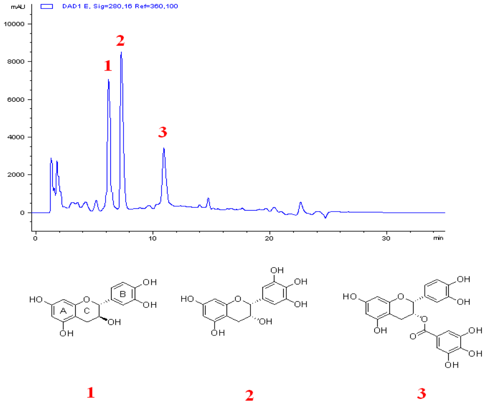
2.1. Combination of C and ECg Produces the Greatest ILSMR Effect in a Screening of Constituents from a Bioactive Fraction of Hawthorn

In the present work, C, ECg and EGC were isolated from hawthorn. A checkerboard method was used to determine which monomer or combination of the three monomers could produce the best ILSMR effect. The combination of C and EGC showed no ILSMR effect, with FICIs higher than 0.5 (Table S1). EGC in combination with ECg also had no ILSMR effect, with most FICIs above 0.5 and only one at 0.5 (Table S2). Significantly, the combination of C and ECg (Table 1) or of C, ECg and EGC showed strong ILSMR effects, with FICIs lower than 0.5 (Table 2), and there was no significant difference between two combinations. Because their pharmacological effects were equivalent, the more simple combination was preferentially chosen. Therefore, the combination of C and ECg was chosen and then its ILSMR effect was further investigated.

2.2. C and ECg Alone Have No ILSMR Effect, But When Combined, They Potentiate Antibacterial Effects of Oxacillin Against WHO-2

The MIC of C against WHO-2 was >1024 mg/L (no antibacterial effect), while that of ECg was 128 mg/L (very low antibacterial effect). C or ECg (32 mg/L) in combination with oxacillin produced FICIs of more than 0.5 or 0.5, demonstrating no or very weak ILSMR effect. Meanwhile, the FICI of C and ECg together with oxacillin was lower than 0.5, suggesting this combination had a strong ILSMR effect.
Two combinations of C and ECg at different concentrations, C (64 mg/L) + ECg (32 mg/L) or C (128 mg/L) + ECg (16 mg/L), combined with oxacillin produced FICIs lower than 0.375 (Table 1). Considering the good solubility of C and poor solubility of ECg in H2O, the latter combination with the lower ECg concentration, C (128 mg/L) + ECg (16 mg/L), was selected and investigated in the subsequent experiments.

2.3. C in Combination with ECg Specifically Potentiate the Antibacterial Effects of β-Lactam Antibiotics Against Clinical MRSA Strains

C in combination with ECg could potentiate the antibacterial effects of oxacillin against WHO-2. Therefore, it was of interest to determine whether C in combination with ECg could potentiate other β-lactam antibiotics or even non-β-lactam antibiotics and to verify the ILSMR effects, not only in WHO-2, but also in clinical MRSA strains. Herein, The 45 clinical MRSA strains were identified by drug susceptibility assay and amplification of the mecA gene. They all are resistant to oxacillin (MIC > 4 mg/L) and other five β-lactams with high levels of resistance. In the present experiment, the effects of C in combination with ECg to potentiate β-lactam antibiotics (oxacillin, ampicillin or ampicillin/sulbactam, cefazolin, cefepime and imipenem/cilastatin) and non-β-lactam antibiotics (vancomycin, linezolid and teicoplanin) against 45 clinical MRSA strains were observed.
The results showed that C or ECg each alone had no ILSMR effect, but C in combination with ECg could potentiate antibacterial effects of six β-lactam antibiotics (Table 3). Three combinations [C (32 mg/L) + ECg (4 mg/L), C (64 mg/L) + ECg (8 mg/L), and C (128 mg/L) + ECg (16 mg/L)] potentiated the effects of all six β-lactam antibiotics against almost all strains. However, none of these combinations could potentiate the effects of non-β-lactam antibiotics (Table S3).

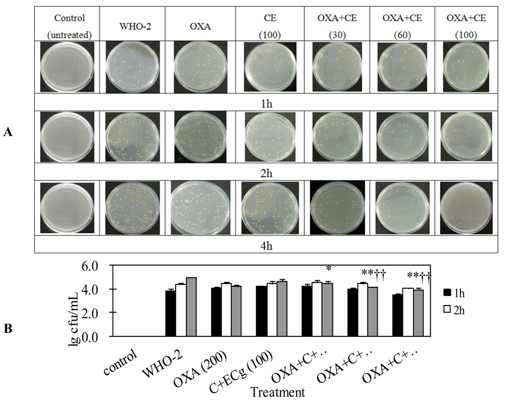
2.4. C and ECg in Combination with Oxacillin Markedly Decrease Whole Blood Bacterial Load Than Oxacillin Alone in Mice Challenged with Sublethal WHO-2

In order to determine whether C in combination with ECg could potentiate β-lactam antibiotics in vivo, the MRSA infection model was established in mice challenged with a sublethal dose of WHO-2. Considering the t1/2 of catechins was very short (approximately 1–4 h), blood bacterial loads were tested at 1, 2 and 4 h time points after the first treatment of C in combination with ECg [22]. The results showed there was no significant difference of bacterial blood load among mice treated with normal saline only, oxacillin only and C in combination with ECg. However, the bacterial load in mice treated with oxacillin (200 mg/kg/day) in combination with C and ECg was significantly lower than that in mice treated with oxacillin alone or C in combination with ECg (p < 0.05). The bacterial loads in mice treated with oxacillin in combination with C and ECg (100 mg/kg/day) was lowest (Figure 1). These results demonstrated C in combination with ECg potentiated oxacillin’s antibacterial effect in vivo, too.

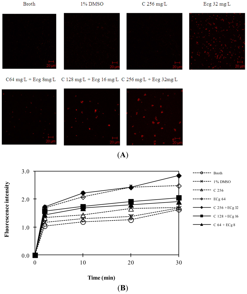
2.5. C in Combination with ECg Increases Accumulation of Daunorubicin within WHO-2

The efflux system could confer multidrug resistance of MRSA. In order to investigate whether C and ECg demonstrated its ILSMR effect via increasing antibiotics accumulation, the influence of C and ECg on drug accumulation was investigated using fluorospectrophotometry assay and laser confocal scanning microscope observation.
Daunorubicin is not a β-lactam antibiotic but with red autofluorescence. It is used as a tracer agent because it can be observed more directly. The influence of daunorubicin was observed using drug susceptibility test and the result was shown by the time-kill curve (Figure S1). After it was confirmed that daunorubicin (20 and 40 mg/L) had no effect on the growth of WHO-2, the effect of C and ECg on the accumulation of daunorubicin within WHO-2 was detected. The results from confocal scanning microscopy showed C (256 mg/L) did not increase the daunorubicin accumulation within WHO-2 but ECg (32 mg/L) increased the drug accumulation. Significantly, C (256 mg/L) in combination with ECg (32 mg/L), increased more daunorubicin accumulation than ECg alone. Interestingly, the higher the concentration of C combined with ECg, the higher the daunorubicin accumulation (Figure 2A). The similar result was also observed from fluorospectrophotometry (Figure 2B). The above two results demonstrated that the ILSMR effect of C in combination with ECg was strongly related to the increased antibiotics’ accumulation.

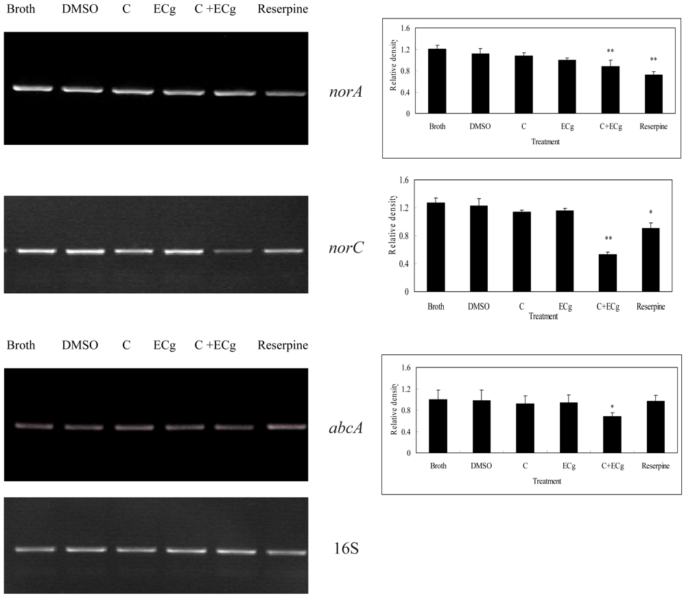
2.6. C in Combination with ECg DownRegulates mRNA Expressions of Efflux Pumps within WHO-2

In order to investigate whether C in combination with ECg manifested its ILSMR effect via inhibiting mRNA expression of efflux pump genes, the influence of C in combination with ECg on mRNA expression of efflux pump genes were investigated using reverse transcription Polymerase Chain Reaction (RT-PCR) method.
The results showed that reserpine as a positive control could downregulate mRNA expressions of norA and norC not abcA, which was in accordance with previous reports [23,24]. C or ECg alone did not influence efflux pump gene expression, but C in combination with ECg could downregulate mRNA expressions of norA, norC and abcA among eight efflux pumps of MRSA (Figure 3), thus suggesting that increased daunorubicin accumulation was related to the inhibition of three efflux pumps’ gene expressions.

2.7. Discussion

In traditional Chinese medicine, hawthorn is used as a peptic agent for stimulating digestion and promoting the function of the stomach, improving blood circulation and removing blood stasis. It is used as the agent for treatment of indigestion with epigastria distension, diarrhea, abdominal pain, amenorrhea, hypertension and hyperlipidemia, as well [25]. To the best of our knowledge, this is the first report demonstrating that hawthorn possesses ILSMR effects.
On the basis of the literature data, there are flavonoids, oligomeric proanthocyanidins, phenolicacids, triterpeneacids, organicacids and sterols within hawthorn fruit. Flavonoids are considered to be the main groups of active constituents in hawthorn extracts [21]. Catechins belong chemically to flavonoids.
Catechins are present in many plants and have many pharmacological effects. Although some of them may have the capacity to sensitize MRSA strains to oxacillin and other β-lactam antibiotics, they only possess weak antibacterial properties [26]. For example, galloy catechins, such as ECg and Cg, reduced the high MIC level of β-lactams to the antibiotic breakpoint or even lower than the breakpoint [14,15,26,27], but non-galloylated catechins, such as C and EC, had no such effect [15]. In the present study, we found ECg only had weak ILSMR effects, which did not accord with the previous reports [14,15], while C had no ILSMR effect as reported. The main reason could be that the ILSMR effect of ECg was influenced by its optical activity, which was affected by the extraction process and purity. Therefore, the discrepancies are likely due to different specific optical activities of ECg from the different sources of ECg and different MRSA strains.
We also found that when C combined with ECg, the ILSMR effect was markedly increased and the ILSMR effect of the combination (C and ECg) was enhanced with the increase in the concentration of C, with a higher concentration of C resulting in a lower FICI. This phenomenon did not occur when the concentration of ECg was increased, suggesting that the ILSMR effect of ECg could be potentiated by C. Previously, the cis form of non-galloylated catechins such as (−)-EC and (−)-EGC was reported to enhance the ILSMR effect of ECg [28], but excluding non-galloylated catechins of the trans form such as C. Herein, we firstly reported the ILSMR effects of C in combination with ECg on multiple classic β-lactam antibiotics against not only a standard MRSA strain but also clinical MRSA strains in vitro and in vivo.
In addition, C was found to be more effective than EC and EGC in potentiating the ILSMR effect of ECg against WHO-2 (data not shown). C and EC are known optical isomers. As the pharmacological effect of drugs requires strict chiral recognition by biological macromolecules [29], the bioactivities of optical isomers are generally different. The difference of chemical structure between C and EGC is that EGC has one more hydroxy group in the B ring than in that of C. Overall, the different abilities of C and EGC to potentiate the ILSMR effect of ECg was presumed to be due to the difference in steric hindrance provided by the hydroxy groups in the B ring of these compounds.
Previously, the ILSMR effect of ECg in combination with β-lactam antibiotics against the standard WHO-2 strain has been described [14,15]. Here, C together with ECg also showed an ILSMR effect on β-lactam antibiotics, not only against the MRSA WHO-2 standard strain, but also against 45 clinical MRSA strains, thereby suggesting the potential clinical value of this combination.
The ILSMR effect of ECg in combination with some β-lactam antibiotics against MRSA has also been described previously [14]. In this study, C in combination with ECg showed ILSMR effects on six β-lactam antibiotics: ampicillin, ampicillin/sulbactam, cefazolin, cefepime and imipenem/cilastatin. These β-lactam antibiotics are both classic and representative β-lactam antibiotics in the clinic. However, they cannot be used in the treatment of MRSA infections due to resistance. Our results showed that C in combination with ECg could significantly reduce the MIC of these six antibiotics against almost all of the clinical MRSA strains. These results are significant since they demonstrate the possibility that these antibiotics can be used in the clinic when combined with C and ECg. Interestingly, C in combination with ECg demonstrated ILSMR effects on β-lactam antibiotics, while they had no such effect on non-β-lactam antibiotics, which has not been reported previously.
Previously, ECg was found to prevent biofilm formation and promoted cell wall thickening, leading to cell aggregation without affecting the rate or extent of growth in culture [30]. Furthermore, ECg could insert into bacterial cytoplasmic membrane to disrupt PBP2a-mediated resistance by delocalizing PBP2 [31]. In the present study, we found that the ILSMR effect of C and ECg was also related to antibiotics accumulation. Efflux pumps were related to drug accumulation.
Efflux pumps were capable of extruding antibiotics, leading to multidrug resistance. There were nine efflux pumps within MRSA; they were norA, norB, norC, mdeA, qacA/B, mepA, smr, sepA[32], and abcA[33]. In a recent study, 50% of the 232 isolates of S. aureus were discovered to pump out at least two structurally unrelated substrates [34]. Frequencies of overexpressed efflux genes were norA (23%), norB (25%), norC (17%), mdeA (11%), and mepA (4%) [34], which meant norA and norB were the most popular pumps in S. aureus. However, abcA was reported to be responsible for the β-lactam resistance in S. aureus[33]. Previously, reserpine (a phytoalkaloid) was reported as efflux pump inhibitor (EPI) against various microbes [23,24]. There was one report that ECg inhibited norA efflux pump expression [35]. Herein, our results showed that reserpine down-regulated mRNA expressions of norA and norC, but not abcA, and C, in combination with ECg, down-regulated mRNA expressions of norA, norC and abcA among eight efflux pumps, while ECg or C alone could not. The above results suggested the ILSMR effect of C and ECg was probably related to the inhibition of three pumps’ mRNA expressions. Which pump(s) played a more important role should be further investigated in the future experiments.
In the present experiments, the synergy was assayed by FICI according to the criterion suitable for combination of only two compounds. Synergism was defined as an FICI ≤ 0.5 [36]. As is known, when more than two compounds were combined against bacteria, FICI would be easily more than 0.5, suggesting there was no synergism. In fact, each agent’s concentration had markedly decreased after combination. Therefore, a criterion suitable for more than two compounds should be investigated in the future.

3. Experimental Section

3.1. Materials

Chemicals used in extraction and isolation, like methanol, ethanol, ethyl acetate, normal butanol, acetone, were from Aladdin Regent Company (Shanghai, China). Macroporous D-101, silica gel and LH20 were from Ouyll Biological Technology Company (Tianjin, China). Oxacillin was purchased from Southwestern Pharmaceutical Corp. LTD. (Guangzhou, China). Ampicillin, cefazolin and cefepime were purchased from North China Pharmaceutical Group Corp. (Shijiazhuang, China). A 2:1 mixture of ampicillin sodium and sulbactam sodium was purchased from Seashore Pharmaceutical Company (Shenzhen, China). A 1:1 mixture of imipenem and cilastatin sodium was purchased from Zhuhai Federal Pharmaceutical Company (Zhuhai, China). Müller-Hinton (MH) agar plates and MH broth were purchased from OXOID Company (Basingstoke, UK).

3.2. Plant Material

The fruits of hawthorn (Crataegus pinnatifida) were collected from Yuzhou Market of Traditional Chinese Herbs, He Nan province in China, and identified by Yimin Zheng (Chongqing University of technology, Chongqing, China). The voucher specimen (2010-0900SZ#) of the hawthorn was deposited in our laboratory for future reference.

3.3. Bacterial Strains

The standard MRSA strain WHO-2 (WHO-2) and 45 clinical MRSA strains were kindly provided by Xiaoxing Luo [37] (The Fourth Military Medical University, Xi’an, China) and Peiyuan Xia (Southwestern Hospital, Chongqing, China), respectively. The properties of WHO-2 and 45 clinical MRSA strains were confirmed by both a drug susceptibility assay according to the Clinical Laboratory Standards Institute (CLSI) guidelines of 2010 (strains were resistant to oxacillin (MIC > 4 mg/L) and amplification of the mecA gene identified by Polymerase Chain Reaction (PCR) in our lab. WHO-2 possessed a high level of resistance to oxacillin (MIC was 512 mg/L) and harbored the mecA gene. Forty-five clinical strains were all resistant to oxacillin (MIC > 4 mg/L) and harbored the mecA gene. The primers of mecA gene and 16S were listed (Table S4).

3.4. Animals

Specific-Pathogen-Free (SPF) Kunming (KM) mice (4–6 weeks old, weighing 18–22 g) were supplied by the Experimental Animal Center of the Third Military Medical University (Chongqing, China). An equal number of male and female mice were used. All animals received humane care in compliance with the Principles of Laboratory Animal Care formulated by the National Society for Medical and Research and the “Guide for the Care and Use of Laboratory Animals” prepared by the National Academy of Sciences (Washington DC, USA). The experiments were approved by a medical ethics committee of The Third Military Medical University.

3.5. Extraction and Isolation

The fruits of hawthorn were crushed and refluxing extracted with 75% EtOH three times (each extraction period lasted 2 h). Merging the extraction together, the 75% EtOH extract was portioned between EtOAc, n-BuOH. Each extract (EtOAc extract, n-BuOH extract, H2O extract) was measured for antibacterial activity and ILSMR effect. The active part was chromatographed on macroporous D-101 column (washed by EtOH-H2O), and 50% EtOH-H2O fraction was collected by the freeze-drying technique. The sample was chromatographed on a silica gel column with chloroform and acetone afforded the fraction1–8 (frs.1–8), and then these fractions were measured for antibacterial effect and ILSMR effect. Fraction 4, which showed a significant ILSMR effect, was then separated by Sephadex LH-20 column chromatography and preparative HPLC (Agilent 1100 Series equipped with G1361A binary pump, G1365B MWD UV detector, G1364B fraction collector), yielding the three monomeric compounds. The three compounds were identified as (+)-catechin, (−)-epicatechin gallate and (−)-epigallatecatechin by the methods of 1H-NMR and spiking with reference compounds which were purchased or available in our laboratory (Figure 4).

3.6. Bacterial Growth

Each single colony from the MH agar plate was inoculated into a 10 mL volume of liquid MH broth (containing 2% NaCl), according to the CLSI 2010 guidelines, and cultivated aerobically at 37 °C in a heated and shaking chamber for 12 h. These cultures were diluted 1:100 (v/v) in another 10 mL of fresh broth and cultivated aerobically until the log growth phase.

3.7. Drug Susceptibility Assay

Bacteria in exponential phase (1 × 10⁵ cfu/mL) were inoculated into 96-well plates. MIC of β-lactam antibiotics, C alone and ECg alone were determined by serial two-fold dilutions in MH broth according to CLSI 2010 guidelines. MIC50 and MIC90 were the half and 90% MICs of β-lactam antibiotics against 45 clinical MRSA strains, respectively.
Synergistic effects were assayed by determining the FICI calculated for each combination using the following formula: FICI = (MIC of combined antibiotic/MIC of antibiotic alone) + (concentration of combined “C”/MIC of “C” alone) + (concentration of combined “ECg”/MIC of “ECg” alone). Synergism was defined as a FICI ≤ 0.5, antagonism was defined as an FICI > 4.0, and no interaction was defined with a FICI 0.5–4.0, according to previously described criteria [36].

3.8. Experimental Animal Model and Drug Treatment

One hundred and five mice, weighing from 18 g to 22 g, were randomly divided into seven groups (15 mice/group) and intravenously injected with a sublethal dose of live WHO-2 (4.0 × 10⁹ cfu/mL). Four hours later, the mice were given the following treatments (C and ECg mixture by intragastric injection and oxacillin by intramuscular injection): Normal saline only, oxacillin (200 mg/kg/day) only; mixture of C and ECg (total dose 100 mg/kg/day); combination of C and ECg mixture (100 mg/kg/day) with oxacillin (200 mg/kg/day); combination of C and ECg mixture (60 mg/kg/day) and oxacillin (200 mg/kg/day); combination of C and ECg mixture (30 mg/kg/day) and oxacillin (200 mg/kg/day). The total injection volume was 0.4 mL/20 g body weight. One milliliter of blood from each of three mice of each group was collected at 1-, 2- and 4-h time points after the first treatment of the agents, and samples were diluted with sterile normal saline immediately. The blood samples were then inoculated onto MH agar plates containing 50 mg/L of oxacillin. After culturing for 24 h at 37 °C, the bacterial colonies were counted and presented as lg cfu/mL of blood.
The dose of oxacillin used in this study was based on a conversion of clinical dosage regimens. Doses of oxacillin in the range from 30–100 mg/kg/day have been proposed to treat bacterial infection in adult patients, and higher doses are given for serious infections. According to the drug conversion principle applied in pharmacological studies, a dose ranging from 30–100 mg/kg/day of oxacillin in adults is equivalent to that of 300–900 mg/kg/day in mice. Therefore, the dose of 200 mg/kg/day of oxacillin chosen for mice in this study was lower than the equivalent doses used for humans in the clinic.
C and ECg were combined at the mass ratio of 8:1 and then mixed with sodium carboxymethylcellulose (0.5%) by milling. The dose of the C and ECg mixture used in mice was based on results of in vitro experiments, which showed that the doses of C at 128 mg/L and ECg at 16 mg/L (144 mg/L total) resulted in the best ILSMR effect. Because the quantity of body fluid in a mouse is generally 70%–80% of its body weight (average 20 g per mouse), the dose of the C and ECg mixture should be about 2 mg per mouse. Therefore, the highest dose of the C and ECg mixture used was calculated at 100 mg/kg/day in mice.

3.9. Accumulation of Daunorubicin within WHO-2

WHO-2 was inoculated into 10 mL of MH broth with seven different treatments: Broth only, 1% (v/v) dimethyl sulfoxide (DMSO), C (256 mg/L), ECg (64 mg/L), the combination of C (64 mg/L) and ECg (8 mg/L), the combination of C (128 mg/L) and ECg (16 mg/L), and the combination of C (256 mg/L) and ECg (64 mg/L). Then the mixtures were cultured for 6 h at 37 °C in a heated and shaking chamber. Then, the bacteria were centrifuged at 3500× g for 5 min to harvest the bacterial pellet. After washing with PBS (0.2 mM, pH 7.2) three times, the bacterial pellet was resuspended and the bacterial suspension was adjusted to an OD600 of 1.0. After co-culture with daunorubicin (40 mg/L) at 37 °C for 2, 10, 20 and 30 min in the dark, daunorubicin accumulation within WHO-2 was observed by the two methods described below.
The first method was fluorospectrophotometry; daunorubicin accumulation within the bacteria was assayed in the absence or presence of C and ECg. The emission wavelength was 467 nm and the excitation wavelength was 588 nm. Additionally, as the second method, at 30 min, 0.5 mL of the bacteria was collected. After washing with PBS three times, the bacteria were fixed on glass cover slips and observed under a 510 Meta confocal microscope (Zeiss, Göttingen, Germany).

3.10. mRNA Expression Assays

WHO-2 was inoculated into 100 mL of MH broth with six different treatments: broth, 1% DMSO (v/v), C (128 mg/L), ECg (16 mg/L), C (128 mg/L) and ECg (16 mg/L) and reserpine (20 mg/L). Bacteria were cultivated to an OD600 of 0.6 and washed with normal saline. Then bacteria were harvested and milled with liquid nitrogen. 1 mL of RNAiso was added to completely lyse bacteria. Chloroform was added to the mixture with shaking for 15 s and stewing for 10 min. Bacteria were harvested by centrifugation at 10,000× g for 15 min in 4 °C. Isopropanol was added with stewing for 15 min. Bacteria were harvested again by centrifugation at 12,000× g for 10 min and washed by 75% (v/v) ethanol, and then 20 μL of RNase-free ddH2O was added. The reverse transcription was performed by PrimeScript RT reagent Kit. The primers for mecA, norA, norB, norC, mepA, mdeA, sepA, qacA/B, abcA, smr and 16S RNA were listed (Table S1). The primers were added to the PCR tubes and subjected to the normal reverse transcription PCR (RT-PCR). RT-PCR products were determined using 1.0% agarose gel electrophoresis and Golden View staining. Images of the gels were analyzed using Quantity One software (Bio-Rad, Hercules, CA, USA).

3.11. Statistical Analysis and Presentation of Data

Each experiment was repeated at least three times, and each data point was represented as the mean, with error bars denoting standard deviations. Differences in bacterial load in the whole blood of mice were examined by repeated measures ANOVA using Statistical Program for Social Sciences (SPSS) 16.0. A p value of 0.05 was considered significant, and a p value of 0.01 was considered highly significant.

4. Conclusions

In conclusion, C in combination with ECg enhanced the antibacterial effect of β-lactam antibiotics against MRSA in vitro and in vivo, which might be related to the increased antibiotics accumulation within MRSA via suppression of the important efflux pumps’ gene expression.

Supplementary Information

ijms-14-01802-s001.pdf

Acknowledgments

The authors thank all colleagues in Medical Research Center for technical assistance. Funding: This study was supported by a grant from the Science Research Foundation of Third Military Medical University 2009XYY07.
  • Conflict of InterestThe authors declare no conflict of interest.

References

  1. Moellering, R.C., Jr. MRSA: The first half century. J. Antimicrob. Chemother. 2012, 67, 4–11. [Google Scholar]
  2. Livermore, D.M. Antibiotic resistance in Staphylococci. Int. J. Antimicrob. Agents 2000, 16, S3–S10. [Google Scholar]
  3. Pantosti, A.; Sanchinim, A.; Monaco, M. Mechanisms of antibiotic resistance in Staphylococcus aureus. Future Microbiol 2007, 2, 323–334. [Google Scholar]
  4. Li, X.Z.; Nikaido, H. Efflux-Mediated drug resistance in bacteria: An update. Drugs 2009, 69, 1555–1623. [Google Scholar]
  5. Poole, K. Efflux pumps as antimicrobial resistance mechanisms. Ann. Med 2007, 39, 162–176. [Google Scholar]
  6. Taylor, P.W.; Stapleton, P.D.; Paul, L.J. New ways to treat bacterial infections. Drug Discov. Today 2002, 7, 1086–1091. [Google Scholar]
  7. Kondo, K.; Takaishi, Y.; Shibata, H.; Higuti, T. ILSMRs (intensifier of β-lactam-susceptibility in methicillin-resistant Staphylococcus aureus) from Tara [Caesalpinia spinosa (Molina) Kuntze]. Phytomedicine 2006, 13, 209–212. [Google Scholar]
  8. Liu, I.X.; Durham, D.G.; Richards, R.M. Baicalin synergy with beta-lactam antibiotics against methicillin-resistant Staphylococcus aureus and other beta-lactam-resistant strains of S. aureus. J. Pharm. Pharmacol 2000, 52, 361–366. [Google Scholar]
  9. Nicolson, K.; Evans, G.; O’Toole, P.W. Potentiation of methicillin activity against methicillin-resistant Staphylococcus aureus by diterpenes. FEMS Microbiol. Lett 1999, 179, 233–239. [Google Scholar]
  10. Shiota, S.; Shimizu, M.; Mizusima, T.; Ito, H.; Hatano, T.; Yoshida, T.; Tsuchiya, T. Restoration of effectiveness of beta-lactams on methicillin-resistant Staphylococcus aureus by tellimagrandin I from rose red. FEMS Microbiol. Lett. 2000, 185, 135–138. [Google Scholar]
  11. Shimizu, M.; Shiota, S.; Mizushima, T.; Ito, H.; Hatano, T.; Yoshida, T.; Tsuchiya, T. Marked potentiation of activity of beta-lactams against methicillin-resistant Staphylococcus aureus by corilagin. Antimicrob. Agents Chemother 2001, 45, 3198–3201. [Google Scholar]
  12. Eid, C.N.; Halligan, N.G.; Nicas, T.I.; Mullen, D.L.; Butler, T.F.; Loncharich, R.J.; Paschal, J.W.; Schofield, C.J.; Westwood, N.J.; Cheng, L. Tripeptide LY301621 and its diastereomers as methicillin potentiators against methicillin resistant Staphylococcus aureus. J. Antibiot (Tokyo) 1997, 50, 283–285. [Google Scholar]
  13. Hu, Z.Q.; Zhao, W.H.; Asano, N.; Yoda, Y.; Hara, Y.; Shimamura, T. Epigallocatechin gallate synergistically enhances the activity of carbapenems against methicillin-resistant Staphylococcus aureus. Antimicrob. Agents Chemother 2002, 46, 558–560. [Google Scholar]
  14. Shiota, S.; Shimizu, M.; Mizushima, T.; Ito, H.; Hatano, T.; Yoshida, T.; Tsuchiya, T. Marked reduction in the minimum inhibitory concentration (MIC) of beta-lactams in methicillin-resistant Staphylococcus aureus produced by epicatechin gallate, an ingredient of green tea (Camellia sinensis). Biol. Pharm. Bull 1999, 22, 1388–1390. [Google Scholar]
  15. Stapleton, P.D.; Shah, S.; Anderson, J.C.; Hara, Y.; Hamilton-Miller, J.M.; Taylor, P.W. Modulation of beta-lactam resistance in Staphylococcus aureus by catechins and gallates. Int. J. Antimicrob. Agents 2004, 23, 462–467. [Google Scholar]
  16. Taylor, P.W.; Hamilton-Miller, J.M.; Stapleton, P.D. Antimicrobial properties of green tea catechins. Food Sci. Technol. Bull 2005, 2, 71–81. [Google Scholar]
  17. Pittler, M.H.; Schmidt, K.; Ernst, E. Hawthorn extract for treating chronic heart failure: Meta-Analysis of randomized trials. Am. J. Med 2003, 114, 665–674. [Google Scholar]
  18. Schüssler, M.; Hölzl, J.; Fricke, U. Myocardial effects of flavonoids from Crataegus species. Arzneimittel-Forschung 1995, 45, 842–845. [Google Scholar]
  19. Schwinger, R.H.; Pietsch, M.; Frank, K.; Brixius, K. Crataegus. special extract WS 1442 increases force of contraction in human myocardium cAMP-independently. J. Cardiovasc. Pharmacol 2000, 35, 700–707. [Google Scholar]
  20. Quettier-Deleu, C.; Voiselle, G.; Fruchart, J.C.; Duriez, P.; Teissier, E.; Bailleul, F.; Vasseur, J.; Trotin, F. Hawthorn extracts inhibit LDL oxidation. Pharmazie 2003, 58, 577–581. [Google Scholar]
  21. Tadić, V.M.; Dobrić, S.; Marković, G.M.; Dordević, S.M.; Arsić, I.A.; Menković, N.R.; Stević, T. Anti-Inflammatory, gastroprotective, free-radical-scavenging, and antimicrobial activities of hawthorn berries ethanol extract. J. Agric. Food Chem 2008, 56, 7700–7709. [Google Scholar]
  22. Liuji, C.; Xianqiang, Y.; Mingxiang, J. Research progress on pharmacokinetics of tea catechins. Cha Ye Ke Xue 2001, 21, 11–16. [Google Scholar]
  23. Schmitz, F.J.; Fluit, A.C.; Lückefahr, M.; Engler, B.; Hofmann, B.; Verhoef, J.; Heinz, H.P.; Hadding, U.; Jones, M.E. The effect of reserpine, an inhibitor of multidrug efflux pumps, on the in vitro activities of ciprofloxacin, sparfloxacin and moxifloxacin against clinical isolates of Staphylococcus aureus. J. Antimicrob. Chemother 1998, 42, 807–810. [Google Scholar]
  24. Gibbons, S.; Udo, E.E. The effect of reserpine, a modulator of multidrug efflux pumps, on the in vitro activity of tetracycline against clinical isolates of methicillin resistant Staphylococcus aureus (MRSA) possessing the tet(K) determinant. Phytother Res 2000, 14, 139–140. [Google Scholar]
  25. Chang, Q.; Zuo, Y.; Harrison, F.; Chow, M.S. Hawthorn-An overview of chemical, pharmacological and clinical studies. J. Clin. Pharmacol 2002, 42, 605–612. [Google Scholar]
  26. Hamilton-Miller, J.M.; Shah, S. Activity of the tea component epicatechin gallate and analogues against methicillin-resistant Staphylococcus aureus. J. Antimicrob. Chemother 2000, 46, 852–853. [Google Scholar]
  27. Zhao, W.H.; Hu, Z.Q.; Okubo, S.; Hara, Y.; Shimamura, T. Mechanism of synergy between epigallocatechin gallate and β-lactams against methicillin-resistant Staphylococcus aureus. Antimicrob. Agents Chemother 2001, 45, 1737–1742. [Google Scholar]
  28. Stapleton, P.D.; Shah, S.; Hara, Y.; Taylor, P.W. Potentiation of catechin gallate-mediated sensitization of Staphylococcus aureus to oxacillin by nongalloylated catechins. Antimicrob. Agents Chemother 2006, 50, 752–755. [Google Scholar]
  29. Qidong, Y.; Guoqiang, L. Chiral Drugs: Research and Evaluation; Chemical Industry Press: Beijing, China, 2011. [Google Scholar]
  30. Stapleton, P.D.; Shah, S.; Ehlert, K.; Hara, Y.; Taylor, P.W. The beta-lactam-resistance modifier (−)-epicatechin gallate alters the architecture of the cell wall of Staphylococcus aureus. Microbiology 2007, 153, 2093–2103. [Google Scholar]
  31. Bernal, P.; Lemaire, S.; Pinho, M.G.; Mobashery, S.; Hinds, J.; Taylor, P.W. Insertion of epicatechin gallate into the cytoplasmic membrane of methicillin-resistant Staphylococcus aureus disrupts penicillin-binding protein (PBP) 2a-mediated beta-lactam resistance by delocalizing PBP2. J. Biol. Chem 2010, 285, 24055–24065. [Google Scholar]
  32. Kosmidis, C.; Schindler, B.D.; Jacinto, P.L.; Patel, D.; Bains, K.; Seo, S.M.; Kaatz, G.W. Expression of multidrug resistance efflux pump genes in clinical and environmental isolates of Staphylococcus aureus. Int. J. Antimicrob. Agents 2012, 40, 204–209. [Google Scholar]
  33. Truong-Bolduc, Q.C.; Hooper, D.C. The transcriptional regulators NorG and MgrA modulate resistance to both quinolones and beta-lactams in Staphylococcus aureus. J. Bacteriol 2007, 189, 2996–3005. [Google Scholar]
  34. DeMarco, C.E.; Cushing, L.A.; Frempong-Manso, E.; Seo, S.M.; Jaravaza, T.A.; Kaatz, G.W. Efflux-Related resistance to norfloxacin, dyes, and biocides in bloodstream isolates of Staphylococcus aureus. Antimicrob. Agents Chemother 2007, 51, 3235–3239. [Google Scholar]
  35. Gibbons, S.; Moser, E.; Kaatz, G.W. Catechin gallates inhibit multidrug resistance (MDR) in Staphylococcus aureus. Planta. Med 2004, 70, 1240–1242. [Google Scholar]
  36. Odds, F.C. Synergy, antagonism, and what the chequerboard puts between them. J. Antimicrob. Chemother 2003, 52, 1. [Google Scholar]
  37. Hou, Z.; Meng, J.R.; Zhao, J.R.; Hu, B.Q.; Liu, J.; Yan, X.J.; Jia, M.; Luo, X.X. Inhibition of beta-lactamase-mediated oxacillin resistance in Staphylococcus aureus by a deoxyribozyme. Acta Pharmacol. Sin 2007, 28, 1775–1782. [Google Scholar]
Figure 1. Effect of oxacillin (OXA) in combination with C and ECg on whole blood bacterial loads in mice treated with a sublethal dose of live MRSA. One hundred and five mice were randomly divided into seven groups (15 mice/group), and all were injected with live WHO-2 (4 × 10⁹ cfu/mL) except the control group (untreated). Four hours later, mice injected with bacteria were intragastrically injected with: (1) normal saline (NS) only; (2) oxacillin (200 mg/kg/day) only; (3) mixture of C and ECg (100 mg/kg/day) only; (4) mixture of C, ECg (30 mg/kg/day) and oxacillin (200 mg/kg/day); (5) mixture of C, ECg (60 mg/kg/day) and oxacillin (200 mg/kg/day); (6) mixture of C, ECg (100 mg/kg/day) and oxacillin (200 mg/kg/day). At indicated time points after the first treatment of C and ECg, 1 mL of blood from 3 mice of each group was collected at 1, 2 and 4 h time points after the first treatment of the agents, and samples were diluted with sterile normal saline immediately. The blood samples were then inoculated onto MüllerHinton (MH) agar plates containing 50 mg/L of oxacillin. After culturing for 24 h at 37 °C, the bacterial colonies were counted and presented as lg cfu/mL of blood. (A) Photograph of MH agar plates containing bacterial colonies; (B) Statistical data of bacterial colonies presented as lg cfu/mL of blood. Data are presented as means ± standard deviation. * p < 0.05, compared to OXA group; ** p < 0.01, compared to OXA group; ††p < 0.01, compared to C and ECg mixture only group.
Ijms 14 01802f1
Figure 2. Daunorubicin accumulation within WHO-2 pretreated with C and ECg. WHO-2 was treated with broth, C (256 mg/L), ECg (32 mg/L) and C and ECg (64 mg/L + 8 mg/L, 128 mg/L + 16 mg/L, 256 mg/L + 32 mg/L) and then cultured for 6 h at 37 °C in a heated, shaking, environmental chamber. Then, the bacteria were centrifuged at 3500× g for 5 min. After washing three times and resuspending, the bacterial suspension was adjusted to an OD600 of 1.0. (A) Bacteria were incubated with daunorubicin (40 mg/L) in the dark at 37 °C for 30 min; 0.5 mL of bacteria was collected. Bacteria were washed three times and resuspended with PBS. Next, the bacteria were fixed on glass cover slips and observed under a 510 Meta confocal microscope (Zeiss, Göttingen, Germany); (B) Bacteria were incubated with daunorubicin (40 mg/L) in the dark at 37 °C for 2, 10, 20 and 30 min; 0.5 mL of bacteria was collected. Bacteria were washed three times and resuspended with phosphate buffered saline (PBS). Then, quantitative determination of daunorubicin accumulation in the absence and presence of C and ECg was performed using fluorospectrophotometry at an emission wavelength of 467 nm and an excitation wavelength of 588 nm.
Ijms 14 01802f2
Figure 3. norA, norC and abcA mRNA expression within WHO-2. WHO-2 was treated with broth, 1% DMSO (v/v), C (128 mg/L), ECg (16 mg/L), C (128 mg/L) and ECg (16 mg/L) and Reserpine (20 mg/L), and cultivated at 37 °C and 100 g until an OD600 of 0.6 (6 h) was reached. After the bacterial harvest, total RNA was extracted and RT-PCR was performed. The signals for norA, norC, abcA and 16S rRNA were integrated on a Gel Doc 1000 Mini-Transilluminator (Bio-Rad, Hercules, CA, USA). 16S was used as a standard to allow semi-quantitative comparisons between samples. One-way analysis of variance (ANOVA) was used to examine the differences among different treatment (n = 3). * p < 0.05; ** p < 0.01 as compared with broth.
Ijms 14 01802f3
Figure 4. The figure showed the HPLC-DAD chromatogram of the effective fraction (fraction 4) which subjected to the silica gel column, and the 3 active compounds were isolated from fraction 4. An Agilent SB C-18 reversed-phase column (150 × 4.6, 5 μm) was used with column temperature set at 30 °C, and the spectrum at absorbance of 280 nm. The mobile phase consisted of methanol (20%) and 0.5% aqueous glacial acetic acid (80%), and the flow rate was 1 mL/min. 1–3 represented (+)-catechin, (−)-epicatechin gallate and (−)-epigallatecatechin, respectively.
Ijms 14 01802f4
Table 1. Minimum inhibitory concentrations (MIC) of oxacillin in combination with C and ECg against WHO-2. Synergistic activities of different concentrations of C, ECg and oxacillin (OXA) on WHO-2 were tested by the checkerboard method. Fractional inhibitory concentration index (FICI) values were calculated as described in Experimental Section.
C (mg/L) aECg (mg/L) bMIC of OXA (mg/L)FICI
00512cannot calculate
85121.625
162560.625
321280.500

160512<1.016
8512<1.073
16256<0 636
3264<0.386

320512<1.031
8512<1.094
16256<0.656
3264<0.406

640512<1.063
8256<0.625
16128<0.438
3232<0.375

1280512<1.125
8128<0.438
1664<0.375
32<4<0.383

2560512<1.250
864<0.438
1616<0.406
32<4<0.508

5120256<1.000
816<0.531
16<4<0.633
32<4<0.758
aFor C, the MIC against WHO-2 was >1024 mg/L;
bFor ECg, the MIC against WHO-2 was 128 mg/L.
Table 2. MIC of oxacillin in combination with C, ECg and EGC against WHO-2. Testing of synergy among C, EGC and oxacillin (OXA) was performed using the checkerboard method. FICI values were calculated as described in Experimental Section.
C aEGC bECg cOXA d MIC (mg/L)FICI
16168256<0.641
16128<0.453
3232<0.391

328256<0.703
16128<0.516
3232<0.454

648256<0.829
16128<0.641
32<4<0.524

32168256<0.656
16128<0.469
3216<0.375

328256<0.719
16128<0.531
3216<0.438

648128<0.594
1664<0.531
32<4<0.535

64168128<0.438
16128<0.475
3216<0.375

328128<0.469
1664<0.406
32<4<0.409

648128<0.594
1632<0.468
32<4<0.534

128168128<0.500
1632<0.438
32<4<0.441

328128<0.375
1632<0.438
32<4<0.503

64816<0.469
16<4<0.316
32<4<0.628

25616864<0.500
1616<0.469
32<4<0.568

32864<0.563
1632<0.563
3216<0.656

648<4<0.565
16<4<0.628
32<4<0.753

51216832<0.688
16<4<0.671
32<4<0.815

32816<0.719
16<4<0.754
32<4<0.878

648<4<0.815
16<4<0.878
32<4<1.003
aFor C, the MIC against WHO-2 was >1024 mg/L;
bFor EGC, the MIC against WHO-2 was 256 mg/L;
cFor ECg, the MIC against WHO-2 was 128 mg/L;
dFor OXA, the MIC against WHO-2 was 512 mg/L.
Table 3. Effect of C and ECg on antibacterial effects of six β-lactam antibiotics against 45 clinical MRSA strains. Synergistic activities of C and ECg in combination with six β-lactams antibiotics (oxacillin, ampicillin, ampicillin/sulbactam, cefazolin, cefepime and imipenem/cilastatin) were tested by checkerboard method. FICI values were calculated as described in Experimental Section.
AntibioticsMIC (mg/L) of β-lactam antibiotics
Concentration of C a + ECg b (mg/L)

016 + 232 + 4


RangeMIC50MIC90RangeMIC50MIC90FICI cRangeMIC50MIC90FICI c
oxacillin128–5122565124–5121282560.56 ± 0.32 (13/45)4–256322560.31 ± 0.22 (36/45)
ampicillin32–512641282–25616640.37 ± 0.26 (30/45)2–2568320.29 ± 0.24 (39/45)
ampicillin/sulbactam8–3232322–3216320.58 ± 0.29 (14/45)2–168160.39 ± 0.23 (33/45)
cefazolin4–5122565124–5121282560.47 ± 0.33 (20/45)4–256321280.25 ± 0.18 (39/45)
cefepime8–2048102410248–10242565120.45 ± 0.23 (18/45)8–10241285120.30 ± 0.20 (39/45)
imipenem/cilastatin4–5121282564–51232640.36 ± 0.25 (29/45)4–5124320.23 ± 0.25 (41/45)

AntibioticsMIC (mg/L) of β-lactam antibiotics
Concentration of Ca+ ECgb(mg/L)

64 + 8128 + 16


RangeMIC50MIC90FICIcRangeMIC50MIC90FICIc

oxacillin4–25641280.28 ± 0.19 (42/45)4–256440.41 ± 0.22 (31/45)
ampicillin2–2562160.27 ± 0.19 (42/45)2–256220.29 ± 0.22 (31/45)
ampicillin/sulbactam2–16240.30 ± 0.19 (42/45)2–16220.44 ± 0.12 (33/45)
cefazolin4–644320.23 ± 0.15 (44/45)4–4440.39 ± 0.21 (32/45)
cefepime8–2568640.25 ± 0.17 (44/45)8–16880.39 ± 0.23 (32/45)
imipenem/cilastatin4–512440.28 ± 0.30 (42/45)4–512440.45 ± 0.26 (32/45)
aFor C, the MIC range, MIC50, MIC90 against 45 MRSA clinical strains were 512–4096 mg/L, 2048 mg/L and 4096 mg/L, respectively;
bFor ECg, the MIC range, MIC50, MIC90 against 45 MRSA clinical strains were 32–1024 mg/L, 64 mg/L and 1024 mg/L, respectively;
cData are expressed as means ± standard deviations. Values in parentheses represent the number of strains for which drug combinations resulted in synergism/number of strains tested.

