<?xml version="1.0" encoding="UTF-8"?>
<!DOCTYPE article PUBLIC "-//NLM//DTD Journal Publishing DTD v2.3 20070202//EN" "journalpublishing.dtd">
<article xmlns:xlink="http://www.w3.org/1999/xlink" xml:lang="en" article-type="research-article">
<front>
<journal-meta>
<journal-id journal-id-type="publisher-id">ijms</journal-id>
<journal-title>International Journal of Molecular Sciences</journal-title>
<abbrev-journal-title>Int. J. Mol. Sci.</abbrev-journal-title>
<issn pub-type="epub">1422-0067</issn>
<publisher>
<publisher-name>Molecular Diversity Preservation International (MDPI)</publisher-name></publisher></journal-meta>
<article-meta>
<article-id pub-id-type="doi">10.3390/ijms131215801</article-id>
<article-id pub-id-type="publisher-id">ijms-13-15801</article-id>
<article-categories>
<subj-group>
<subject>Article</subject></subj-group></article-categories>
<title-group>
<article-title>Cholesterol-Dependent Energy Transfer between Fluorescent Proteins—Insights into Protein Proximity of APP and BACE1 in Different Membranes in Niemann-Pick Type C Disease Cells</article-title></title-group>
<contrib-group>
<contrib contrib-type="author">
<name><surname>von Einem</surname><given-names>Bjoern</given-names></name><xref ref-type="aff" rid="af1-ijms-13-15801">1</xref></contrib>
<contrib contrib-type="author">
<name><surname>Weber</surname><given-names>Petra</given-names></name><xref ref-type="aff" rid="af2-ijms-13-15801">2</xref></contrib>
<contrib contrib-type="author">
<name><surname>Wagner</surname><given-names>Michael</given-names></name><xref ref-type="aff" rid="af2-ijms-13-15801">2</xref></contrib>
<contrib contrib-type="author">
<name><surname>Malnar</surname><given-names>Martina</given-names></name><xref ref-type="aff" rid="af3-ijms-13-15801">3</xref></contrib>
<contrib contrib-type="author">
<name><surname>Kosicek</surname><given-names>Marko</given-names></name><xref ref-type="aff" rid="af3-ijms-13-15801">3</xref></contrib>
<contrib contrib-type="author">
<name><surname>Hecimovic</surname><given-names>Silva</given-names></name><xref ref-type="aff" rid="af3-ijms-13-15801">3</xref></contrib>
<contrib contrib-type="author">
<name><surname>von Arnim</surname><given-names>Christine A. F.</given-names></name><xref ref-type="aff" rid="af1-ijms-13-15801">1</xref></contrib>
<contrib contrib-type="author">
<name><surname>Schneckenburger</surname><given-names>Herbert</given-names></name><xref ref-type="aff" rid="af2-ijms-13-15801">2</xref><xref ref-type="corresp" rid="c1-ijms-13-15801">*</xref></contrib></contrib-group>
<aff id="af1-ijms-13-15801">
<label>1</label>Department of Experimental Neurology, Ulm University, Helmholtz Str. 8/1, 89081 Ulm, Germany; E-Mails: <email>bjoern.von-einem@uni-ulm.de</email> (B.E.); <email>christine.arnim@uni-ulm.de</email> (C.A.F.A.)</aff>
<aff id="af2-ijms-13-15801">
<label>2</label>Institut für Angewandte Forschung, Hochschule Aalen, Anton-Huber Str. 21, 73430 Aalen, Germany; E-Mails: <email>petra.weber@htw-aalen.de</email> (P.W.); <email>michael.wagner@htw-aalen.de</email> (M.W.)</aff>
<aff id="af3-ijms-13-15801">
<label>3</label>Division of Molecular Medicine, Rudjer Boskovic Institute, Bijenicka 54, 10000 Zagreb, Croatia; E-Mails: <email>martina.malnar@irb.hr</email> (M.M.); <email>marko.kosicek@irb.hr</email> (M.K.); <email>silva.hecimovic@irb.hr</email> (S.H.)</aff>
<author-notes>
<corresp id="c1-ijms-13-15801">
<label>*</label>Author to whom correspondence should be addressed; E-Mail: <email>herbert.schneckenburger@htw-aalen.de</email>; Tel.: +49-7361-576-3401.</corresp></author-notes>
<pub-date pub-type="collection">
<year>2012</year></pub-date>
<pub-date pub-type="epub">
<day>26</day>
<month>11</month>
<year>2012</year></pub-date>
<volume>13</volume>
<issue>12</issue>
<fpage>15801</fpage>
<lpage>15812</lpage>
<history>
<date date-type="received">
<day>18</day>
<month>10</month>
<year>2012</year></date>
<date date-type="rev-recd">
<day>14</day>
<month>11</month>
<year>2012</year></date>
<date date-type="accepted">
<day>15</day>
<month>11</month>
<year>2012</year></date></history>
<permissions>
<copyright-statement>© 2012 by the authors; licensee Molecular Diversity Preservation International, Basel, Switzerland.</copyright-statement>
<copyright-year>2012</copyright-year>
<license license-type="open-access" xlink:href="http://creativecommons.org/licenses/by/3.0">
<p>This article is an open-access article distributed under the terms and conditions of the Creative Commons Attribution license (http://creativecommons.org/licenses/by/3.0/).</p></license></permissions>
<abstract>
<p>Förster resonance energy transfer (FRET) -based techniques have recently been applied to study the interactions between β-site APP-cleaving enzyme-GFP (BACE1-GFP) and amyloid precursor protein-mRFP (APP-mRFP) in U373 glioblastoma cells. In this context, the role of APP-BACE1 proximity in Alzheimer’s disease (AD) pathogenesis has been discussed. FRET was found to depend on intracellular cholesterol levels and associated alterations in membrane stiffness. Here, NPC1 null cells (CHO-<italic>NPC1</italic><sup>−/−</sup>), exhibiting increased cholesterol levels and disturbed cholesterol transport similar to that observed in Niemann-Pick type C disease (NPC), were used to analyze the influence of altered cholesterol levels on APP-BACE1 proximity. Fluorescence lifetime measurements of whole CHO-wild type (WT) and CHO-<italic>NPC1</italic><sup>−/−</sup> cells (EPI-illumination microscopy), as well as their plasma membranes (total internal reflection fluorescence microscopy, TIRFM), were performed. Additionally, generalized polarization (GP) measurements of CHO-WT and CHO-<italic>NPC1</italic><sup>−/−</sup> cells incubated with the fluorescence marker laurdan were performed to determine membrane stiffness of plasma- and intracellular-membranes. CHO-<italic>NPC1</italic><sup>−/−</sup> cells showed higher membrane stiffness at intracellular- but not plasma-membranes, equivalent to cholesterol accumulation in late endosomes/lysosomes. Along with higher membrane stiffness, the FRET efficiency between BACE1-GFP and APP-mRFP was reduced at intracellular membranes, but not within the plasma membrane of CHO-<italic>NPC1</italic><sup>−/−</sup>. Our data show that FRET combined with TIRF is a powerful technique to determine protein proximity and membrane fluidity in cellular models of neurodegenerative diseases.</p></abstract>
<kwd-group>
<kwd>FRET</kwd>
<kwd>cholesterol</kwd>
<kwd>APP</kwd>
<kwd>BACE1</kwd>
<kwd>NPC</kwd>
<kwd>TIRFM</kwd>
<kwd>neurodegeneration</kwd>
<kwd>laurdan</kwd>
<kwd>generalized polarization</kwd></kwd-group></article-meta></front>
<body>
<sec sec-type="intro">
<title>1. Introduction</title>
<p>Brain cholesterol is the main component of cellular membranes in the central nervous system (CNS) and plays an essential role in regulation of membrane fluidity, and thereby, structural integrity and functional specificity at various cellular locations is important for neuronal development and survival [<xref ref-type="bibr" rid="b1-ijms-13-15801">1</xref>–<xref ref-type="bibr" rid="b4-ijms-13-15801">4</xref>], synapse maturation and optimal synaptic activity [<xref ref-type="bibr" rid="b5-ijms-13-15801">5</xref>,<xref ref-type="bibr" rid="b6-ijms-13-15801">6</xref>]. As a consequence, brain cholesterol homeostasis is essential for the physiological function of the CNS, and imbalanced brain cholesterol levels are linked to neurodegenerative diseases including Huntington’s disease (HD) [<xref ref-type="bibr" rid="b7-ijms-13-15801">7</xref>,<xref ref-type="bibr" rid="b8-ijms-13-15801">8</xref>], Parkinson’s disease (PD) [<xref ref-type="bibr" rid="b9-ijms-13-15801">9</xref>–<xref ref-type="bibr" rid="b11-ijms-13-15801">11</xref>], Alzheimer’s disease (AD) [<xref ref-type="bibr" rid="b12-ijms-13-15801">12</xref>–<xref ref-type="bibr" rid="b15-ijms-13-15801">15</xref>] and Niemann–Pick type C disease (NPC) [<xref ref-type="bibr" rid="b16-ijms-13-15801">16</xref>–<xref ref-type="bibr" rid="b18-ijms-13-15801">18</xref>].</p>
<p>The amyloid hypothesis of AD pathogenesis favors amyloid-β peptide (Aβ), a product of the sequential proteolytic cleavage of the transmembrane amyloid protein precursor (APP) by β-site APP-cleaving enzyme (β-secretase or BACE1) and γ-secretase, as a trigger for AD [<xref ref-type="bibr" rid="b19-ijms-13-15801">19</xref>–<xref ref-type="bibr" rid="b22-ijms-13-15801">22</xref>]. The proteolytic processing of APP by β- and γ-secretase occurs predominantly in cholesterol-rich microdomains, so called lipid rafts, at intracellular membranes [<xref ref-type="bibr" rid="b23-ijms-13-15801">23</xref>–<xref ref-type="bibr" rid="b27-ijms-13-15801">27</xref>]. Several risk factors for AD are associated with cholesterol metabolism, including dyslipidaemia, coronary artery and cerebrovascular disease [<xref ref-type="bibr" rid="b28-ijms-13-15801">28</xref>]. Apart from age, the ε4 allele of Apolipoprotein E (ApoE), a cholesterol carrier, is the major risk factor for sporadic AD [<xref ref-type="bibr" rid="b29-ijms-13-15801">29</xref>–<xref ref-type="bibr" rid="b32-ijms-13-15801">32</xref>]. Several independent studies have shown that changes in cholesterol level lead to altered APP processing by BACE1 and, subsequently, altered levels of APP fragments (Aβ and secreted APPβ (sAPPβ)) [<xref ref-type="bibr" rid="b27-ijms-13-15801">27</xref>,<xref ref-type="bibr" rid="b33-ijms-13-15801">33</xref>–<xref ref-type="bibr" rid="b35-ijms-13-15801">35</xref>]. Niemann–Pick type C disease (NPC) is a lysosomal storage disorder caused by mutations in the genes <italic>NPC1</italic> or <italic>NPC2</italic>. The resulting dysfunction in proteins involved in intracellular cholesterol transport and homeostasis leads to intracellular accumulation of cholesterol and sphingolipids in late endosomes and lysosomes [<xref ref-type="bibr" rid="b18-ijms-13-15801">18</xref>,<xref ref-type="bibr" rid="b36-ijms-13-15801">36</xref>,<xref ref-type="bibr" rid="b37-ijms-13-15801">37</xref>]. NPC symptoms include progressive ataxia, dystonia and dementia. An increasing number of studies in cell culture, as well as in animal models, revealed parallels in the molecular mechanisms of NPC and AD pathology [<xref ref-type="bibr" rid="b38-ijms-13-15801">38</xref>–<xref ref-type="bibr" rid="b41-ijms-13-15801">41</xref>]. Like AD, NPC goes along with impaired cholesterol homeostasis, altered APP metabolism and abnormal tau phosphorylation [<xref ref-type="bibr" rid="b42-ijms-13-15801">42</xref>].</p>
<p>In previous experiments, we and others demonstrated that APP processing by BACE1, and subsequently sAPPβ secretion, is increased with rising intracellular cholesterol levels, whereas sAPPα secretion is reduced [<xref ref-type="bibr" rid="b33-ijms-13-15801">33</xref>]. Cholesterol depletion by MβCD leads to the opposite effect [<xref ref-type="bibr" rid="b33-ijms-13-15801">33</xref>–<xref ref-type="bibr" rid="b35-ijms-13-15801">35</xref>,<xref ref-type="bibr" rid="b43-ijms-13-15801">43</xref>,<xref ref-type="bibr" rid="b44-ijms-13-15801">44</xref>]. Using EPI-illumination microscopy (for detection of whole cells) combined with FRET measurements, we have recently shown [<xref ref-type="bibr" rid="b33-ijms-13-15801">33</xref>] that APP-mRFP and BACE1-GFP proximity was decreased at intracellular compartments associated with increased cholesterol level and membrane stiffness, whereas no changes could be observed at the plasma membrane, as measured by FRET experiments under TIRFM conditions (further abbreviated as TIRET). Increased APP cleavage by BACE1 and sAPPβ secretion was likewise found in CHO-<italic>NPC1</italic><sup>−</sup><italic><sup>/</sup></italic><sup>−</sup> cells [<xref ref-type="bibr" rid="b35-ijms-13-15801">35</xref>]. As these cells are also enriched in intracellular cholesterol, we used them to extend our previous study regarding membrane stiffness and altered proximity of APP and BACE1 upon increased cholesterol levels.</p></sec>
<sec sec-type="results|discussion">
<title>2. Results and Discussion
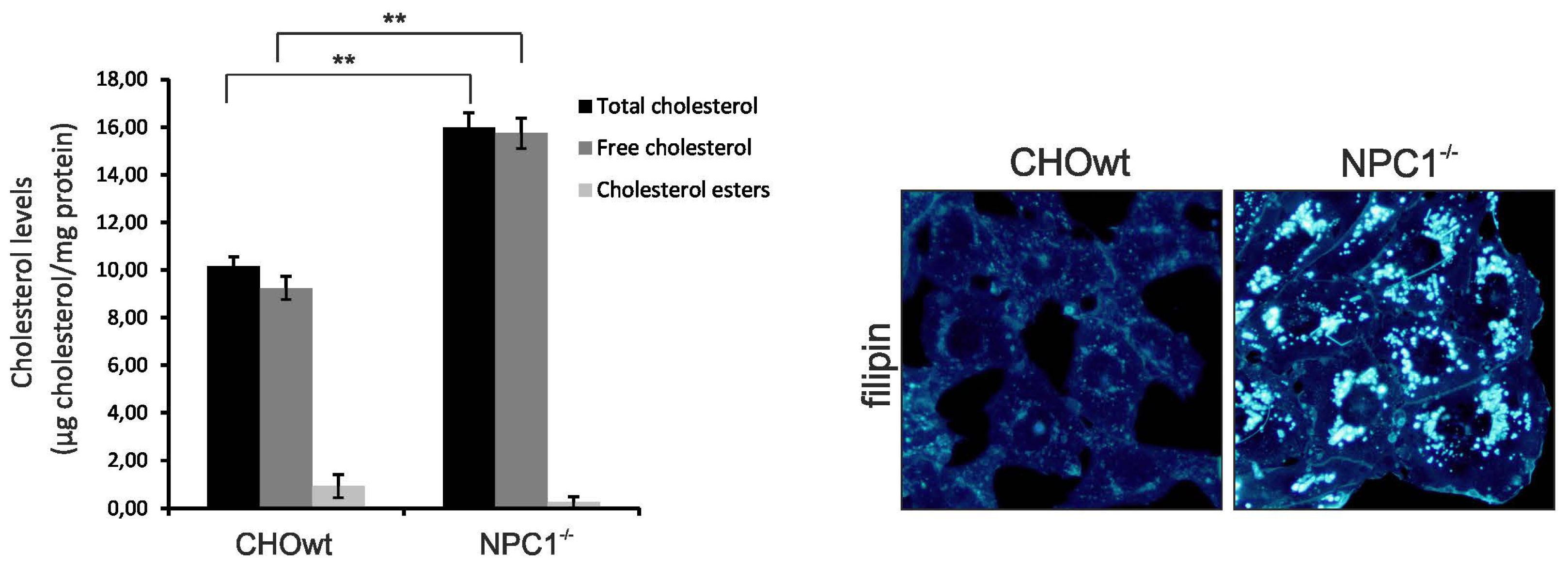
</title>
<sec>
<title>2.1. CHO-<italic>NPC1</italic><sup>−/−</sup> Cells are Enriched in Total, as well as, Vesicular Cholesterol
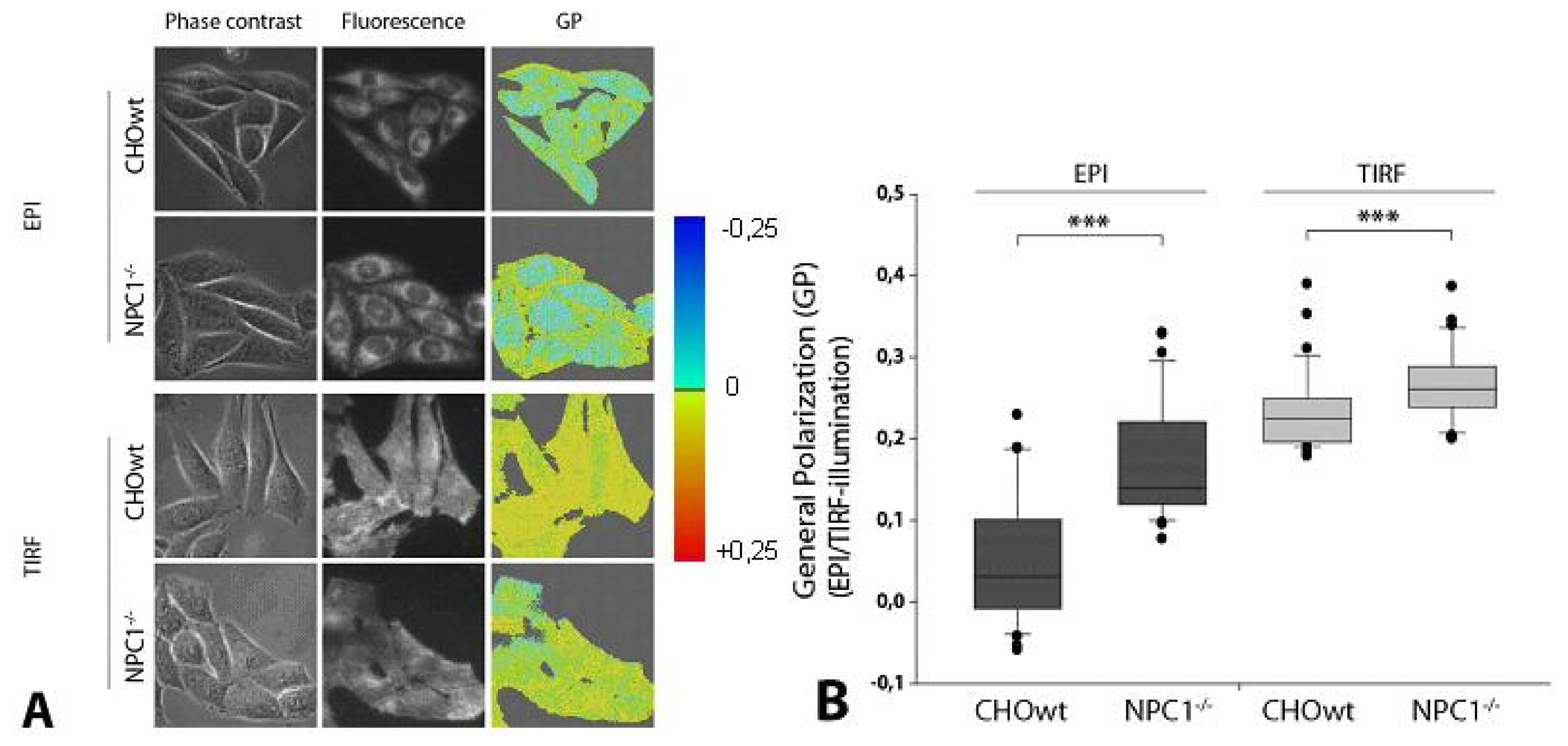
</title>
<p>As we wanted to compare the measurements with our recently published data of cholesterol loaded and depleted glioblastoma astrocytoma U373 cells [<xref ref-type="bibr" rid="b33-ijms-13-15801">33</xref>], we first performed cholesterol level measurements of CHO-WT and CHO-<italic>NPC1</italic><sup>−</sup><italic><sup>/</sup></italic><sup>−</sup> cells (<xref ref-type="fig" rid="f1-ijms-13-15801">Figure 1</xref>). As expected, CHO-<italic>NPC1</italic><sup>−</sup><italic><sup>/</sup></italic><sup>−</sup> cells had a significantly higher free and total cholesterol levels compared to CHO-WT. To further visualize the distribution of free cholesterol within the cells, we performed filipin staining (Sigma-Aldrich). As described for NPC, we found highly increased vesicular distribution of free cholesterol in CHO-<italic>NPC1</italic><sup>−</sup><italic><sup>/</sup></italic><sup>−</sup> cells compared to CHO-WT (<xref ref-type="fig" rid="f1-ijms-13-15801">Figure 1</xref>).</p>
<p>Interestingly, we found that CHO-<italic>NPC1</italic><sup>−</sup><italic><sup>/</sup></italic><sup>−</sup> cells had a cholesterol level comparable with cholesterol depleted U373 glioblastoma cells (<xref ref-type="supplementary-material" rid="s1-ijms-13-15801">Supplementary Table S1</xref>). This can be explained by the high cholesterol levels in astrocytes. Astrocytes supply neurons with cholesterol, as neurons do not produce enough cholesterol on their own.</p></sec>
<sec>
<title>2.2. <italic>NPC1</italic><sup>−/−</sup> Cells Display Higher Membrane Stiffness at Intracellular Membranes</title>
<p>To further analyze differences in membrane stiffness associated with altered cholesterol levels, we used the previously described method of wide-field fluorescence microscopy for measuring membrane dynamics of living cells based on 6-dodecanoyl-2-dimethylamino naphthalene (laurdan) staining and generalized polarization (GP) measurements [<xref ref-type="bibr" rid="b45-ijms-13-15801">45</xref>]. As reported previously [<xref ref-type="bibr" rid="b45-ijms-13-15801">45</xref>,<xref ref-type="bibr" rid="b46-ijms-13-15801">46</xref>], GP represents a normalized value for the difference of fluorescence intensity in two spectral bands, with the first one being predominant in a rather stiff-, and the second one in a more fluid environment. Using TIRF and EPI-illumination, we were able to distinguish between alterations of membrane stiffness at the plasma membrane and intracellular membranes.</p>
<p>As observed before in cholesterol loaded U373 cells, we found only slightly increased stiffness of the plasma membrane as measured by TIRFM. However, using EPI-illumination of whole cells, we found highly increased membrane stiffness at intracellular membranes in CHO-<italic>NPC1</italic><sup>−</sup><italic><sup>/</sup></italic><sup>−</sup> cells compared to CHO-WT (<xref ref-type="fig" rid="f2-ijms-13-15801">Figure 2</xref>). This is most likely due to the massive cholesterol load of intracellular compartments in <italic>NPC1</italic><sup>−</sup><italic><sup>/</sup></italic><sup>−</sup> cells (<xref ref-type="fig" rid="f1-ijms-13-15801">Figure 1</xref>). These results are in line with our earlier measurements of U373 cells, where cholesterol loading increased membrane stiffness and also occurred mainly at intracellular compartments, but not at the plasma membrane [<xref ref-type="bibr" rid="b33-ijms-13-15801">33</xref>].</p></sec>
<sec>
<title>
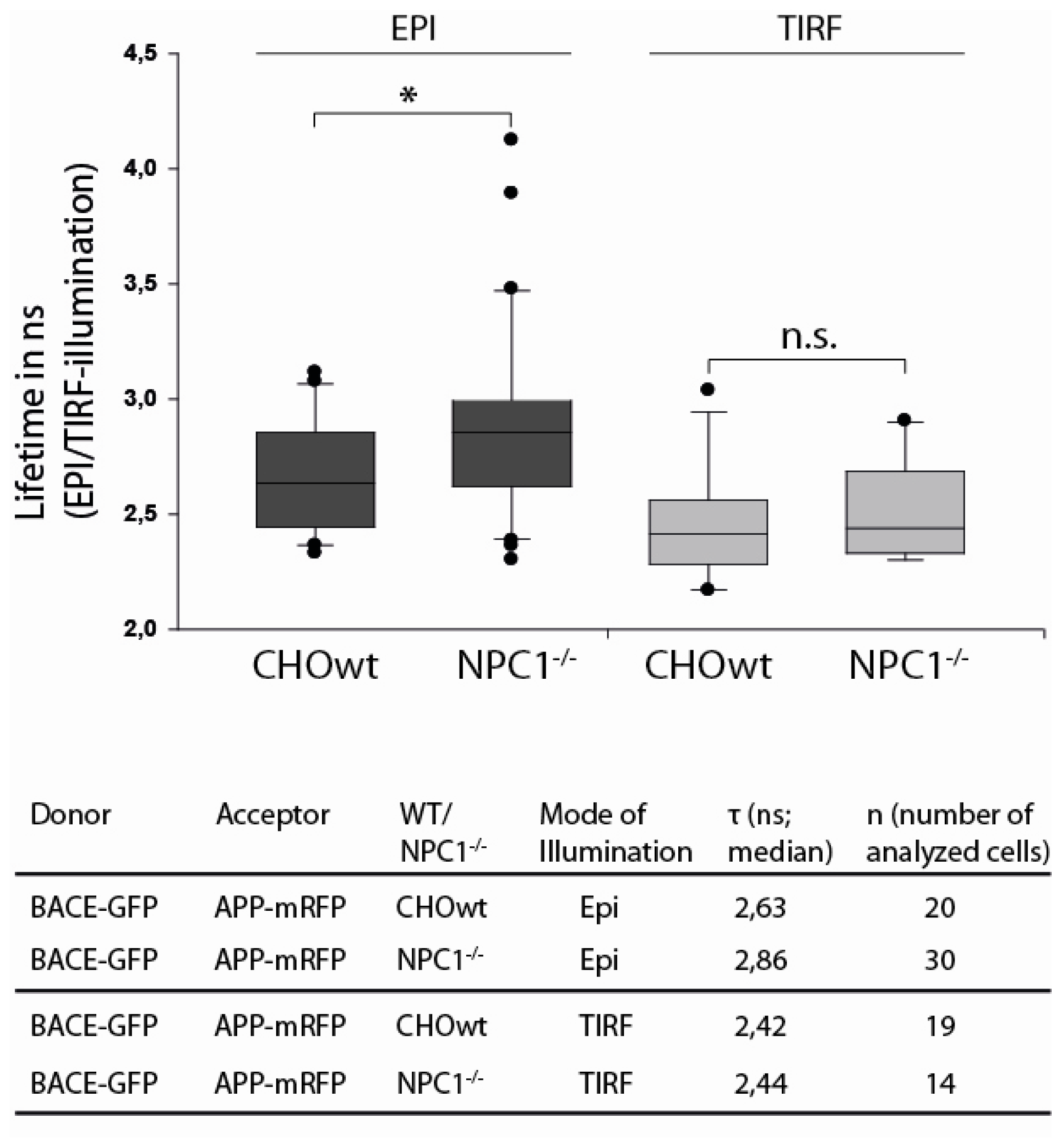
2.3. APP and BACE1 Proximity is Decreased in <italic>NPC1</italic><sup>−/−</sup> Cells, but Not at the Cell Surface</title>
<p>To address the question whether APP and BACE1 proximity is altered at the cell surface or at intracellular membranes due to an increased cholesterol level in CHO-<italic>NPC1</italic><sup>−</sup><italic><sup>/</sup></italic><sup>−</sup> cells, we combined EPI or TIRF microscopy with fluorescence lifetime measurements, as described earlier [<xref ref-type="bibr" rid="b33-ijms-13-15801">33</xref>]. Following excitation by picosecond laser pulses, fluorescence decrease of the donor BACE1-GFP was mono-exponential with the lifetimes depicted in <xref ref-type="fig" rid="f3-ijms-13-15801">Figure 3</xref>.</p>
<p>Non-radiative energy transfer from a donor to an acceptor molecule, according to the Förster mechanism [<xref ref-type="bibr" rid="b47-ijms-13-15801">47</xref>], is often determined from fluorescence lifetime experiments, since the lifetime τ of an excited molecular state corresponds to the reciprocal of the sum of its deactivation rates, including fluorescence (<italic>k</italic><sub>F</sub>), internal conversion (<italic>k</italic><sub>IC</sub>), intersystem crossing (<italic>k</italic><sub>ISC</sub>) and intermolecular energy transfer (<italic>k</italic><sub>ET</sub>). If τ is the lifetime of a donor molecule in presence of, and τ<sub>0</sub> in absence of an acceptor molecule, the difference 1/τ −1/τ<sub>0</sub> reflects the energy transfer rate <italic>k</italic><sub>ET</sub> from the donor to the acceptor. For dipole-dipole interaction, this rate is proportional to R<sup>−6</sup>, with R representing the intermolecular distance, which generally is smaller than about 10 nm. An increase of the fluorescence lifetime τ of the donor can be related to a decrease of <italic>k</italic><sub>ET</sub>, and therefore, to a decrease of donor-acceptor (in this case BACE1-GFP and APP-mRFP) proximity.</p>
<p>In agreement with previous results [<xref ref-type="bibr" rid="b33-ijms-13-15801">33</xref>] (and in contrast to an APP-BACE positive control [<xref ref-type="bibr" rid="b33-ijms-13-15801">33</xref>]), we found no significant changes in donor lifetime and, therefore, in APP-BACE1 proximity at the cell surface as measured by TIRFM. However, under EPI illumination, we observed a significant increase of donor lifetime in CHO-<italic>NPC1</italic><sup>−</sup><italic><sup>/</sup></italic><sup>−</sup> cells compared to CHO-WT (<xref ref-type="fig" rid="f3-ijms-13-15801">Figure 3</xref>). This indicates that APP and BACE1 come less often into close proximity in CHO-<italic>NPC1</italic><sup>−</sup><italic><sup>/</sup></italic><sup>−</sup> cells compared to CHO-WT and that that enhanced membrane stiffness due to cholesterol enrichment is associated with decreased proximity of APP and BACE1 in CHO-<italic>NPC1</italic><sup>−</sup><italic><sup>/</sup></italic><sup>−</sup> cells.</p>
<p>Data on how cholesterol influences APP processing are quite inconsistent. Age of the cells, as well as cell lines used for the experiments, seem to influence the results. However, consistent with our previous findings, most data from <italic>in vitro</italic> and <italic>in vivo</italic> experiments show increased APP β-cleavage upon cholesterol increase, and reduced β-cleavage and increased α-cleavage upon cholesterol depletion [<xref ref-type="bibr" rid="b27-ijms-13-15801">27</xref>,<xref ref-type="bibr" rid="b34-ijms-13-15801">34</xref>,<xref ref-type="bibr" rid="b44-ijms-13-15801">44</xref>,<xref ref-type="bibr" rid="b48-ijms-13-15801">48</xref>–<xref ref-type="bibr" rid="b51-ijms-13-15801">51</xref>]. To understand how BACE1-cleavage of APP is increased, while at the same time APP-BACE1 proximity is reduced, seems to be quite challenging. But one has to keep in mind that, besides its influence on cellular membranes, altered cholesterol levels induce complex alterations in cell metabolism and might influence different membranes to different extent. It is possible that, in some compartments, APP-BACE1 proximity is decreased, whereas in others, APP processing by BACE1 is increased. Although in CHO-<italic>NPC1</italic><sup>−</sup><italic><sup>/</sup></italic><sup>−</sup> cells APP and BACE1 were shown to accumulate in the same compartments (transferrin receptor-positive vesicles) [<xref ref-type="bibr" rid="b52-ijms-13-15801">52</xref>], their routing to and out of these compartments could be quite different, which in turn could result in their overall decreased proximity within the cell. Indeed, we showed recently a strong colocalization of APP, and not BACE1, with early endosomal marker EEA1, and a marked accumulation of APP <italic>C</italic>-terminal fragments (APP-CTFs), and not BACE1, within late endosomes in CHO-<italic>NPC1</italic><sup>−</sup><italic><sup>/</sup></italic><sup>−</sup> cells [<xref ref-type="bibr" rid="b52-ijms-13-15801">52</xref>]. Regarding transport and endocytosis rates and mechanisms, there is little data about alterations upon cholesterol enrichment, but it might lead to accelerated turnover and endocytosis of APP and BACE1 in lipid rafts [<xref ref-type="bibr" rid="b53-ijms-13-15801">53</xref>], thereby promoting APP processing by BACE1. We have previously reported that APP localization is shifted to lipid rafts in CHO-<italic>NPC1</italic><sup>−</sup><italic><sup>/</sup></italic><sup>−</sup> cells [<xref ref-type="bibr" rid="b54-ijms-13-15801">54</xref>]. Another explanation might be the direct impact of cholesterol environment on BACE1 activity. In living cells, BACE1 seems to require intact rafts for activity [<xref ref-type="bibr" rid="b23-ijms-13-15801">23</xref>], and BACE1 outside rafts seems to be inactive. Cholesterol enrichment, as found in CHO-<italic>NPC1</italic><sup>−</sup><italic><sup>/</sup></italic><sup>−</sup>, might change raft composition and size, thereby altering BACE1 activity. Additionally, FRET efficiency is strongly dependent on the spatial orientation of donor and acceptor molecules and, in particular, orientation of donor and acceptor dipole moments. Besides cholesterol, lipid rafts are also enriched in large molecules, like sphingolipids, and proteins that project beyond the phospholipid bilayer and might influence the spatial orientation of the donor and acceptor molecules. Therefore, we cannot completely rule out that increased membrane stiffness and altered raft composition due to cholesterol enrichment might promote APP-BACE1 interaction, but at the same time, influence GFP-RFP FRET efficiency due to sterical hindrance.</p></sec></sec>
<sec>
<title>3. Experimental Section</title>
<sec>
<title>3.1. Expression Constructs</title>
<p>We utilized previously described APP695-rfp and BACE1-gfp expression constructs [<xref ref-type="bibr" rid="b55-ijms-13-15801">55</xref>–<xref ref-type="bibr" rid="b57-ijms-13-15801">57</xref>].</p></sec>
<sec>
<title>3.2. Cell Culture and Stable Transfection</title>
<p>Chinese hamster ovary wild type (CHO-WT) cells and CHO-<italic>NPC1</italic><sup>−</sup><italic><sup>/</sup></italic><sup>−</sup> cells (kindly provided by Dr. Daniel Ory) were maintained as described previously [<xref ref-type="bibr" rid="b52-ijms-13-15801">52</xref>]. Cell lines stably expressing APP-mRFP and BACE1-GFP were established by co-transfection using Lipofectamine LTX (Invitrogen, Darmstadt, Germany) and selection in the media supplemented with an appropriate selective antibiotic.</p>
<p>Both cell lines were seeded on microscope slides and maintained in DMEM (Gibco) supplemented with 10% fetal bovine serum (FBS) and 1x penicillin/streptomycin (P/S). Cells were incubated in 5% CO<sub>2</sub> and 37 °C for 24 h to 36 h before analysis.</p></sec>
<sec>
<title>3.3. Cholesterol Quantification</title>
<p>CHO-WT and CHO-<italic>NPC1</italic><sup>−</sup><italic><sup>/</sup></italic><sup>−</sup> cells were plated in 6-well plates and grown for 48 h in 10% FBS DMEM/F12 medium until confluent. Cells from each well were collected and lysed in CoIP lysis buffer (50 mM Tris, pH 7.6, 150 mM NaCl, 2 mM EDTA, 1% NP40) containing protease inhibitor cocktail (Roche Applied Science). Protein concentration in cell lysates (mg protein/mL) was determined with the DC Protein Assay (BioRad). The protein concentration in cell lysate is proportional to the number of cells collected for lysis. Concentration of free cholesterol, cholesterol esters and total cholesterol in cell lysates (μg cholesterol/mL) was measured using Amplex Red Cholesterol Assay Kit (Molecular Probes), following the manufacturer’s instructions. Cholesterol concentration was normalized according to protein concentration of the matching sample. Thus, cholesterol levels were expressed in μg cholesterol/mg protein.</p></sec>
<sec>
<title>3.4. Filipin Staining</title>
<p>Filipin (Sigma Aldrich) staining was done as previously described [<xref ref-type="bibr" rid="b58-ijms-13-15801">58</xref>]. The cells were mounted and were viewed by fluorescence microscope (Olympus BX51).</p></sec>
<sec>
<title>3.5. Laurdan Staining and GP Measurements</title>
<p>The hydrophobic membrane marker 6-dodecanoyl-2-dimethylamino naphthalene (laurdan) was obtained from Molecular Probes (Göttingen, Germany) and prepared as a 2 mM stock solution in ethanol. For GP measurement, cells were grown on microscope object slides for 48 h prior to rinsing with Earl’s balanced salt solution (EBSS) and incubation with laurdan (8 μM in culture medium) for 60 min at 37 °C. GP measurement and calculation was performed as described previously [<xref ref-type="bibr" rid="b45-ijms-13-15801">45</xref>] using the following equation: GP = (I<sub>440</sub> − I<sub>490</sub>)/(I<sub>440</sub> + I<sub>490</sub>), with I<sub>440</sub> and I<sub>490</sub> representing the fluorescence intensities measured at 440 nm and 490 nm, respectively.</p></sec>
<sec sec-type="methods">
<title>3.6. Microscopic Imaging and FRET Analysis</title>
<p>Microscopic experiments were performed with a picosecond laser diode emitting light at 470 nm (LDH 470 with driver PDL 800 B, Picoquant, Berlin, Germany; average power 1 mW, pulse duration 70 ps, repetition rate 40 MHz) as the excitation source of GFP. This laser diode was adapted to a fluorescence microscope (Axioplan 1, Carl Zeiss Jena, Germany) with either a multimode fiber and a dichroic mirror (reflecting light at wavelengths λ ≤ 510 nm) for EPI-illumination, or with a single mode fiber (Point Source, kineFlex, Southampton, UK) for illumination by an evanescent electromagnetic field (penetration depth around 130 nm) with a specific device for total internal reflection (TIR) fluorescence microscopy [<xref ref-type="bibr" rid="b59-ijms-13-15801">59</xref>]. In the first case, whole cells were illuminated, whereas in the second case, selective illumination of the plasma membranes and adjacent cellular sites occurred. Using a purpose-made optical setup [<xref ref-type="bibr" rid="b33-ijms-13-15801">33</xref>,<xref ref-type="bibr" rid="b45-ijms-13-15801">45</xref>,<xref ref-type="bibr" rid="b60-ijms-13-15801">60</xref>], one could easily switch between the two excitation modes. For measuring fluorescence decay kinetics of single cells, a long pass filter transmitting light above 515 nm and a time-gated image-intensifying CCD camera system (Picostar HR 12; LaVision, Göttingen, Germany) with a time resolution of 200 ps were used, as described in detail elsewhere [<xref ref-type="bibr" rid="b61-ijms-13-15801">61</xref>]. Fluorescence decay kinetics of individual cells were recorded by shifting the time gate of 200 ps over an axis of 20 ns. Since the fluorescence intensity of mRFP was very low, all fluorescence was related to GFP.</p>
<p>For GP evaluation, cells incubated with laurdan (see above) were used and exposed to a laser diode emitting light at 391 nm (LDH 400 with driver PDL 800 B; Picoquant, Berlin) upon EPI-illumination and a laser diode at 375 nm (LDH 375; Picoquant, Berlin) upon TIR illumination. Simultaneous use of both laser diodes facilitated parallel measurements under maintenance of similar extinction coefficients [<xref ref-type="bibr" rid="b46-ijms-13-15801">46</xref>]. Fluorescence spectra of single cells were recorded at λ ≥ 420 nm with a polychromator and an image intensifying detection unit (IMD 4562, Hamamatsu Photonics, Ichino-Cho, Japan) [<xref ref-type="bibr" rid="b62-ijms-13-15801">62</xref>] fixed on top of the microscope. In addition, fluorescence images were recorded at 450 ± 20 nm and 490 ± 20 nm using appropriate interference filters and an electron multiplying (EM-)CCD camera (DV887DC, ANDOR Technology, Belfast, U.K.) with Peltier cooling and a sensitivity below 10<sup>−16</sup> W/pixel.</p></sec></sec>
<sec>
<title>4. Conclusion</title>
<p>Combination of FRET and TIRF (TIRET) (in comparison with FRET under EPI illumination) is an appropriate and very selective technique to examine molecular interactions in the plasma membrane of living cells, since illumination by an evanescent electromagnetic field arising at a cell-substrate surface is limited to less than 100 nm. By combining FRET with EPI illumination, we were able to confirm previously observed cholesterol-associated alterations in proximity of AD relevant proteins APP and BACE1 at intracellular compartments, in contrast to the plasma membranes. In the future, alterations in protein proximity may be further localized within the cells by fluorescence lifetime imaging microscopy (FLIM), where the same experimental setup can be used. This may permit more detailed information about processes involved in the formation of neurodegenerative disorders and other diseases.</p></sec>
<sec sec-type="supplementary-material">
<title>Supplementary Information</title>
<supplementary-material id="s1-ijms-13-15801" content-type="local-data">
<media xlink:href="ijms-13-15801-s001.pdf" mimetype="application" mime-subtype="pdf"/></supplementary-material></sec></body>
<back>
<ack>
<title>Acknowledgment</title>
<p>This work was supported by the grant of the Ministry of Science, Education and Sports (MSES) of the Republic of Croatia (098-0982522-2525 to S.H.) and the bilateral project between DAAD and MSES (to S.H.). The authors thank Claudia Hintze for skillful technical assistance.</p></ack>
<ref-list>
<title>References</title>
<ref id="b1-ijms-13-15801"><label>1</label><citation citation-type="journal"><person-group person-group-type="author"><name><surname>Dietschy</surname><given-names>J.M.</given-names></name><name><surname>Turley</surname><given-names>S.D.</given-names></name></person-group><article-title>Cholesterol metabolism in the brain</article-title><source>Curr. Opin. Lipidol</source><year>2001</year><volume>12</volume><fpage>105</fpage><lpage>112</lpage><pub-id pub-id-type="doi">10.1097/00041433-200104000-00003</pub-id><pub-id pub-id-type="pmid">11264981</pub-id></citation></ref>
<ref id="b2-ijms-13-15801"><label>2</label><citation citation-type="journal"><person-group person-group-type="author"><name><surname>Simons</surname><given-names>K.</given-names></name><name><surname>Ikonen</surname><given-names>E.</given-names></name></person-group><article-title>How cells handle cholesterol</article-title><source>Science</source><year>2000</year><volume>290</volume><fpage>1721</fpage><lpage>1726</lpage><pub-id pub-id-type="doi">10.1126/science.290.5497.1721</pub-id><pub-id pub-id-type="pmid">11099405</pub-id></citation></ref>
<ref id="b3-ijms-13-15801"><label>3</label><citation citation-type="journal"><person-group person-group-type="author"><name><surname>Bjorkhem</surname><given-names>I.</given-names></name></person-group><article-title>Crossing the barrier: Oxysterols as cholesterol transporters and metabolic modulators in the brain</article-title><source>J. Int. Med</source><year>2006</year><volume>260</volume><fpage>493</fpage><lpage>508</lpage><pub-id pub-id-type="doi">10.1111/j.1365-2796.2006.01725.x</pub-id></citation></ref>
<ref id="b4-ijms-13-15801"><label>4</label><citation citation-type="journal"><person-group person-group-type="author"><name><surname>Jurevics</surname><given-names>H.</given-names></name><name><surname>Morell</surname><given-names>P.</given-names></name></person-group><article-title>Cholesterol for synthesis of myelin is made locally, not imported into brain</article-title><source>J. Neurochem</source><year>1995</year><volume>64</volume><fpage>895</fpage><lpage>901</lpage><pub-id pub-id-type="pmid">7830084</pub-id></citation></ref>
<ref id="b5-ijms-13-15801"><label>5</label><citation citation-type="journal"><person-group person-group-type="author"><name><surname>Pfrieger</surname><given-names>F.W.</given-names></name></person-group><article-title>Role of cholesterol in synapse formation and function</article-title><source>Biochim. Biophys. Acta</source><year>2003</year><volume>1610</volume><fpage>271</fpage><lpage>280</lpage><pub-id pub-id-type="doi">10.1016/S0005-2736(03)00024-5</pub-id><pub-id pub-id-type="pmid">12648780</pub-id></citation></ref>
<ref id="b6-ijms-13-15801"><label>6</label><citation citation-type="journal"><person-group person-group-type="author"><name><surname>Mauch</surname><given-names>D.H.</given-names></name><name><surname>Nagler</surname><given-names>K.</given-names></name><name><surname>Schumacher</surname><given-names>S.</given-names></name><name><surname>Goritz</surname><given-names>C.</given-names></name><name><surname>Muller</surname><given-names>E.C.</given-names></name><name><surname>Otto</surname><given-names>A.</given-names></name><name><surname>Pfrieger</surname><given-names>F.W.</given-names></name></person-group><article-title>Cns synaptogenesis promoted by glia-derived cholesterol</article-title><source>Science</source><year>2001</year><volume>294</volume><fpage>1354</fpage><lpage>1357</lpage><pub-id pub-id-type="doi">10.1126/science.294.5545.1354</pub-id><pub-id pub-id-type="pmid">11701931</pub-id></citation></ref>
<ref id="b7-ijms-13-15801"><label>7</label><citation citation-type="journal"><person-group person-group-type="author"><name><surname>Valenza</surname><given-names>M.</given-names></name><name><surname>Rigamonti</surname><given-names>D.</given-names></name><name><surname>Goffredo</surname><given-names>D.</given-names></name><name><surname>Zuccato</surname><given-names>C.</given-names></name><name><surname>Fenu</surname><given-names>S.</given-names></name><name><surname>Jamot</surname><given-names>L.</given-names></name><name><surname>Strand</surname><given-names>A.</given-names></name><name><surname>Tarditi</surname><given-names>A.</given-names></name><name><surname>Woodman</surname><given-names>B.</given-names></name><name><surname>Racchi</surname><given-names>M.</given-names></name><etal/></person-group><article-title>Dysfunction of the cholesterol biosynthetic pathway in huntington’s disease</article-title><source>J. Neurosci</source><year>2005</year><volume>25</volume><fpage>9932</fpage><lpage>9939</lpage><pub-id pub-id-type="doi">10.1523/JNEUROSCI.3355-05.2005</pub-id><pub-id pub-id-type="pmid">16251441</pub-id></citation></ref>
<ref id="b8-ijms-13-15801"><label>8</label><citation citation-type="journal"><person-group person-group-type="author"><name><surname>Leoni</surname><given-names>V.</given-names></name><name><surname>Mariotti</surname><given-names>C.</given-names></name><name><surname>Nanetti</surname><given-names>L.</given-names></name><name><surname>Salvatore</surname><given-names>E.</given-names></name><name><surname>Squitieri</surname><given-names>F.</given-names></name><name><surname>Bentivoglio</surname><given-names>A.R.</given-names></name><name><surname>Bandettini del Poggio</surname><given-names>M.</given-names></name><name><surname>Piacentini</surname><given-names>S.</given-names></name><name><surname>Monza</surname><given-names>D.</given-names></name><name><surname>Valenza</surname><given-names>M.</given-names></name><etal/></person-group><article-title>Whole body cholesterol metabolism is impaired in huntington’s disease</article-title><source>Neurosci. Lett</source><year>2011</year><volume>494</volume><fpage>245</fpage><lpage>249</lpage><pub-id pub-id-type="doi">10.1016/j.neulet.2011.03.025</pub-id><pub-id pub-id-type="pmid">21406216</pub-id></citation></ref>
<ref id="b9-ijms-13-15801"><label>9</label><citation citation-type="journal"><person-group person-group-type="author"><name><surname>Hu</surname><given-names>G.</given-names></name></person-group><article-title>Total cholesterol and the risk of parkinson’s disease: A review for some new findings</article-title><source>Parkinson’s Dis</source><year>2010</year><volume>2010</volume><fpage>836962</fpage></citation></ref>
<ref id="b10-ijms-13-15801"><label>10</label><citation citation-type="journal"><person-group person-group-type="author"><name><surname>Bar-On</surname><given-names>P.</given-names></name><name><surname>Crews</surname><given-names>L.</given-names></name><name><surname>Koob</surname><given-names>A.O.</given-names></name><name><surname>Mizuno</surname><given-names>H.</given-names></name><name><surname>Adame</surname><given-names>A.</given-names></name><name><surname>Spencer</surname><given-names>B.</given-names></name><name><surname>Masliah</surname><given-names>E.</given-names></name></person-group><article-title>Statins reduce neuronal alpha-synuclein aggregation in <italic>in vitro</italic> models of parkinson’s disease</article-title><source>J. Neurochem</source><year>2008</year><volume>105</volume><fpage>1656</fpage><lpage>1667</lpage><pub-id pub-id-type="doi">10.1111/j.1471-4159.2008.05254.x</pub-id><pub-id pub-id-type="pmid">18248604</pub-id></citation></ref>
<ref id="b11-ijms-13-15801"><label>11</label><citation citation-type="journal"><person-group person-group-type="author"><name><surname>Bosco</surname><given-names>D.A.</given-names></name><name><surname>Fowler</surname><given-names>D.M.</given-names></name><name><surname>Zhang</surname><given-names>Q.</given-names></name><name><surname>Nieva</surname><given-names>J.</given-names></name><name><surname>Powers</surname><given-names>E.T.</given-names></name><name><surname>Wentworth</surname><given-names>P.</given-names><suffix>Jr</suffix></name><name><surname>Lerner</surname><given-names>R.A.</given-names></name><name><surname>Kelly</surname><given-names>J.W.</given-names></name></person-group><article-title>Elevated levels of oxidized cholesterol metabolites in lewy body disease brains accelerate alpha-synuclein fibrilization</article-title><source>Nat. Chem. Biol.</source><year>2006</year><volume>2</volume><fpage>249</fpage><lpage>253</lpage><pub-id pub-id-type="doi">10.1038/nchembio782</pub-id><pub-id pub-id-type="pmid">16565714</pub-id></citation></ref>
<ref id="b12-ijms-13-15801"><label>12</label><citation citation-type="journal"><person-group person-group-type="author"><name><surname>Puglielli</surname><given-names>L.</given-names></name><name><surname>Tanzi</surname><given-names>R.E.</given-names></name><name><surname>Kovacs</surname><given-names>D.M.</given-names></name></person-group><article-title>Alzheimer’s disease: The cholesterol connection</article-title><source>Nat. Neurosci</source><year>2003</year><volume>6</volume><fpage>345</fpage><lpage>351</lpage><pub-id pub-id-type="doi">10.1038/nn0403-345</pub-id><pub-id pub-id-type="pmid">12658281</pub-id></citation></ref>
<ref id="b13-ijms-13-15801"><label>13</label><citation citation-type="journal"><person-group person-group-type="author"><name><surname>Reitz</surname><given-names>C.</given-names></name><name><surname>Tang</surname><given-names>M.X.</given-names></name><name><surname>Schupf</surname><given-names>N.</given-names></name><name><surname>Manly</surname><given-names>J.J.</given-names></name><name><surname>Mayeux</surname><given-names>R.</given-names></name><name><surname>Luchsinger</surname><given-names>J.A.</given-names></name></person-group><article-title>Association of higher levels of high-density lipoprotein cholesterol in elderly individuals and lower risk of late-onset Alzheimer disease</article-title><source>Arch. Neurol</source><year>2010</year><volume>67</volume><fpage>1491</fpage><lpage>1497</lpage><pub-id pub-id-type="doi">10.1001/archneurol.2010.297</pub-id><pub-id pub-id-type="pmid">21149810</pub-id></citation></ref>
<ref id="b14-ijms-13-15801"><label>14</label><citation citation-type="journal"><person-group person-group-type="author"><name><surname>Notkola</surname><given-names>I.L.</given-names></name><name><surname>Sulkava</surname><given-names>R.</given-names></name><name><surname>Pekkanen</surname><given-names>J.</given-names></name><name><surname>Erkinjuntti</surname><given-names>T.</given-names></name><name><surname>Ehnholm</surname><given-names>C.</given-names></name><name><surname>Kivinen</surname><given-names>P.</given-names></name><name><surname>Tuomilehto</surname><given-names>J.</given-names></name><name><surname>Nissinen</surname><given-names>A.</given-names></name></person-group><article-title>Serum total cholesterol, apolipoprotein e epsilon 4 allele, and Alzheimer’s disease</article-title><source>Neuroepidemiology</source><year>1998</year><volume>17</volume><fpage>14</fpage><lpage>20</lpage><pub-id pub-id-type="doi">10.1159/000026149</pub-id><pub-id pub-id-type="pmid">9549720</pub-id></citation></ref>
<ref id="b15-ijms-13-15801"><label>15</label><citation citation-type="journal"><person-group person-group-type="author"><name><surname>Kivipelto</surname><given-names>M.</given-names></name><name><surname>Helkala</surname><given-names>E.L.</given-names></name><name><surname>Laakso</surname><given-names>M.P.</given-names></name><name><surname>Hanninen</surname><given-names>T.</given-names></name><name><surname>Hallikainen</surname><given-names>M.</given-names></name><name><surname>Alhainen</surname><given-names>K.</given-names></name><name><surname>Iivonen</surname><given-names>S.</given-names></name><name><surname>Mannermaa</surname><given-names>A.</given-names></name><name><surname>Tuomilehto</surname><given-names>J.</given-names></name><name><surname>Nissinen</surname><given-names>A.</given-names></name><etal/></person-group><article-title>Apolipoprotein e epsilon4 allele, elevated midlife total cholesterol level, and high midlife systolic blood pressure are independent risk factors for late-life alzheimer disease</article-title><source>Ann. Intern. Med</source><year>2002</year><volume>137</volume><fpage>149</fpage><lpage>155</lpage><pub-id pub-id-type="pmid">12160362</pub-id></citation></ref>
<ref id="b16-ijms-13-15801"><label>16</label><citation citation-type="journal"><person-group person-group-type="author"><name><surname>Lloyd-Evans</surname><given-names>E.</given-names></name><name><surname>Morgan</surname><given-names>A.J.</given-names></name><name><surname>He</surname><given-names>X.</given-names></name><name><surname>Smith</surname><given-names>D.A.</given-names></name><name><surname>Elliot-Smith</surname><given-names>E.</given-names></name><name><surname>Sillence</surname><given-names>D.J.</given-names></name><name><surname>Churchill</surname><given-names>G.C.</given-names></name><name><surname>Schuchman</surname><given-names>E.H.</given-names></name><name><surname>Galione</surname><given-names>A.</given-names></name><name><surname>Platt</surname><given-names>F.M.</given-names></name></person-group><article-title>Niemann-pick disease type c1 is a sphingosine storage disease that causes deregulation of lysosomal calcium</article-title><source>Nat. Med</source><year>2008</year><volume>14</volume><fpage>1247</fpage><lpage>1255</lpage><pub-id pub-id-type="doi">10.1038/nm.1876</pub-id><pub-id pub-id-type="pmid">18953351</pub-id></citation></ref>
<ref id="b17-ijms-13-15801"><label>17</label><citation citation-type="journal"><person-group person-group-type="author"><name><surname>Sturley</surname><given-names>S.L.</given-names></name><name><surname>Patterson</surname><given-names>M.C.</given-names></name><name><surname>Pentchev</surname><given-names>P.</given-names></name></person-group><article-title>Unraveling the sterol-trafficking defect in niemann-pick c disease</article-title><source>Proc. Natl. Acad. Sci. USA</source><year>2009</year><volume>106</volume><fpage>2093</fpage><lpage>2094</lpage><pub-id pub-id-type="doi">10.1073/pnas.0812934106</pub-id><pub-id pub-id-type="pmid">19211789</pub-id></citation></ref>
<ref id="b18-ijms-13-15801"><label>18</label><citation citation-type="journal"><person-group person-group-type="author"><name><surname>Vance</surname><given-names>J.E.</given-names></name></person-group><article-title>Lipid imbalance in the neurological disorder, niemann-pick c disease</article-title><source>FEBS Lett</source><year>2006</year><volume>580</volume><fpage>5518</fpage><lpage>5524</lpage><pub-id pub-id-type="doi">10.1016/j.febslet.2006.06.008</pub-id><pub-id pub-id-type="pmid">16797010</pub-id></citation></ref>
<ref id="b19-ijms-13-15801"><label>19</label><citation citation-type="journal"><person-group person-group-type="author"><name><surname>Hardy</surname><given-names>J.A.</given-names></name><name><surname>Higgins</surname><given-names>G.A.</given-names></name></person-group><article-title>Alzheimer’s disease: The amyloid cascade hypothesis</article-title><source>Science</source><year>1992</year><volume>256</volume><fpage>184</fpage><lpage>185</lpage><pub-id pub-id-type="doi">10.1126/science.1566067</pub-id><pub-id pub-id-type="pmid">1566067</pub-id></citation></ref>
<ref id="b20-ijms-13-15801"><label>20</label><citation citation-type="journal"><person-group person-group-type="author"><name><surname>Hardy</surname><given-names>J.</given-names></name><name><surname>Selkoe</surname><given-names>D.J.</given-names></name></person-group><article-title>The amyloid hypothesis of Alzheimer’s disease: Progress and problems on the road to therapeutics</article-title><source>Science</source><year>2002</year><volume>297</volume><fpage>353</fpage><lpage>356</lpage><pub-id pub-id-type="doi">10.1126/science.1072994</pub-id><pub-id pub-id-type="pmid">12130773</pub-id></citation></ref>
<ref id="b21-ijms-13-15801"><label>21</label><citation citation-type="journal"><person-group person-group-type="author"><name><surname>Selkoe</surname><given-names>D.J.</given-names></name></person-group><article-title>The molecular pathology of Alzheimer’s disease</article-title><source>Neuron</source><year>1991</year><volume>6</volume><fpage>487</fpage><lpage>498</lpage><pub-id pub-id-type="doi">10.1016/0896-6273(91)90052-2</pub-id><pub-id pub-id-type="pmid">1673054</pub-id></citation></ref>
<ref id="b22-ijms-13-15801"><label>22</label><citation citation-type="journal"><person-group person-group-type="author"><name><surname>Hardy</surname><given-names>J.</given-names></name><name><surname>Allsop</surname><given-names>D.</given-names></name></person-group><article-title>Amyloid deposition as the central event in the aetiology of Alzheimer’s disease</article-title><source>Trends Pharmacol. Sci</source><year>1991</year><volume>12</volume><fpage>383</fpage><lpage>388</lpage><pub-id pub-id-type="doi">10.1016/0165-6147(91)90609-V</pub-id><pub-id pub-id-type="pmid">1763432</pub-id></citation></ref>
<ref id="b23-ijms-13-15801"><label>23</label><citation citation-type="journal"><person-group person-group-type="author"><name><surname>Ehehalt</surname><given-names>R.</given-names></name><name><surname>Keller</surname><given-names>P.</given-names></name><name><surname>Haass</surname><given-names>C.</given-names></name><name><surname>Thiele</surname><given-names>C.</given-names></name><name><surname>Simons</surname><given-names>K.</given-names></name></person-group><article-title>Amyloidogenic processing of the alzheimer beta-amyloid precursor protein depends on lipid rafts</article-title><source>J. Cell Biol</source><year>2003</year><volume>160</volume><fpage>113</fpage><lpage>123</lpage><pub-id pub-id-type="doi">10.1083/jcb.200207113</pub-id><pub-id pub-id-type="pmid">12515826</pub-id></citation></ref>
<ref id="b24-ijms-13-15801"><label>24</label><citation citation-type="journal"><person-group person-group-type="author"><name><surname>Rouvinski</surname><given-names>A.</given-names></name><name><surname>Gahali-Sass</surname><given-names>I.</given-names></name><name><surname>Stav</surname><given-names>I.</given-names></name><name><surname>Metzer</surname><given-names>E.</given-names></name><name><surname>Atlan</surname><given-names>H.</given-names></name><name><surname>Taraboulos</surname><given-names>A.</given-names></name></person-group><article-title>Both raft- and non-raft proteins associate with chaps-insoluble complexes: Some app in large complexes</article-title><source>Biochem. Biophys. Res. Commun</source><year>2003</year><volume>308</volume><fpage>750</fpage><lpage>758</lpage><pub-id pub-id-type="doi">10.1016/S0006-291X(03)01470-0</pub-id><pub-id pub-id-type="pmid">12927782</pub-id></citation></ref>
<ref id="b25-ijms-13-15801"><label>25</label><citation citation-type="journal"><person-group person-group-type="author"><name><surname>Bouillot</surname><given-names>C.</given-names></name><name><surname>Prochiantz</surname><given-names>A.</given-names></name><name><surname>Rougon</surname><given-names>G.</given-names></name><name><surname>Allinquant</surname><given-names>B.</given-names></name></person-group><article-title>Axonal amyloid precursor protein expressed by neurons <italic>in vitro</italic> is present in a membrane fraction with caveolae-like properties</article-title><source>J. Biol. Chem</source><year>1996</year><volume>271</volume><fpage>7640</fpage><lpage>7644</lpage><pub-id pub-id-type="doi">10.1074/jbc.271.13.7640</pub-id><pub-id pub-id-type="pmid">8631800</pub-id></citation></ref>
<ref id="b26-ijms-13-15801"><label>26</label><citation citation-type="journal"><person-group person-group-type="author"><name><surname>Riddell</surname><given-names>D.R.</given-names></name><name><surname>Christie</surname><given-names>G.</given-names></name><name><surname>Hussain</surname><given-names>I.</given-names></name><name><surname>Dingwall</surname><given-names>C.</given-names></name></person-group><article-title>Compartmentalization of beta-secretase (asp2) into low-buoyant density, noncaveolar lipid rafts</article-title><source>Curr. Biol</source><year>2001</year><volume>11</volume><fpage>1288</fpage><lpage>1293</lpage><pub-id pub-id-type="doi">10.1016/S0960-9822(01)00394-3</pub-id><pub-id pub-id-type="pmid">11525745</pub-id></citation></ref>
<ref id="b27-ijms-13-15801"><label>27</label><citation citation-type="journal"><person-group person-group-type="author"><name><surname>Simons</surname><given-names>M.</given-names></name><name><surname>Keller</surname><given-names>P.</given-names></name><name><surname>de Strooper</surname><given-names>B.</given-names></name><name><surname>Beyreuther</surname><given-names>K.</given-names></name><name><surname>Dotti</surname><given-names>C.G.</given-names></name><name><surname>Simons</surname><given-names>K.</given-names></name></person-group><article-title>Cholesterol depletion inhibits the generation of beta-amyloid in hippocampal neurons</article-title><source>Proc. Natl. Acad. Sci. USA</source><year>1998</year><volume>95</volume><fpage>6460</fpage><lpage>6464</lpage><pub-id pub-id-type="doi">10.1073/pnas.95.11.6460</pub-id><pub-id pub-id-type="pmid">9600988</pub-id></citation></ref>
<ref id="b28-ijms-13-15801"><label>28</label><citation citation-type="journal"><person-group person-group-type="author"><name><surname>Tolppanen</surname><given-names>A.M.</given-names></name><name><surname>Solomon</surname><given-names>A.</given-names></name><name><surname>Soininen</surname><given-names>H.</given-names></name><name><surname>Kivipelto</surname><given-names>M.</given-names></name></person-group><article-title>Midlife vascular risk factors and Alzheimer’s disease: Evidence from epidemiological studies</article-title><source>J. Alzheimer’s Dis</source><year>2012</year><volume>32</volume><fpage>531</fpage><lpage>540</lpage></citation></ref>
<ref id="b29-ijms-13-15801"><label>29</label><citation citation-type="journal"><person-group person-group-type="author"><name><surname>Corder</surname><given-names>E.H.</given-names></name><name><surname>Saunders</surname><given-names>A.M.</given-names></name><name><surname>Strittmatter</surname><given-names>W.J.</given-names></name><name><surname>Schmechel</surname><given-names>D.E.</given-names></name><name><surname>Gaskell</surname><given-names>P.C.</given-names></name><name><surname>Small</surname><given-names>G.W.</given-names></name><name><surname>Roses</surname><given-names>A.D.</given-names></name><name><surname>Haines</surname><given-names>J.L.</given-names></name><name><surname>Pericak-Vance</surname><given-names>M.A.</given-names></name></person-group><article-title>Gene dose of apolipoprotein e type 4 allele and the risk of Alzheimer’s disease in late onset families</article-title><source>Science</source><year>1993</year><volume>261</volume><fpage>921</fpage><lpage>923</lpage><pub-id pub-id-type="doi">10.1126/science.8346443</pub-id><pub-id pub-id-type="pmid">8346443</pub-id></citation></ref>
<ref id="b30-ijms-13-15801"><label>30</label><citation citation-type="journal"><person-group person-group-type="author"><name><surname>Poirier</surname><given-names>J.</given-names></name><name><surname>Davignon</surname><given-names>J.</given-names></name><name><surname>Bouthillier</surname><given-names>D.</given-names></name><name><surname>Kogan</surname><given-names>S.</given-names></name><name><surname>Bertrand</surname><given-names>P.</given-names></name><name><surname>Gauthier</surname><given-names>S.</given-names></name></person-group><article-title>Apolipoprotein e polymorphism and alzheimer’s disease</article-title><source>Lancet</source><year>1993</year><volume>342</volume><fpage>697</fpage><lpage>699</lpage><pub-id pub-id-type="doi">10.1016/0140-6736(93)91705-Q</pub-id><pub-id pub-id-type="pmid">8103819</pub-id></citation></ref>
<ref id="b31-ijms-13-15801"><label>31</label><citation citation-type="journal"><person-group person-group-type="author"><name><surname>Rebeck</surname><given-names>G.W.</given-names></name><name><surname>Reiter</surname><given-names>J.S.</given-names></name><name><surname>Strickland</surname><given-names>D.K.</given-names></name><name><surname>Hyman</surname><given-names>B.T.</given-names></name></person-group><article-title>Apolipoprotein e in sporadic Alzheimer’s disease: Allelic variation and receptor interactions</article-title><source>Neuron</source><year>1993</year><volume>11</volume><fpage>575</fpage><lpage>580</lpage><pub-id pub-id-type="doi">10.1016/0896-6273(93)90070-8</pub-id><pub-id pub-id-type="pmid">8398148</pub-id></citation></ref>
<ref id="b32-ijms-13-15801"><label>32</label><citation citation-type="journal"><person-group person-group-type="author"><name><surname>Strittmatter</surname><given-names>W.J.</given-names></name><name><surname>Saunders</surname><given-names>A.M.</given-names></name><name><surname>Schmechel</surname><given-names>D.</given-names></name><name><surname>Pericak-Vance</surname><given-names>M.</given-names></name><name><surname>Enghild</surname><given-names>J.</given-names></name><name><surname>Salvesen</surname><given-names>G.S.</given-names></name><name><surname>Roses</surname><given-names>A.D.</given-names></name></person-group><article-title>Apolipoprotein e: High-avidity binding to beta-amyloid and increased frequency of type 4 allele in late-onset familial Alzheimer disease</article-title><source>Proc. Natl. Acad. Sci. USA</source><year>1993</year><volume>90</volume><fpage>1977</fpage><lpage>1981</lpage><pub-id pub-id-type="doi">10.1073/pnas.90.5.1977</pub-id><pub-id pub-id-type="pmid">8446617</pub-id></citation></ref>
<ref id="b33-ijms-13-15801"><label>33</label><citation citation-type="journal"><person-group person-group-type="author"><name><surname>Von Arnim</surname><given-names>C.A.</given-names></name><name><surname>von Einem</surname><given-names>B.</given-names></name><name><surname>Weber</surname><given-names>P.</given-names></name><name><surname>Wagner</surname><given-names>M.</given-names></name><name><surname>Schwanzar</surname><given-names>D.</given-names></name><name><surname>Spoelgen</surname><given-names>R.</given-names></name><name><surname>Strauss</surname><given-names>W.L.</given-names></name><name><surname>Schneckenburger</surname><given-names>H.</given-names></name></person-group><article-title>Impact of cholesterol level upon app and bace proximity and app cleavage</article-title><source>Biochem. Biophys. Res. Commun</source><year>2008</year><volume>370</volume><fpage>207</fpage><lpage>212</lpage><pub-id pub-id-type="doi">10.1016/j.bbrc.2008.03.047</pub-id><pub-id pub-id-type="pmid">18374657</pub-id></citation></ref>
<ref id="b34-ijms-13-15801"><label>34</label><citation citation-type="journal"><person-group person-group-type="author"><name><surname>Fassbender</surname><given-names>K.</given-names></name><name><surname>Simons</surname><given-names>M.</given-names></name><name><surname>Bergmann</surname><given-names>C.</given-names></name><name><surname>Stroick</surname><given-names>M.</given-names></name><name><surname>Lutjohann</surname><given-names>D.</given-names></name><name><surname>Keller</surname><given-names>P.</given-names></name><name><surname>Runz</surname><given-names>H.</given-names></name><name><surname>Kuhl</surname><given-names>S.</given-names></name><name><surname>Bertsch</surname><given-names>T.</given-names></name><name><surname>von Bergmann</surname><given-names>K.</given-names></name><etal/></person-group><article-title>Simvastatin strongly reduces levels of Alzheimer’s disease beta -amyloid peptides abeta 42 and abeta 40 <italic>in vitro</italic> and <italic>in vivo</italic></article-title><source>Proc. Natl. Acad. Sci. USA</source><year>2001</year><volume>98</volume><fpage>5856</fpage><lpage>5861</lpage><pub-id pub-id-type="doi">10.1073/pnas.081620098</pub-id><pub-id pub-id-type="pmid">11296263</pub-id></citation></ref>
<ref id="b35-ijms-13-15801"><label>35</label><citation citation-type="journal"><person-group person-group-type="author"><name><surname>Malnar</surname><given-names>M.</given-names></name><name><surname>Kosicek</surname><given-names>M.</given-names></name><name><surname>Mitterreiter</surname><given-names>S.</given-names></name><name><surname>Omerbasic</surname><given-names>D.</given-names></name><name><surname>Lichtenthaler</surname><given-names>S.F.</given-names></name><name><surname>Goate</surname><given-names>A.</given-names></name><name><surname>Hecimovic</surname><given-names>S.</given-names></name></person-group><article-title>Niemann-pick type c cells show cholesterol dependent decrease of app expression at the cell surface and its increased processing through the beta-secretase pathway</article-title><source>Biochim. Biophys. Acta</source><year>2010</year><volume>1802</volume><fpage>682</fpage><lpage>691</lpage><pub-id pub-id-type="doi">10.1016/j.bbadis.2010.05.006</pub-id><pub-id pub-id-type="pmid">20493254</pub-id></citation></ref>
<ref id="b36-ijms-13-15801"><label>36</label><citation citation-type="journal"><person-group person-group-type="author"><name><surname>Vanier</surname><given-names>M.T.</given-names></name><name><surname>Millat</surname><given-names>G.</given-names></name></person-group><article-title>Niemann-pick disease type c</article-title><source>Clin. Genet</source><year>2003</year><volume>64</volume><fpage>269</fpage><lpage>281</lpage><pub-id pub-id-type="doi">10.1034/j.1399-0004.2003.00147.x</pub-id><pub-id pub-id-type="pmid">12974729</pub-id></citation></ref>
<ref id="b37-ijms-13-15801"><label>37</label><citation citation-type="journal"><person-group person-group-type="author"><name><surname>Walkley</surname><given-names>S.U.</given-names></name><name><surname>Suzuki</surname><given-names>K.</given-names></name></person-group><article-title>Consequences of npc1 and npc2 loss of function in mammalian neurons</article-title><source>Biochim. Biophys. Acta</source><year>2004</year><volume>1685</volume><fpage>48</fpage><lpage>62</lpage><pub-id pub-id-type="doi">10.1016/j.bbalip.2004.08.011</pub-id><pub-id pub-id-type="pmid">15465426</pub-id></citation></ref>
<ref id="b38-ijms-13-15801"><label>38</label><citation citation-type="journal"><person-group person-group-type="author"><name><surname>Jin</surname><given-names>L.W.</given-names></name><name><surname>Shie</surname><given-names>F.S.</given-names></name><name><surname>Maezawa</surname><given-names>I.</given-names></name><name><surname>Vincent</surname><given-names>I.</given-names></name><name><surname>Bird</surname><given-names>T.</given-names></name></person-group><article-title>Intracellular accumulation of amyloidogenic fragments of amyloid-beta precursor protein in neurons with niemann-pick type c defects is associated with endosomal abnormalities</article-title><source>Am. J. Pathol</source><year>2004</year><volume>164</volume><fpage>975</fpage><lpage>985</lpage><pub-id pub-id-type="doi">10.1016/S0002-9440(10)63185-9</pub-id><pub-id pub-id-type="pmid">14982851</pub-id></citation></ref>
<ref id="b39-ijms-13-15801"><label>39</label><citation citation-type="journal"><person-group person-group-type="author"><name><surname>Kodam</surname><given-names>A.</given-names></name><name><surname>Maulik</surname><given-names>M.</given-names></name><name><surname>Peake</surname><given-names>K.</given-names></name><name><surname>Amritraj</surname><given-names>A.</given-names></name><name><surname>Vetrivel</surname><given-names>K.S.</given-names></name><name><surname>Thinakaran</surname><given-names>G.</given-names></name><name><surname>Vance</surname><given-names>J.E.</given-names></name><name><surname>Kar</surname><given-names>S.</given-names></name></person-group><article-title>Altered levels and distribution of amyloid precursor protein and its processing enzymes in niemann-pick type c1-deficient mouse brains</article-title><source>Glia</source><year>2010</year><volume>58</volume><fpage>1267</fpage><lpage>1281</lpage><pub-id pub-id-type="doi">10.1002/glia.21001</pub-id><pub-id pub-id-type="pmid">20607864</pub-id></citation></ref>
<ref id="b40-ijms-13-15801"><label>40</label><citation citation-type="journal"><person-group person-group-type="author"><name><surname>Burns</surname><given-names>M.</given-names></name><name><surname>Gaynor</surname><given-names>K.</given-names></name><name><surname>Olm</surname><given-names>V.</given-names></name><name><surname>Mercken</surname><given-names>M.</given-names></name><name><surname>LaFrancois</surname><given-names>J.</given-names></name><name><surname>Wang</surname><given-names>L.</given-names></name><name><surname>Mathews</surname><given-names>P.M.</given-names></name><name><surname>Noble</surname><given-names>W.</given-names></name><name><surname>Matsuoka</surname><given-names>Y.</given-names></name><name><surname>Duff</surname><given-names>K.</given-names></name></person-group><article-title>Presenilin redistribution associated with aberrant cholesterol transport enhances beta-amyloid production <italic>in vivo</italic></article-title><source>J. Neurosci</source><year>2003</year><volume>23</volume><fpage>5645</fpage><lpage>5649</lpage><pub-id pub-id-type="pmid">12843267</pub-id></citation></ref>
<ref id="b41-ijms-13-15801"><label>41</label><citation citation-type="journal"><person-group person-group-type="author"><name><surname>Yamazaki</surname><given-names>T.</given-names></name><name><surname>Chang</surname><given-names>T.Y.</given-names></name><name><surname>Haass</surname><given-names>C.</given-names></name><name><surname>Ihara</surname><given-names>Y.</given-names></name></person-group><article-title>Accumulation and aggregation of amyloid beta-protein in late endosomes of niemann-pick type c cells</article-title><source>J. Biol. Chem</source><year>2001</year><volume>276</volume><fpage>4454</fpage><lpage>4460</lpage><pub-id pub-id-type="doi">10.1074/jbc.M009598200</pub-id><pub-id pub-id-type="pmid">11085995</pub-id></citation></ref>
<ref id="b42-ijms-13-15801"><label>42</label><citation citation-type="journal"><person-group person-group-type="author"><name><surname>Mattsson</surname><given-names>N.</given-names></name><name><surname>Olsson</surname><given-names>M.</given-names></name><name><surname>Gustavsson</surname><given-names>M.K.</given-names></name><name><surname>Kosicek</surname><given-names>M.</given-names></name><name><surname>Malnar</surname><given-names>M.</given-names></name><name><surname>Mansson</surname><given-names>J.E.</given-names></name><name><surname>Blomqvist</surname><given-names>M.</given-names></name><name><surname>Gobom</surname><given-names>J.</given-names></name><name><surname>Andreasson</surname><given-names>U.</given-names></name><name><surname>Brinkmalm</surname><given-names>G.</given-names></name><etal/></person-group><article-title>Amyloid-beta metabolism in niemann-pick c disease models and patients</article-title><source>Metab. Brain Dis</source><year>2012</year><volume>27</volume><fpage>573</fpage><lpage>585</lpage><pub-id pub-id-type="doi">10.1007/s11011-012-9332-8</pub-id><pub-id pub-id-type="pmid">22935999</pub-id></citation></ref>
<ref id="b43-ijms-13-15801"><label>43</label><citation citation-type="journal"><person-group person-group-type="author"><name><surname>Kalvodova</surname><given-names>L.</given-names></name><name><surname>Kahya</surname><given-names>N.</given-names></name><name><surname>Schwille</surname><given-names>P.</given-names></name><name><surname>Ehehalt</surname><given-names>R.</given-names></name><name><surname>Verkade</surname><given-names>P.</given-names></name><name><surname>Drechsel</surname><given-names>D.</given-names></name><name><surname>Simons</surname><given-names>K.</given-names></name></person-group><article-title>Lipids as modulators of proteolytic activity of bace: Involvement of cholesterol, glycosphingolipids, and anionic phospholipids <italic>in vitro</italic></article-title><source>J. Biol. Chem</source><year>2005</year><volume>280</volume><fpage>36815</fpage><lpage>36823</lpage><pub-id pub-id-type="doi">10.1074/jbc.M504484200</pub-id><pub-id pub-id-type="pmid">16115865</pub-id></citation></ref>
<ref id="b44-ijms-13-15801"><label>44</label><citation citation-type="journal"><person-group person-group-type="author"><name><surname>Kojro</surname><given-names>E.</given-names></name><name><surname>Gimpl</surname><given-names>G.</given-names></name><name><surname>Lammich</surname><given-names>S.</given-names></name><name><surname>Marz</surname><given-names>W.</given-names></name><name><surname>Fahrenholz</surname><given-names>F.</given-names></name></person-group><article-title>Low cholesterol stimulates the nonamyloidogenic pathway by its effect on the alpha -secretase adam 10</article-title><source>Proc. Natl. Acad. Sci. USA</source><year>2001</year><volume>98</volume><fpage>5815</fpage><lpage>5820</lpage><pub-id pub-id-type="doi">10.1073/pnas.081612998</pub-id><pub-id pub-id-type="pmid">11309494</pub-id></citation></ref>
<ref id="b45-ijms-13-15801"><label>45</label><citation citation-type="journal"><person-group person-group-type="author"><name><surname>Weber</surname><given-names>P.</given-names></name><name><surname>Wagner</surname><given-names>M.</given-names></name><name><surname>Schneckenburger</surname><given-names>H.</given-names></name></person-group><article-title>Fluorescence imaging of membrane dynamics in living cells</article-title><source>J. Biomed. Optic</source><year>2010</year><volume>15</volume><fpage>046017</fpage><pub-id pub-id-type="doi">10.1117/1.3470446</pub-id></citation></ref>
<ref id="b46-ijms-13-15801"><label>46</label><citation citation-type="journal"><person-group person-group-type="author"><name><surname>Parasassi</surname><given-names>T.</given-names></name><name><surname>De Stasio</surname><given-names>G.</given-names></name><name><surname>Ravagnan</surname><given-names>G.</given-names></name><name><surname>Rusch</surname><given-names>R.M.</given-names></name><name><surname>Gratton</surname><given-names>E.</given-names></name></person-group><article-title>Quantitation of lipid phases in phospholipid vesicles by the generalized polarization of laurdan fluorescence</article-title><source>Biophys. J</source><year>1991</year><volume>60</volume><fpage>179</fpage><lpage>189</lpage><pub-id pub-id-type="doi">10.1016/S0006-3495(91)82041-0</pub-id><pub-id pub-id-type="pmid">1883937</pub-id></citation></ref>
<ref id="b47-ijms-13-15801"><label>47</label><citation citation-type="journal"><person-group person-group-type="author"><name><surname>Förster</surname><given-names>T.</given-names></name></person-group><article-title>Zwischenmolekulare energiewanderung und fluoreszenz</article-title><source>Ann. Physik</source><year>1948</year><volume>6</volume><fpage>55</fpage><lpage>75</lpage></citation></ref>
<ref id="b48-ijms-13-15801"><label>48</label><citation citation-type="journal"><person-group person-group-type="author"><name><surname>Bodovitz</surname><given-names>S.</given-names></name><name><surname>Klein</surname><given-names>W.L.</given-names></name></person-group><article-title>Cholesterol modulates alpha-secretase cleavage of amyloid precursor protein</article-title><source>J. Biol. Chem</source><year>1996</year><volume>271</volume><fpage>4436</fpage><lpage>4440</lpage><pub-id pub-id-type="doi">10.1074/jbc.271.8.4436</pub-id><pub-id pub-id-type="pmid">8626795</pub-id></citation></ref>
<ref id="b49-ijms-13-15801"><label>49</label><citation citation-type="journal"><person-group person-group-type="author"><name><surname>Frears</surname><given-names>E.R.</given-names></name><name><surname>Stephens</surname><given-names>D.J.</given-names></name><name><surname>Walters</surname><given-names>C.E.</given-names></name><name><surname>Davies</surname><given-names>H.</given-names></name><name><surname>Austen</surname><given-names>B.M.</given-names></name></person-group><article-title>The role of cholesterol in the biosynthesis of beta-amyloid</article-title><source>Neuroreport</source><year>1999</year><volume>10</volume><fpage>1699</fpage><lpage>1705</lpage><pub-id pub-id-type="doi">10.1097/00001756-199906030-00014</pub-id><pub-id pub-id-type="pmid">10501560</pub-id></citation></ref>
<ref id="b50-ijms-13-15801"><label>50</label><citation citation-type="journal"><person-group person-group-type="author"><name><surname>Abad-Rodriguez</surname><given-names>J.</given-names></name><name><surname>Ledesma</surname><given-names>M.D.</given-names></name><name><surname>Craessaerts</surname><given-names>K.</given-names></name><name><surname>Perga</surname><given-names>S.</given-names></name><name><surname>Medina</surname><given-names>M.</given-names></name><name><surname>Delacourte</surname><given-names>A.</given-names></name><name><surname>Dingwall</surname><given-names>C.</given-names></name><name><surname>De Strooper</surname><given-names>B.</given-names></name><name><surname>Dotti</surname><given-names>C.G.</given-names></name></person-group><article-title>Neuronal membrane cholesterol loss enhances amyloid peptide generation</article-title><source>J. Cell Biol</source><year>2004</year><volume>167</volume><fpage>953</fpage><lpage>960</lpage><pub-id pub-id-type="doi">10.1083/jcb.200404149</pub-id><pub-id pub-id-type="pmid">15583033</pub-id></citation></ref>
<ref id="b51-ijms-13-15801"><label>51</label><citation citation-type="journal"><person-group person-group-type="author"><name><surname>Guardia-Laguarta</surname><given-names>C.</given-names></name><name><surname>Coma</surname><given-names>M.</given-names></name><name><surname>Pera</surname><given-names>M.</given-names></name><name><surname>Clarimon</surname><given-names>J.</given-names></name><name><surname>Sereno</surname><given-names>L.</given-names></name><name><surname>Agullo</surname><given-names>J.M.</given-names></name><name><surname>Molina-Porcel</surname><given-names>L.</given-names></name><name><surname>Gallardo</surname><given-names>E.</given-names></name><name><surname>Deng</surname><given-names>A.</given-names></name><name><surname>Berezovska</surname><given-names>O.</given-names></name><etal/></person-group><article-title>Mild cholesterol depletion reduces amyloid-beta production by impairing app trafficking to the cell surface</article-title><source>J. Neurochem</source><year>2009</year><volume>110</volume><fpage>220</fpage><lpage>230</lpage><pub-id pub-id-type="doi">10.1111/j.1471-4159.2009.06126.x</pub-id><pub-id pub-id-type="pmid">19457132</pub-id></citation></ref>
<ref id="b52-ijms-13-15801"><label>52</label><citation citation-type="journal"><person-group person-group-type="author"><name><surname>Malnar</surname><given-names>M.</given-names></name><name><surname>Kosicek</surname><given-names>M.</given-names></name><name><surname>Lisica</surname><given-names>A.</given-names></name><name><surname>Posavec</surname><given-names>M.</given-names></name><name><surname>Krolo</surname><given-names>A.</given-names></name><name><surname>Njavro</surname><given-names>J.</given-names></name><name><surname>Omerbasic</surname><given-names>D.</given-names></name><name><surname>Tahirovic</surname><given-names>S.</given-names></name><name><surname>Hecimovic</surname><given-names>S.</given-names></name></person-group><article-title>Cholesterol-depletion corrects app and bace1 misstrafficking in npc1-deficient cells</article-title><source>Biochim. Biophys. Acta</source><year>2012</year><volume>1822</volume><fpage>1270</fpage><lpage>1283</lpage><pub-id pub-id-type="doi">10.1016/j.bbadis.2012.04.002</pub-id><pub-id pub-id-type="pmid">22551668</pub-id></citation></ref>
<ref id="b53-ijms-13-15801"><label>53</label><citation citation-type="journal"><person-group person-group-type="author"><name><surname>Marquer</surname><given-names>C.</given-names></name><name><surname>Devauges</surname><given-names>V.</given-names></name><name><surname>Cossec</surname><given-names>J.C.</given-names></name><name><surname>Liot</surname><given-names>G.</given-names></name><name><surname>Lecart</surname><given-names>S.</given-names></name><name><surname>Saudou</surname><given-names>F.</given-names></name><name><surname>Duyckaerts</surname><given-names>C.</given-names></name><name><surname>Leveque-Fort</surname><given-names>S.</given-names></name><name><surname>Potier</surname><given-names>M.C.</given-names></name></person-group><article-title>Local cholesterol increase triggers amyloid precursor protein-bace1 clustering in lipid rafts and rapid endocytosis</article-title><source>FASEB J</source><year>2011</year><volume>25</volume><fpage>1295</fpage><lpage>1305</lpage><pub-id pub-id-type="doi">10.1096/fj.10-168633</pub-id><pub-id pub-id-type="pmid">21257714</pub-id></citation></ref>
<ref id="b54-ijms-13-15801"><label>54</label><citation citation-type="journal"><person-group person-group-type="author"><name><surname>Kosicek</surname><given-names>M.</given-names></name><name><surname>Malnar</surname><given-names>M.</given-names></name><name><surname>Goate</surname><given-names>A.</given-names></name><name><surname>Hecimovic</surname><given-names>S.</given-names></name></person-group><article-title>Cholesterol accumulation in niemann pick type c (npc) model cells causes a shift in app localization to lipid rafts</article-title><source>Biochem. Biophys. Res. Commun</source><year>2010</year><volume>393</volume><fpage>404</fpage><lpage>409</lpage><pub-id pub-id-type="doi">10.1016/j.bbrc.2010.02.007</pub-id><pub-id pub-id-type="pmid">20138836</pub-id></citation></ref>
<ref id="b55-ijms-13-15801"><label>55</label><citation citation-type="journal"><person-group person-group-type="author"><name><surname>Von Arnim</surname><given-names>C.A.</given-names></name><name><surname>Tangredi</surname><given-names>M.M.</given-names></name><name><surname>Peltan</surname><given-names>I.D.</given-names></name><name><surname>Lee</surname><given-names>B.M.</given-names></name><name><surname>Irizarry</surname><given-names>M.C.</given-names></name><name><surname>Kinoshita</surname><given-names>A.</given-names></name><name><surname>Hyman</surname><given-names>B.T.</given-names></name></person-group><article-title>Demonstration of bace (beta-secretase) phosphorylation and its interaction with gga1 in cells by fluorescence-lifetime imaging microscopy</article-title><source>J. Cell. Sci</source><year>2004</year><volume>117</volume><fpage>5437</fpage><lpage>5445</lpage><pub-id pub-id-type="doi">10.1242/jcs.01422</pub-id><pub-id pub-id-type="pmid">15466887</pub-id></citation></ref>
<ref id="b56-ijms-13-15801"><label>56</label><citation citation-type="journal"><person-group person-group-type="author"><name><surname>Von Arnim</surname><given-names>C.A.</given-names></name><name><surname>Spoelgen</surname><given-names>R.</given-names></name><name><surname>Peltan</surname><given-names>I.D.</given-names></name><name><surname>Deng</surname><given-names>M.</given-names></name><name><surname>Courchesne</surname><given-names>S.</given-names></name><name><surname>Koker</surname><given-names>M.</given-names></name><name><surname>Matsui</surname><given-names>T.</given-names></name><name><surname>Kowa</surname><given-names>H.</given-names></name><name><surname>Lichtenthaler</surname><given-names>S.F.</given-names></name><name><surname>Irizarry</surname><given-names>M.C.</given-names></name><etal/></person-group><article-title>Gga1 acts as a spatial switch altering amyloid precursor protein trafficking and processing</article-title><source>J. Neurosci</source><year>2006</year><volume>26</volume><fpage>9913</fpage><lpage>9922</lpage><pub-id pub-id-type="doi">10.1523/JNEUROSCI.2290-06.2006</pub-id><pub-id pub-id-type="pmid">17005855</pub-id></citation></ref>
<ref id="b57-ijms-13-15801"><label>57</label><citation citation-type="journal"><person-group person-group-type="author"><name><surname>Kinoshita</surname><given-names>A.</given-names></name><name><surname>Whelan</surname><given-names>C.M.</given-names></name><name><surname>Smith</surname><given-names>C.J.</given-names></name><name><surname>Mikhailenko</surname><given-names>I.</given-names></name><name><surname>Rebeck</surname><given-names>G.W.</given-names></name><name><surname>Strickland</surname><given-names>D.K.</given-names></name><name><surname>Hyman</surname><given-names>B.T.</given-names></name></person-group><article-title>Demonstration by fluorescence resonance energy transfer of two sites of interaction between the low-density lipoprotein receptor-related protein and the amyloid precursor protein: Role of the intracellular adapter protein fe65</article-title><source>J. Neurosci</source><year>2001</year><volume>21</volume><fpage>8354</fpage><lpage>8361</lpage><pub-id pub-id-type="pmid">11606623</pub-id></citation></ref>
<ref id="b58-ijms-13-15801"><label>58</label><citation citation-type="journal"><person-group person-group-type="author"><name><surname>Neufeld</surname><given-names>E.B.</given-names></name><name><surname>Wastney</surname><given-names>M.</given-names></name><name><surname>Patel</surname><given-names>S.</given-names></name><name><surname>Suresh</surname><given-names>S.</given-names></name><name><surname>Cooney</surname><given-names>A.M.</given-names></name><name><surname>Dwyer</surname><given-names>N.K.</given-names></name><name><surname>Roff</surname><given-names>C.F.</given-names></name><name><surname>Ohno</surname><given-names>K.</given-names></name><name><surname>Morris</surname><given-names>J.A.</given-names></name><name><surname>Carstea</surname><given-names>E.D.</given-names></name><etal/></person-group><article-title>The niemann-pick c1 protein resides in a vesicular compartment linked to retrograde transport of multiple lysosomal cargo</article-title><source>J. Biol. Chem</source><year>1999</year><volume>274</volume><fpage>9627</fpage><lpage>9635</lpage><pub-id pub-id-type="doi">10.1074/jbc.274.14.9627</pub-id><pub-id pub-id-type="pmid">10092649</pub-id></citation></ref>
<ref id="b59-ijms-13-15801"><label>59</label><citation citation-type="journal"><person-group person-group-type="author"><name><surname>Stock</surname><given-names>K.</given-names></name><name><surname>Sailer</surname><given-names>R.</given-names></name><name><surname>Strauss</surname><given-names>W.S.</given-names></name><name><surname>Lyttek</surname><given-names>M.</given-names></name><name><surname>Steiner</surname><given-names>R.</given-names></name><name><surname>Schneckenburger</surname><given-names>H.</given-names></name></person-group><article-title>Variable-angle total internal reflection fluorescence microscopy (va-tirfm): Realization and application of a compact illumination device</article-title><source>J. Microsc</source><year>2003</year><volume>211</volume><fpage>19</fpage><lpage>29</lpage><pub-id pub-id-type="doi">10.1046/j.1365-2818.2003.01200.x</pub-id><pub-id pub-id-type="pmid">12839547</pub-id></citation></ref>
<ref id="b60-ijms-13-15801"><label>60</label><citation citation-type="journal"><person-group person-group-type="author"><name><surname>Von Einem</surname><given-names>B.</given-names></name><name><surname>Schwanzar</surname><given-names>D.</given-names></name><name><surname>Rehn</surname><given-names>F.</given-names></name><name><surname>Beyer</surname><given-names>A.S.</given-names></name><name><surname>Weber</surname><given-names>P.</given-names></name><name><surname>Wagner</surname><given-names>M.</given-names></name><name><surname>Schneckenburger</surname><given-names>H.</given-names></name><name><surname>von Arnim</surname><given-names>C.A.</given-names></name></person-group><article-title>The role of low-density receptor-related protein 1 (lrp1) as a competitive substrate of the amyloid precursor protein (app) for bace1</article-title><source>Exp. Neurol</source><year>2010</year><volume>225</volume><fpage>85</fpage><lpage>93</lpage><pub-id pub-id-type="doi">10.1016/j.expneurol.2010.05.017</pub-id><pub-id pub-id-type="pmid">20685197</pub-id></citation></ref>
<ref id="b61-ijms-13-15801"><label>61</label><citation citation-type="journal"><person-group person-group-type="author"><name><surname>Schneckenburger</surname><given-names>H.</given-names></name><name><surname>Wagner</surname><given-names>M.</given-names></name><name><surname>Kretzschmar</surname><given-names>M.</given-names></name><name><surname>Strauss</surname><given-names>W.S.</given-names></name><name><surname>Sailer</surname><given-names>R.</given-names></name></person-group><article-title>Laser-assisted fluorescence microscopy for measuring cell membrane dynamics</article-title><source>Photochem. Photobiol. Sci</source><year>2004</year><volume>3</volume><fpage>817</fpage><lpage>822</lpage><pub-id pub-id-type="doi">10.1039/b317047k</pub-id><pub-id pub-id-type="pmid">15295640</pub-id></citation></ref>
<ref id="b62-ijms-13-15801"><label>62</label><citation citation-type="journal"><person-group person-group-type="author"><name><surname>Schneckenburger</surname><given-names>H.</given-names></name><name><surname>Gschwend</surname><given-names>M.H.</given-names></name><name><surname>Sailer</surname><given-names>R.</given-names></name><name><surname>Mock</surname><given-names>H.P.</given-names></name><name><surname>Strauss</surname><given-names>W.S.</given-names></name></person-group><article-title>Time-gated fluorescence microscopy in cellular and molecular biology</article-title><source>Cell. Mol. Biol. (Noisy-le-grand)</source><year>1998</year><volume>44</volume><fpage>795</fpage><lpage>805</lpage></citation></ref></ref-list>
<sec sec-type="display-objects">
<title>Figures</title>
<fig id="f1-ijms-13-15801" position="float">
<label>Figure 1</label>
<caption>
<p>CHO-<italic>NPC1</italic><sup>−</sup><italic><sup>/</sup></italic><sup>−</sup> cells show increased levels of free and total cholesterol and vesicular accumulation of free cholesterol. Cholesterol levels (total cholesterol, free cholesterol and cholesterol esters) and free cholesterol accumulation were analyzed in CHO-<italic>NPC1</italic><sup>−</sup><italic><sup>/</sup></italic><sup>−</sup><italic>vs.</italic> CHO-WT cells. Cholesterol levels were determined in cell lysates by AmplexRed Cholesterol Assay (Invitrogen, Darmstadt, Germany) (<bold>left panel</bold>). Note that total cholesterol in CHO-<italic>NPC1</italic><sup>−</sup><italic><sup>/</sup></italic><sup>−</sup> cells is mainly comprised of free cholesterol, with only negligible amounts of esterified cholesterol. Shown are the mean and SEM of three independent experiments. Statistical analysis was performed using Student’s <italic>t</italic>-test: <bold>**</bold><italic>p</italic> &lt; 0.01. Filipin staining shows punctuate accumulation of free cholesterol in CHO-<italic>NPC1</italic><sup>−</sup><italic><sup>/</sup></italic><sup>−</sup> cells compared to CHO-WT cells (<bold>right panel</bold>).</p></caption>
<graphic xlink:href="ijms-13-15801f1.gif"/></fig>
<fig id="f2-ijms-13-15801" position="float">
<label>Figure 2</label>
<caption>
<p>CHO-<italic>NPC1</italic><sup>−</sup><italic><sup>/</sup></italic><sup>−</sup> cells show significantly increased membrane stiffness. (<bold>A</bold>) from right to left: generalized polarization (GP) values (with a color code from blue to red corresponding to fluid-stiff), corresponding fluorescence intensities (recorded at λ ≥ 420 nm) and phase contrast images of CHO-WT and CHO-<italic>NPC1</italic><sup>−</sup><italic><sup>/</sup></italic><sup>−</sup> cells, incubated with laurdan as a function of intracellular cholesterol amount for whole cells (EPI-illumination) and plasma membranes (TIRF illumination) at T = 24 °C; (<bold>B</bold>) GP at the plasma membranes of CHO-<italic>NPC1</italic><sup>−</sup><italic><sup>/</sup></italic><sup>−</sup> cells (TIRF) is only slightly increased compared to CHO-WT cells (<italic>n</italic> = 32; <bold>***</bold><italic>p</italic> &lt; 0.001). In contrast, GP is strongly increased at intracellular compartments of CHO-<italic>NPC1</italic><sup>−</sup><italic><sup>/</sup></italic><sup>−</sup> cells compared to CHO-WT (EPI) (<italic>n</italic> = 32; <bold>***</bold><italic>p</italic> &lt; 0.001).</p></caption>
<graphic xlink:href="ijms-13-15801f2.gif"/></fig>
<fig id="f3-ijms-13-15801" position="float">
<label>Figure 3</label>
<caption>
<p>Fluorescence lifetime of GFP for probing GFP-RFP proximity in BACE1-GFP and APP-mRFP double stably transfected CHO cells. Lifetimes at plasma membranes, as shown by TIRF, are not significantly altered. Under EPI-illumination, BACE1-GFP lifetimes of CHO-<italic>NPC1</italic><sup>−</sup><italic><sup>/</sup></italic><sup>−</sup> cells are significantly higher compared to CHO-WT cells (Mann-Whitney rank sum test: * <italic>p</italic> = 0.039).</p></caption>
<graphic xlink:href="ijms-13-15801f3.gif"/></fig></sec></back></article>
