Enhancing Single Molecule Imaging in Optofluidics and Microfluidics

Abstract
:1. Introduction
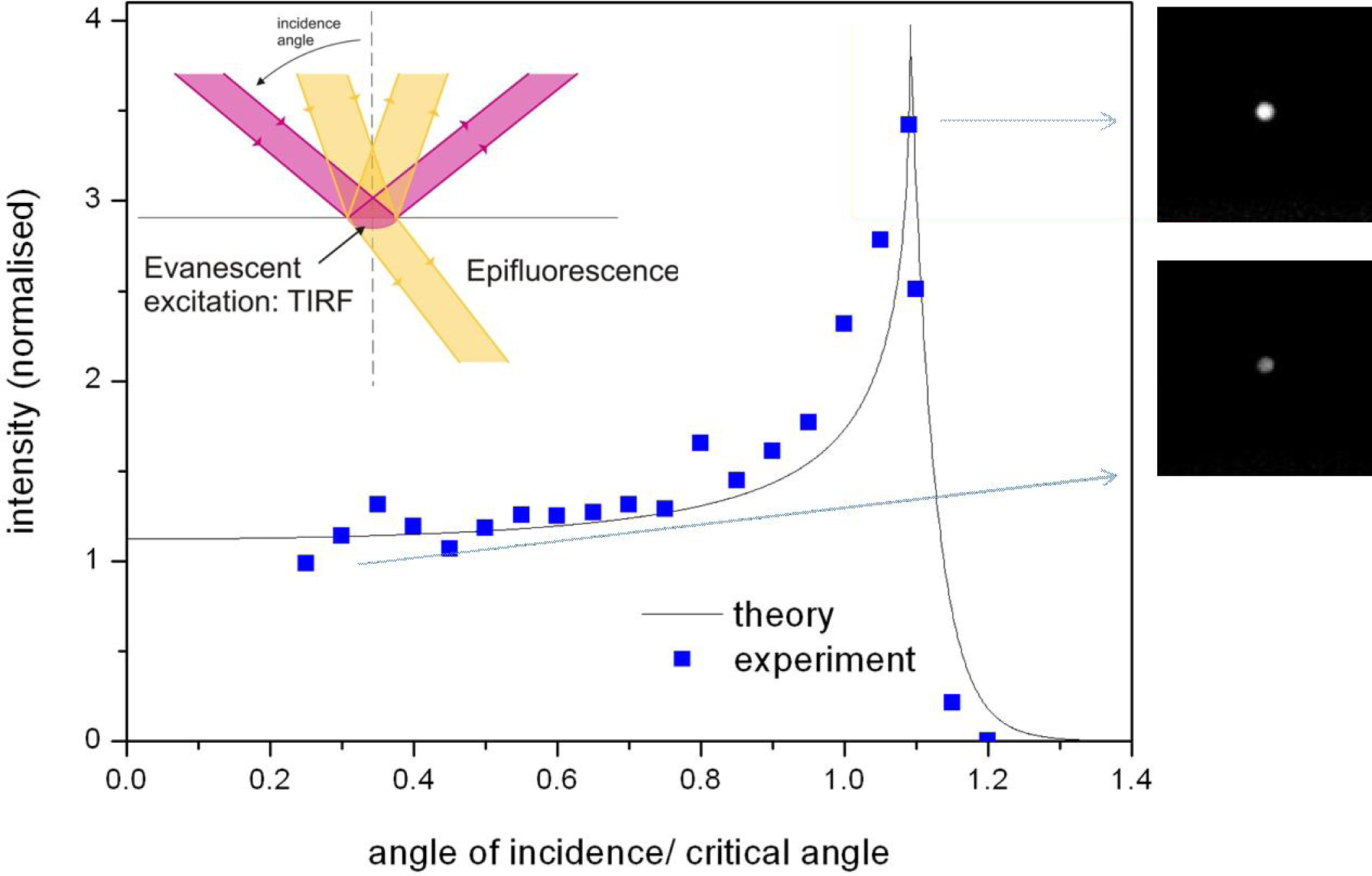
2. Signal to Noise Ratio Enhancement in Optofluidics
- Confinement of the excitation volume to reduce the background;
- Confinement of the excitation density to enhance the pumping rate;
- Modification of the radiative and non-radiative rates of the chromophores;
- Scattering enhancement and optical loss minimization to increase the signal reaching the detector;
- Modification of the emission pattern in order to improve the collection efficiency.
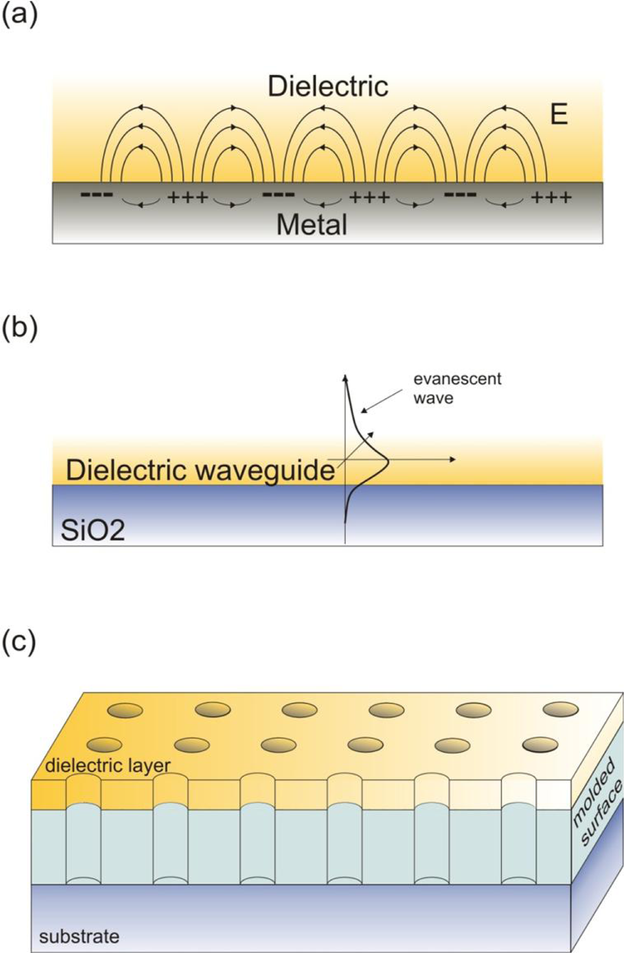
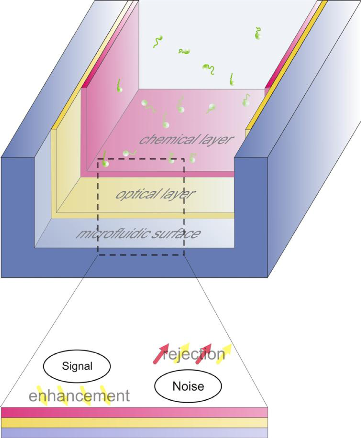
3. Microfluidic Architectures for Minimizing Photobleaching and the Excitation Volume
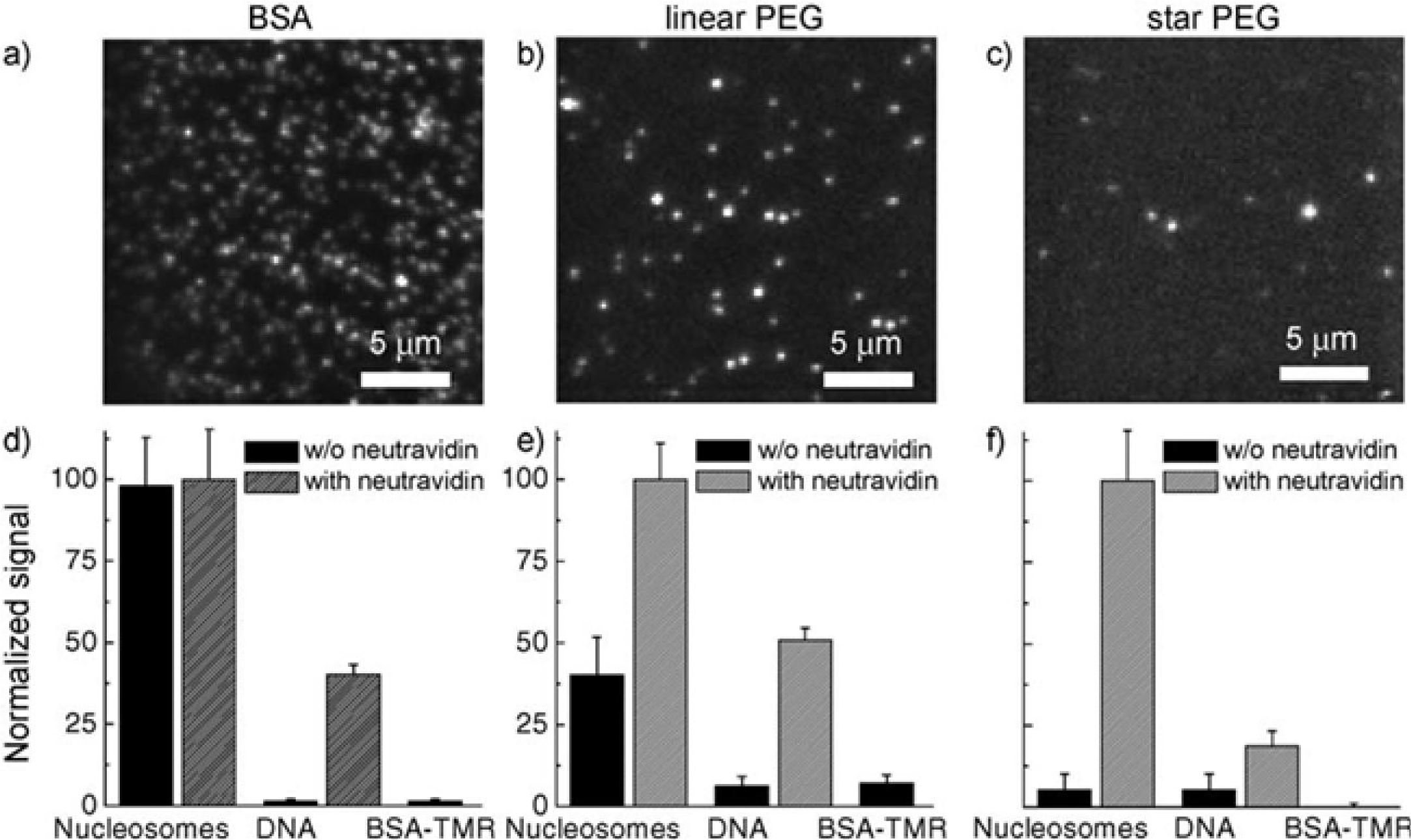
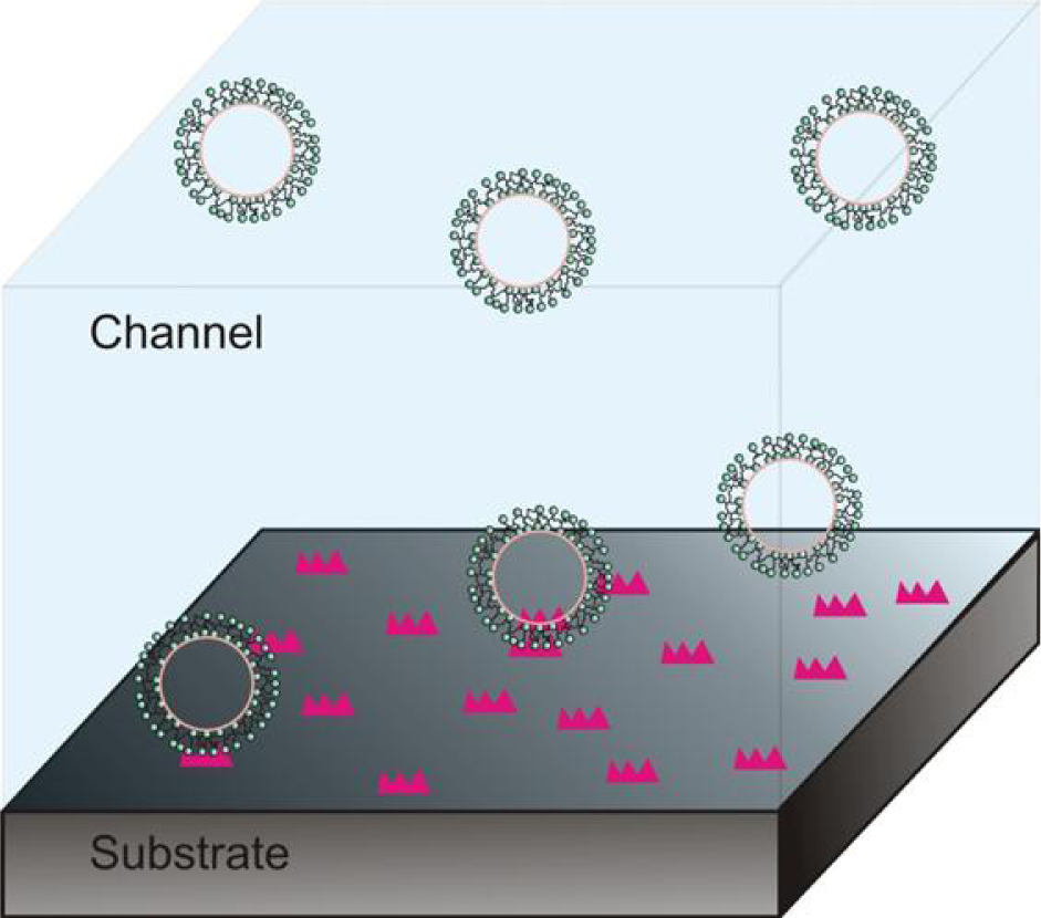
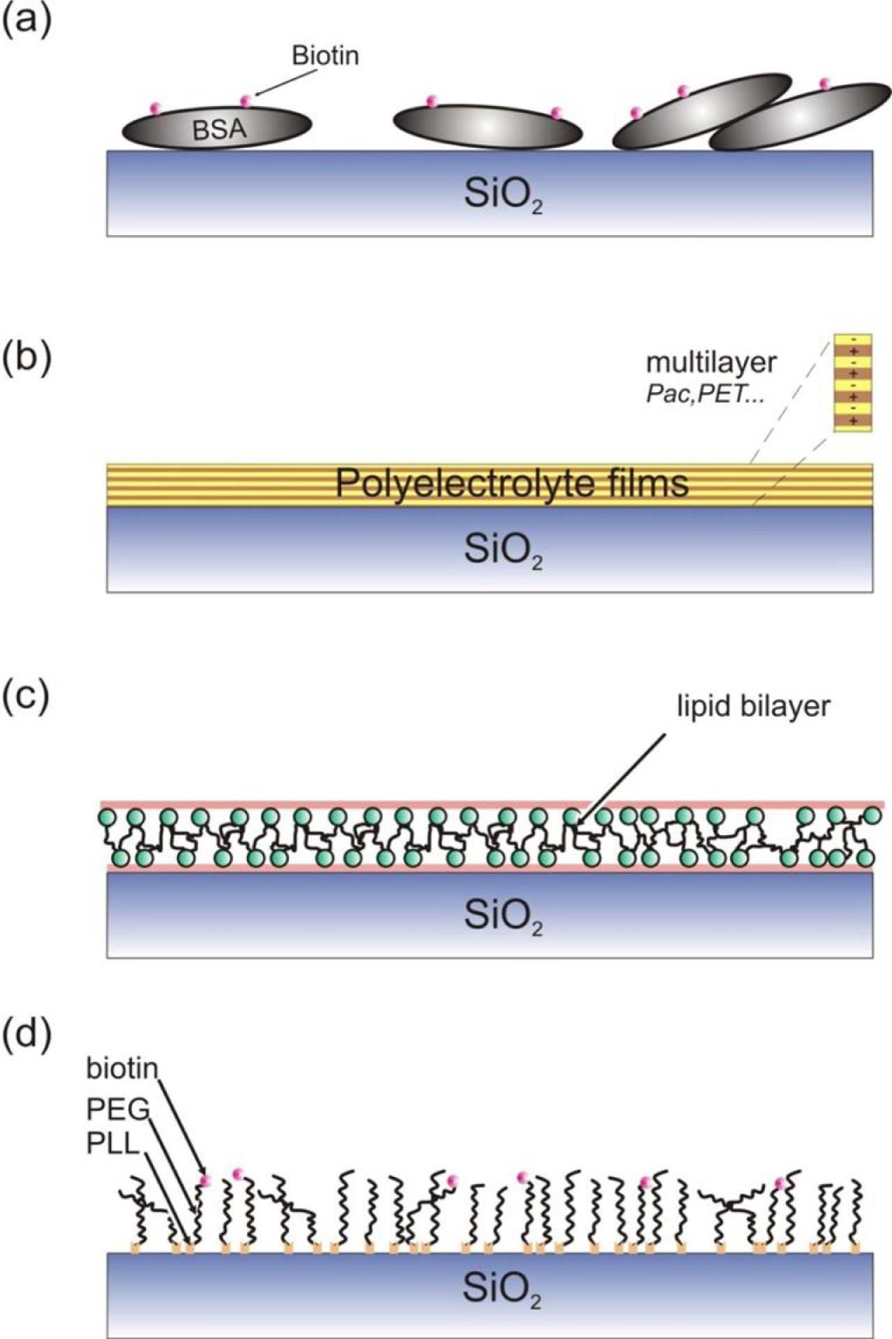
4. Noise Reduction Using Microfluidic Compatible Surface Passivation Strategies
- Simplicity and minimal preparation requirements: Single molecule imaging and detection is becoming a substantially interdisciplinary field, comprised of biologists, physicists and chemists. Hence, synergetic also to microfluidics, the chemical modification needs to be simple and require few implementation steps;
- Compatibility with microfluidic fabrication: this necessitates that the surface chemistry is not compromised during fabrication, but also that the microfluidics retain their properties (e.g., mechanical) during surface treatment;
- Bioactivity: The immobilized biomolecules must retain their activity, for example a protein that can unfold and refold, or a nucleic acid that can be recognized by site-specific proteins.
5. Conclusions
Acknowledgments
References
- Moerner, WE; Kador, L. Optical detection and spectroscopy of single molecules in a solid. Phys. Rev. Lett 1989, 62, 2535–2538. [Google Scholar]
- Orrit, M; Bernard, J. Single pentacene molecules detected by fluorescence excitation in a para-terpenyl crystal. Phys. Rev. Lett 1990, 65, 2716–2719. [Google Scholar]
- Betzig, E; Chichester, RJ. Single molecules observed by near-field scanning optical microscopy. Science 1993, 262, 1422–1425. [Google Scholar]
- Nie, SM; Chiu, DT; Zare, RN. Probing individual molecules with confocal fluorescence microscopy. Science 1994, 266, 1018–1021. [Google Scholar]
- Macklin, JJ; Trautman, JK; Harris, TD; Brus, LE. Imaging and time-resolved spectroscopy of single molecules at an interface. Science 1996, 272, 255–258. [Google Scholar]
- Xie, XS. Single-molecule spectroscopy and dynamics at room temperature. Acc. Chem. Res 1996, 29, 598–606. [Google Scholar]
- Weiss, S. Measuring conformational dynamics of biomolecules by single molecule fluorescence spectroscopy. Nat. Struct. Biol 2000, 7, 724–729. [Google Scholar]
- Lupton, JM. Single-molecule spectroscopy for plastic electronics: Materials analysis from the bottom-up. Adv. Mater 2010, 22, 1689–1721. [Google Scholar]
- Moerner, WE. New directions in single-molecule imaging and analysis. Proc. Natl. Acad. Sci. USA 2007, 104, 12596–12602. [Google Scholar]
- Joo, C; Balci, H; Ishitsuka, Y; Buranachai, C; Ha, T. Advances in single-molecule fluorescence methods for molecular biology. Annu. Rev. Biochem 2008, 77, 51–76. [Google Scholar]
- Lakowicz, JR. Principles of Fluorescence Spectroscopy; Springer: New York, NY, USA, 2006. [Google Scholar]
- Shimomura, O; Johnson, FH; Saiga, Y. Extraction, purification and properties of Aequorin, a bioluminescent protein from muminous hydromedusan Aequorea. J. Cell. Comp. Physiol 1962, 59, 223–239. [Google Scholar]
- Chalfie, M; Tu, Y; Euskirchen, G; Ward, WW; Prasher, DC. Green fluorescent protein as a marker for gene expression. Science 1994, 263, 802–805. [Google Scholar]
- Giepmans, BNG; Adams, SR; Ellisman, MH; Tsien, RY. The fluorescent toolbox for assessing protein location and function. Science 2006, 312, 217–224. [Google Scholar]
- Braslavsky, I; Hebert, B; Kartalov, E; Quake, SR. Sequence information can be obtained from single DNA molecules. Proc. Natl. Acad. Sci. USA 2003, 100, 3960–3964. [Google Scholar]
- Fuller, CW; Middendorf, LR; Benner, SA; Church, GM; Harris, T; Huang, XH; Jovanovich, SB; Nelson, JR; Schloss, JA; Schwartz, DC; et al. The challenges of sequencing by synthesis. Nat. Biotechnol 2009, 27, 1013–1023. [Google Scholar]
- Flusberg, BA; Webster, DR; Lee, JH; Travers, KJ; Olivares, EC; Clark, TA; Korlach, J; Turner, SW. Direct detection of DNA methylation during single-molecule, real-time sequencing. Nat. Methods 2010, 7, 461–465. [Google Scholar]
- van Oijen, AM; Kohler, J; Schmidt, J; Muller, M; Brakenhoff, GJ. 3-Dimensional super-resolution by spectrally selective imaging. Chem. Phys. Lett 1998, 292, 183–187. [Google Scholar]
- Thompson, RE; Larson, DR; Webb, WW. Precise nanometer localization analysis for individual fluorescent probes. Biophys. J 2002, 82, 2775–2783. [Google Scholar]
- Yildiz, A; Forkey, JN; McKinney, SA; Ha, T; Goldman, YE; Selvin, PR. Myosin V walks hand-over-hand: Single fluorophore imaging with 1.5-nm localization. Science 2003, 300, 2061–2065. [Google Scholar]
- Hell, SW; Wichmann, J. Breaking the diffraction resolution limit by stimulated emission-stimulated emission depletion fluorescence microscopy. Opt. Lett 1994, 19, 780–782. [Google Scholar]
- Testa, I; Schonle, A; Middendorff, CV; Geisler, C; Medda, R; Wurm, CA; Stiel, AC; Jakobs, S; Bossi, M; Eggeling, C; et al. Nanoscale separation of molecular species based on their rotational mobility. Opt. Express 2008, 16, 21093–21104. [Google Scholar]
- Betzig, E. Proposed method for molecular optical imaging. Opt. Lett 1995, 20, 237–239. [Google Scholar]
- Hess, ST; Girirajan, TPK; Mason, MD. Ultra-high resolution imaging by fluorescence photoactivation localization microscopy. Biophys. J 2006, 91, 4258–4272. [Google Scholar]
- Rust, MJ; Bates, M; Zhuang, XW. Sub-diffraction-limit imaging by stochastic optical reconstruction microscopy (STORM). Nat. Methods 2006, 3, 793–795. [Google Scholar]
- Sharonov, A; Hochstrasser, RM. Wide-field subdiffraction imaging by accumulated binding of diffusing probes. Proc. Natl. Acad. Sci. USA 2006, 103, 18911–18916. [Google Scholar]
- Goldsmith, RH; Moerner, WE. Watching conformational- and photodynamics of single fluorescent proteins in solution. Nat. Chem 2010, 2, 179–186. [Google Scholar]
- Rudenko, MI; Kuhn, S; Lunt, EJ; Deamer, DW; Hawkins, AR; Schmidt, H. Ultrasensitive Q beta phage analysis using fluorescence correlation spectroscopy on an optofluidic chip. Biosens. Bioelectron 2009, 24, 3258–3263. [Google Scholar]
- Taniguchi, Y; Choi, PJ; Li, GW; Chen, HY; Babu, M; Hearn, J; Emili, A; Xie, XS. Quantifying E-coli Proteome and Transcriptome with Single-Molecule Sensitivity in Single Cells. Science 2010, 329, 533–538. [Google Scholar]
- Kim, S; Streets, AM; Lin, RR; Quake, SR; Weiss, S; Majumdar, DS. High-throughput single-molecule optofluidic analysis. Nat. Methods 2011, 8, 242–245. [Google Scholar]
- Squires, TM; Quake, SR. Microfluidics: Fluid physics at the nanoliter scale. Rev. Mod. Phys 2005, 77, 977–1026. [Google Scholar]
- Reyes, DR; Iossifidis, D; Auroux, PA; Manz, A. Micro total analysis systems. 1. Introduction, theory, and technology. Anal. Chem 2002, 74, 2623–2636. [Google Scholar]
- Stone, HA; Stroock, AD; Ajdari, A. Engineering flows in small devices: Microfluidics toward a lab-on-a-chip. Annu. Rev. Fluid Mech 2004, 36, 381–411. [Google Scholar]
- Burns, MA; Johnson, BN; Brahmasandra, SN; Handique, K; Webster, JR; Krishnan, M; Sammarco, TS; Man, PM; Jones, D; Heldsinger, D; et al. An integrated nanoliter DNA analysis device. Science 1998, 282, 484–487. [Google Scholar]
- Effenhauser, CS; Manz, A; Widmer, HM. Glass chips for hisgh speed capillary electrophoresis separations with submicrometer plate heights. Anal. Chem 1993, 65, 2637–2642. [Google Scholar]
- Maerkl, SJ. Next generation microfluidic platforms for high-throughput protein biochemistry. Curr. Opin. Biotechnol 2011, 22, 59–65. [Google Scholar] [Green Version]
- Prakash, M; Gershenfeld, N. Microfluidic bubble logic. Science 2007, 315, 832–835. [Google Scholar]
- Weaver, JA; Melin, J; Stark, D; Quake, SR; Horowitz, MA. Static control logic for microfluidic devices using pressure-gain valves. Nat. Phys 2010, 6, 218–223. [Google Scholar]
- Di Carlo, D; Wu, LY; Lee, LP. Dynamic single cell culture array. Lab Chip 2006, 6, 1445–1449. [Google Scholar]
- Vasdekis, AE; O'Neil, CP; Hubbell, JA; Psaltis, D. Microfluidic assays for DNA manipulation based on a block copolymer immobilization strategy. Biomacromolecules 2010, 11, 827–831. [Google Scholar]
- Chung, K; Kim, Y; Kanodia, JS; Gong, E; Shvartsman, SY; Lu, H. A microfluidic array for large-scale ordering and orientation of embryos. Nat. Methods 2011, 8, 171–176. [Google Scholar]
- Huebner, A; Sharma, S; Srisa-Art, M; Hollfelder, F; Edel, JB; Demello, AJ. Microdroplets: A sea of applications? Lab Chip 2008, 8, 1244–1254. [Google Scholar]
- Stjernstrom, M; Roeraade, J. Method for fabrication of microfluidic systems in glass. J. Micromech. Microeng 1998, 8, 33–38. [Google Scholar]
- Qin, D; Xia, YN; Whitesides, GM. Soft lithography for micro- and nanoscale patterning. Nat. Protoc 2010, 5, 491–502. [Google Scholar]
- Unger, MA; Chou, HP; Thorsen, T; Scherer, A; Quake, SR. Monolithic microfabricated valves and pumps by multilayer soft lithography. Science 2000, 288, 113–116. [Google Scholar]
- Psaltis, D; Quake, SR; Yang, CH. Developing optofluidic technology through the fusion of microfluidics and optics. Nature 2006, 442, 381–386. [Google Scholar]
- Monat, C; Domachuk, P; Eggleton, BJ. Integrated optofluidics: A new river of light. Nat. Photonics 2007, 1, 106–114. [Google Scholar]
- Song, WZ; Vasdekis, AE; Li, ZY; Psaltis, D. Optofluidic evanescent dye laser based on a distributed feedback circular grating. Appl Phys Lett 2009, 94. [Google Scholar] [CrossRef]
- Cuennet, JG; Vasdekis, AE; De Sio, L; Psaltis, D. Optofluidic modulator based on peristaltic nematogen microflows. Nat. Photonics 2011, 5, 234–238. [Google Scholar]
- Liu, GL; Kim, J; Lu, Y; Lee, LP. Optofluidic control using photothermal nanoparticles. Nat. Mater 2006, 5, 27–32. [Google Scholar]
- Cui, XQ; Lee, LM; Heng, X; Zhong, WW; Sternberg, PW; Psaltis, D; Yang, CH. Lensless high-resolution on-chip optofluidic microscopes for Caenorhabditis elegans and cell imaging. Proc. Natl. Acad. Sci. USA 2008, 105, 10670–10675. [Google Scholar]
- Yang, AHJ; Moore, SD; Schmidt, BS; Klug, M; Lipson, M; Erickson, D. Optical manipulation of nanoparticles and biomolecules in sub-wavelength slot waveguides. Nature 2009, 457, 71–75. [Google Scholar]
- Vasdekis, AE; Cuennet, JG; Song, WZ; Choi, J-W; De Sio, L; O'Neil, CP; Hubbell, JA; Psaltis, D. Surface optofluidics. Proceedings of SPIE Optics and Photonics, San Diego, CA, USA, 1–5 August 2010; p. 776224.
- Wenger, J; Rigneault, H. Photonic methods to enhance fluorescence correlation spectroscopy and single molecule fluorescence detection. Int. J. Mol. Sci 2010, 11, 206–221. [Google Scholar]
- Walter, NG; Huang, CY; Manzo, AJ; Sobhy, MA. Do-it-yourself guide: how to use the modern single-molecule toolkit. Nat. Methods 2008, 5, 475–489. [Google Scholar]
- Hill, EK; de Mello, AJ. Single-molecule detection using confocal fluorescence detection: Assessment of optical probe volumes. Analyst 2000, 125, 1033–1036. [Google Scholar]
- Levene, MJ; Korlach, J; Turner, SW; Foquet, M; Craighead, HG; Webb, WW. Zero-mode waveguides for single-molecule analysis at high concentrations. Science 2003, 299, 682–686. [Google Scholar]
- Schmidt, H; Hawkins, AR. Optofluidic waveguides: I. Concepts and implementations. Microfluid. Nanofluid 2008, 4, 3–16. [Google Scholar]
- Armani, AM; Kulkarni, RP; Fraser, SE; Flagan, RC; Vahala, KJ. Label-free, single-molecule detection with optical microcavities. Science 2007, 317, 783–787. [Google Scholar]
- Zhu, HY; White, IM; Suter, JD; Zourob, M; Fan, XD. Opto-fluidic micro-ring resonator for sensitive label-free viral detection. Analyst 2008, 133, 356–360. [Google Scholar]
- Paige, MF; Bjerneld, EJ; Moerner, WE. A comparison of through-the-objective total internal reflection microscopy and epifluorescence microscopy for single-molecule fluorescence imaging. Single Mol 2001, 2, 191–201. [Google Scholar]
- De Fornel, F. Evanescent Waves: from Newtonian Optics to Atomic Optics; Rhodes, WT, Ed.; Springer-Verlag: Berlin, Germany, 1997. [Google Scholar]
- Axelrod, D. Total internal reflection fluorescence microscopy in cell biology. Traffic 2001, 2, 764–774. [Google Scholar]
- Axelrod, D; Burghardt, TP; Thompson, NL. Total internal reflection fluorescence. Annu. Rev. Biophys. Bioeng 1984, 13, 247–268. [Google Scholar]
- Axelrod, D. Selective imaging of surface fluorescence with very high aperture microscope objectives. J. Biomed. Opt 2001, 6, 6–13. [Google Scholar]
- Carniglia, CK; Mandel, L; Drexhage, KH. Absorption and emission of evanescent photons. J. Opt. Soc. Am 1972, 62, 479–486. [Google Scholar]
- Lotsch, HKV. Beam displacement at total reflection - Goos Hanchen effect 1. Optik 1970, 32, 116–137. [Google Scholar]
- Lai, HM; Cheng, FC; Tang, WK. Goos-Hanchen effect around and off the critical angle. J. Opt. Soc. Am. A 1986, 3, 550–557. [Google Scholar]
- Barnes, WL; Dereux, A; Ebbesen, TW. Surface plasmon subwavelength optics. Nature 2003, 424, 824–830. [Google Scholar]
- Anger, P; Bharadwaj, P; Novotny, L. Enhancement and quenching of single-molecule fluorescence. Phys Rev Lett 2006, 96. [Google Scholar] [CrossRef]
- Kuhn, S; Hakanson, U; Rogobete, L; Sandoghdar, V. Enhancement of single-molecule fluorescence using a gold nanoparticle as an optical nanoantenna. Phys Rev Lett 2006, 97. [Google Scholar] [CrossRef]
- Tam, F; Goodrich, GP; Johnson, BR; Halas, NJ. Plasmonic enhancement of molecular fluorescence. Nano Lett 2007, 7, 496–501. [Google Scholar]
- Zhang, J; Fu, Y; Chowdhury, MH; Lakowicz, JR. Metal-enhanced single-molecule fluorescence on silver particle monomer and dimer: Coupling effect between metal particles. Nano Lett 2007, 7, 2101–2107. [Google Scholar]
- Cang, H; Labno, A; Lu, CG; Yin, XB; Liu, M; Gladden, C; Liu, YM; Zhang, XA. Probing the electromagnetic field of a 15-nanometre hotspot by single molecule imaging. Nature 2011, 469, 385–388. [Google Scholar]
- Taminiau, TH; Stefani, FD; Segerink, FB; Van Hulst, NF. Optical antennas direct single-molecule emission. Nat. Photonics 2008, 2, 234–237. [Google Scholar]
- Burghardt, TP; Charlesworth, JE; Halstead, MF; Tarara, JE; Ajtai, K. In situ fluorescent protein imaging with metal film-enhanced total internal reflection microscopy. Biophys. J 2006, 90, 4662–4671. [Google Scholar]
- Rigneault, H; Capoulade, J; Dintinger, J; Wenger, J; Bonod, N; Popov, E; Ebbesen, TW; Lenne, PF. Enhancement of single-molecule fluorescence detection in subwavelength apertures. Phys Rev Lett 2005, 95. [Google Scholar] [CrossRef]
- Kinkhabwala, A; Yu, ZF; Fan, SH; Avlasevich, Y; Mullen, K; Moerner, WE. Large single-molecule fluorescence enhancements produced by a bowtie nanoantenna. Nat. Photonics 2009, 3, 654–657. [Google Scholar]
- Aouani, H; Itzhakov, S; Gachet, D; Devaux, E; Ebbesen, TW; Rigneault, H; Oron, D; Wenger, J. Colloidal quantum dots as probes of excitation field enhancement in photonic antennas. ACS Nano 2010, 4, 4571–4578. [Google Scholar]
- Song, JH; Atay, T; Shi, SF; Urabe, H; Nurmikko, AV. Large enhancement of fluorescence efficiency from CdSe/ZnS quantum dots induced by resonant coupling to spatially controlled surface plasmons. Nano Lett 2005, 5, 1557–1561. [Google Scholar]
- Kim, K; Kim, DJ; Cho, EJ; Suh, JS; Huh, YM; Kim, D. Nanograting-based plasmon enhancement for total internal reflection fluorescence microscopy of live cells. Nanotechnology 2009, 20. [Google Scholar] [CrossRef]
- Kravets, VG; Zoriniants, G; Burrows, CP; Schedin, F; Geim, AK; Barnes, WL; Grigorenko, AN. Composite Au nanostructures for fluorescence studies in visible light. Nano Lett 2010, 10, 874–879. [Google Scholar]
- Barnes, WL. Fluorescence near interfaces: the role of photonic mode density. J. Mod. Opt 1998, 45, 661–699. [Google Scholar]
- Liebermann, T; Knoll, W. Surface-plasmon field-enhanced fluorescence spectroscopy. Colloids Surf. A 2000, 171, 115–130. [Google Scholar]
- Lakowicz, JR; Geddes, CD; Gryczynski, I; Malicka, J; Gryczynski, Z; Aslan, K; Lukomska, J; Matveeva, E; Zhang, JA; Badugu, R; et al. Advances in surface-enhanced fluorescence. J. Fluoresc 2004, 14, 425–441. [Google Scholar]
- Fort, E; Gresillon, S. Surface enhanced fluorescence. J Phys D 2008, 41. [Google Scholar] [CrossRef]
- Purcell, EM. Spontaneous emission probabilities at radio frequencies. Phys Rev 1946, 69, 681–681. [Google Scholar]
- Pu, Y; Grange, R; Hsieh, CL; Psaltis, D. Nonlinear optical properties of core-shell nanocavities for enhanced second-harmonic generation. Phys Rev Lett 2010. [Google Scholar] [CrossRef]
- Sun, G; Khurgin, JB; Soref, RA. Practical enhancement of photoluminescence by metal nanoparticles. Appl Phys Lett 2009. [Google Scholar] [CrossRef]
- Xie, XS; Dunn, RC. Probing single molecule dynamics. Science 1994, 265, 361–364. [Google Scholar]
- Huang, L; Maerkl, SJ; Martin, OJF. Integration of plasmonic trapping in a microfluidic environment. Opt. Express 2009, 17, 6018–6024. [Google Scholar]
- Gerard, JM; Sermage, B; Gayral, B; Legrand, B; Costard, E; Thierry-Mieg, V. Enhanced spontaneous emission by quantum boxes in a monolithic optical microcavity. Phys. Rev. Lett 1998, 81, 1110–1113. [Google Scholar]
- Badolato, A; Hennessy, K; Atature, M; Dreiser, J; Hu, E; Petroff, PM; Imamoglu, A. Deterministic coupling of single quantum dots to single nanocavity modes. Science 2005, 308, 1158–1161. [Google Scholar]
- Kaiser, R; Levy, Y; Vansteenkiste, N; Aspect, A; Seifert, W; Leipold, D; Mlynek, J. Resonant enhancement of evanescent waves with a thin dielectric waveguide. Opt. Commun 1994, 104, 234–240. [Google Scholar]
- Ke, PC; Szajman, J; Gan, XAS; Gu, M. Optimization of the enhanced evanescent wave for near-field microscopy. In Three-Dimensional Microscopy: Image Acquisition and Processing IV; Cogswell, CJ, Conchello, JA, Wilson, T, Katzir, A, Eds.; SPIE-International Society for Optical Engine: Bellingham WA, USA, 1997; Volume 2984, pp. 42–49. [Google Scholar]
- Tsang, M; Psaltis, D. Reflectionless evanescent-wave amplification by two dielectric planar waveguides. Opt. Lett 2006, 31, 2741–2743. [Google Scholar]
- Kim, K; Cho, EJ; Huh, YM; Kim, D. Thin-film-based sensitivity enhancement for total internal reflection fluorescence live-cell imaging. Opt. Lett 2007, 32, 3062–3064. [Google Scholar]
- Soboleva, IV; Descrovi, E; Summonte, C; Fedyanin, AA; Giorgis, F. Fluorescence emission enhanced by surface electromagnetic waves on one-dimensional photonic crystals. Appl Phys Lett 2009, 94. [Google Scholar] [CrossRef]
- Hassanzadeh, A; Nitsche, M; Mittler, S; Armstrong, S; Dixon, J; Langbein, U. Waveguide evanescent field fluorescence microscopy: Thin film fluorescence intensities and its application in cell biology. Appl Phys Lett 2008, 92. [Google Scholar] [CrossRef]
- Budach, W; Abel, AP; Bruno, AE; Neuschafer, D. Planar waveguides as high performance sensing platforms for fluorescence-based multiplexed oligonucleotide hybridization assays. Anal. Chem 1999, 71, 3347–3355. [Google Scholar]
- Lee, KG; Chen, XW; Eghlidi, H; Kukura, P; Lettow, R; Renn, A; Sandoghdar, V; Gotzinger, S. A planar dielectric antenna for directional single-photon emission and near-unity collection efficiency. Nat. Photonics 2011, 5, 166–169. [Google Scholar]
- Toninelli, C; Delley, Y; Stoferle, T; Renn, A; Gotzinger, S; Sandoghdar, V. A scanning microcavity for in situ control of single-molecule emission. Appl Phys Lett 2010, 97. [Google Scholar] [CrossRef]
- Begon, C; Rigneault, H; Jonsson, P; Rarity, JG. Spontaneous emission control with planar dielectric structures: An asset for ultrasensitive fluorescence analysis. Single Mol 2000, 1, 207–214. [Google Scholar]
- Neuschafer, D; Budach, W; Wanke, C; Chibout, SD. Evanescent resonator chips: A universal platform with superior sensitivity for fluorescence-based microarrays. Biosens. Bioelectron 2003, 18, 489–497. [Google Scholar]
- Ganesh, N; Zhang, W; Mathias, PC; Chow, E; Soares, J; Malyarchuk, V; Smith, AD; Cunningham, BT. Enhanced fluorescence emission from quantum dots on a photonic crystal surface. Nat. Nanotechnol 2007, 2, 515–520. [Google Scholar]
- Zhang, W; Cunningham, BT. Fluorescence enhancement by a photonic crystal with a nanorod-structured high index layer. Appl Phys Lett 2008, 93. [Google Scholar] [CrossRef]
- Kaji, T; Yamada, T; Ueda, R; Xu, XS; Otomo, A. Fabrication of two-dimensional Ta2O5 photonic crystal slabs with ultra-low background emission toward highly sensitive fluorescence spectroscopy. Opt. Express 2011, 19, 1422–1428. [Google Scholar]
- Liu, YM; Wang, S; Park, YS; Yin, XB; Zhang, X. Fluorescence enhancement by a two-dimensional dielectric annular Bragg resonant cavity. Opt. Express 2010, 18, 25029–25034. [Google Scholar]
- Norris, DJ; KuwataGonokami, M; Moerner, WE. Excitation of a single molecule on the surface of a spherical microcavity. Appl. Phys. Lett 1997, 71, 297–299. [Google Scholar]
- Gerard, D; Wenger, J; Devilez, A; Gachet, D; Stout, B; Bonod, N; Popov, E; Rigneault, H. Strong electromagnetic confinement near dielectric microspheres to enhance single-molecule fluorescence. Opt. Express 2008, 16, 15297–15303. [Google Scholar]
- Schwartz, JJ; Stavrakis, S; Quake, SR. Colloidal lenses allow high-temperature single-molecule imaging and improve fluorophore photostability. Nat. Nanotechnol 2010, 5, 127–132. [Google Scholar]
- Rasnik, I; McKinney, SA; Ha, T. Nonblinking and longlasting single-molecule fluorescence imaging. Nat. Methods 2006, 3, 891–893. [Google Scholar]
- Campos, LA; Liu, JW; Wang, XA; Ramanathan, R; English, DS; Munoz, V. A photoprotection strategy for microsecond-resolution single-molecule fluorescence spectroscopy. Nat. Methods 2011, 8, 143–146. [Google Scholar]
- Lemke, EA; Gambin, Y; Vandelinder, V; Brustad, EM; Liu, HW; Schultz, PG; Groisman, A; Deniz, AA. Microfluidic device for single-molecule experiments with enhanced photostability. J. Am. Chem. Soc 2009, 131, 13610–13612. [Google Scholar]
- Gambin, Y; VanDelinder, V; Ferreon, ACM; Lemke, EA; Groisman, A; Deniz, AA. Visualizing a one-way protein encounter complex by ultrafast single-molecule mixing. Nat. Methods 2011, 8, 239–241. [Google Scholar]
- Osborne, MA; Balasubramanian, S; Furey, WS; Klenerman, D. Optically biased diffusion of single molecules studied by confocal fluorescence microscopy. J. Phys. Chem. B 1998, 102, 3160–3167. [Google Scholar]
- Reisner, W; Larsen, NB; Silahtaroglu, A; Kristensen, A; Tommerup, N; Tegenfeldt, JO; Flyvbjerg, H. Single-molecule denaturation mapping of DNA in nanofluidic channels. Proc. Natl. Acad. Sci. USA 2010, 107, 13294–13299. [Google Scholar]
- Eijkel, JCT; van den Berg, A. Nanofluidics: What is it and what can we expect from it? Microfluid. Nanofluid 2005, 1, 249–267. [Google Scholar]
- Wang, YC; Stevens, AL; Han, JY. Million-fold preconcentration of proteins and peptides by nanofluidic filter. Anal. Chem 2005, 77, 4293–4299. [Google Scholar]
- Schoch, RB; Han, JY; Renaud, P. Transport phenomena in nanofluidics. Rev. Mod. Phys 2008, 80, 839–883. [Google Scholar]
- Song, H; Chen, DL; Ismagilov, RF. Reactions in droplets in microflulidic channels. Angew. Chem. Int. Ed 2006, 45, 7336–7356. [Google Scholar]
- Rhoades, E; Gussakovsky, E; Haran, G. Watching proteins fold one molecule at a time. Proc. Natl. Acad. Sci. USA 2003, 100, 3197–3202. [Google Scholar]
- Okumus, B; Wilson, TJ; Lilley, DMJ; Ha, T. Vesicle encapsulation studies reveal that single molecule ribozyme heterogeneities are intrinsic. Biophys. J 2004, 87, 2798–2806. [Google Scholar]
- Gunnarsson, A; Jonsson, P; Marie, R; Tegenfeldt, JO; Hook, F. Single-molecule detection and mismatch discrimination of unlabeled DNA targets. Nano Lett 2008, 8, 183–188. [Google Scholar]
- Visnapuu, ML; Duzdevich, D; Greene, EC. The importance of surfaces in single-molecule bioscience. Mol. Biosyst 2008, 4, 394–403. [Google Scholar]
- Koopmans, WJA; Schmidt, T; van Noort, J. Nucleosome immobilization microscopy. Chemphyschem 2008, 9, 2002–2009. [Google Scholar]
- Rasnik, I; McKinney, SA; Ha, T. Surfaces and orientations: Much to FRET about? Acc. Chem. Res 2005, 38, 542–548. [Google Scholar]
- Horgan, AM; Moore, JD; Noble, JE; Worsley, GJ. Polymer- and colloid-mediated bioassays, sensors and diagnostics. Trends Biotechnol 2010, 28, 485–494. [Google Scholar]
- Dickson, RM; Cubitt, AB; Tsien, RY; Moerner, WE. On/off blinking and switching behaviour of single molecules of green fluorescent protein. Nature 1997, 388, 355–358. [Google Scholar]
- Chrambac, A; Rodbard, D. Polyacrylamide gel electrophoresis. Science 1971, 172, 440–451. [Google Scholar]
- Ostuni, E; Chapman, RG; Holmlin, RE; Takayama, S; Whitesides, GM. A survey of structure-property relationships of surfaces that resist the adsorption of protein. Langmuir 2001, 17, 5605–5620. [Google Scholar]
- van Poll, ML; Khodabakhsh, S; Brewer, PJ; Shard, AG; Ramstedt, M; Huck, WTS. Surface modification of PDMS via self-organization of vinyl-terminated small molecules. Soft Matter 2009, 5, 2286–2293. [Google Scholar]
- Zhou, JW; Voelcker, NH; Ellis, AV. Simple surface modification of poly(dimethylsiloxane) for DNA hybridization. Biomicrofluidics 2010, 4. [Google Scholar] [CrossRef]
- Kim, HD; Nienhaus, GU; Ha, T; Orr, JW; Williamson, JR; Chu, S. Mg2+-dependent conformational change of RNA studied by fluorescence correlation and FRET on immobilized single molecules. Proc. Natl. Acad. Sci. USA 2002, 99, 4284–4289. [Google Scholar]
- Tan, E; Wilson, TJ; Nahas, MK; Clegg, RM; Lilley, DMJ; Ha, T. A four-way junction accelerates hairpin ribozyme folding via a discrete intermediate. Proc. Natl. Acad. Sci. USA 2003, 100, 9308–9313. [Google Scholar]
- Bentley, DR; Balasubramanian, S; Swerdlow, HP; Smith, GP; Milton, J; Brown, CG; Hall, KP; Evers, DJ; Barnes, CL; Bignell, HR; et al. Accurate whole human genome sequencing using reversible terminator chemistry. Nature 2008, 456, 53–59. [Google Scholar]
- Krieg, A; Ruckstuhl, T; Seeger, S. Towards single-molecule DNA sequencing: Assays with low nonspecific adsorption. Anal. Biochem 2006, 349, 181–185. [Google Scholar]
- Kartalov, EP; Unger, MA; Quake, SR. Polyelectrolyte surface interface for single-molecule fluorescence studies of DNA polymerase. Biotechniques 2003, 34, 505–510. [Google Scholar]
- Xiao, M; Wan, E; Chu, C; Hsueh, WC; Cao, Y; Kwok, PY. Direct determination of haplotypes from single DNA molecules. Nat. Methods 2009, 6, 199–201. [Google Scholar]
- Chan, TF; Ha, C; Phong, A; Cai, DM; Wan, E; Leung, L; Kwok, PY; Xiao, M. A simple DNA stretching method for fluorescence imaging of single DNA molecules. Nucleic Acids Res 2006, 34. [Google Scholar] [CrossRef]
- Gomez-Sjoberg, R; Leyrat, AA; Pirone, DM; Chen, CS; Quake, SR. Versatile, fully automated, microfluidic cell culture system. Anal. Chem 2007, 79, 8557–8563. [Google Scholar]
- Liu, VA; Jastromb, WE; Bhatia, SN. Engineering protein and cell adhesivity using PEO-terminated triblock polymers. J. Biomed. Mater. Res 2002, 60, 126–134. [Google Scholar]
- Sofia, SJ; Premnath, V; Merrill, EW. Poly(ethylene oxide) grafted to silicon surfaces: Grafting density and protein adsorption. Macromolecules 1998, 31, 5059–5070. [Google Scholar]
- Groll, J; Amirgoulova, EV; Ameringer, T; Heyes, CD; Rocker, C; Nienhaus, GU; Moller, M. Biofunctionalized, ultrathin coatings of cross-linked star-shaped poly(ethylene oxide) allow reversible folding of immobilized proteins. J. Am. Chem. Soc 2004, 126, 4234–4239. [Google Scholar]
- Lee, JN; Park, C; Whitesides, GM. Solvent compatibility of poly(dimethylsiloxane)-based microfluidic devices. Anal. Chem 2003, 75, 6544–6554. [Google Scholar]
- Glasmastar, K; Larsson, C; Hook, F; Kasemo, B. Protein adsorption on supported phospholipid bilayers. J. Colloid Interface Sci 2002, 246, 40–47. [Google Scholar]
- Keller, CA; Glasmastar, K; Zhdanov, VP; Kasemo, B. Formation of supported membranes from vesicles. Phys. Rev. Lett 2000, 84, 5443–5446. [Google Scholar]
- Yoshina-Ishii, C; Boxer, SG. Arrays of mobile tethered vesicles on supported lipid bilayers. J. Am. Chem. Soc 2003, 125, 3696–3697. [Google Scholar]
- Graneli, A; Yeykal, CC; Prasad, TK; Greene, EC. Organized arrays of individual DIVA molecules tethered to supported lipid bilayers. Langmuir 2006, 22, 292–299. [Google Scholar]
- Graneli, A; Yeykal, CC; Robertson, RB; Greene, EC. Long-distance lateral diffusion of human Rad51 on double-stranded DNA. Proc. Natl. Acad. Sci. USA 2006, 103, 1221–1226. [Google Scholar]
- Greene, EC; Wind, S; Fazio, T; Gorman, J; Visnapuu, ML. DNA curtains for high-throughput single molecule optical imaging. Methods Enzymol 2010, 472, 293–315. [Google Scholar]
- Harris, JM. Poly(ethylene glycol) Chemistry-Biotechnical and Biomedical Applications; Harris, JM, Ed.; Plenum: New York, NY, USA, 1992. [Google Scholar]
- Elbert, DL; Hubbell, JA. Self-assembly and steric stabilization at heterogeneous, biological surfaces using adsorbing block copolymers. Chem. Biol 1998, 5, 177–183. [Google Scholar]
- Kim, D; Park, S; Lee, JH; Jeong, YY; Jon, S. Antibiofouling polymer-coated gold nanoparticles as a contrast agent for in vivo x-ray computed tomography imaging. J. Am. Chem. Soc 2007, 129, 7661–7665. [Google Scholar]
- Huang, NP; Voros, J; De Paul, SM; Textor, M; Spencer, ND. Biotin-derivatized poly(l-lysine)-g-poly(ethylene glycol): A novel polymeric interface for bioaffinity sensing. Langmuir 2002, 18, 220–230. [Google Scholar]
- Papahadjopoulos, D; Allen, TM; Gabizon, A; Mayhew, E; Matthay, K; Huang, SK; Lee, KD; Woodle, MC; Lasic, DD; Redemann, C; et al. Sterically stabilized liposomes-improvements in pharmacokinetics and antitumour therapeutic efficacy. Proc. Natl. Acad. Sci. USA 1991, 88, 11460–11464. [Google Scholar]
- Tanner, NA; van Oijen, AM. Visualizing DNA replication at the single-molecule level. Methods Enzymol 2010, 475, 259–278. [Google Scholar]
- Lee, S; Voros, J. An aqueous-based surface modification of poly(dimethylsiloxane) with poly(ethylene glycol) to prevent biofouling. Langmuir 2005, 21, 11957–11962. [Google Scholar]
- Kenausis, GL; Voros, J; Elbert, DL; Huang, NP; Hofer, R; Ruiz-Taylor, L; Textor, M; Hubbell, JA; Spencer, ND. Poly(l-lysine)-g-poly(ethylene glycol) layers on metal oxide surfaces: Attachment mechanism and effects of polymer architecture on resistance to protein adsorption. J. Phys. Chem. B 2000, 104, 3298–3309. [Google Scholar]
- Scott, EA; Nichols, MD; Cordova, LH; George, BJ; Jun, YS; Elbert, DL. Protein adsorption and cell adhesion on nanoscale bioactive coatings formed from poly(ethylene glycol) and albumin microgels. Biomaterials 2008, 29, 4481–4493. [Google Scholar]
- Yu, L; Li, CM; Liu, YS; Gao, J; Wang, W; Gan, Y. Flow-through functionalized PDMS microfluidic channels with dextran derivative for ELISAs. Lab Chip 2009, 9, 1243–1247. [Google Scholar]
- Massia, SP; Stark, J; Letbetter, DS. Surface-immobilized dextran limits cell adhesion and spreading. Biomaterials 2000, 21, 2253–2261. [Google Scholar]
- Zhang, ZW; Feng, XJ; Xu, F; Liu, X; Liu, BF. “Click” chemistry-based surface modification of poly(dimethylsiloxane) for protein separation in a microfluidic chip. Electrophoresis 2010, 31, 3129–3136. [Google Scholar]
- Wang, AJ; Xu, JJ; Chen, HY. In-situ grafting hydrophilic polymer on chitosan modified poly (dimethylsiloxane) microchip for separation of biomolecules. J. Chromatogr 2007, 1147, 120–126. [Google Scholar]
- Zhang, ZW; Feng, XJ; Luo, QM; Liu, BF. Environmentally friendly surface modification of PDMS using PEG polymer brush. Electrophoresis 2009, 30, 3174–3180. [Google Scholar]
- Heyes, CD; Groll, J; Moller, M; Nienhaus, GU. Synthesis, patterning and applications of star-shaped poly(ethylene glycol) biofunctionalized surfaces. Mol. Biosyst 2007, 3, 419–430. [Google Scholar]
- Xiao, Y; Yu, XD; Xu, JJ; Chen, HY. Bulk modification of PDMS microchips by an amphiphilic copolymer. Electrophoresis 2007, 28, 3302–3307. [Google Scholar]
- Zhou, JH; Yan, H; Ren, KN; Dai, W; Wu, HK. Convenient method for modifying poly(dimethylsiloxane) with poly(ethylene glycol) in microfluidics. Anal. Chem 2009, 81, 6627–6632. [Google Scholar]
- Hu, SW; Ren, XQ; Bachman, M; Sims, CE; Li, GP; Allbritton, N. Surface modification of poly(dimethylsiloxane) microfluidic devices by ultraviolet polymer grafting. Anal. Chem 2002, 74, 4117–4123. [Google Scholar]
- Sebra, RP; Masters, KS; Cheung, CY; Bowman, CN; Anseth, KS. Detection of antigens in biologically complex fluids with photografted whole antibodies. Anal. Chem 2006, 78, 3144–3151. [Google Scholar]
- Kim, P; Jeong, HE; Khademhosseini, A; Suh, KY. Fabrication of non-biofouling polyethylene glycol micro- and nanochannels by ultraviolet-assisted irreversible sealing. Lab Chip 2006, 6, 1432–1437. [Google Scholar]
- Han, J-H; Yoon, J-Y. Reusable, polyethylene glycol-structured microfluidic channels for particle immunoassays. J. Biol. Eng 2009, 3, 1–6. [Google Scholar]
- Xu, YH; Yang, XR; Wang, EK. Review: Aptamers in microfluidic chips. Anal. Chim. Acta 2010, 683, 12–20. [Google Scholar]


© 2011 by the authors; licensee MDPI, Basel, Switzerland. This article is an open-access article distributed under the terms and conditions of the Creative Commons Attribution license (http://creativecommons.org/licenses/by/3.0/).
Share and Cite
Vasdekis, A.E.; Laporte, G.P.J. Enhancing Single Molecule Imaging in Optofluidics and Microfluidics. Int. J. Mol. Sci. 2011, 12, 5135-5156. https://doi.org/10.3390/ijms12085135
Vasdekis AE, Laporte GPJ. Enhancing Single Molecule Imaging in Optofluidics and Microfluidics. International Journal of Molecular Sciences. 2011; 12(8):5135-5156. https://doi.org/10.3390/ijms12085135
Chicago/Turabian StyleVasdekis, Andreas E., and Gregoire P.J. Laporte. 2011. "Enhancing Single Molecule Imaging in Optofluidics and Microfluidics" International Journal of Molecular Sciences 12, no. 8: 5135-5156. https://doi.org/10.3390/ijms12085135