Crystal and molecular structure of cis-Dichlorobis(triphenylphosphite) Platinum(II)

Chemistry. Dept., Science Faculty, Bu-Ali-Sina University Hamadan, Iran
*
Author to whom correspondence should be addressed.
Molecules 2001, 6(9), 777-783; https://doi.org/10.3390/60900777
Submission received: 5 December 2000
/
Revised: 26 August 2001
/
Accepted: 27 August 2001
/
Published: 31 August 2001
Abstract
:The single crystal structure of cis-dichlorobis(triphenyphosphite) platinum(II), [PtCl2(P(OPh)3)2] has been determined. This complex crystallises in the orthorhombic space group, P212121 with cell constant, a=10.4135(13), b=14.0635(16), c=23.505(3) Ǻ and v=3448.3(7) Ǻ 3. The Pt-Cl1 and Pt-Cl2 distances are 2.3390(10) Ǻ and 2.3256 Ǻ, which are longer than Pt-P1 and Pt-P2 with 2.1985(12) Ǻ and 2.1998(10) Ǻ respectively. These data together with bond angles suggest a distorted square planar geometry for this complex with two chlorine ligands in a cis configuration.
Introduction
In spite of the extensive structural studies on platinum and palladium complexes containing a variety of P-donor ligands [1,2], X-ray structure analyses of platinum derivatives containing triphenylphosphite derivatives are scarce. To the best of our knowledge no such determinations of the title compound have been reported so far. We have recently reported a new procedure for the synthesis of a palladium complex [3] and herein we describe the synthesis and structure determination of an analogous platinum complex.
Results and discussion
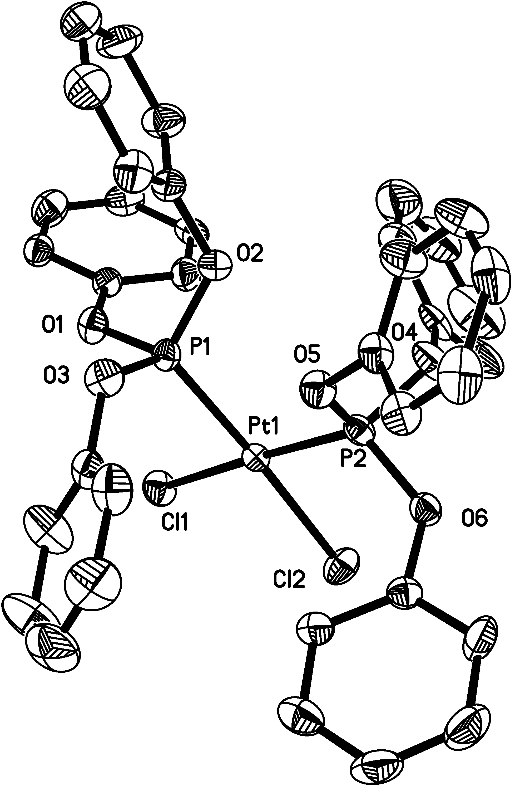
Cis-dichlorobis(triphenylphophite)platinum, [PtCl2(P(OPh)3)2], (1) has been studied in solution This complex was synthesised using a new method but the IR and NMR data are in excellent agreement with an earlier conclusion [1]. However, relatively little work has been done on structural determinations of this type of complex and this lack of information is probably due to difficulty experienced in crystallising the compound from the ethanol reaction solution [4,5]. This work describes the new method of the preparation and characterisation of (1). The structure of (1) is shown in Figure 1.
Figure 1.
Molecular structure of [PtCl2(P(OPh)3)2] showing the atom numbering scheme used.
Crystal data, structural refinement are summarised in Table 1.

| Formula weight | 886.59 |
| Temperature | 213(2) K |
| Wavelength | 0.71073 A |
| Crystal system, space group | orthorhombic, P 21 21 21 |
| Unit cell dimensions | a = 10.4315 (13 ) Ǻ alpha = 90 deg. |
| b = 14.0635 (16) Ǻ beta = 90 deg | |
| c = 23.505 ( 3 ) Ǻ gamma = 90 deg. | |
| Volume | 3448.3 ( 7 ) Ǻ 3 |
| Z, calculated density | 4, 1.708 Mg/m3 |
| Absorption coefficient | 4.164 mm-1 |
| F (000) | 1744 |
| Crystal size | 0.68 × 0.20 × 0.16 mm |
| Theta range for data collection | 1.73 to 24.26 deg. |
| Index ranges | -11<=h<11, -16<=k<=16, -26<=1<=26 |
| Reflections collected / unique | 22003/5449 [R(int)=0.0534] |
| Completeness to 2theta = 24.26 | 98.0% |
| Max. and min. transmission | 0.5554 and 0.1640 |
| Refinement method | full-matrix least-squares on F2 |
| Data / restraints / parameters | 5449 / 0 /424 |
| Goodness-of-fit on F∩2 | 0.943 |
| Final r indices [I>2sigma(I)] | R1 = 0.0203, WR2 = 0.0410 |
| R indices (all data) | R1 = 0.0280, WR2 = 0.0425 |
| Absolute structure parameter | -0.018(5) |
| Largest diff. peak and hole | 0.559 and -0.783 e.A−3 |
| Absorption correction | numerical (X-SHAPE : Stoe, 1997) |
| Tmin =0.1640, Tmax =0.5554 | |
| 22003 measured reflections | |
| 5449 independent reflections | |
| 4677 reflections with> 2sigma(I |
The more important bond lengths and angles together with references are presented in Table 2.
| Distances (Ǻ) | |||
| O1-P1 | 1.579(3) | O6-P2 | 1.577(3) |
| Angles (°) | |||
| O1-P1-O2 | 104.69(17) | O4-P2-Pt1 | 117.27(14) |
Atomic coordinates are given in Table 3.
Table 3.
Atomic coordinates (x 104) and equivalent isotropic displacement parameters (A2 x 103) U(eq) is defined as one third of the trace of the orthogonalized Uij tensor.
| X | Y | Z | U(eq) | |
| C(1) | -2590(4) | 790(3) | 676(2) | 23(1) |
The structural analysis proved a cis-distorted square planar arrangement for 1, and different interatomic distances and bond angles in comparison with [PtCl4]2-(2) was observed. The Pt-P and Pt-Cl bonds are approximately within the same range of other phosphine complexes [1,2], but the Pt-Cl distances are significantly shorter than Pt-P (see Table 3). This causes a big distortion which leads to asymmetric structure and is much bigger than what we observed in cis-[PdCl2(P(OPh)3)2] (3) structure [3]. Evidently the small difference between Cl2-Pt1-P1 [177.20° (4)] and Cl1-Pt1-P2 [176.93°(4)] angles also confirms the distorted square planar geometry of 1. This is in contrast with what has been observed in solution [5]. Discussion of bond-length in 1 [in comparison with 2 containing a C2 axis] in relation to the above configuration is quite interesting. We noticed that 1 with the different P1-Pt1-P2 [93.56° (4)], P1-Pt1-Cl1 [89.38° (4)], P2-Pt1-Cl2 [88.58° (4)] and Cl1Pt1-Cl2 [88.51° (4)] angles is completely asymmetrical. Not only it changes from D4h and C2v symmetry from 2 and 3 respectively but it is also consistent with C1 symmetry structure. These can be taken probably as a structural confirmation of the effect of different metal atoms in 1 and 3 or a larger proportion of σ -donor character in the Pt-P bonds for 1 in comparison with the Pd-P bonds of 3, which results in a greater strict effect of the phosphite ligands. All the bond lengths (Table 2) are approximately within the normal range compared with other phosphine complexes [1,2]. Finally as one can see the P1-Pt1-Cl2 angle [88.51°(4)] is narrower than the P1-Pt1-P2 [93.56°(4)] angle and this is apparently due to the steric effect of phosphite ligands.
Experimental
Refluxing PtCl2 and POPh3 at 60 ºC in a molar ratio of 1:2 in toluene for 6 h. gave a yellowish precipitate, which was extracted from the obtained suspension. Colourless crystals suitable for X-ray diffraction analysis were obtained by slow addition of methanol to a chloroform solution containing the compound. Crystallographic data was recorded on a Stoe IPDS diffractometer using graphite monochromated Mokα-radiation (λ = 0.71073 Å), T = 200 K. Structures were solved by direct methods and refined by full-matrix least squares against F2 using all data. Supplementary material, comprising crystallographic experimental details, positional parameters for all atoms, bond distances and angles, anisotropic thermal parameters and hydrogen atoms coordinates has been deposited with the Cambridge Crystallographic Database. The deposition code is CCDC144750.
Acknowledgements
We thank the University of Bu-Ali-Sina for grant support to A.N and Dr. Jamie F. Bickley, University of Liverpool, UK, for the crystal structure analysis.
References
- Kitano, Y.; Ashida, T. Acta Crystallogr. 1983, C39, 1015.
- Davies, J.A.; Pinkerton, A.A.; Staples, R.J. Acta Crystallogr. 1990, C48, 48.
- Sabounchei, S.J.; Naghipour, A; Bickley, J.F. Acta Crystallgr. 2000, C56, 280.
- Allen, F.A.; Pidcok, A.; Waterhouse, C.R. J. Chem. Soc. (A) 1970, 2087.
- Ahmed, N.; Ainscough, E.W.; James, T.A.; Robinson, S.D. J. Chem. Soc. Dalton Trans. 1973, 1148.
- EXPOS, Stoe IPDS Software for Publication, version 2.79. Stoe IPDS: Darmstadt, Germany, Stoe 1997a.
- Cell Program for Cell Refinement; Version 2.79. Stoe IPDS: Darmstadt, Germany, Stoe 1997b.
- INTEGRATE Program for Reduction of IPDS Data, Version 2.79. Stoe IPDS: Darmstadt, Germany, Stoe 1997c.
- Sheldrick, G.M. Acta Crytallogr. 1990, A46, 467.
- Sheldrick, G.M. SHELXL 97 release 97-1; Program for Refinement of [3d] Crystal Structure; University of Göttingen: Germany, 1997.
- Sample Availability: The product reported in this paper is available from MDPI
© 2001 by MDPI (http://www.mdpi.org). Reproduction is permitted for noncommercial purposes.
Share and Cite
MDPI and ACS Style
Sabounchei, S.J.; Naghipour, A. Crystal and molecular structure of cis-Dichlorobis(triphenylphosphite) Platinum(II). Molecules 2001, 6, 777-783. https://doi.org/10.3390/60900777
AMA Style
Sabounchei SJ, Naghipour A. Crystal and molecular structure of cis-Dichlorobis(triphenylphosphite) Platinum(II). Molecules. 2001; 6(9):777-783. https://doi.org/10.3390/60900777
Chicago/Turabian StyleSabounchei, Seyyed Javad, and Ali Naghipour. 2001. "Crystal and molecular structure of cis-Dichlorobis(triphenylphosphite) Platinum(II)" Molecules 6, no. 9: 777-783. https://doi.org/10.3390/60900777