Article

A Tri-O-Bridged Diels-Alder Adduct from Cortex Mori Radicis

1
State Key Laboratory of Bioactive Substance and Function of Natural Medicines, Institute of Materia Medica, Chinese Academy of Medical Sciences and Peking Union Medical College, Beijing 100050, China
2
Institute of Medicinal of Biotechnology, Chinese Academy of Medical Sciences and Peking Union Medical College, Beijing 100050, China
3
GRU Cancer Center, Augusta University, Augusta, GA 30912, USA
*
Author to whom correspondence should be addressed.
These authors contributed equally to this paper.
Molecules 2018, 23(1), 133; https://doi.org/10.3390/molecules23010133
Submission received: 11 December 2017 / Revised: 7 January 2018 / Accepted: 7 January 2018 / Published: 9 January 2018
(This article belongs to the Section Natural Products Chemistry)

Abstract

:
Sanggenon X, an unusual tri-O-bridged Diels-Alder adduct, was isolated from Cortex Mori Radicis. Its structure was established by spectroscopic analysis, including NMR and HR-MS (High Resolution Mass Spectrometry). Sanggenon X contained three O-bridged rings, where the oxygenated bridgeheads were all quaternary carbons. Chemical methylation was carried out to deduce the linkages of the three O-bridges. The absolute configuration was determined by calculating the ECD (Electronic Circular Dichroism) using the TDDFT (Time-Dependent Density Functional Theory) method. Sanggenon X showed significant antioxidant activity against Fe2+-Cys-induced lipid peroxidation in rat liver microsomes, and was as effective as the positive control, curcumin.

Graphical Abstract

1. Introduction

Cortex Mori Radicis is the root bark of some Morus species (e.g., M. alba, M. mongolica, M. cathayana, and M. australis), and has been used in traditional Chinese medicine as an antidiabetic, a diuretic, and an expectorant agent. Various compounds have been identified from Morus plants, such as Diels-Alder (D-A) adducts, stilbenes, flavonoids, and alkaloids. Their antioxidant [1,2,3], anti-inflammatory [4,5], antimicrobial [6,7,8,9], anticarcinogenic [10,11,12], and antidiabetic [13] activities have been widely reported. In our previous studies, some analgesic benzofuran-type stilbenes related to the traditional antirheumatic usage of Cortex Mori Radicis were reported [14]. Our ongoing research led to the discovery of an unusual tri-O-bridged D-A compound in which oxygenated bridgeheads were all quaternary carbons. Herein, we report the isolation, structure elucidation, and the absolute configuration of the previously undescribed compound named Sanggenon X (1).

2. Results and Discussion

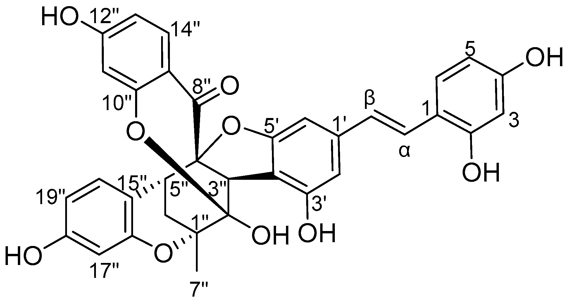
Sanggenon X (Figure 1) was obtained as a yellowish-brown amorphous powder. Its IR spectrum showed absorption bands assigned to carbonyl (1685 cm−1) and aromatic (1605, 1509 and 1459 cm−1) groups. The molecular formula C34H26O10 was determined by (+)-ESI HR-MS (electrospray ionization high resolution mass spectrometry) at m/z 595.1586 [M + H]+ (calcd for C34H27O10+, 595.1599). The 1H-NMR spectrum of 1 (Table 1) showed three aromatic moieties as follows: (a) a trisubstituted benzoyl at δH 7.44 (d, J = 8.7 Hz, H-14″), 6.48 (d, J = 8.7 Hz, H-13″), and 6.09 (s, H-11″); (b) a trisubstituted phenyl ring at δH 6.51 (d, J = 9.0 Hz, H-20″), 6.22 (d, J = 9.0 Hz, H-19″), and 6.23 (s, H-17″); (c) a stilbene moiety at δH 7.30 (d, J = 8.4 Hz, H-6), 6.23 (d, J = 8.4 Hz, H-5), 6.30 (s, H-3), 6.44 (s, H-6′), 6.16 (s, H-2′), 7.09 (d, J = 16.2 Hz, H-α), and 6.74 (d, J = 16.2 Hz, H-β). These fragments in the downfield region were similar to a known D–A adduct, kuwanon Y [15,16]. In addition, the spectrum showed five singlets assigned to active hydroxyl protons at δH 9.55 (OH-2), 9.38 (OH-4), 9.29 (OH-18″), 8.85 (OH-3′), and 6.64 (OH-2″). In the upfield region, there were two methines at δH 3.17 (s, H-3″) and 2.66 (s, H-5″); one methylene at δH 2.51 (d, J = 13.8 Hz, H-6″) and 1.77 (dd, J = 13.8, 3.0 Hz, H-6″); and one methyl group at δH 1.61 (s, H-7″). Combined with the seven aliphatic carbons δC 109.1 (C-2″), 91.4 (C-4″), 74.4 (C-1″), 47.3 (C-3″), 36.6 (C-5″), 30.1 (C-6″), 22.1 (C-7″) in the 13C-NMR spectrum, the spectroscopic data established that the structure was a methylcyclohexane D-A skeleton, as shown in Figure 1.
In the HMBC spectrum (Figure 2), the cross-peaks from methyl protons H-7″ to C-1″/C-2″/C-6″; from methine H-3″ to C-2″; from methane H-5″ to C-1″/C-3″/C-4″; and from methylene H-6″ to C-1″/C-2″/C-4″ established that the D-A skeleton was 1″,2″,4″-trioxymethylcyclohexane. The proton H-3″ was correlated with C-3′/C-4′/C-5′ (δC 154.2, 110.7, and 159.2) of stilbene, suggesting that the stilbene was attached to the C-3″ of the D-A skeleton at C-4′ position. The cross-peaks from H-3″/H-5″ to C-8″ (δC 194.9) confirmed the linkage from benzoyl C-8″ to C-4″. The correlations from H-5″ to C-16″/C-20″ (δC 154.6 and 133.3) showed that the phenyl group was connected at C-15″ to C-5″. The cross-peaks from an unusual active proton OH-2″ to C-2″ and C-3″, combined with the chemical shift of C-2″ (δC 109.1), demonstrated that the C-2″ was a hemiketal carbon. Because four phenolic hydroxyl protons (OH-2, 4, 3′, 18″) were correlated with their own adjacent carbons, there must be three oxygen-bridges connecting C-1″, C-2″, or C-4″ of cyclohexane to C-5′, C-10″, C-12″, or C-16″ of the aromatic moieties, given the molecular formula C34H26O10.
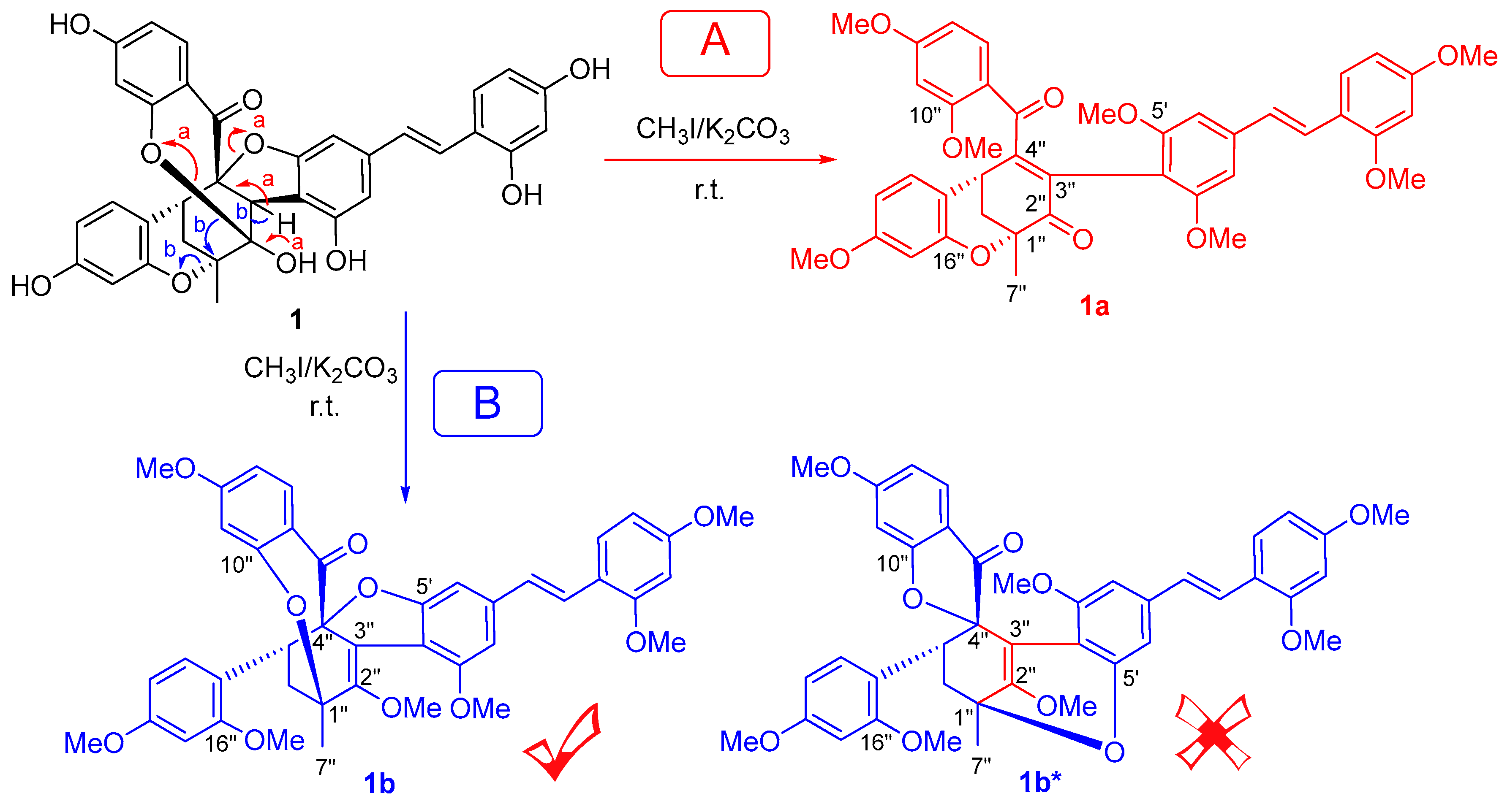
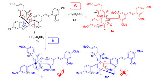
Because all the oxygenated bridgeheads (C-1″, 2″, 4″) were quaternary carbons, the methylation of compound 1 with CH3I/K2CO3 was carried out to confirm the linkages of three O-bridges. Two products—1a and 1b as shown in Figure 3—were identified by the 1D and 2D-NMR spectra.
In 1a, the 13C-NMR spectrum showed two carbonyl carbons at δC 193.6 (C-2″) and 193.0 (C-8″), two olefinic carbons at δC 127.7 (C-3″) and 160.4 (C-4″), four aliphatic carbons at δC 75.9 (C-1″), 33.7(C-5″), 35.6 (C-6″), and 22.7 (C-7″), and the aromatic moieties. The 1H-NMR spectrum showed one methyl at δH 1.57 (s, H-7″), one methine at δH 3.84 (t, J = 3.0 Hz, H-5″), and one methylene at δH 2.65, 2.23 (each dd, J = 13.3, 3.0 Hz, H-6″). The HMBC showed correlations from H-5″ to C-1″/C-3″, from H-6″ to C-1″/C-2″/C-5″, and from H-7″ to C-1″/C-2″/C-6″, establishing that the D-A skeleton was 1″-oxymethylcyclohex-3″-en-2″-one. In addition, the cross-peaks from H-2′ (δH 6.42) to C-3′ (δC 157.7) and from H-6′ (δH 6.41) to C-5′ (δC 157.8) provided the assignments for C-3′ and C-5′ of the stilbene. The cross-peaks from H-11″ (δH 6.39)/H-14″ (δH 7.04) to C-10″ (δC 159.7) and from H-11″/H-13″ (δH 6.33)/H-14″ (δH 7.04) to C-12″ (δC 164.2) provided the assignments for C-10″ and C-12″ of the benzoyl group. The cross-peaks from H-17″ (δH 6.47)/H-20″ (δH 6.97) to C-16″ (δC 153.8)/C-18″ (δC 160.2) provided the assignments of the oxygenated carbons (C-16″ and C-18″) of trisubstituted benzene. All of the methoxylated carbons were assigned by the cross-peaks from methyl groups to their ipso carbons, and only C-1″ and C-16″ were not substituted by a methyl group. Therefore, one O-bridge was assigned between C-1″ and C-16″.
In 1b, the A-D skeleton was determined to be 1″,2″,4″-trioxymethylcyclohexene, which was deduced from one methyl at δH 1.57 (s, H-7″), one methylene at δH 2.28 (dd, J = 13.5, 1.2 Hz, H-6″a) and 1.87 (dd, J = 13.5, 4.2 Hz, H-6″e), and seven carbons at δC 76.4 (C-1″), 148.0 (C-2″), 122.4 (C-3″), 99.4 (C-4″), 33.9 (C-5″), 31.3 (C-6″), and 23.2 (C-7″). This was further confirmed by the HMBC correlations from the H-6″ to C-1″/C-2″/C-4″/C-5″ and from H-7″ to C-1″/C-2″/C-6″. In addition, the cross-peaks from H-2′ (δH 6.56) to C-3′ (δC 155.7) and from H-6′ (δH 6.36) to C-5′ (δC 161.8) provided the assignments for C-3′ and C-5′ of the stilbene moiety. The cross-peaks from H-11″ (δH 6.62)/H-14″ (δH 7.24) to C-10″ (δC 159.6)/C-12″ (δC 163.6) were used to assign C-10″ and C-12″ of the benzoyl group. The cross-peaks from H-17″ (δH 6.27)/H-20″ (δH 7.12) to C-16″ (δC 154.9)/C-18″ (δC 160.0) provided the assignments for the oxygenated aromatic carbon C-16″ and C-18″. All methoxylated carbons were assigned by the cross-peaks from methyl groups to their ipso carbons. Four carbons C-1″, C-4″, C-5′, and C-10″ were not substituted by a methyl group. Given the molecular formula C41H40O10 as calculated by HRMS, there should be two O-bridges in 1b between C-1″/C-10″ and C-4″/C-5′, or between C-1″/C-5′ and C-4″/C-10″. Finally, the O-bridges were attributed at C-1″/C-10″ and C-4″/C-5′ due to the weak NOESY cross-peak between H-6″a (δH 2.28)/H-14″ (δH 7.24). The structure of 1b could be further confirmed by the unreasonably twisted double bond C2″-C3″ that would be present if the O-bridges were located on C-1″/C-5′ and C-4″/C-10″ (1b* in Figure 3).
Given the structures of 1a and 1b, the three O-bridges in 1 were suggested to be at C-1″/C-16″, C-2″/C-10″, and C-4″/C-5′, depending on the proposed reaction mechanism. In the methylation of 1, there were two reactive centers: the hemiketal at C-2″ and its adjacent benzyl proton H-3″. In pathway A, deprotonation at C-3″ under alkali conditions formed a ketone from the hemiketal. Subsequently, the two O-bridges at C-2″ and C-4″ were broken to form a 1,4-butenedione. In pathway B, the hydroxyl group at C-2″ hemiketal was first methylated before deprotonation at C-3″ under alkali conditions. A double bond was formed as the O-bridge at C-2″ migrated to C-1″ with an intramolecular 1,2-rearrangement, and the O-bridge between C-1″/C-16″ was broken. Meanwhile, a configuration inversion of the C-7″ methyl group was observed from 1 to 1b. This phenomenon was further confirmation of the intramolecular O-bridge migration from C-2″ to C-1″.
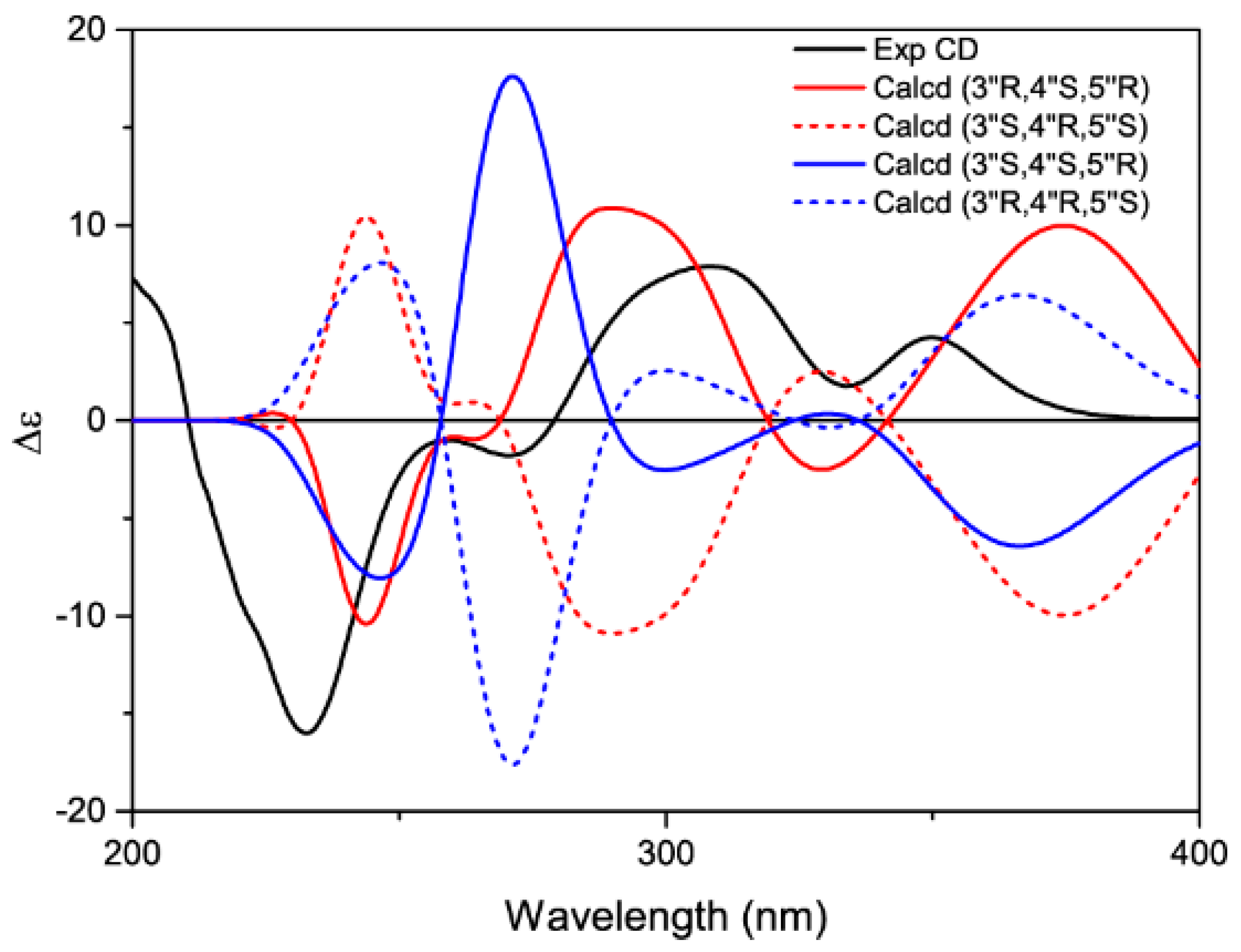
Because the two bridged rings on C1″/C5″ and C2″/C4″ were adjacent to each other, they must be on opposite sides of the hexane plane. Thus, the orientation of C-3″ yielded two sets of epimers—cis-trans or all-trans, in agreement with the biosynthesis pathway [17] of the D-A adducts in the genus Morus. Although the benzyl carbonyl of 1 was coplanar with the aromatic ring, its CD (Circular Dichroism) spectrum did not show the split Cotton effects typical of non-O-bridged D-A adducts, such as mulberrofurans C and J [16]. Two positive Cotton effect peaks at 349 nm and 308 nm were observed, in accordance with the calculated ECD (Electronic Circular Dichroism) spectrum (Figure 4) of one 3″H-α epimer—i.e., (3″R, 4″S, 5″R)—by using the TDDFT (Time-Dependent Density Functional Theory) method. Therefore, the absolute configuration of 1 was determined to be (1″R, 2″R, 3″R, 4″S, 5″R).
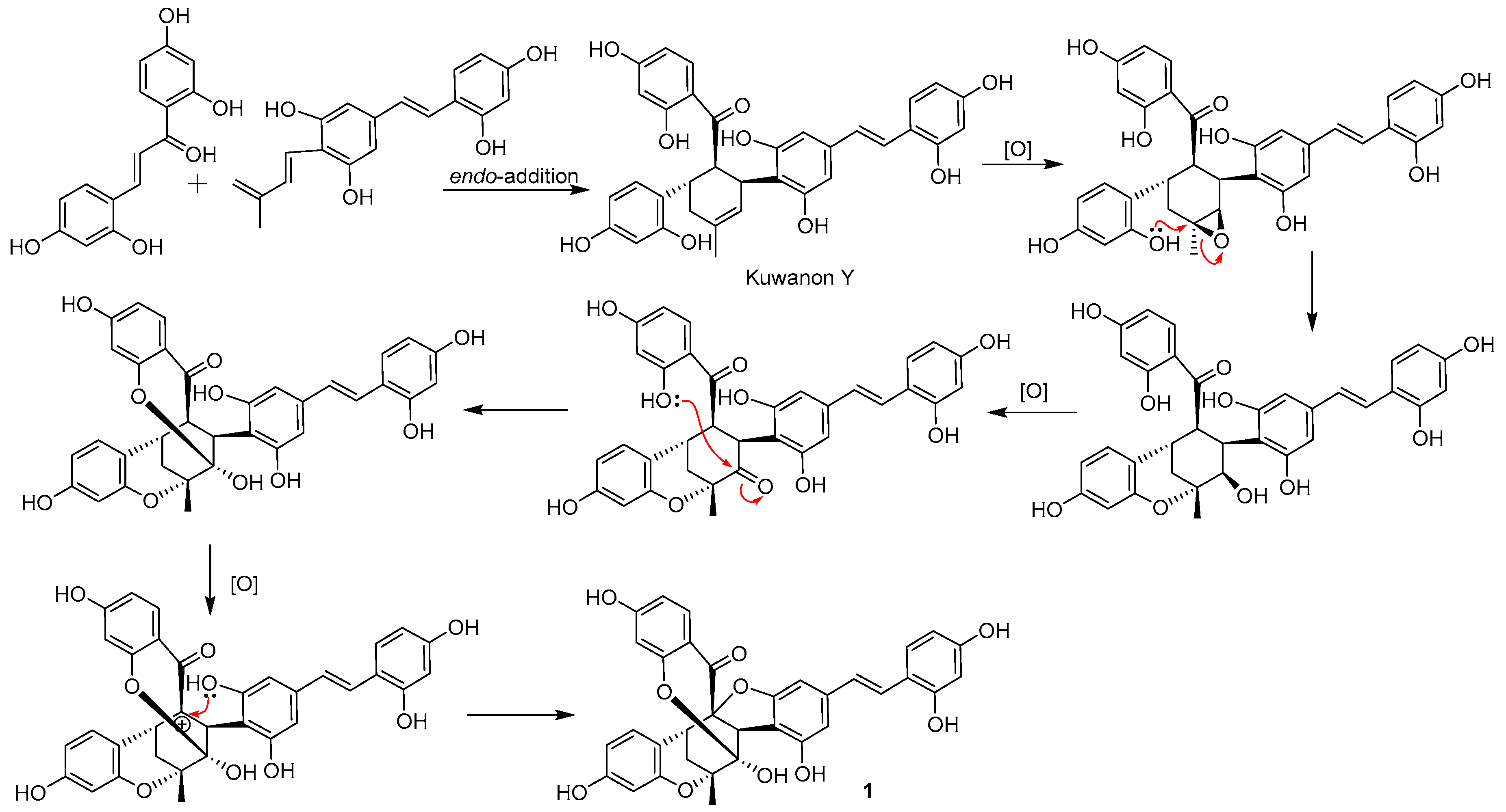
The genus Morus is a plant source with rich D-A adducts. More than 50 D-A adducts have been found in the previous studies [18]. However, a natural product with a highly oxygenated D-A skeleton is rarely reported [19]. A plausible biosynthetic pathway for 1 was postulated in Figure 5, based on the KEGG pathway prediction. Kuwanon Y, a D-A adduct found in genus Morus [16], afforded 1 through three oxidization steps. First, the double bond of the D-A skeleton was oxidized to an epoxide by an oxidase [20] or putative Cyt P450 monooxygenase [19], then the epoxide was attacked by 16″-OH at C-1″, and 2″-OH was formed. Sequentially, the newly formed 2″-OH was oxidized to a carbonyl by an oxidoreductase [21] and was attracted by 10″-OH to form a hemiketal [22]. Finally, the α-position of the 8″-carbonyl was oxidized to form an electrophilic center and was trapped by 5′-OH [23] to afford 1.
In in vitro bioassays, sanggenon X (1) showed significant antioxidant activity against Fe2+-Cys-induced lipid peroxidation in rat liver microsomes with 81.25% inhibition of malondialdehyde (MDA) release, similar to the positive control, curcumin, with an 81.75% inhibition ratio.

3. Experimental

3.1. General Experimental Procedures

Melting points were determined on an XT5B melting point apparatus (Beijing Keyi Electric Light Instrument Factory, Beijing, China) and were uncorrected. Optical rotations were measured with a P-2000 polarimeter (Jasco, Tokyo, Japan). ECD spectra were recorded at room temperature with a J-815 spectropolarimeter (Jasco, Tokyo, Japan). UV spectra were collected in MeOH on a V-650 spectrophotometer (Jasco, Tokyo, Japan). IR spectra were recorded on a Nicolet 5700 spectrometer (Thermo, Madison, WI, USA) by the FT-IR transmission electron microscopy method. 1H- and 13C-NMR spectra were acquired using an AVIIIHD 600 spectrometer (Bruker, Billerica, MA, USA). ESI HR-MS were recorded on a 1200 series LC/6520 quadrupole time of flight (QTOF) spectrometer (Agilent). Column chromatography (CC) purification was performed using silica gel (160–200 mesh), Sephadex LH-20 (GE, Boston, MA, USA), and C18 (50 μm, YMC, Kyoto, Japan). CC fractions were analyzed by thin-layer chromatography (TLC) using silica gel GF254.

3.2. Plant Material

The Cortex Mori Radicis were bought from Anguo herb market, Hebei, China, and were collected from Hunan Province, China, in 2012. These samples were identified by Professor Lin Ma, Institute of Materia Medica, Chinese Academy of Medical Science and Peking Union Medical College, China. A voucher specimen (ID-S-2604) was deposited in the Institute of Materia Medica, Chinese Academy of Medical Science and Peking Union Medical College, China.

3.3. Extraction and Isolation

The powdered Cortex Mori Radicis (50 kg) were soaked with 50% EtOH for 24 h and percolated with 300 L 50% EtOH. Then evaporation of the solvent under reduced pressure gave a liquid extract, which was suspended in H2O and partitioned with EtOAc. The EtOAc extract (ca. 1 kg) was applied to a silica gel (100–200 mesh, 2 kg) column, eluting with a gradient of increasing MeOH concentration (0–100%) in CHCl3, to yield 22 fractions A–V. Fraction M–O (50 g) was applied to a Sephadex LH-20 (3 L) column, using 90% MeOH as eluent, to give subfractions MO-1 to 13. Fraction MO-11 (8 g) was loaded on a silica gel (100–200 mesh, 160 g) column and eluted with a gradient of increasing MeOH concentration (0–100%) in CH2Cl2 to yield five subfractions. The second fraction (3.2 g) was chromatographed over Sephadex LH-20 (400 mL, eluted by MeOH), MPLC over C18 (eluted by MeOH:H2O 10–60%), and HPLC (YMC C18 20 × 250 mm, 5 μm, 65% MeOH in H2O, flow rate 5 mL/min) to give 1 (68 mg, tR = 39 min).
Sanggenon X (1): Yellowish-brown amorphous powder; m.p. 199.0–200.3 °C (d); [ α ] D 20 = −8.76° (c = 1.00, MeOH); UV (MeOH) λmax (logε) 208.5 (4.73), 285 (4.40), 326 (4.51) nm; CD (MeOH) 232.5 (∆ε −16.00), 308 (∆ε +7.88), 349.5 (∆ε +4.24) nm; IR υmax 3392, 1685, 1605, 1509, 1459, 1279, 1217, 1165, 1125, 1064, 995, 973, 838, 767, 661, 636, 525 cm−1; 1H-NMR (DMSO-d6, 600 MHz) 9.55 (1H, s 2-OH), 9.38 (1H, s 4-OH), 8.85 (1H, s 3′-OH), 6.64 (1H, s 2″-OH), 9.29 (1H, s 18″-OH), other data see Table 1; 13C-NMR (DMSO-d6, 150 MHz) data, see Table 1; (+)-ESIMS m/z 595 [M + H]+, 617 [M + Na]+; (+)HR-ESIMS m/z 595.1586 [M + H]+ (calcd. for C34H27O10+, 595.1599). (Supplementary Materials Figure S1a–j, Table S1).
1a: (+)HR-ESIMS m/z 693.2708 [M + H]+ (calcd for C41H41O10+, 693.2694). 1H-NMR (DMSO-d6, 600 MHz) 3.44 s (2-OMe), 3.77 (3H, s, 4-OMe), 3.37 (3H, s, 3′-OMe), 3.82 (3H, s, 5′-OMe), 3.71 3H, s, 10″-OMe), 3.71 (3H, s, 12″-OMe), 3.71 (3H, s, 18″-OMe), other data see Table 1. 13C-NMR (DMSO-d6, 150 MHz) 55.2 (2-OMe), 55.3 (4-OMe), 55.7 (3′-OMe), 55.6 (5′,10″,12″,18″-OMe), other data see Table 1. (Supplementary Materials Figure S2a–f, Table S1).
1b: (+)HR-ESIMS m/z 693.2708 [M + H]+ (calcd. for C41H41O10+, 693.2694). 1H-NMR (DMSO-d6, 600 MHz) 3.77 (3H, s, 2-OMe), 3.73 (3H, s, 4-OMe), 3.76 (9H, s, 3′,12″,16″-OMe), 3.32 (3H, s, 2″-OMe), 3.60 (3H, s, 18″-OMe), other data see Table 1. 13C-NMR (DMSO-d6, 150 MHz) 56.0 (2-OMe), 55.8 (4-OMe), 56.1 (3′,16″-OMe), 60.7 (2″-OMe), 56.2 (12″-OMe), 55.5 (18″-OMe), other data see Table 1. (Supplementary Materials Figure S3a–h, Table S1).

3.4. Methylation of 1

Twenty milligrams of 1 was dissolved in dried acetone, 200 mg K2CO3 and 400 μL CH3I were added and then stirred for 24 h. Then, the solution was dried and purified by RP-HPLC (Grace Adsorbosphere XL C18 10 × 250 mm, 5 μm, 90% MeOH in H2O, flow rate 2 mL/min) to yield compounds 1a and 1b (1a: 3.5 mg, 11.7%, tR = 11.9 min; 1b: 3.4 mg, 11.3%, tR = 25.9 min).

3.5. Calculation of ECD

Calculated ECD was performed on the 3″H-α (1″R, 2″R, 3″R, 4″S, 5″R), 3″H-β (1″R, 2″R, 3″S, 4″S, 5″R), and their enantiomers of 1. Conformation search was done with the MMFF94 molecular mechanics force field via the MOE software package (MOE2009.10, Chemical Computing Group Inc., Montreal, QC, Canada). Calculated ECD was performed using the TDDFT method (Gaussian 09 B.01, Gaussian, Wallingford, CT, USA, 2009) at B3LYP/6-31+G(d,p)//B3LYP/6-311+G(d,p) level for the configurations within an energy window of 5 kcal/mol. The conductor-like polarizable continuum model was used with MeOH (ε = 32.613) in order to take the solvent effects into consideration. The Boltzmann distribution was calculated based on the relative free energy (ΔG) and the final ECD (σ = 0.25 eV, UV shift = 10 nm) was simulated by using SpecDis (V1.64, University of Wuerzburg, Germany, 2015).

3.6. Lipid Peroxidation Assay

Antioxidative activity was evaluated as the inhibitory activity of compounds against Fe2+-Cys-induced lipid peroxidation in rat liver microsomes by the formation of malondialdehyde-thiobarbituric acid (MDA-TBA) adduct. Microsomes were isolated from SD rat livers and suspended in 100 mM TMS buffer (pH 7.4). The microsomal suspension (1 mg protein/mL), different concentrations of compound or vehicle, and 0.2 mM cysteine in 0.1 M PBS (pH 7.4) were incubated at 37 °C for 15 min, 50 µM FeSO4 was added, and the reaction mixture was then incubated at 37 °C for 15 min again. An equal volume of 20% (w/v) TCA (Trichloroacetic Acid) and 0.6% (w/v) TBA were added and kept in a boiling water bath for 10 min. After the mixture was centrifuged at 3000× g for 10 min, the absorbance of supernatant was measured at 532 nm and the concentration of MDA was calculated as C = (OD − 0.006)/0.07 × 10 nmol/mL. Lipid peroxidation inhibitory activity was calculated as follows: [1 − (T − B)/(C − B)] × 100%, in which T, C, and B are MDA concentrations of the sample treated, the control without sample, and the zero time control, respectively. Curcumin (10−4 M) was used as the positive control.

4. Conclusions

In this paper, a tri-O-bridged D-A adduct, sanggenon X (1), was isolated from a 55% alcohol extract of Cortex Mori Radicis. Given its complex structure with several quaternary carbons in the bridgeheads, it was fortunate for us to determine the exact structure with the help of chemical methylation and calculated ECD. The structure of 1, with highly oxygenated D-A skeleton, adds a new skeletal entity to the natural D-A adducts and provides a new framework for synthesis and biological evaluation in the future.

Supplementary Materials

The supplementary materials are available online. Copies of MS, UV, ECD, IR, and NMR spectra of compounds 1, 1a, and 1b are available online.

Acknowledgments

We gratefully acknowledge the financial support from the CAMS Innovation Fund for Medical Sciences (CIFMS) No. 2016-I2M-3-011. We thank for Dan Zhang and Xiu-Qi Bao for determining the lipid peroxidation assay.

Author Contributions

A.-Q.L. performed the isolation and purification; M.-H.C. calculated the ECD spectra; J.G. analyzed the data; L.W. and H.-Y.Y. carried out the antioxidant assay; L.L., B.Z., and H.-K.H. contributed the extraction of raw material. S.-J.W. wrote the paper and was responsible for the whole work.

Conflicts of Interest

The authors declare no conflict of interest.

References

  1. Abbas, G.M.; Abdel Bar, F.M.; Baraka, H.N.; Gohar, A.A.; Lahloub, M.F. A new antioxidant stilbene and other constituents from the stem bark of Morus nigra L. Nat. Prod. Res. 2014, 28, 952–959. [Google Scholar] [CrossRef] [PubMed]
  2. Ahmad, A.; Gupta, G.; Afzal, M.; Kazmi, I.; Anwar, F. Antiulcer and antioxidant activities of a new steroid from Morus alba. Life Sci. 2013, 92, 202–210. [Google Scholar] [CrossRef] [PubMed]
  3. Kapche, G.D.W.F.; Amadou, D.; Waffo Teguo, P.; Donfack, J.H.; Fozing, C.D.; Harakat, D.; Tchana, A.N.; Merillon, J.M.; Moundipa, P.F.; Ngadjui, B.T.; et al. Hepatoprotective and antioxidant arylbenzofurans and flavonoids from the twigs of Morus mesozygia. Planta Med. 2011, 77, 1044–1047. [Google Scholar] [CrossRef] [PubMed]
  4. Lim, H.J.; Jin, H.G.; Woo, E.R.; Lee, S.K.; Kim, H.P. The root barks of Morus alba and the flavonoid constituents inhibit airway inflammation. J. Ethnopharmacol. 2013, 149, 169–175. [Google Scholar] [CrossRef] [PubMed]
  5. Riviere, C.; Krisa, S.; Pechamat, L.; Nassra, M.; Delaunay, J.C.; Marchal, A.; Badoc, A.; Waffo-Teguo, P.; Merillon, J.M. Polyphenols from the stems of Morus alba and their inhibitory activity against nitric oxide production by lipopolysaccharide-activated microglia. Fitoterapia 2014, 97, 253–260. [Google Scholar] [CrossRef] [PubMed]
  6. Sohn, H.Y.; Son, K.H.; Kwon, C.S.; Kwon, G.S.; Kang, S.S. Antimicrobial and cytotoxic activity of 18 prenylated flavonoids isolated from medicinal plants: Morus alba L., Morus mongolica Schneider, Broussnetia papyrifera (L.) Vent, Sophora flavescens Ait and Echinosophora koreensis Nakai. Phytomedicine 2004, 11, 666–672. [Google Scholar] [CrossRef] [PubMed]
  7. Grienke, U.; Richter, M.; Walther, E.; Hoffmann, A.; Kirchmair, J.; Makarov, V.; Nietzsche, S.; Schmidtke, M.; Rollinger, J.M. Discovery of prenylated flavonoids with dual activity against influenza virus and Streptococcus pneumoniae. Sci. Rep. 2016, 6, 27156. [Google Scholar] [CrossRef] [PubMed]
  8. Pethakamsetty, L.; Ganapaty, S.; Bharathi, K.M. Phytochemical and antimicrobial examination of the root extracts of Morus Indica. Int. J. Pharm. Sci. Rev. Res. 2013, 21, 75–80. [Google Scholar]
  9. Fukai, T.; Oku, Y.; Hano, Y.; Terada, S. Antimicrobial activities of hydrophobic 2-arylbenzofurans and an isoflavone against vancomycin-resistant enterococci and methicillin-resistant Staphylococcus aureus. Planta Med. 2004, 70, 685–687. [Google Scholar] [CrossRef] [PubMed]
  10. Lim, S.L.; Park, S.Y.; Kang, S.; Park, D.; Kim, S.H.; Um, J.Y.; Jang, H.J.; Lee, J.H.; Jeong, C.H.; Jang, J.H.; et al. Morusin induces cell death through inactivating STAT3 signaling in prostate cancer cells. Am. J. Cancer Res. 2015, 5, 289–299. [Google Scholar] [PubMed]
  11. Zhu, J.J.; Yan, G.R.; Xu, Z.J.; Hu, X.; Wang, G.H.; Wang, T.; Zhu, W.L.; Hou, A.J.; Wang, H.Y. Inhibitory effects of (2′R)-2′,3′-dihydro-2′-(1-hydroxy-1-methylethyl)-2,6′-bibenzofuran-6,4′-diol on mushroom tyrosinase and melanogenesis in B16-F10 melanoma cells. Phytother. Res. 2015, 29, 1040–1045. [Google Scholar] [CrossRef] [PubMed]
  12. Tan, Y.X.; Liu, C.; Chen, R.Y. New 2-arylbenzofurans with selective cytotoxicity from Morus wittiorum. Phytochem. Lett. 2012, 5, 419–422. [Google Scholar] [CrossRef]
  13. Almeida, J.R.G.D.S.; Souza, G.R.; Araujo, E.C.D.C.; Silva, F.S.; Tolentino De Lima, J.; Ribeiro, L.A.D.A.; Nunes, X.P.; Barbosa Filho, J.M.; Junior, L.J.Q.; Viana Dos Santos, M.R. Medicinal plants and natural compounds from the genus Morus (Moraceae) with hypoglycemic activity: A review. In Glucose Tolerance; InTech: Rijeka, Croatia, 2012; pp. 189–206. [Google Scholar]
  14. Wang, Y.-N.; Liu, M.-F.; Hou, W.-Z.; Xu, R.-M.; Gao, J.; Lu, A.-Q.; Xie, M.-P.; Li, L.; Zhang, J.-J.; Peng, Y.; et al. Bioactive Benzofuran Derivatives from Cortex Mori Radicis, and Their Neuroprotective and Analgesic Activities Mediated by mGluR1. Molecules 2017, 22, 236. [Google Scholar] [CrossRef] [PubMed]
  15. Gao, L.; Han, J.; Lei, X. Enantioselective Total Syntheses of Kuwanon X, Kuwanon Y, and Kuwanol A. Org. Lett. 2016, 18, 360–363. [Google Scholar] [CrossRef] [PubMed]
  16. Hano, Y.; Suzuki, S.; Nomura, T.; Iitaka, Y. Absolute configuration of natural Diels-Alder type adducts from the morus root bark. Heterocycles 1988, 27, 2315–2325. [Google Scholar]
  17. Nomura, T.; Hano, Y.; Fukai, T. Chemistry and biosynthesis of isoprenylated flavonoids from Japanese mulberry tree. Proc. Jpn. Acad. Ser. B Phys. Biol. Sci. 2009, 85, 391–408. [Google Scholar] [CrossRef] [PubMed]
  18. Yang, Y.; Tan, Y.-X.; Chen, R.-Y.; Kang, J. The latest review on the polyphenols and their bioactivities of Chinese Morus plants. J. Asian Nat. Prod. Res. 2014, 16, 690–702. [Google Scholar] [CrossRef] [PubMed]
  19. Schümann, J.; Hertweck, C. Molecular Basis of Cytochalasan Biosynthesis in Fungi:  Gene Cluster Analysis and Evidence for the Involvement of a PKS-NRPS Hybrid Synthase by RNA Silencing. J. Am. Chem. Soc. 2007, 129, 9564–9565. [Google Scholar] [CrossRef] [PubMed]
  20. Ward, D.A.; MacMillan, J.; Gong, F.; Phillips, A.L.; Hedden, P. Gibberellin 3-oxidases in developing embryos of the southern wild cucumber, Marah macrocarpus. Phytochemistry 2010, 71, 2010–2018. [Google Scholar] [CrossRef] [PubMed]
  21. Nakajima, K.; Hashimoto, T.; Yamada, Y. Two tropinone reductases with different stereospecificities are short-chain dehydrogenases evolved from a common ancestor. Proc. Natl. Acad. Sci. USA 1993, 90, 9591–9595. [Google Scholar] [CrossRef] [PubMed]
  22. Xiong, L.; Zhou, Q.-M.; Zou, Y.; Chen, M.-H.; Guo, L.; Hu, G.-Y.; Liu, Z.-H.; Peng, C. Leonuketal, a Spiroketal Diterpenoid from Leonurus japonicus. Org. Lett. 2015, 17, 6238–6241. [Google Scholar] [CrossRef] [PubMed]
  23. Markwell-Heys, A.W.; George, J.H. Some chemical speculation on the biosynthesis of corallidictyals A-D. Org. Biomol. Chem. 2016, 14, 5546–5549. [Google Scholar] [CrossRef] [PubMed]
Sample Availability: Samples of the compounds 1, 1a, and 1b are available from the authors.
Figure 1. Structure of sanggenon X (1).
Molecules 23 00133 g001
Figure 2. Key correlations of compounds 1, 1a, and 1b in HMBC and NOESY spectra.
Molecules 23 00133 g002
Figure 3. Methylation of compound 1.
Molecules 23 00133 g003
Figure 4. Experimental and calculated ECD (Electronic Circular Dichroism) of compound 1.
Molecules 23 00133 g004
Figure 5. Plausible biosynthetic pathway of compound 1.
Molecules 23 00133 g005
Table 1. NMR spectroscopic data for compounds 1, 1a, and 1b in DMSO-d6 (J in Hz).
Position11a1b
δHδC δHδC δHδC
1 115.2 118.2 118.4
2 156.0 156.8 158.4
36.30 s102.56.57 d (2.2)98.46.51 s c98.7
4 158.1 160.4 161.1
56.23 d (8.4) a107.16.54 dd (8.8, 2.2)105.66.50 d (8.4) c106.2
67.30 d (8.4)127.27.49 d (8.8)127.67.45 d (8.4)128.3
α7.09 d (16.2)123.17.16 d (16.5)123.17.15 d (16.8)124.6
β6.74 d (16.2)124.76.91 d (16.5)127.06.89 d (16.8)126.6
1′ 139.3 139.3 141.8
2′6.16 s105.76.42 s b101.16.56 s104.5
3′ 154.2 157.7 155.7
4′ 110.7 110.7 110.7
5′ 159.2 157.8 161.8
6′6.44 s98.86.41 s101.76.36 s100.1
1″ 74.4 75.9 76.4
2″ 109.1 193.6 148.0
3″3.17 s47.3 127.7 122.4
4″ 91.4 160.4 99.4
5″2.66 brs36.63.84 t (3.0)33.7N.D.33.9
6″2.51 d (13.8) *
1.77 dd (13.8, 3.0)
30.12.65 dd (13.3, 3.0)
2.23 dd (13.3, 3.0)
35.62.28 dd (13.5, 1.2)
1.87 dd (13.5, 4.2)
31.3
7″1.61 s22.11.57 s22.71.57 s23.2
8″ 194.9 193.0 202.1
9″ 112.8 119.4 120.7
10″ 166.2 159.7 159.6
11″6.09 s97.66.39 d (1.9)97.56.62 d (2.1)99.0
12″ 171.6 164.2 163.6
13″6.48 d (8.7)111.46.33 dd (8.8, 1.9)105.36.50 dd (8.7, 2.1) c105.8
14″7.44 d (8.7)125.77.04 d (8.8)131.37.24 d (8.7)131.5
15″ 107.9 115.1 112.9
16″ 154.6 153.8 154.9
17″6.23 s a106.56.47 d (2.4)101.46.27 d (2.4)101.5
18″ 157.7 160.2 160.0
19″6.22 d (9.0) a101.96.43 dd (8.4, 2.4) b106.76.43 dd (8.4, 2.4)106.9
20″6.51 d (9.0)133.36.97 d (8.4)129.57.12 d (8.4)133.0
a–c The signals overlapped with each other. * Half of this signal was overlapped by a solvent peak. Measured at 150 MHz or 125 MHz for 13C.