Article

Anti-Influenza Virus Activity and Constituents Characterization of Paeonia delavayi Extracts

Institute of Medicinal Plant Development, Peking Union Medical College & Chinese Academy of Medical Sciences, Beijing 100193, China
*
Author to whom correspondence should be addressed.
Molecules 2016, 21(9), 1133; https://doi.org/10.3390/molecules21091133
Submission received: 21 June 2016 / Revised: 29 July 2016 / Accepted: 23 August 2016 / Published: 26 August 2016
(This article belongs to the Section Natural Products Chemistry)

Abstract

:
Paeonia delavayi, an endemic species in southwestern China, has been widely used as a traditional remedy for cardiovascular, extravasated blood, stagnated blood and female diseases in traditional Chinese medicine (TCM). However, there are no reports on the anti-influenza virus activity of this species. Here, the anti-influenza virus activity of P. delavayi root extracts was first evaluated by an influenza virus neuraminidase (NA) inhibition assay. Meantime, constituents in the active extracts were identified using ultra-high performance liquid coupled with quadrupole time-of-flight mass spectrometry (UPLC-Q-TOF-MS) and seven major identified constituents were used to further evaluate the NA inhibitory activity. The results showed that the ethyl acetate fraction (EA) and the ethanol fraction (E) of P. delavayi both presented strong NA inhibitory activity with IC50 values of 75.932 μg/mL and 83.550 μg/mL, respectively. Twenty-seven constituents were characterized in these two active extracts by UPLC-Q-TOF-MS analysis, and seven major identified constituents exhibited high activity against the influenza virus. Among them, Benzoylpaeoniflorin (IC50 = 143.701 µM) and pentagalloylglucose (IC50 = 62.671 µM) exhibited the highest activity against the influenza virus, even far stronger than oseltamivir acid (IC50 = 281.308 µM). This study indicated that P. delavayi was a strong NA inhibitor, but cell-based inhibition, anti-influenza virus activity in vivo and anti-influenza virus mechanism still need to be tested and explored.

Graphical Abstract

1. Introduction

The Paeonia genus, known for its ornamental value and medicinal properties, has a long medicinal history in Traditional Chinese Medicine (TCM) [1]. Pharmacological studies of this genus have shown a variety of activities, including analgesic, sedative, anti-inflammatory, antimicrobial, antitumor, antiviral, cardiovascular protection activities and so on [2,3,4]. The pharmacological activities are primarily attributed to monoterpenes, which has a “cage-like” pinane skeleton. Paeoniflorin and its derivatives represent major monoterpene glycosides in the plants of Paeonia. Furthermore, previous chemical investigations on this genus have led to the identification of seven classes, including monoterpenoid glycosides, flavonoids, tannins, stilbenes, triterpenoids and steroids, paeonols, and phenols [3,4]. Paeonia delavayi—one of the most important species in the Paeonia genus—is protected as an endemic species in southwestern China, which is mainly distributed in Yunnan, Sichuan and Tibet areas [5,6]. Its root, one of the main sources of the Chinese medicine “mudanpi”, has been used as a remedy for cardiovascular, extravasated blood, stagnated blood and female diseases [7,8].
The influenza virus, which belongs to the Orthomyxoviridae family, is a respiratory pathogen that reproduces rapidly, mutates frequently, and occasionally crosses the species barrier with high morbidity and mortality [9,10,11]. So far, two classes of drugs are available for influenza infections: M2 protein blockers (adamantanes), neuraminidase (NA) and inhibitors (zanamivir and oseltamivir) [12]. The M2 protein blockers, amantadine and rimantadine, are exclusively active against the influenza A virus, and possess adverse drug resistance effects. As a result, this class of drugs was strongly resisted by the Centers for Disease Control and Prevention (CDC) [13]. NA inhibitors are effective on both influenza A and B infections, and recommended by the WHO [14]. Nevertheless, the use of this class of antiviral drug has its limitations, such as drug-resistance, toxicity and cost [15,16,17,18]. Hence, new antiviral agents with high efficiency and low toxicity against the influenza virus are urgently needed.
Plants have been used for treating infectious diseases caused by viruses in traditional Chinese medicine for a long time. Much research has shown that plant extracts and natural products have a wide range of pharmacological activities, including anti-inflammatory, antibacterial, anticancer and antiviral [12]. For instance, polyphenols from green tea have the ability to inhibit influenza virus replication [19,20] and polyphenolic compounds derived from Phellinus baumii could inhibit influenza virus infection [10]. In recent years, the antiviral activity of some species of Paeonia has been explored. It has been reported that the extract of P. lactiflora possesses anti-influenza activity and has the potential for development as an anti-influenza agent [2]. In addition, literature also reported that P. lactiflora and P. suffruticosa both showed antiviral activities against respiratory syncytial virus (RSV) [21]. However, there are no reports of anti-influenza virus activity in P. delavayi. In the present study, an influenza virus NA inhibition assay was used to screen the anti-influenza virus activity of the extracts and UPLC-Q-TOF-MS was used to identify chemical constituents of the active extracts. Then, the anti-influenza virus activity of seven major constituents of extracts was further verified by determining their NA inhibitory capacity.

2. Results and Discussion

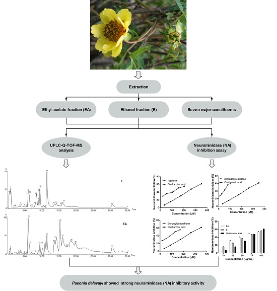
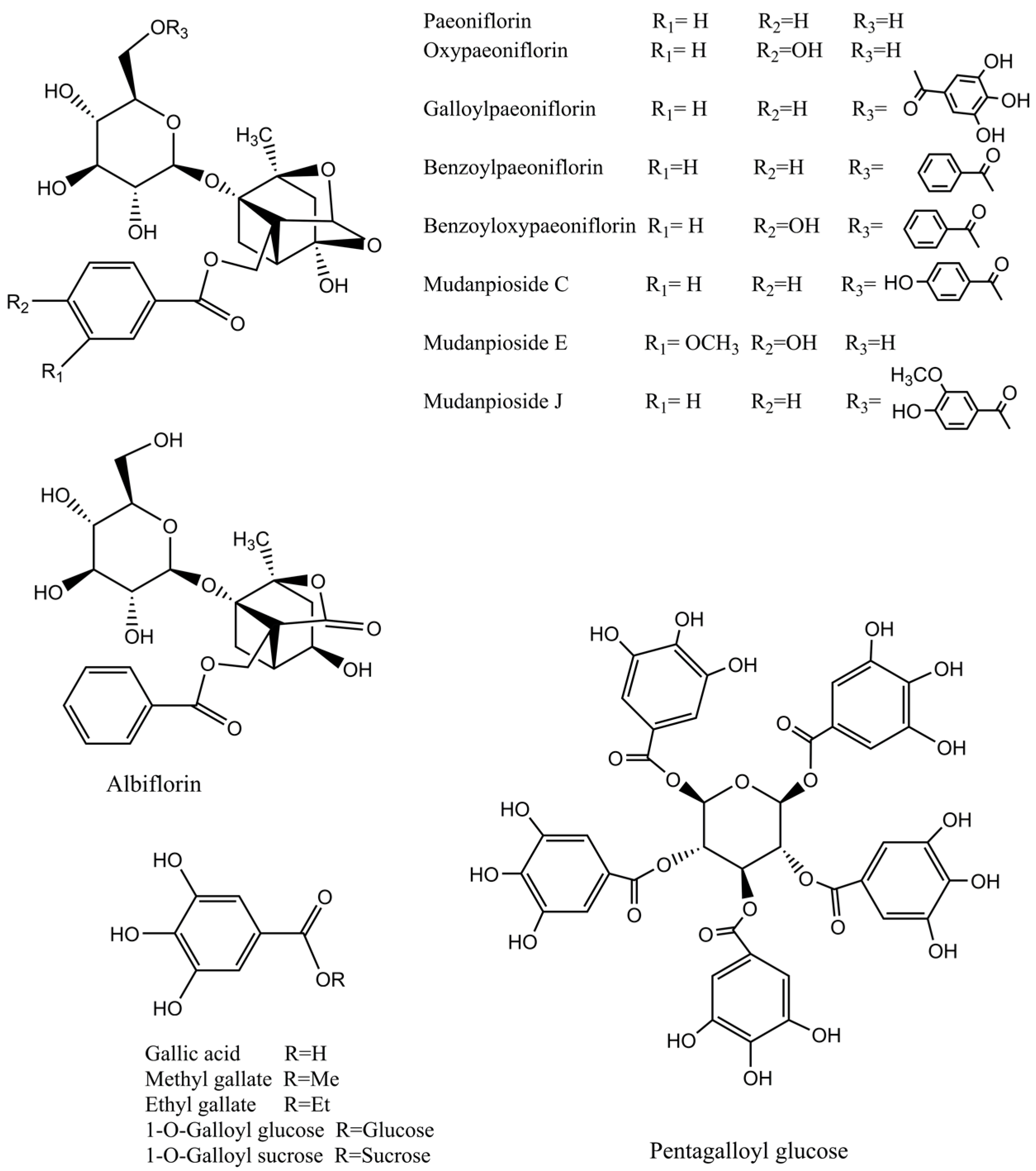
2.1. UPLC-Q-TOF-MS Analysis

According to the retention time and the molecular ions, together with the major fragments observed in MS spectra and followed by searching the literature, 27 compounds were tentatively identified. The identified compounds were definitely classified into three groups: phenols, tannins and monoterpene glycosides. The retention time, molecular formula, exact mass, experimental mass, the major fragments and identified compounds were listed in Table 1. The base peak ion (BPI) chromatogram and the chemical structures of the identified compounds were presented in Figure 1 and Figure 2, respectively.
Based on accurate molecular ions, fragment ions and the literature, three phenols and fourteen tannins were characterized in the extracts, all of which belong to gallic acid derivatives showing similar fragments, such as m/z at 169[gallic acid–H], 125[gallic acid–H–CO2]. The ion [M–H] at m/z 183, 197 combined with neutral losses of 15 Da (CH3) and 29 Da (CH2CH3), respectively. The fragment ions of gallic acid, methy gallte and ethy gallte are consistent with previous reports [1,25]. Moreover, twelve galloylglucoses containing a glucose and different numbers of gallic acid moieties were detected, including the isomers of trigalloyl glucose, tetragalloyl glucose and hexagalloyl glucose. These results revealed that tannins were rich in the extracts.
Monoterpene glycosides are the major bioactive constituents of Paeonia species that have been reported by numerous literatures [4,26,27]. In this work, ten monoterpene glycosides were characterized, including albiflorin, paeoniflorin, oxypaeoniflorin, 4-O-ethylpaeoniflorin, benzoyloxypaeoniflorin, benzoylpaeoniflorin, galloylpaeoniflorin, mudanpioside C, mudanpioside E and mudanpioside J. These monoterpene glycosides have a pinane skeleton and a mono-glucose moiety and their fragmentation pattern is consistent with previous reports [22,23].

2.2. Influenza Virus Neuraminidase (NA) Activity Assay

NA has been regarded as one of the most important targets to screen the drugs of anti-influenza viruses A and B. The anti-influenza virus activity of the extracts and seven standard compounds were presented in Figure 3. They all showed dose-dependent activity and their IC50 values were shown in Table 2, and a lower IC50 value indicates a higher activity. Based on IC50 value, the activity of extracts was in the order of EA > Oseltamivir acid > E, and the activity of seven compounds was in the order of pentagalloylglucose > benzoylpaeoniflorin > albiflorin > paeoniflorin > oseltamivir acid > ethyl gallate > methyl gallat > gallic acid.
Although P. delavayi is one of the main sources of the Chinese traditional medicine “mudanpi”, which treats cardiovascular, extravasated blood, stagnated blood and female diseases; no research has reported its anti-viral activity. In the present study, the inhibitory activity of two extracts from P. delavayi (E and EA fractions) to NA was explored. Our findings, for the first time, showed that the extracts of P. delavayi display anti-influenza virus activity in vitro based on enzyme-based assay, especially EA fractions. Therefore, this study implies that P. delavayi may be used as a promising anti-viral drug resource, but its anti-influenza virus activity still needs to be further evaluated.
In order to confirm the bioactive constituents of the extracts, seven standard compounds that were identified in the qualitative assay (gallic acid, methyl gallate, ethyl gallate, pentagalloylglucose, benzoylpaeoniflorin, albiflorin, paeoniflorin) were used to analyse NA inhibitory capacity in vitro. These compounds belong to phenols, tannins and monoterpene glycosides (shown in Table 2). Oseltamivir acid, a metabolite of Oseltamivir, was used as a positive control. The result of the enzyme inhibition assay showed that all seven standard compounds exhibited anti-influenza virus activity, and pentagalloylglucose, benzoylpaeoniflorin, albiflorin and paeoniflorin showed stronger activity against NA than Oseltamivir acid. In several literature reports [28,29,30], the antiviral activities of pentagalloylglucose and gallic acid have been reported, but there are few reports regarding the antiviral activities of methyl gallate, ethyl gallate, benzoylpaeoniflorin, albiflorin and paeoniflorin. In this study, the NA inhibitory activity of methyl gallate, ethyl gallate, benzoylpaeoniflorin, albiflorin and paeoniflorin were reported for the first time.
Influenza is a highly contagious disease and can cause high morbidity and mortality in an epidemic. Antiviral agents are an important part of a rational approach to epidemic influenza and are critical to prevent pandemic influenza, but the resistance of influenza to antiviral drugs is now widespread due to mutations in the influenza viruses [15]. Hence, development of new natural anti-influenza drugs becomes necessary and urgent. Data presented here shows that extracts from P. delavayi are potent NA inhibitors. However, false-positive results in the commonly used method of enzyme-based NA inhibition assays were reported in several literatures and the reliability of a large number of flavonoid-based NA inhibitors reported in the literature is under question [31,32]. So, cell-based assay and anti-influenza virus activity, in vivo of the extracts and major components, need to be further tested and evaluated. Furthermore, high-throughput biology has greatly contributed to natural product-based drug discovery and some natural products were shown to be able to lead to structures for drug development, but these were false positives in most cases and those compounds acted as pan-assay interference compounds (PAINS) [33,34]. Gallic acid and some other phenolic natural products have been characterized promiscuous inhibitors because their target-based activities were linked to cell-based activities and activities in vivo, but there may be no common mechanisms involved [34]. Therefore, the study of the anti-influenza virus mechanism of P. delavayi root becomes quite necessary.

3. Experimental Section

3.1. Chemicals and Standard Substances

Analytical grade reagents that were used for extraction were obtained from Beijing Chemical Plant Co. Ltd. (Beijing, China). LC-MS grade acetonitrile was purchased from Fisher Scientific (Beijing, China). De-ionized water was purified using a Milli-Q system (Millipore, Bedford, MA, USA).
Gallic acid, methyl gallate, ethyl gallate, pentagalloylglucose, paeoniflorin, albiflorin and benzoylpaeoniflorin were purchased from Chengdu Must Biotechnology Co. Ltd. (Sichuan, China). Oseltamivir acid was purchased from Medchem Express, LLC (Monmouth Junction, NJ, USA).

3.2. Neuraminidase Inhibitors Screen Kit

Neuraminidase Inhibitors Screen Kit (NO. P0309) was purchased from Beyotime Institute of Biotechnology Co. Ltd. (Shanghai, China), which contains 10 mL buffer, 1 mL NA, 1 mL fluorescent substrate and 1.2 mL Milli-Q water.

3.3. Plant Material Collection and Sample Preparation

Samples of P. delavayi were collected in Dali, located in central Yunnan, China, in September 2010. The samples were identified by one of the authors, Professor Linfang Huang, and the voucher specimens (NO. P201009-188) were deposited in the Herbarium of the Chinese Academy of Medical Science & Peaking Union Medicinal College.
The samples were powdered to a fine powder in a grinder. A total of 500 g of powder was extracted by infusion with 2.5 L of petroleum ether for 24 h. Then, the residue was extracted with 80% ethanol using reflux extraction 3 times, 2 h each time. The filtered extracted solutions were concentrated using a rotary evaporator to yield a crude extract. The crude extract was dissolved in ethanol and successively extracted with ethyl acetate using the method of liquid-liquid extraction, and the solvent was removed to obtain a dry form of the ethyl acetate fraction (EA) and ethanol fraction (E) using a rotary evaporator. These two fractions were used for assays because these fractions exhibited greater influenza NA inhibitory activity than other fractions in our preliminary experiments. These fractions were dissolved in methanol and filtered through 0.22 μm Nylon micropore membranes and used for UHPLC-Q-TOF-MS assays at concentrations (m/v) of 5 mg/mL.

3.4. UPLC-Q-TOF-MS Analysis

3.4.1. Liquid Chromatography

UPLC analysis was performed using a Waters Acquity UPLC system, equipped with a binary solvent system, an automatic sample manger and a photodiode-array (PDA) detector. The column that was used for the chromatographic separation was an ACQUITY UPLC BEH C18 2.1 mm × 100 mm, 1.7 μm (Waters, Milford, MA, USA) at 30 °C. The conditions consisted of a gradient elution using aqueous formic acid 0.1% (v/v) as mobile phase A and acetonitrile as phase B at a flow rate of 0.3 mL/min. The following gradient was applied: 0–2 min, 5% B; 2–4 min, 5%–10% B; 4–10 min, 10%–15% B; 10–30 min, 15%–20% B and 30–35 min, 20%–30% B. The injection volume was 2 μL and the injection temperature was 15 ℃.

3.4.2. Mass Spectrometry

Tandem mass spectrometry was performed with a hybrid quadrupole orthogonal time-of-flight mass spectrometer (Q-TOF-MS) (Waters, Milford, MA, USA) using an electrospray ionization source for the ionization of the target compounds. The operating parameters were as follows: capillary voltage of 3.0 kV (ESI+) or 2.2 kV (ESI); sample cone voltage 35 kV; extraction cone voltage 4 kV, ion source temperature 100 °C, desolvation temperature 450 °C and desolvation gas (N2) flow of 800 L/h, and scan range, m/z 100–1200. The mass spectrometer was calibrated with sodium formate. Leucine-enkephalin was used as an external reference at a constant flow of 5 µL/min. The data was processed with Masslynx V4.1 software (Waters, Milford, MA, USA).

3.5. Neuraminidase (NA) Inhibition Assay

The NA inhibition assay was carried out in a 96-well microplate reader using a procedure given by kit instruction [35]. Reaction mixture containing 70 μL of reaction buffer solution, 10 μL of NA and 10 μL of standard samples of the extracts in 10% DMSO were added to each well. Vibration mixing was carried out for about 1 min and incubation at a temperature of 37 °C for 2 min so that the NA and standard samples of the extracts can be fully interacted. After which, 10 μL of fluorescent substrate was added to give a total of 100 μL reaction mixture. The entire mixture was thoroughly mixed by vibration for about 1 min and the plate was incubated at 37 °C for 20 min. The fluorescence was read on a microplate spectrophotometer (Molecular Device, Gemini EM, USA) with an excitation wavelength at 322 nm and an emission wavelength at 450 nm. Oseltamivoir acid was used as a positive control. The inhibition (%) was calculated using the formula:
NA Inhibitory activity (%) = [1 − (Fs − F0)/(Fm − F0)] × 100%
Fs was fluorescence intensity in the presence of the sample, F0 was the absorbance in the presence of the sample background, and Fm was the absorbance of the negative control (without the sample). The 50% inhibitory concentration (IC50) was determined by probit regression in SPSS.

3.6. Statistical Analysis

NA inhibition experiments were performed in triplicate. SPSS 19 (BM SPSS, Chicago, IL, USA) and Graph Pad Prism 6 (GraphPad, San Diego, CA, USA) were used for the statistical analysis of the data.

4. Conclusions

In summary, the anti-influenza virus effect of P. delavayi root extracts was first evaluated by an influenza virus neuraminidase (NA) inhibition assay and chemical constituents of the active extract were characterized by UPLC-Q-TOF-MS in this study. As a result, the extracts showed strong NA inhibition and twenty-seven compounds were identified. In addition, seven major constituents in the extracts all showed favorable NA inhibitory activity. Among them, pentagalloylglucose, benzoylpaeoniflorin, albiflorin and paeoniflorin displayed stronger inhibitory activity than Oseltamivir acid. This study indicates that P. delavayi is a strong NA inhibitor, and provides scientific evidence and a new idea for anti-influenza virus drug discovery, but cell-based inhibition, anti-influenza virus activity in vivo, as well as anti-influenza virus mechanism, still need to be tested and explored.

Acknowledgments

This study was supported by grants from the National Natural Science Foundation of China (No. 81274013; No. 81130069; No. 81473315).

Author Contributions

Linfang Huang designed research and revised the manuscript; Xianying Yang performed the NA inhibition assay; Jinhua Li performed the UPLC-Q-TOF-MS analysis and wrote the paper. All authors read and approved the final manuscript.

Conflicts of Interest

The authors declare no conflict of interest.

References

  1. He, C.; Peng, B.; Dan, Y.; Peng, Y.; Xiao, P. Chemical taxonomy of tree peony species from China based on root cortex metabolic fingerprinting. Phytochemistry 2014, 107, 69–79. [Google Scholar] [CrossRef]
  2. Ho, J.Y.; Chang, H.W.; Lin, C.F.; Liu, C.J.; Hsieh, C.F.; Horng, J.T. Characterization of the anti-influenza activity of the Chinese herbal plant Paeonia lactiflora. Viruses 2014, 6, 1861–1875. [Google Scholar] [CrossRef]
  3. Wu, S.H.; Wu, D.G.; Chen, Y.W. Chemical constituents and bioactivities of plants from the genus Paeonia. Chem. Biodivers. 2010, 7, 90–104. [Google Scholar] [CrossRef]
  4. He, C.N.; Peng, Y.; Zhang, Y.C.; Xu, L.J.; Gu, J.; Xiao, P.G. Phytochemical and biological studies of Paeoniaceae. Chem. Biodivers. 2010, 7, 805–838. [Google Scholar] [CrossRef] [PubMed]
  5. Jiang, Y.; Li, B.T. Flora of China; Science Press: Beijing, China, 1979; Volume 27, p. 47. [Google Scholar]
  6. Wang, D.X.; Ma, H.; Zhang, Y.L.; Duan, A.A.; Li, W.J.; Li, Z.H. Paeonia (Paeoniaceae) expressed sequence tag-derived microsatellite markers transferred to Paeonia delavayi. GMR 2013, 12, 1278–1282. [Google Scholar] [CrossRef] [PubMed]
  7. Wu, S.H.; Luo, X.D.; Ma, Y.B.; Hao, X.J.; Wu, D.G. Monoterpenoid derivatives from Paeonia delavayi. J. Asian Nat. Prod. Res. 2002, 4, 135–140. [Google Scholar] [CrossRef] [PubMed]
  8. Wu, S.H.; Chen, Y.W.; Yang, L.Y.; Li, S.L.; Li, Z.Y. Monoterpene glycosides from Paeonia delavayi. Fitoterapia 2007, 78, 76–78. [Google Scholar] [CrossRef] [PubMed]
  9. Gao, H.; Guo, W.; Wang, Q.; Zhang, L.; Zhu, M.; Zhu, T.; Gu, Q.; Wang, W.; Li, D. Aspulvinones from a mangrove rhizosphere soil-derived fungus aspergillus terreus Gwq-48 with anti-influenza A viral (H1N1) activity. Bioorg. Med. Chem. Lett. 2013, 23, 1776–1778. [Google Scholar] [CrossRef] [PubMed]
  10. Hwang, B.S.; Lee, I.K.; Choi, H.J.; Yun, B.S. Anti-influenza activities of polyphenols from the medicinal mushroom Phellinus baumii. Bioorg. Med. Chem. Lett. 2015, 25, 3256–3260. [Google Scholar] [CrossRef] [PubMed]
  11. Yuan, L.; Zhang, C.; Sun, H.; Liu, Q.; Huang, J.; Sheng, L.; Lin, B.; Wang, J.; Chen, L. The semi-synthesis of novel andrographolide analogues and anti-influenza virus activity evaluation of their derivatives. Bioorg. Med. Chem. Lett. 2016, 26, 769–773. [Google Scholar] [CrossRef] [PubMed]
  12. Zu, M.; Yang, F.; Zhou, W.; Liu, A.; Du, G.; Zheng, L. In vitro anti-influenza virus and anti-inflammatory activities of theaflavin derivatives. Antiviral Res. 2012, 94, 217–224. [Google Scholar] [CrossRef] [PubMed]
  13. Centers for Disease Control and Prevention (CDC). High levels of adamantane resistance among influenza A (H3N2) viruses and interim guidelines for use of antiviral agents--United States, 2005–06 influenza season. MMWR 2006, 20, 44–46. [Google Scholar]
  14. Pizzorno, A.; Abed, Y.; Boivin, G. Influenza drug resistance. Semin. Respir.Crit. Care Med. 2011, 32, 409–422. [Google Scholar] [CrossRef] [PubMed]
  15. Pielak, R.M.; Chou, J.J. Flu channel drug resistance: A tale of two sites. Protein Cell 2010, 1, 246–258. [Google Scholar] [CrossRef] [PubMed]
  16. Dolin, R. Resistance to neuraminidase inhibitors. In Clinical Infectious Diseases: An Official Publication of the Infectious Diseases Society of America; University of Chicago Press: Chicago, IL, USA, 2011; Volume 52, pp. 438–439. [Google Scholar]
  17. Stephenson, I.; Democratis, J.; Lackenby, A.; McNally, T.; Smith, J.; Pareek, M.; Ellis, J.; Bermingham, A.; Nicholson, K.; Zambon, M. Neuraminidase inhibitor resistance after oseltamivir treatment of acute influenza A and B in children. In Clinical Infectious Diseases: An Official Publication of the Infectious Diseases Society of America; University of Chicago Press: Chicago, IL, USA, 2009; Volume 48, pp. 389–396. [Google Scholar]
  18. Moscona, A. Neuraminidase Inhibitors for Influenza. N. Engl. J. Med. 2005, 353, 1363–1373. [Google Scholar] [CrossRef] [PubMed]
  19. Song, J.M.; Lee, K.H.; Seong, B.L. Antiviral effect of catechins in green tea on influenza virus. Antiviral Res. 2005, 68, 66–74. [Google Scholar] [CrossRef] [PubMed]
  20. Kuzuhara, T.; Iwai, Y.; Takahashi, H.; Hatakeyama, D.; Echigo, N. Green tea catechins inhibit the endonuclease activity of influenza A virus RNA polymerase. PLoS Curr. 2009, 1, RRN1052. [Google Scholar] [CrossRef]
  21. Ma, S.C.; Du, J.; But, P.; Deng, X.L. Antiviral Chinese medicinal herbs against respiratory syncytial virus. J. Ethnopharmacol. 2002, 79, 205–211. [Google Scholar] [CrossRef]
  22. Xiao, C.; Wu, M.; Chen, Y.; Zhang, Y.; Zhao, X.; Zheng, X. Revealing metabolomic variations in Cortex Moutan from different root parts using HPLC-MS method. Phytochem. Anal. PCA 2015, 26, 86–93. [Google Scholar] [CrossRef] [PubMed]
  23. Su, S.; Cui, W.; Zhou, W.; Duan, J.A.; Shang, E.; Tang, Y. Chemical fingerprinting and quantitative constituent analysis of Siwu decoction categorized formulae by UPLC-QTOF/MS/MS and HPLC-DAD. Chin. Med. 2013, 8, 1–15. [Google Scholar] [CrossRef] [PubMed]
  24. Wu, M.; Gu, Z. Screening of bioactive compounds from moutan cortex and their anti-inflammatory activities in rat synoviocytes. eCAM 2009, 6, 57–63. [Google Scholar] [CrossRef] [PubMed]
  25. Hollecker, L.; Pinna, M.; Filippino, G.; Scrugli, S.; Pinna, B.; Argiolas, F.; Murru, M. Simultaneous determination of polyphenolic compounds in red and white grapes grown in Sardinia by high performance liquid chromatography-electron spray ionisation-mass spectrometry. J. Chromatogr. A 2009, 1216, 3402–3408. [Google Scholar] [CrossRef] [PubMed]
  26. Shi, Y.H.; Zhu, S.; Ge, Y.W.; He, Y.M.; Kazuma, K.; Wang, Z.; Yoshimatsu, K.; Komatsu, K. Monoterpene derivatives with anti-allergic activity from red peony root, the root of Paeonia lactiflora. Fitoterapia 2016, 108, 55–61. [Google Scholar] [CrossRef] [PubMed]
  27. Li, P.; Zhang, Z.M.; Li, T.; Zhang, Y.B.; Sze, S.C.; Wang, G.C.; Li, Y.L.; Ye, W.C. Monoterpene derivatives from the roots of Paeonia lactiflora and their anti-proliferative activity. Fitoterapia 2014, 98, 124–129. [Google Scholar] [CrossRef] [PubMed]
  28. Perez, R.M. Antiviral activity of compounds isolated from plants. Pharm. Biol. 2008, 41, 107–157. [Google Scholar] [CrossRef]
  29. Shi, L.; Lei, Y.; Srivastava, R.; Qin, W.; Chen, J.J. Gallic acid induces apoptosis in human cervical epithelial cells containing human papillomavirus type 16 episomes. J. Med. Virol. 2016, 88, 127–134. [Google Scholar] [CrossRef] [PubMed]
  30. Raheel, R.; Ashraf, M.; Ejaz, S.; Javeed, A.; Altaf, I. Assessment of the cytotoxic and anti-viral potential of aqueous extracts from different parts of Acacia nilotica (Linn) Delile against Peste des petits ruminants virus. Environ. Toxicol. Pharmacol. 2013, 35, 72–81. [Google Scholar] [CrossRef] [PubMed]
  31. Chamni, S.; De-Eknamkul, W. Recent progress and challenges in the discovery of new neuraminidase inhibitors. Exp. Opin. Ther. Patents 2013, 23, 409–423. [Google Scholar] [CrossRef] [PubMed]
  32. Martina, R.; Lilia, S.; Elisabeth, W.; Anja, H.; Heike, B.; Ulrike, G.; Judith, M.R.; Susanne, V.G.; Klaus, R.L.; Johannes, K.; et al. Complementary assays helping to overcome challenges for identifying neuraminidase inhibitors. Future Virol. 2015, 10, 77–88. [Google Scholar]
  33. Bisson, J.; McAlpine, J.B.; Friesen, J.B.; Chen, S.N.; Graham, J.; Pauli, G.F. Can Invalid Bioactives Undermine Natural Product-Based Drug Discovery? J. Med. Chem. 2016, 59, 1671–1690. [Google Scholar] [CrossRef] [PubMed]
  34. Baell, J.B. Feeling Nature’s PAINS: Natural Products, Natural Product Drugs, and Pan Assay Interference Compounds (PAINS). J. Nat. Prod. 2016, 79, 616–628. [Google Scholar] [CrossRef] [PubMed]
  35. Gubareva, L.V.; Webster, R.G.; Hayden, F.G. Detection of influenza virus resistance to neuraminidase inhibitors by an enzyme inhibition assay. Antiviral Res. 2002, 53, 47–61. [Google Scholar] [CrossRef]
  • Sample Availability: Samples of the compounds 2, 4, 9, 11, 12, 19, 27 are available from the authors.
Figure 1. Base peak chromatogram (BPC) of the ethanol fraction (E) and the ethyl acetate fraction (EA) of P. delavayi root (negative mode).
Molecules 21 01133 g001
Figure 2. Chemical structures of the main identified constituents in the extracts from P. delavayi root.
Molecules 21 01133 g002
Figure 3. NA inhibitory activity of seven major constituents and the extracts of P. delavayi root. EA, the ethyl acetate fraction; E, the ethanol fraction.
Molecules 21 01133 g003
Table 1. Twenty-seven chemical constituents identified in the ethyl acetate fraction (EA) and the ethanol fraction (E) of P. delavayi root extracts using UPLC-Q-TOF-MS.
PeakTR (min)Formulam/z Calculatedm/z ExperimentalAssigned IdentityFragment IonsExtractsReference
10.48C13H16O10331.0660331.08261-O-Galloyl glucose169[M–H–glucosyl], 125[M–H–glucosyl–CO2]E,EA[1]
20.82C7H6O5169.0132169.0360Gallic acid *125[M–H–CO2]E,EA[1,22]
31.01C19H26O15493.3928493.12341-O-Galloyl sucrose457[M–H–2H2O], 331[M–2H–glucosyl], 169[M–H–2glucosyl], 125[M–H–2glucosyl–CO2]E,EA[22,23]
43.37C8H8O5183.0288183.0517Methyl gallate *169 [M–CH3], 168[M–H–CH3], 124[M–H–CH3–CO2], 125[M–CH3–CO2]E,EA[1,22]
53.93C20H20O14483.0769483.0833Digalloyl-hexose331[M–H–galloyl], 313[M–H–galloyl–H2O], 271, 169[gallic acid–H], 125[gallic acid–H–CO2]E,EA[23]
64.38C27H24O18635.0879635.0823Trigalloyl-glucose483[M–H–galloyl], 395[M–H–galloyl–2CO2], 169[gallic acid–H], 125[gallic acid–H–CO2]E,EA[1]
74.65C24H29O13525.1595525.1633Mudanpioside E363[M–H–glucosyl], 359[M–benzoic acid–OH–CO], 277, 121[benzoic acid–H]E,EA[1]
84.97C27H24O18635.0879635.0812Trigalloyl-glucose isomer483[M–H–galloyl], 465[M–H–gallic acid], 313[M–H–2galloyl–H2O], 169[gallic acid–H], 125[gallic acid–H–CO2]E,EA[1]
95.57C23H28O11479.1548479.1605Albiflorin *449[M–H–CH2O], 435[M–H–CO2] , 357[M–H–benzoic acid], 283[M–glucose–OH], 121[benzoic acid–H]E,EA[1,23]
105.98C27H24O18635.0879635.0818Trigalloyl-glucose isomer465[M–H–gallic acid], 448, 197[ethyl gallate–H], 169[M169[gallic acid–H], 125[gallic acid–H–CO2]E,EA[1]
116.02C9H10O5197.0445197.0670Ethyl gallate *169[M–CH3CH2], [M–CH3CH2–OH], 125[M–CH3CH2–CO2]E,EA[1]
126.32C23H28O11479.1548479.1591Paeoniflorin *449[M–H–CH2O], 431[M–H–CH2O–H2O], 327[M–H–benzoic acid–CH2O], 309[M–H–benzoic acid–CH2O–H2O], 165[M–H–benzoic acid–CH2O–glucosyl], 121[benzoic acid–H]E,EA[22,24]
136.86C23H28O12495.1497495.1548Oxypaeoniflorin449[M–OH–CH2O], 465[M–H–CH2O], 327[M–(p-hydroxybenzoyl)–CO], 165[M–(p-hydroxybenzoyl)–CO–glucosyl], 137[p-hydroxybenzoyl], 121[benzoic acid–H]E,EA[1,23]
148.21C41H30O26937.0947937.0488Dihydroxymethyl benzoyl tetragalloyl glucose787[M–H–dihydroxymethylbenzoyl], 615[M–H–2galloyl–H2O], 477, 393, 183[ethyl gallate–H], 169[gallic acid–H], 125[gallic acid–H–CO2]E[1]
158.39C30H32O18787.0989787.0790Tetragalloyl glucose635[M–H–galloyl], 617[M–H–gallic acid], 477, 465[M–H–galloyl–gallic acid], 393, 301, 169[gallic acid–H], 125[gallic acid–H–CO2]E,EA[1]
168.94C30H32O19787.0989787.0802Tetragalloyl glucose isomer635[M–H–galloyl], 617[M–H–gallic acid], 477, 465[M–H–galloyl–gallic acid], 393 , 331 [M–H–3galloyl], 301, 169[gallic acid–H], 125[gallic acid–H–CO2]E,EA[1]
179.45C30H32O15631.1658631.1658Galloylpaeoniflorin449[M–H–galloyl–CH2O], 169[gallic acid–H], 125[gallic acid–H–CO2], 121[benzoic acid–H]E,EA[22,23]
189.46C34H28O22787.0989787.0787Tetragalloyl glucose isomer635[M–H–galloyl], 617[M–H–gallic acid], 477, 465[M–H–galloyl–gallic acid], 393 , 331[M–H–3galloyl] 301, 169[gallic acid–H], 125[gallic acid–H–CO2]E,EA[1]
1911.48C41H32O26939.1098939.0750Pentagalloyl glucose *787[M–H–galloyl], 769[M–H–gallic acid], 635[M–H–2galloyl], 617[M–H–galloyl–gallic acid], 469[M–3H–2galloyl–glucosyl], 393, 169[gallic acid–H], 125[gallic acid–H–CO2]E,EA[23]
2013.63C48H34O301091.12081091.0715Hexagalloyl glucose939[M–H–galloyl], 769[M–H–galloyl–gallic acid], 469[M–3H–3galloyl–glucosyl], 393, 169[gallic acid–H], 125[gallic acid–H–CO2]E,EA[1]
2114.25C48H34O311091.12081091.0712Hexagalloyl glucose isomer769[M–H–galloyl–gallic acid], 469[M–3H–3galloyl–glucosyl], 393, 197[ethyl gallate–H], 169[gallic acid–H], 125[gallic acid–H–CO2]E,EA[1]
2216.12C48H34O331091.12081091.0708Hexagalloyl glucose isomer769[M–H–galloyl–gallic acid], 469[M–3H–3galloyl–glucosyl], 393, 197[ethyl gallate–H], 169[gallic acid–H], 125[gallic acid–H–CO2]E,EA[1]
2316.98C30H32O13599.1765599.1752Mudanpioside C477[M–benzoic acid], 257 , 137[p-hydroxybenzoyl], 121[benzoic acid–H]E,EA[1]
2417.42C31H34O14629.1870629.1805Mudanpioside J599[M–H–CH2O], 257, 121[benzoic acid–H]EA[1,22]
2519.34C25H32O11507.1861507.18924-O-Ethylpaeoniflorin385[M–benzoic acid], 121[benzoic acid–H], 103[benzoic acid–H–H2O]EA[4,7]
2620.18C30H32O13599.1765599.1741Benzoyloxypaeoniflorin522, 447[M–H–CH2O–benzoic acid], 137[p-hydroxybenzoyl], 121[benzoic acid–H]EA[1,24]
2730.63C30H31O12583.1816583.1792Benzoylpaeoniflorin *553[M–H–CH2O], 431[M–H–benzoic acid–CH2O] 165[M–H–benzoic acid–benzoyl–CH2O–glucosyl], 121[benzoic acid–H]E,EA[24]
* Identified with a reference.
Table 2. The IC50 values of P. delavayi root extracts (E and EA) and seven major constituents in neuraminidase (NA) inhibition assay.
CompoundsIC50Compound Classified
Paeoniflorin210.786 µMMonoterpene glycosides
Albiflorin167.115 µMMonoterpene glycosides
Benzoylpaeoniflorin143.701 µMMonoterpene glycosides
Gallic acid373.289 µMPhenols
pentagalloylglucose62.671 µMTannins
Methyl gallate338.285 µMPhenols
Ethyl gallate274.195 µMPhenols
the ethyl acetate fraction (EA)75.932 µg/mL
the ethanol fraction (E)83.550 µg/mL
Oseltamivir acid (Postive control)281.308 µM (79.990 µg/mL)

