Article

Factors Affecting the Formation of 2:1 Host:Guest Inclusion Complexes of 2-[(R-Phenyl)amine]-1,4-naphthalenediones (PAN) in β- and γ-Cyclodextrins

by
Christopher K. Jankowski
1,
Christine Lamouroux
1,
Manuel Jiménez-Estrada
2,
Sebastien Arseneau
1 and
Brian D. Wagner
3,*
1
Département de Chimie et Biochimie, Université de Moncton, Moncton, NB E1A 3E9, Canada
2
Instituto de Química, Universidad Nacional Autónoma de México, Ciudad Universitaria, México D.F. 04510, Mexico
3
Department of Chemistry, University of Prince Edward Island, Charlottetown, PEI C1A 4P3, Canada
*
Author to whom correspondence should be addressed.
Molecules 2016, 21(11), 1568; https://doi.org/10.3390/molecules21111568
Submission received: 14 October 2016 / Revised: 7 November 2016 / Accepted: 11 November 2016 / Published: 18 November 2016
(This article belongs to the Special Issue Cyclodextrin Chemistry)

Abstract

:
The molecular hosts cyclodextrins form inclusion complexes with a wide variety of guests, resulting in complexes with various host:guest stoichiometries. In the case of a series of 19 1,4-naphthoquinolines as guests with either β- or γ-cyclodextrin studied using electrospray mass spectroscopy, in most cases only 1:1 complexes were observed, with 2:1 host:guest complexes observed in just 6 out of 38 host:guest combinations. It is shown that these higher-order complexes were observed only in the case of small (or no) electronically withdrawing substituents, and were much less likely in the case of the larger γ-cyclodextrin host. The size and electronic properties of the substituents involved shows that both steric and electronic factors must be taken into account in predicting which cyclodextrin host:guest stoichiometries will be stable enough to form (or once formed, be robust enough to be observed in the ESI-MS experiments). It is clear that the prediction of host-guest stoichiometry for a specific host-guest pair is complicated, and involves a subtle interplay of both electronic and steric factors. However, there are definite trends, which can be used to help predict host:guest stoichiometry for a given host-guest pair.

Graphical Abstract

1. Introduction

Cyclodextrins (CDs), the cyclic oligomers of glucopyranose, are by far the most commonly used organic host compounds for the inclusion of organic guest molecules [1,2,3]. The inclusion of guest molecules into cyclodextrin host cavities results in significant changes to the guest properties, including solubility [4], spectroscopic properties [5] and stability [6]. These effects of cyclodextrin inclusion have been utilized in a wide variety of research and commercial applications [1,2,3], including in chromatography [7], trace analysis [8], drug delivery [9], guest stabilization [6], food science [6] and industrial applications [10].
However, it remains challenging to predict the stoichiometry of cyclodextrin host:guest inclusion complexes. In a broad sense, consideration of the size of the guest will to some extent help to predict this stoichiometry, as guests that are small relative to the native cyclodextrin host cavity diameter will tend to form 1:2 CD:guest complexes [11]; those that are similar in size to the CD cavity will form 1:1 complexes [12]; and those that are significantly larger (or in particular significantly longer) than the host cavity will tend to form 2:1 cyclodextrin:guest complexes [13,14]. However, small changes in guest shape can have a significant effect on the observed stoichiometry. In addition, higher order complexes have also been reported, including 2:2 [13] and even 2:3 [14], and in the solid state, even higher order cyclodextrin complexes have been reported, involving 3 CD hosts [15], and even 4:3 [16] and 4:5 [17] overall CD:guest stoichiometries! Moreover, in some cases, a mixture of complexes with different stoichiometries can be observed for a single host-guest pair [13,18,19], resulting in non-integer average stoichiometries (e.g., 1.3:1) [19]. In this paper, we report an example of a cyclodextrin:guest system in which varying the specific substituent on the guest, in this case 1,4-naphthoquinoline, results in a change in stoichiometry from 1:1 to 2:1. These results will be analyzed and discussed with the aim of furthering the current understanding of the host:guest inclusion phenomenon, and the prediction of host:guest stoichiometry, for cyclodextrin hosts.
The host:guest inclusion complexes of 1,4-naphthoquinolines have been extensively studied because of their applications in medicine and biology [20]. The p-quinone system is quite popular as a part of many natural products [18,21] Furthermore, the electrochemistry of these and related systems has been well studied [22,23,24,25,26]. For example, this system allows for electron transfer to occur in bacteria [20,22], mitochondria [20,23] and chloroplasts [20,24]. In addition, 1,4-naphthoquinolines have found many practical applications, including as fungicides [27] and antimalarial agents [28]. Recently, some newer 1,4-quinone structures have been prepared and studied by one of our co-authors and his co-workers, most interestingly those having an N-mono substituted amine group in position 2 of the quinone ring [
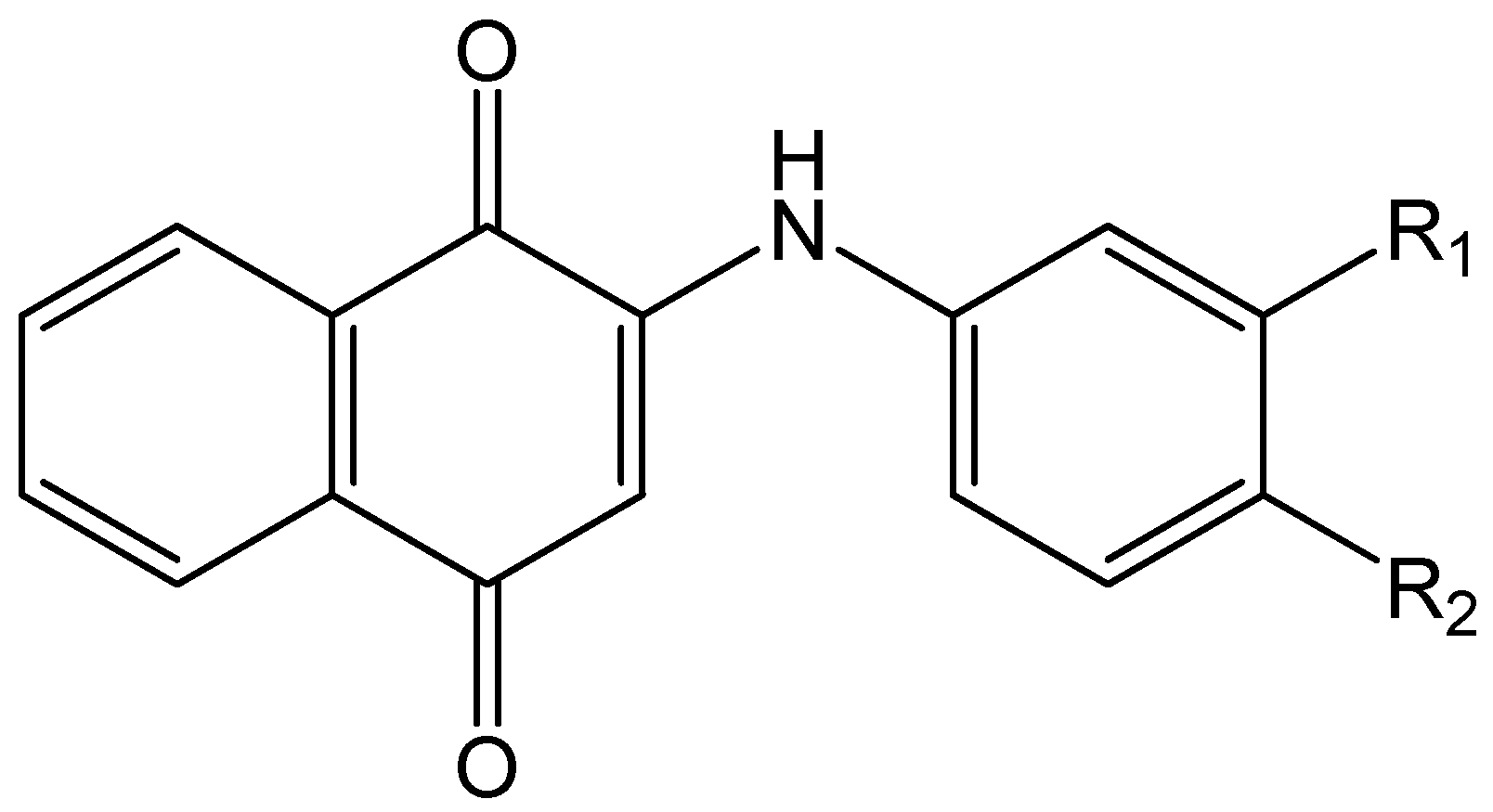
29,30,31]. These compounds, in particular the 2-[(R-phenyl)amine]-1,4-naphthoquinone (PAN) (R = substituent) (1) and its eighteen derivatives were synthesised by a member of our group [29,30] and studied as dyes for their effects on various systems [29,30,31]. These syntheses were designed in order to vary the size of the substituent on the benzene ring and to assure different electronic effects of these substituents (Figure 1).
We decided to undertake further study of this family of compounds using a new approach, namely supramolecular host-guest inclusion, whereby the PAN derivatives will be the guest molecules and enter the internal cavity of cyclodextrin (CD) hosts, in particular β- and γ-CD (α-CD was not used due to its small cavity size relative to PAN). The transport of PAN encapsulated in the CD hosts in biological systems is of significant interest. In their recent studies Gomez-Sandoval [29], Wen [32] and Kobetic [33] studied the host-guest inclusion and the stoichiometry of the resulting complexes of various biologically active compounds. Interestingly, it was found that such complexes often involved a 2:1 host:guest ratio, for example with the antiviral bioactive agent acyclovir [34]. This observation was subsequently related to central issues in molecular recognition and inclusion phenomenon, namely the size-fit and geometric complementarity factors involved in determining the inclusion complex stoichiometry for a specific host-guest pair [35].
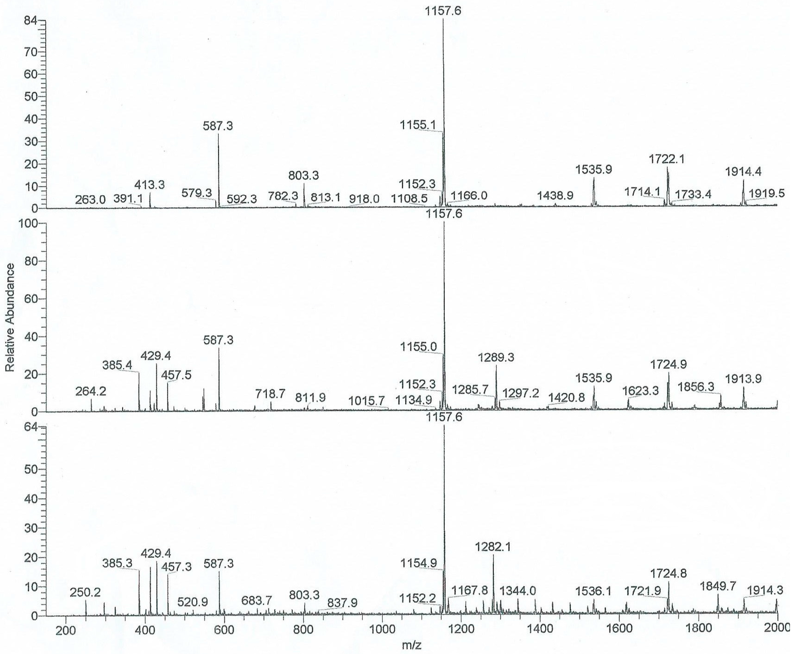
For this investigation the most important technique for studying the inclusion phenomena was ESI mass spectroscopy [36,37,38], which is a relatively straightforward and rapid technique, which directly and rapidly determines the CD:guest stoichiometry (from the observed mass of the complexes), without using complex analysis and fitting techniques, as are required for example for microcalorimetry or fluorescence spectroscopy. We [39] and others [40] have recently studied, using ESI-MS, the use of various CD hosts, of different sizes, in the context of the transport of drugs through the digestive system [39] or to encapsulate drugs such as analgesics [40]. We have also previously used ESI mass spectroscopy, in combination with fluorescence spectroscopy, to study and characterize the binding of the fluorescent probe Nile Red in various cyclodextrins [
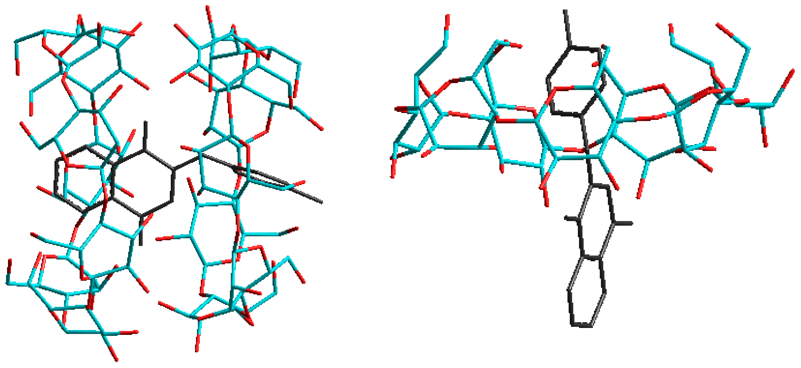
41]. In this case the formation in solution of CD: guest complexes at 1:1 or 2:1 could easily be detected. In parallel, we use molecular modelling, which has been shown to produce quite reliable data for the stability of such complexes [32,39,41,42,43,44], and also allows for the calculation of the energies of potential 1:1 or 2:1 host:guest complexes.
The relative abundance of complexes with varying stoichiometries in the ESI-MS experiments is directly related to the energy and thus stability of these complexes. Wen et al. [32] calculated the energies of such complexes in the case of the malarial drug chinchonine using molecular modeling, to explain their ESI-MS results. We will use a similar approach in this work; however, we were also fascinated by the observed persistence of 2:1 complexes in the case of certain derivative guests, and their rather unusual behaviour under ESI conditions, and the reasons why some substituted PAN derivatives showed 2:1 complexation in the ESI-MS results, but the majority did not. In order to follow this trend we used in our ESI experiments the condition of excess of cyclodextrin in order to assure the formation and observation of only the most stable multi-cyclodextrin adduct ions. When the Wen group used protected CDs [32], many unexpected reactions took place, for example dealkylation or sharing of guests between cyclodextrin hosts. For this reason we preferred, as suggested by Kobetic [33], using the “native” CDs of differing size, namely β- and γ-CD, which have an appropriate cavity size for these guests. Furthermore, we decided to perform molecular modelling studies of the 1:1 and 2:1 host:guest complexes, with the goal of observing some significant differences justifying the stability of some of these complexes and the absence of others. Overall, our interest in unusual, uncommon or simply strange structures, proposed for ion structures observed under ESI conditions, was undertaken to rationalise the observed complexes using the thermodynamic calculations via molecular modelling (MM+ in particular as a first step) to develop a hypothesis for their formation and in particular to explain the unusual stability in solution of some of these complexes.
We present herein the comparison of the data obtained from the mass spectrometry of the 19 PAN dyes (shown in Figure 1 and Table 1) and two CDs, namely β- and γ-CD, and the corresponding data of calculated energies for 1:1 and 2:1 complexes obtained via molecular modelling. The experimental and calculated data and results will be discussed in the following sections. Surprisingly, the 1:1 complexes between PAN and CD are not easily formed under the standard experimental conditions for ESI/PI (methanol, drop of formic acid, optimized ionization energy to observed the complex ions), and were only observed at a relatively high excess of CD. As will be discussed below, under such excess CD conditions, 1:1 and in the case of some host-guest pairs 2:1 host:guest complexes were observed.

2. Results

2.1. Electrospray Ionization Mass Spectroscopy (ESI-MS) Studies

ESI-MS experiments were performed for all 19 guests in both β- and γ-CD; the results of these experiments in terms of the observation of host:guest inclusion complexes, and their stoichiometry, are presented in Table 1. Representative ESI-MS spectra for the host β-CD itself, as well as the two specific host-guest pairs involving PAN derivative 10 (2-[(p-methyl-phenyl)amine]-1,4-naphthalenedione) or the parent PAN 1 both in the presence of β-CD are shown in Figure 2. In all 38 cases, host-guest inclusion was observed. In the majority of these 38 pair combinations, only 1:1 host:guest complexes were observed (32 out of 38 cases). Only in a total of six cases were 2:1 host:guest complexes observed, four of them for β-CD (namely m-CH3, p-CH3, m-ethyl and un-substituted PAN, two of which are shown in Figure 2) and two for γ-CD (namely m-CH3 and p-CH3). This lack of higher-order complexes in all but six CD:PAN pairs was reproducible and clearly established by these experiments.
In all six cases in which 2:1 host:guest inclusion complexes were observed, 1:1 complexes were also observed to varying relative concentrations. This indicates a stepwise equilibrium formation of these 2:1 complexes, as follows:
CD + PAN ⇆ {CD:PAN}
CD + {CD:PAN} ⇆ {CD2:PAN}
with equilibrium binding constants K1 and K2 for Equilibria 1 and 2, respectively. The magnitude of the binding (formation) constants K1 and K2 cannot be determined from ESI-MS experiments, only the presence (or absence) of these complexes.
The ESI-MS experiments provide direct and unequivocal determination of not only the formation of inclusion complexes but also the varying host:guest ratios (stoichiometries) observed. However, no experimental information is provided by this technique on the nature of these complexes (nor the magnitude of the binding constants, as discussed above), for example the specific orientation in which all or part of the PAN guest fits inside the CD cavity. In the case of PAN, as shown in Figure 1, it is possible to form 1:1 complexes by inclusion by the CD of either the anilino or quinone moiety ends. Complexation of either of these anilino or quinine complexes by a second host CD would then result in the same 2:1 complex formed. This results in a somewhat complex mechanism for the stepwise formation of the 2:1 complexes from initial 1:1 complexes as proposed above, due to the possibility of two different 1:1 intermediate complexes. This full set of equilibria is illustrated in Figure 3; in this case two different 1:1 binding constants, K1 and K1’ (as indicated by Equilibrium 1 above), are needed, for the formation of anilino or quinone complexes, respectively, and also two pairs of 2:1 binding constants, K2 and K2’ (as indicted by Equilibrium 2), for the addition of the second host CD to either the anilino or quinone 1:1 complexes, respectively.
Considering the shape of the PAN guests, as shown in Figure 1, it is most likely that the phenyl ring of the aniline moiety is being included in the 1:1 complexes, due to the large steric hindrance caused by the two carbonyls on the quinone moiety. In other words, K1 is expected to be larger than K1’. This preference of inclusion of the anilino moiety over the quinone end is also supported by our molecular mechanics calculations, as will be discussed in Section 2.2.
It is observed that the formation of 2:1 complexes is more likely with β-CD (occurs with four PAN derivative guests) than with γ-CD (occurs only with two PAN derivative guests). This makes sense, considering the relative size of these two hosts: γ-CD is much larger than β-CD, so there is more likely to be unfavourable steric interaction between the two hosts in the case of γ-CD, making the encapsulation of the guest by a second host less likely. Furthermore, the cavity of γ-CD is larger than that of β-CD, making it more likely that the guest can be completely encapsulated within a single host cavity. The reason why only certain guests are encapsulated by β- and γ-CD will be discussed in Section 3, by considering the electronic properties of these different guests.

2.2. Molecular Modeling Studies

Molecular modeling calculations were performed for all 19 PAN derivatives as guests encapsulated in the β-CD, to estimate the stabilization energy obtained by complexation (which provides an indication of the relative binding constants). Both modes of encapsulation for 1:1 inclusion were examined and compared, i.e., encapsulation of the anilino moiety (anilino complex) vs. encapsulation of the quinone moiety (quinone complex). Table 2 lists the results of the molecular modeling calculations for the stability of each of the two types of 1:1 complex with β-CD for each of the 19 guests, in terms of ΔE, the difference in energy between the complex and the free host and guest. Also listed is the value of ΔE for the formation of the 2:1 complex, in which both ends of the guest PAN are encapsulated by a β-CD host.
As can be seen from Table 2, in most cases there is a greater stabilization (more negative ΔE) for anilino end encapsulation than for quinone end encapsulation, namely in 14 of the 19 cases (only PAN guests 1, 2, 3 and 4 showed a lower energy for the quinone mode of encapsulation, while PAN 16 showed identical energy for the two modes). This provides support for our proposal in Section 2.1 that the 1:1 complexes most likely involve inclusion of the anilino end of the PAN guest.
Since only PAN derivatives 1, 4, 7 and 10 showed a 2:1 complex under ESI conditions, we further investigated the results indicated in Table 2 to understand why only 1:1 complexes were present for the other PAN derivatives. The PAN derivatives 1, 4, 7 and 10 contain a relatively small substituent (H, m-Et, m-Me and p-Me respectively) on the aniline end of the molecule. Those four substituents being very small, they have little steric effect on the stability of the complexation. They also do not cause strong electronic effects, since they are only very weakly electron donating (the electronic effects will be discussed in Section 3). Since no considerable steric or electronic effects are present, these 2:1 complexes are stable and are observed in the ESI spectra (except for derivative 9 with a p-Et group). Figure 4 shows the structures calculated via molecular modeling for the 2:1 as well as the 1:1 anilino complex of β-CD with PAN derivative 10 (p-methyl). This figure illlustrates the minimal steric interaction between the CD cavity and the small p-methyl substituent on the anilino ring, and the deep anilino penetration into the CD cavity, in both cases.
PAN derivatives 5 and 6 (p-n-Bu and p-n-Hexyl respectively) did not show a 2:1 complex since the n-butyl and n-hexyl groups are quite bulky and under the energy of the ESI, the complexation on the aniline side of the molecule would be lost.
For the other PAN derivatives that did not show a 2:1 complex in ESI, we examined the electronic effects of these substituents since none of them is really bulky enough to cause a significant steric effect. For all the other PAN derivatives (2, 11, 15 and 18) with meta substituents (m-NO2, m-CO2H, m-Cl, m-F), the energy difference between the 1:1 quinone and 1:1 anilino complexes are relatively identical (see Table 2). In this case, under ESI energy if one of the β-CD is lost it is difficult to determine which of the two complexations will be lost.
This leaves PAN derivatives 3, 8, 12, 13, 14, 16, 17, and 19. All of these derivatives have a para substituent. Comparing derivatives 16 and 18 (p-Cl and p-Br respectively) shows that the 1:1 anilino complexation for p-Br (derivative 17) is favored. This is due to the inductive effect of the p-Br which forces the nitrogen atom of the aniline group to push its electronic density into the aromatic. This further stabilizes the aniline moiety versus the quinone. Derivatives 8, 12, 13, 14 and 19 (p-NO2, p-CO2H, p-CN, p-CF3 and p-COCH3), all show the same trend since these groups are electron withdrawing and also force the nitrogen atom to donate its electronic density to the aniline side of the molecule. Derivative 3 (p-OMe) shows the exact opposite effect since it is an electron donating group. The electronic density of this group forces the nitrogen atom to push its electronic density into the quinone group. In this case, the 1:1 quinone complexation will be favored.
Thus, when a derivative does not show a 2:1 complexation in the ESI, the absence of these 2:1 complex are explained by a combination steric and electronic effects. Furthermore, the preference for either the 1:1 quinone or 1:1 anilino is correlated by the difference of energy from Table 2.

3. Discussion

From the molecular modeling studies described above, it is clear that the electron withdrawing/donating abilities of the various substituents on this set of guests plays a significant role in the nature of the inclusion complexes obtained. This effect can of course be investigated quantitatively using the well-known set of Hammett σm and σp substituent constants, which have been determined and tabulated based on the effect of meta or para substitution of a substituent the acid equilibrium constant of benzoic acid [45,46]. These parameters take into account the inductive and resonance effect of the substituent on the electron distribution of the aromatic ring(s). In general, substituents can be considered as either electron-donating groups (increased aromatic electron density) or electron-withdrawing groups (decreased aromatic electron density). Electron-donating groups have negative σm and σp substituent constant values, whereas electron-withdrawing groups have positive σm and σp substituent constant values.
It is clear from Table 3 that, other than the unsubstituted PAN itself, only PAN derivatives with substituents with negative σm values, i.e., with electron donating groups, exhibit 2:1 CD:PAN complexes with β-CD in the ESI-MS spectrum. No PAN derivative with a meta substituent with a positive σm forms 2:1 complexes with β-CD. A similar result is observed for para-substituted PAN derivatives with β-CD, as shown in Table 4: the only ones that form 2:1 complexes are those with substituents with negative σp values, i.e., with electron donating groups. Once again, no PAN derivative with a para substituent with a positive σp forms 2:1 complexes with β-CD. Thus, a relatively high aromatic electron density is required for the formation of 2:1 complexes, which is supported by the presence of electron-donating substituents. However, not all derivatives with negative σm meta substituents show 2:1 complexes: ethyl, n-butyl, and n-hexyl all have negative values but do not form 2:1 complexes. In the latter two cases, the steric effect of these larger alkyl substituents must play a role. In the case of ethyl substitution, the steric effect is seen to be intermediate between the small methyl group and the larger n-butyl, and n-hexyl groups: ethyl does form 2:1 complexes with β-CD when substituted in the meta position, but not in the para. It is interesting and informative to note that the only substituent which gave 2:1 complexes in all four cases (meta and para substitution, β- and γ-CD) was the smallest alkyl substituent, the methyl group.
Table 5 and Table 6 show the analogous results in the case of the larger γ-CD; in this case, only methyl substituents show 2:1 complexes. As discussed in Section 2.1 above, it is less likely that 2:1 host:guest complexes will form with the much larger γ-CD, as there would be much more steric interference between the two hosts encapsulating the same PAN guest. In this case, the electron donating requirement of the guest is more important, to help overcome the host steric effect, as evidenced by the fact that the unsubstituted PAN does not form 2:1 complexes (contrary to the result in β-CD). It is, however, surprising that the larger alkyl substituents do not encourage 2:1 complexation, as a larger guest would be more likely to be encapsulated by two hosts.
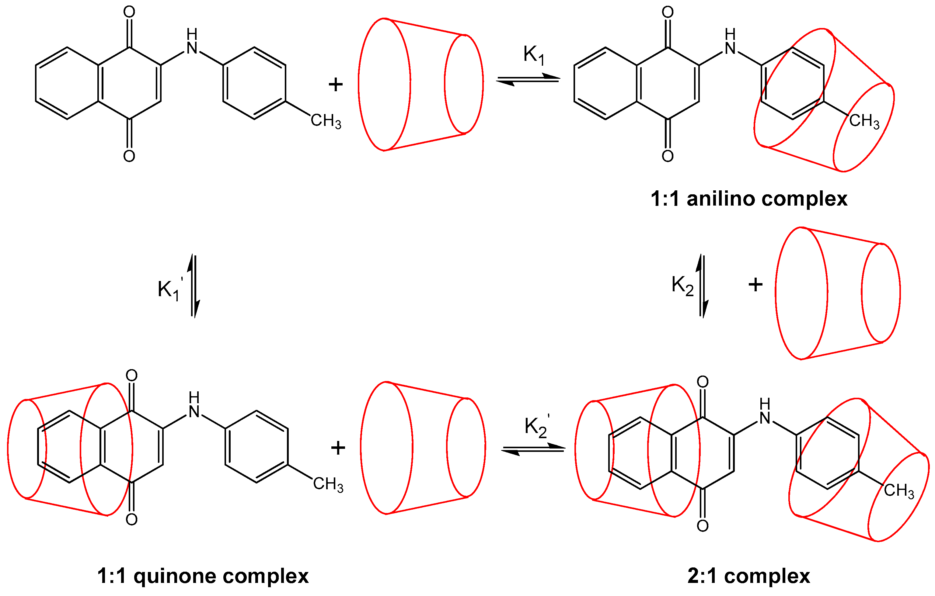
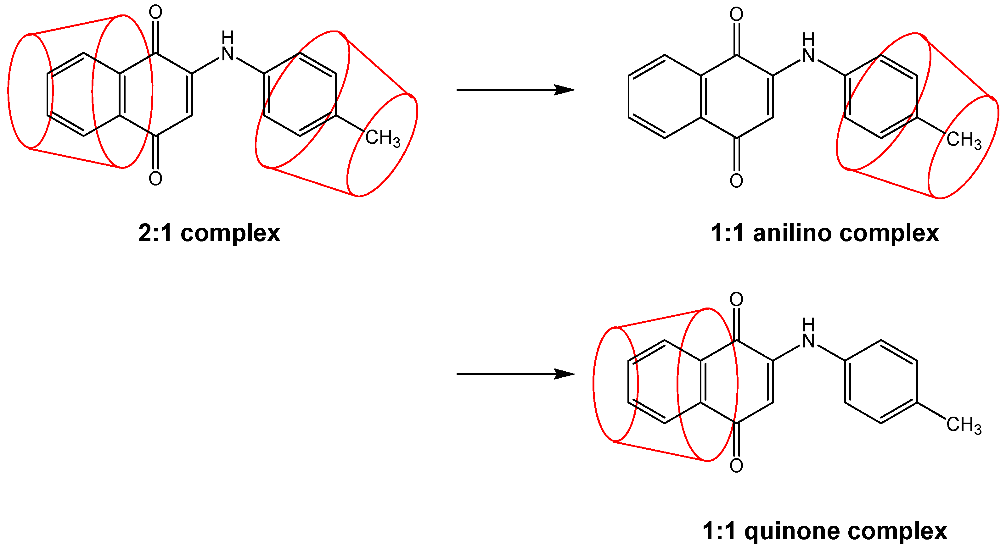
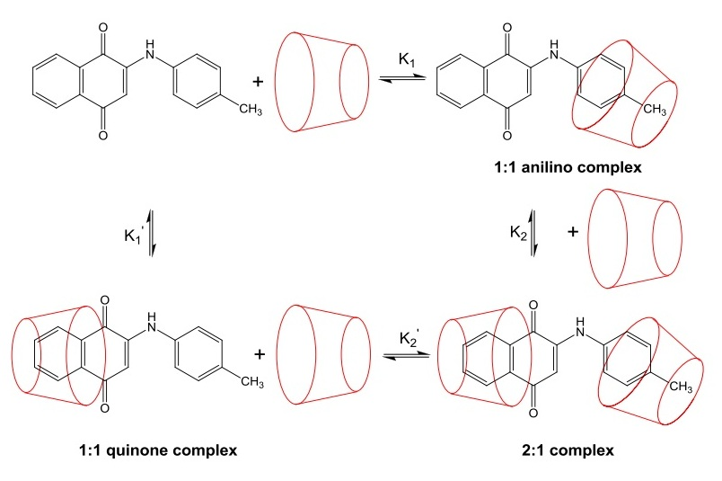
It should be mentioned that in the ESI-MS experiments, only complexes which survive the ionization process are observed. Thus, there is a small possibility that 2:1 complexes form in most cases (especially considering the experimental conditions of a large excess of cyclodextrin), but that such complexes fall apart under MS-ESI conditions for all but a few of the PAN derivatives, resulting in only 1:1 complexes, either anilino (most likely), quinone, or both, being observed in the ESI experiments. This possibility is depicted in Figure 5.

4. Materials and Methods

PAN derivatives 119 were synthesized as previously described [28]. β- and γ-CD were obtained from Sigma-Aldrich (St. Louis, MO, USA), and used as received.
Electrospray ionization mass spectroscopy (ESI-MS) experiments were performed on a Quattro II Micromass (Waters, Elstree, Hertfordshire, UK) spectrometer (CEN de Saclay, Gif-sur-Yvette, France). Solutions contained 10−6 to 10−7 M of the PAN derivative, with a large (three- to ten-fold) excess of cyclodextrin). Samples were injected at 10 mL/min, with a source temperature of 80 °C and capillary voltage maintained at +3.35 KV. The cone voltage ranged from 10–70 V; at 20 V the skimmer voltage was 1.9 V. In a typical experimental setting, 100 μL of an ethanol solution of the CD and PAN (10:1 mole ratio; large excess of CD used to allow for formation of higher-order host:guest complexes ) was prepared, in some cases a drop of formic acid was added, and introduced through a Harvard Apparatus syringe pump. The ions were detected by scanning the first quadrupole and the mass range was monitored from m/z = 80–2000 in 7 s. At least 50 scans were averaged to obtain representative spectra.
Molecular modeling calculations were performed on HyperChem 6 MM+ (U de M) in gas phase and in solvent box (ethanol) modes. The minimization of energy was performed using the technique described in the HyperChem instruction manual, and in references [39] and [41]. Energies obtained were minimized potential energies as defined in the HyperChem 6 MM+ approach, and include conformational motions for the obtaining the lowest energy complex configuration. These calculations were done in the same way across the entire set of host:guest pairs, allowing for relatiave stability comparisons to be made. In a first phase, 1600 Polak Ribiere iterations were applied. A small incremental energy was applied in a second phase (0.001 ps for a total of 1 ps, 370 K) for selected insertion models only. All calculated complex structures (1:1, 2:1 and 1:2 stoichiometry) and energy data are available upon request from C.K. Jankowski (U de M).

5. Conclusions

2-[(R-phenyl)amine]-1,4-naphthoquinones (PAN) form host-guest inclusion complexes with both β- and γ-CD, as observed by ESI-MS experiments. For the majority of the 19 PAN derivatives studied in these two CDs, only 1:1 CD:PAN complexes were observed (32 out of 38 host:guest pairs). In six cases, 2:1 host:guest complexes were also observed: 4 cases for β-CD (parent PAN, m-methyl, m-ethyl, and p-methyl) and 2 cases for the larger γ-CD (m- and p-methyl). Thus, the specific nature of the PAN derivative, and the effect of the specific substituents, has a major effect on determining the resulting host-guest stoichiometry, as does the size of the host CD cavity. Molecular modeling studies showed that 1:1 complexation most likely involves encapsulation of the aniline rather than the quinone moiety, and indicated large stabilization upon 2:1 complexation. Consideration of the Hammett parameters of the substituents showed that 2:1 host-guest complexes formed only with PAN derivatives with small substituents with negative σm or σp values, i.e., for guests with electron donating substituents. This trend indicates that higher electron density on the aromatic ring encourages complexation by 2 host molecules. However, larger alkyl substituents with negative σm and σp parameters did not yield 2:1 complexes, even for the larger γ-CD host, showing that steric effects in terms of guest size are also highly important. It is clear that the prediction of host-guest stoichiometry for a specific host-guest pair is complicated, and involves a subtle interplay of both electronic and steric factors. However, as demonstrated in this paper, there are trends in terms of the electronic nature of the guest and the size of both the guest and host, which can be used to help predict the expected host:guest stoichiometry for a given host-guest pair.

Acknowledgments

Financial support for this work was provided by the Natural Sciences and Engineering Research Council of Canada (NSERC), by the Faculty of Research and Graduate Studies at the Université de Moncton, and by the Office of Research at the University of Prince Edward Island. M. Jiménez-Estrada would like to thank Drs. Martha A. Martinez and Norma M. Rubalcava, Faculdad de Quimica, Universidad Nacional Autónoma de México, for help with the synthesis of some of the guest compounds.

Author Contributions

B.D.W., C.K.J. and M.J.-E. conceived this project, analysed the results and wrote the manuscript. M.J.-E prepared most of the PAN compounds used, C.L. recorded the ESI mass spectra and S.A. and C.K.J. performed the molecular modelling and computational studies.

Conflicts of Interest

The authors declare no conflict of interest.

References

  1. Szejtli, J. Introduction and general overview of cyclodextrin chemistry. Chem. Rev. 1998, 98, 1743–1753. [Google Scholar] [CrossRef] [PubMed]
  2. Del Valle, E.M.M. Cyclodextrins and their uses: A review. Proc. Biochem. 2004, 39, 1033–1046. [Google Scholar] [CrossRef]
  3. Crini, G. Review: A history of cyclodextrins. Chem. Rev. 2014, 114, 10940–10975. [Google Scholar] [CrossRef] [PubMed]
  4. Süle, A.; Szente, L.; Csempesz, F. Enhancement of drug solubility in supramolecular and colloidal systems. J. Pharm. Sci. 2009, 98, 484–494. [Google Scholar] [CrossRef] [PubMed]
  5. Wagner, B.D. The effects of cyclodextrins on guest fluorescence. In Cyclodextrin Photochemistry, Photophysics and Photobiology; Douhal, A., Ed.; Elsevier B.V.: Amsterdam, The Netherlands, 2006; pp. 27–59. [Google Scholar]
  6. Marques, H.M.C. A review on cyclodextrin encapsulation of essential oils and volatiles. Flavour Fragr. J. 2010, 25, 313–326. [Google Scholar] [CrossRef]
  7. Oros, G.; Cserháti, T.; Szőgyi, M. Cyclodextrins in chromotography. Part 1. Liquid chromotographic methods. Eur. Chem. Bull. 2013, 2, 920–926. [Google Scholar]
  8. Wagner, B.D. Recent applications of host-guest inclusion in fluorescence-based trace analysis. Curr. Anal. Chem. 2007, 3, 183–195. [Google Scholar] [CrossRef]
  9. Li, J.; Loh, X.J. Cyclodextrin-based supramolecular architectures: Synthesis, structures, and applications for drug and gene delivery. Adv. Drug Deliv. Rev. 2008, 60, 1000–1017. [Google Scholar] [CrossRef] [PubMed]
  10. Hedges, A.R. Industrial applications of cyclodextrins. Chem. Rev. 1998, 98, 2035–2044. [Google Scholar] [CrossRef] [PubMed]
  11. Yorozu, T.; Hoshino, M.; Imamura, M. Fluorescence studies of pyrene inclusion complexes with α-, β- and γ-cyclodextrins in aqueous solutions. Evidence for formation of pyrene dimer in γ-cyclodextrin cavity. J. Phys. Chem. 1982, 86, 4426–4429. [Google Scholar] [CrossRef]
  12. Ali, S.M.; Shamim, S. Analysis of computational models of β-cyclodextrin complexes: Structural studies of morniflumate hydrochloride and β-cyclodextrin complex in aqueous solution by quantitative ROESY analysis. J. Incl. Phenom. Macrocycl. Chem. 2015, 83, 19–26. [Google Scholar] [CrossRef]
  13. Sau, S.; Solanki, B.; Oprecio, R.; van Stam, J.; Evans, C.H. Higher-order cyclodextrin complexes: The naphthalene system. J. Incl. Phenom. Macrocycl. Chem. 2004, 48, 173–180. [Google Scholar] [CrossRef]
  14. Christoforides, E.; Mentzafos, D.; Bethanis, K. Structural studies of the inclusion complexes of the (+)- and (−)-borneol enantiomers in α- and β-cyclodextrin. J. Incl. Phenom. Macrocycl. Chem. 2015, 81, 193–203. [Google Scholar] [CrossRef]
  15. Chatziefthimiou, S.D.; Yannakopoulou, K.; Mavridis, I.M. β-Cyclodextrin trimers enclosing an unusual organization of guest: The inclusion complex β-cyclodextrin/4-pyridinealdazine. CrystEngComm 2007, 9, 976–979. [Google Scholar] [CrossRef]
  16. Caira, M.R.; Dodds, D.R.; Nassimbeni, L.R. Diverse modes of guest inclusion in a cyclodextrin: X-ray structurual and thermal characterization of a 4:3 β-cyclodextrin-cyclizine complex. Supramol. Chem. 2001, 13, 61–70. [Google Scholar] [CrossRef]
  17. Liu, Y.; Zhong, R.-Q.; Zhang, H.-Y.; Song, H.-B. A unique tetramer of 4:5 β-cyclodxtrin-ferrocene in the solid state. Chem. Commun. 2005, 2211–2213. [Google Scholar] [CrossRef] [PubMed]
  18. Biernacka, J.; Betlejewska-Kielak, K.; Wiltowska-Jarosz, J.; Klosińska-Szmurlo, E.; Mazurek, A.P. Mass spectrometry and molecular modeling studies on the inclusion complexes between alendronate and β-cyclodextrin. J. Incl. Phenom. Macrocycl. Chem. 2014, 78, 437–443. [Google Scholar] [CrossRef] [PubMed]
  19. Gurarslan, A.; Shen, J.; Caydamli, Y.; Tonelli, A.E. Pyripoxyfen cyclodextrin inclusion compounds. J. Incl. Phenom. Macrocycl. Chem. 2015, 82, 489–496. [Google Scholar] [CrossRef]
  20. Morton, R.A. Biochemistry of Quinones; Acad. Press: New York, NY, USA, 1965. [Google Scholar]
  21. Bentley, R.; Campbell, I.M. Biological reactions of quinones. In Quinoid Compounds; Patai, S., Ed.; John Wiley: Chichester, UK, 1974; Volume 2, pp. 683–736. [Google Scholar]
  22. Huntington, J.L.; Davis, D.G. The effect of molecular structure on the electrode kinetics of aminoquinones and quinone thioethers. J. Electrochem. Soc. 1971, 118, 57–63. [Google Scholar] [CrossRef]
  23. Li, C.Y.; Caspar, M.L.; Dixon, D.W., Jr. The effects of substitution on the electrochemical reduction of dihydroxynaphthaceneqinone in N,N-dimethylformamide. Electrochim. Acta 1980, 25, 1135–1142. [Google Scholar] [CrossRef]
  24. Qureshi, G.A. The correlation between the polarographic and structural parameters of alizarin and its substituents. Bull. Soc. Chim. Belg. 1981, 90, 223–227. [Google Scholar] [CrossRef]
  25. Stradins, J.; Glezer, V.; Turovska, B. Electrochemical reduction of N-aryl- and N-arylsulphonylbenzoquinoneimines in acetonitrile—II. Electron affinity and relationship between oxidation and reduction potentials. Electrochim. Acta 1991, 36, 1219–1225. [Google Scholar] [CrossRef]
  26. Glezer, V.; Turovska, B.; Stradins, J.; Friemanis, J. Electrochemical reduction of N-aryl- and N-arylsulphonylbenzoquinonemonoimines in acetonitrile—I. Electroreduction mechanism and effects of proton donors on electrochemical process. Electrochim. Acta 1990, 35, 1933–1940. [Google Scholar] [CrossRef]
  27. Clark, N.G. The fungicidal activity of substituted 1,4-naphthoquininones. Part III: Amino, anilino and acylcamino derivatives. Pestic. Sci. 1985, 16, 23–32. [Google Scholar] [CrossRef]
  28. Kapadia, G.J.; Azuine, M.A.; Balasubramanian, V.; Sridhar, R. Aminonaphthoquinones—A Novel Class of Compounds with Potent Antimalarial Activity against Plasmodium Falciparum. Pharmacol. Res. 2001, 43, 363–367. [Google Scholar] [CrossRef] [PubMed]
  29. Gómez-Sandoval, Z.; Calaminici, P.; Köster, A.M.; Lotina-Hennsen, B.; King-Díaz, B.; Macías-Ruvalcaba, N.; Aguilar-Martínez, M.; Jiménez-Estrada, M. Density functional study of 2-[(R-phenyl)amine]-1,4-naphtalenediones. J. Chem. Theory Comput. 2007, 3, 894–904. [Google Scholar] [CrossRef] [PubMed]
  30. Aguilar-Martínez, M.; Cuevas, G.; Jiménez-Estrada, M.; González, I.; Lotina-Hennsen, B.; Macías-Ruvalcaba, N. An experimental and theoretical study of the substituent effects on the redox properties of 2-[(R-phenyl)amine]-1,4-naphtalenediones in acetonitrile. J. Org. Chem. 1999, 64, 3684–3694. [Google Scholar] [CrossRef] [PubMed]
  31. King-Díaz, B.; Macías-Ruvalcaba, N.A.; Aguilar-Martínez, M.; Calaminici, P.; Köster, A.M.; Gómez-Sandoval, Z.; Reveles, J.U.; Lotine-Hennsen, B. 2-[(R-phenyl)amine]-1,4-naphtalenediones as Photosystem I electron acceptors. Structure-activity Relationships of m- and p-PAN compounds with QSAR Analysis. J. Photochem. Photobiol. B Biol. 2006, 83, 105–113. [Google Scholar] [CrossRef] [PubMed]
  32. Wen, X.; Liu, Z.; Zhu, T.; Zhu, M.; Jiang, K.; Huang, Q. Evidence for the 2:1 molecular recognition and inclusion behaviour between β- and γ-cyclodextrins and cinchonine. Bioorg. Chem. 2004, 32, 223–233. [Google Scholar] [CrossRef] [PubMed]
  33. Kobetić, R.; Jursic, B.S.; Bonnette, S.; Tsai, J.S.-C.; Savatore, S.J. Study of the lorazepam:cyclodextrin inclusion complexes using electrospray ionization mass spectrometry. Tet. Lett. 2001, 42, 6077–6082. [Google Scholar] [CrossRef]
  34. Dubes, A.; Degobert, G.; Fessi, H.; Parrot-Lopez, H. Synthesis and characterisation of sulfated amphiphilic α-, β- and γ-cyclodextrins: Application to the complexation of acyclovir. Carbohydr. Res. 2003, 338, 2185–2193. [Google Scholar] [CrossRef]
  35. Liu, Y.; Li, L.; Zhang, H.-Y.; Fan, Z.; Guan, X.-D. Selective binding of chiral molecules of cinchona alkaloid by β- and γ-cyclodextrins and organoselenium-bridged bis(β-cyclodextrin)s. Bioorg. Chem. 2003, 31, 11–23. [Google Scholar] [CrossRef]
  36. Schalley, C.A. Supramolecular chemistry goes gas phase: The mass spectrometric examination of noncovalent interactions in host-guest chemistry and molecular recognition. Int. J. Mass Spectrom. 2000, 194, 11–39. [Google Scholar] [CrossRef]
  37. Gabelica, V.; Galic, N.; Rosu, F.; Houssier, C.; de Pauw, E. Influence of response factors on determining equilibrium association constants of non-covalent complexes by electrospray mass spectrometry. J. Mass Spectrom. 2003, 38, 491–501. [Google Scholar] [CrossRef] [PubMed]
  38. Gabelica, V.; Galic, N.; de Pauw, E. On the specificity of cyclodextrin complexes detected by electrospray mass spectrometry. J. Am. Soc. Mass Spectrom. 2002, 13, 946–953. [Google Scholar] [CrossRef]
  39. Jankowska, A.; Jankowski, C.K.; Chiasson, J.B. On chloralose-cyclodextrin complexes by ESI-mass spectrometry. J. Incl. Phenom. Macrocycl. Chem. 2005, 52, 213–221. [Google Scholar] [CrossRef]
  40. Guo, M.Q.; Zhang, S.; Song, F.; Wang, D.; Liu, Z.; Liu, S. Studies on the non-covalent complexes between oleanic acid and cyclodextrins using electrospray ionization tandem mass spectrometry. J. Mass Spectrom. 2003, 38, 723–731. [Google Scholar] [CrossRef] [PubMed]
  41. Wagner, B.D.; Stojanovic, N.; LeClair, G.; Jankowski, C.K. A spectroscopic and molecular modeling study of the nature of the association complexes of Nile Red with cyclodextrins. J. Incl. Phenom. Macrocycl. Chem. 2003, 45, 275–283. [Google Scholar] [CrossRef]
  42. Göschl, M.; Crouzy, S.I.; Chapron, Y. Molecular dynamics study of an α-cyclodxtrin-phosphatidylinosiol inclusion complex. Eur. Biophys. J. 1996, 24, 300–310. [Google Scholar] [CrossRef] [PubMed]
  43. Ali, S.M.; Falma, K.; Dhokale, S. Structure elucidation of β-cyclodextrin-xylazine complex by a combination of quantitative 1H-1H ROESY and molecular dynamics studies. Beilstein J. Org. Chem. 2013, 9, 1917–1924. [Google Scholar] [CrossRef] [PubMed]
  44. Jana, M.; Bandyopadhyay, S. Molecular dynamics study of β-cyclodextrin-phenylalanine (1:1) inclusion complex in aqueous medium. J. Phys. Chem. B 2013, 117, 9280–9287. [Google Scholar] [CrossRef] [PubMed]
  45. Hansch, C.; Leo, A.; Taft, R.W. A Survey of Hammett Substituent Constants and Resonance and Field Parameters. Chem. Rev. 1991, 91, 165–195. [Google Scholar] [CrossRef]
  46. Williams, A. Free Energy Relationships in Organic and Bio-Organic Chemistry; Royal Society of London: Cambridge, UK, 2003; Appendix 3; pp. 258–277. [Google Scholar]
  • Sample Availability: Samples of the compounds 119 are available from MJE.
Figure 1. General structure of the 2-[(R-phenyl)amine]-1,4-naphthalenedione guests of interest. The list of the 19 specific guests studied and their R1 and R2 substituents is given in Table 1.
Molecules 21 01568 g001
Figure 2. ESI-MS spectra of 2-[(p-methyl-phenyl)amine]-1,4-naphthalenedione and β-CD. Upper spectrum: β-CD control: MNa+ ion at m/z 1157.6; Middle spectrum: β-CD (2 × 10⁻⁵ M) complexed with PAN 10 (2 × 10⁻⁶ M), ratio 10:1; 2 β-CD + PAN 10 ion at m/z 1289.2 (MNa2)2+; PAN 10 MH+ ion at m/z 264.2; β-CD MNa+ ion at m/z 1157.6; Bottom Spectrum: β-CD (2 × 10⁻⁵ M) complex with PAN 1 (2 × 10⁻⁶ M), ratio 10:1; 2 β-CD + PAN 1 ion at m/z 1282.1 (MNa2)2+; PAN 1 MH+ ion at m/z 250.2; β-CD MNa+ ion at m/z 1157.6. When the MS/MS experiments were performed on (CD2MNa2)2+ ion formation of daughter ions at (CDMNa)+ and (CD2MH)+ were not observed; the only daughter ions recorded were (CDNa)+ and MH+.
Molecules 21 01568 g002
Figure 3. Illustration of the full proposed mechanism for the reversible stepwise formation of the 1:1 and 2:1 host:guest inclusion complexes between β-CD and 2-[(p-methyl-phenyl)amine]-1,4-naphthalenedione (PAN derivative 10) observed in the ESI-MS spectrum shown in Figure 2.
Molecules 21 01568 g003
Figure 4. Molecular modeling predicted energy structures for the 2:1 (left) and 1:1 anilino (right) host:guest complexes formed between β-CD and 2-[(p-methyl-phenyl)amine]-1,4-naphthalenedione (PAN derivative 1) and observed in the ESI-MS spectrum shown in Figure 2.
Molecules 21 01568 g004
Figure 5. Hypothesis for the lack of observed 2:1 complexes in the case of most PAN derivatives: loss of one CD host to form 1:1 complexes, either anilino or quinone (consistent with the overall scheme shown in Figure 3).
Molecules 21 01568 g005
Table 1. Substituents and substitution pattern for 2-[(R-phenyl)amine]-1,4-naphthalenedione (PAN) derivatives 119, and their CD:PAN complex stoichiometries as observed by MS-ESI spectrometry.
GuestR1 (meta)R2 (para)1:1 β-CD2:1 β-CD1:1 γ-CD2:1 γ-CD
1HH
2NO2H
3HOMe
4EtH
5HnBu
6Hn-hexyl
7MeH
8H NO2
9HEt
10HMe
11CO2HH
12HCO2H
13HCN
14HCF3
15ClH
16HCl
17HBr
18FH
19HCOCH3
Table 2. Molecular modeling calculations for the energy stabilization upon complexation (ΔE, in kcal/mol) for the inclusion of the 19 PAN derivatives, as 1:1 (anilino encapsulation), 1:1 (quinone encapsulation), or 2:1 (both ends encapsulated) complexes.
Guest R1 (meta)R2 (para)ΔE 1:1 (Anilino)ΔE 1:1 (Quinone)ΔE 2:1 (Both Ends)
1HH−19.82−21.77−31.37
2NO2H−20.95−21.51−29.69
3HOMe−19.23−25.16−37.79
4EtH−20.99−22.80−33.73
5HnBu−30.04−21.51−48.26
6Hn-hexyl−30.92−24.44−52.03
7MeH−31.09−28.71−40.63
8HNO2−23.44−19.06−34.86
9HEt−22.61−20.61−49.07
10HMe−21.76−20.01−48.72
11CO2HH−23.07−22.41−31.10
12HCO2H−30.22−25.15−42.80
13HCN−24.3−16.43−39.65
14HCF3−19.52−17.64−36.38
15ClH−30.18−27.45−38.21
16HCl−22.27−22.27−27.92
17HBr−30.43−28.66−54.34
18FH−27.23−27.94−44.17
19HCOCH3−23.61−22.16−34.70
Table 3. Comparison of meta-substituted 2-[(R-Phenyl)amine]-1,4-naphthalenediones which form 2:1 and 1:1 vs. only 1:1 complexes with β-CD.
GuestStoichiometryR1 (meta)σm (R1)
11:1 and 2:1H0
41:1 and 2:1Et−0.07
71:1 and 2:1Me−0.06
21:1 onlyNO20.72
111:1 onlyCO2H0.35
151:1 onlyCl0.37
181:1 onlyF0.34
Table 4. Comparison of para-substituted 2-[(R-Phenyl)amine]-1,4-naphthalenediones which show 2:1 and 1:1 vs. only 1:1 complexes with β-CD.
GuestStoichiometryR2 (para)σp (R2)
11:1 and 2:1H0
101:1 and 2:1Me−0.14
31:1 onlyOMe0.27
51:1 onlynBu0.16
61:1 onlyn-hexyl0.16
81:1 onlyNO20.78
91:1 onlyEt0.15
121:1 onlyCO2H0.44
131:1 onlyCN0.66
141:1 onlyCF30.53
161:1 onlyCl0.23
171:1 onlyBr0.23
191:1 onlyCOCH30.50
Table 5. Comparison of meta-substituted 2-[(R-Phenyl)amine]-1,4-naphthalenediones which form 2:1 and 1:1 vs. only 1:1 complexes with γ-CD.
GuestStoichiometryR1 (meta)σm (R1)
71:1 and 2:1Me−0.06
11:1 onlyH0
41:1 onlyEt−0.07
21:1 onlyNO20.72
111:1 onlyCO2H0.35
151:1 onlyCl0.37
181:1 onlyF0.34
Table 6. Comparison of para-substituted 2-[(R-Phenyl)amine]-1,4-naphthalenediones which form 2:1 and 1:1 vs. only 1:1 complexes with γ-CD.
GuestStoichiometryR2 (para)σp (R2)
101:1 and 2:1Me−0.14
11:1 onlyH0
31:1 onlyOMe−0.27
51:1 onlynBu−0.16
61:1 onlyn-hexyl−0.16
81:1 onlyNO20.78
91:1 onlyEt−0.15
121:1 onlyCO2H0.44
131:1 onlyCN0.66
141:1 onlyCF30.53
161:1 onlyCl0.23
171:1 onlyBr0.23
191:1 onlyCOCH30.50

