Article

Anti-Complementary Components of Helicteres angustifolia

1
State Key Laboratory of Natural Medicines, China Pharmaceutical University, Nanjing 210009, China
2
Department of Pharmacognosy, School of Pharmacy, Fudan University, Shanghai 201203, China
*
Author to whom correspondence should be addressed.
Molecules 2016, 21(11), 1506; https://doi.org/10.3390/molecules21111506
Submission received: 11 October 2016 / Revised: 5 November 2016 / Accepted: 6 November 2016 / Published: 10 November 2016

Abstract

:
A first phenalenon derivative with an acetyl side chain at C-8, 8-acetyl-9-hydroxy-3-methoxy-7-methyl-1-phenalenon (compound 1), and a pair of new sesquilignan epimers at C-7″ of hedyotol C and hedyotol D analogs, hedyotol C 7″-O-β-d-glucopyranoside (compound 2) and hedyotol D 7″-O-β-d-glucopyranoside (compound 3) were isolated from the aerial parts of Helicteres angustifolia together with nine known compounds (412). Their structures were elucidated on the basis of spectroscopic methods, including mass spectroscopy, and 1D and 2D nuclear magnetic resonance. Eleven isolates exhibited anti-complementary activity. In particular, compounds 4 and 5 exhibited potent anti-complementary activities against the classical and alternative pathways with CH50 values of 0.040 ± 0.009 and 0.009 ± 0.002 mM, and AP50 values of 0.105 ± 0.015 and 0.021 ± 0.003 mM, respectively. The targets of compounds 4 and 5 in the complement activation cascade were also identified. In conclusion, the anti-complementary components of H. angustifolia possessed chemical diversity and consisted mostly of flavonoids and lignans in this study.

1. Introduction

The complement system, as a chief component of innate immunity, plays a significant role in host immune defense against infection and in the clearance of antigen-antibody complexes from the bloodstream. It can be activated by a cascade mechanism through the classical pathway (CP), alternative pathway (AP) or the lectin pathway [1]. Generally, the normal activation of the complement system leads to elimination of invading pathogens and optimal host response. However, excessive activation of the complement system may induce tissue damage, inflammation and a number of pathological situations such as systemic lupus erythematosus, rheumatoid arthritis and acute respiratory distress syndrome [2,3]. Thus, inhibition of the complement system is potentially therapeutic in diseases resulting from uncontrolled or overshooting complement activation. The desirable properties of a valuable anti-complementary therapeutic agent are that it should be inexpensive and highly specific, either having a long plasma half-life or being active orally [2]. However, none of the currently available agents meet all these criteria. A wide array of specific complement inhibitors, for instance cobra venom factor (CVF), synthetic nafamastat mesilate (FUT-175), a recombinant humanized antibody fragment, and a monoclonal antibody, Eculizumab, have been developed to target various components of the complement cascade [4,5,6,7,8]. However, the clinical development of this plethora of complement therapeutics has exhibited some side effects. Therefore, there is an urgent need to search for naturally occurring and low-toxicity anti-complementary agents from traditional Chinese medicines.
The dried roots or whole plants of Helicteres angustifolia L. (Sterculiaceae) have been used in Chinese folk medicine as anti-inflammatory, antidotal, analgesic, anti-bacterial and anti-cancer agents [9]. Previous phytochemical studies revealed that this plant mainly contains triterpenoids, penylpropanoids, quinones, lignans and flavonoids [10,11,12]. It is commonly accepted that the complement system is a crucial trigger for inflammation [13,14]. Therefore, one may hypothesize that complement inhibition intercepts the process of complement-dependent inflammation diseases. In our effort to search for anti-complementary agents from Chinese herb medicines and to clarify their anti-complementary constituents [3,15,16], an ethanolic extract of the aerial parts of H. angustifolia was found to show a potent anti-complementary activity, which encouraged us to investigate its anti-complementary constituents systematically. In this study, one new skeleton compound (1) and two new compounds (2 and 3) were isolated from the aerial parts of this plant, together with nine known compounds, on the basis of MS, 1D and 2D NMR, as well as comparison with the literature. Herein, the isolation, characterization, and anti-complementary activity of these compounds are reported. In addition, the target identification in the complement activation cascade has been investigated for anti-complementary constituents.

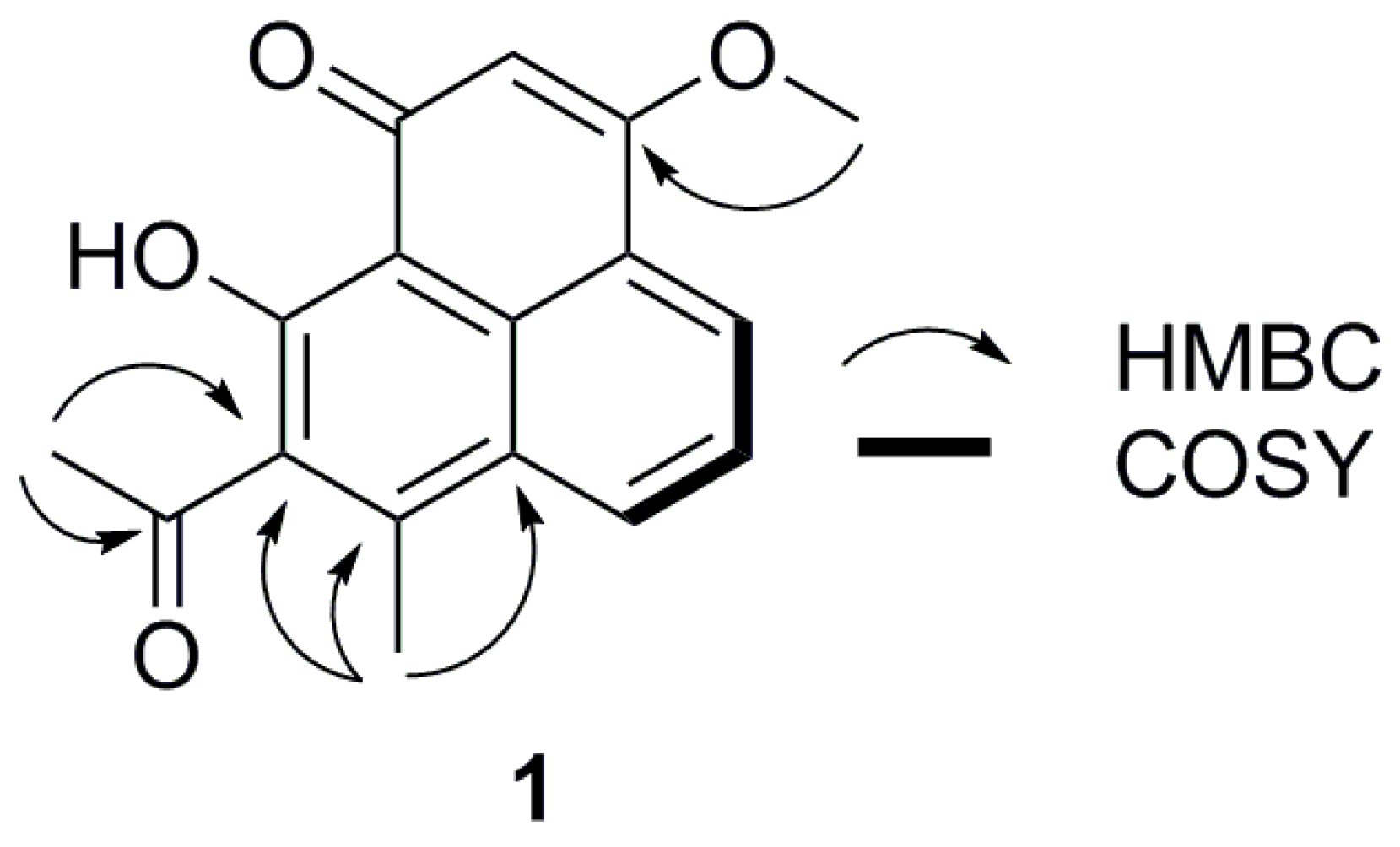
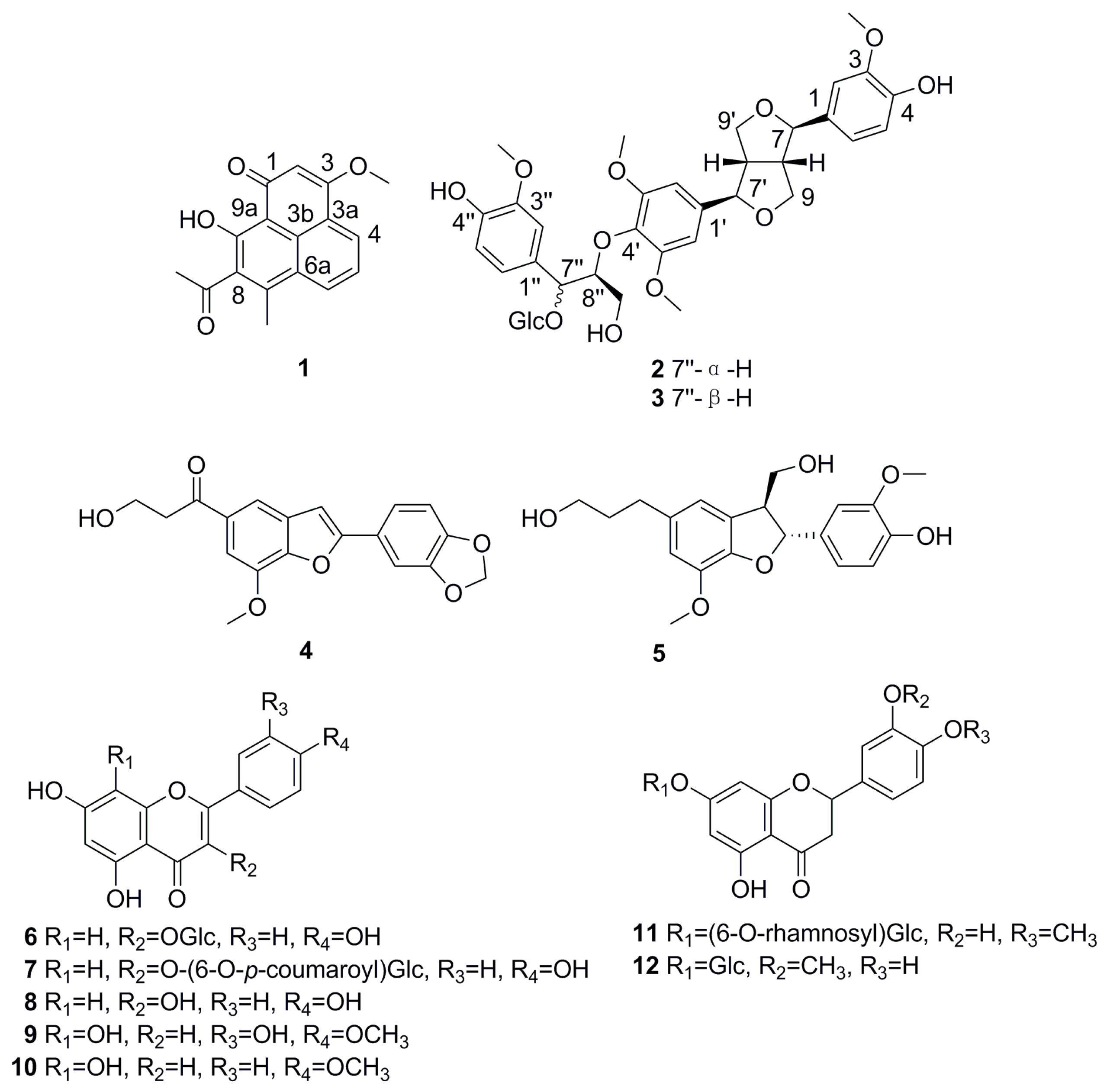
2. Results and Discussion

In our search for anti-complementary activity compounds from plants, the 95% ethanolic extract of H. angustifolia possessed a potent anti-complementary activity. Thus, the EtOAc-soluble fraction was concentrated under reduced pressure to produce a residue that was subjected to multiple chromatographies. From this active fraction, 12 compounds (112) including one new skeleton compound (1) and two new compounds (2 and 3) were isolated and identified.
Compound 1 (Figure 1) was obtained as yellow crystals (CHCl3), and its molecular formula was determined as C17H14O4 from HR-ESI-MS (m/z 283.0962 [M + H]+, calcd. for C17H15O4+, 283.0965). The 1H-NMR spectrum (Table 1) exhibited signals for three aromatic protons (δH 8.43 (1H, d, J = 7.8 Hz) 8.24 (1H, d, J = 7.8 Hz) and 7.58 (1H, t, J = 7.8 Hz)), an olefinic methine (δH 6.66 (1H, s)), two methyls (δH 2.72 and 2.58 (each 3H, s)), a methoxyl (δH 3.92 (3H, s)) and a peri-hydroxy (δH 17.36 (1H, s)), as well as corresponding with 13C-NMR resonances at δC 127.9, 130.8, 123.7, 100.2, 32.1, 16.0, 56.4 and 174.2, respectively. Furthermore, the 13C-NMR spectrum (Table 1) showed two carbonyl carbon signals (δC 204.1 and 181.1) and eight quaternary carbon signals. The 1H-1H COSY spectrum revealed the presence of a subunit C-4–C-5–C-6 (Figure 2). These NMR signals indicated that compound 1 had a phenalenone skeleton as in the known compounds 7-methyl-1-phenalenon [17] and myeloconone A2 [18]. The HMBC (Figure 2) correlations from 7-CH3H 2.58) to C-7 (δC 144.1), C-8 (δC 136.3) and C-6a (δC 125.4) indicated that the methyl was attached to C-7. Different from 7-methyl-1-phenalenon, compound 1 possessed a methoxyl, an acetyl and a peri-hydroxy group in its structure. The HMBC (Figure 2 and see Supplementary Materials) correlations from 3-OCH3H 3.92) to C-3 (δC 166.6), 8-COCH3H 2.72) to 8-COCH3C 204.1) and C-8 (δC 136.3) further confirmed that the methoxyl was connected to C-3, and the acetyl group was connected to C-8. Consequently, the structure of 1 was deduced as 8-acetyl-9-hydroxy-3-methoxy-7-methyl-1-phenalenon. To our knowledge, 1 is the first phenalenon derivative with an acetyl side chain connected to C-8.
Compound 2 (Figure 1) was obtained as white amorphous powder, and the molecular formula was determined as C37H46O16 by HR-ESI-MS (m/z 769.2662 [M + Na]+, calcd. for C37H46NaO16+, 769.2678). The 1H- and 13C-NMR spectra (Table 2) showed characteristic signals for a sesquilignan similar to hedyotol C [19], including eight aromatic protons (two sets of 1,2,4-trisubstituted phenyl rings and a 1,2,3,5-tetrasubstituted phenyl ring), four methoxyl groups, two phenolic and five alcoholic hydroxyl groups, one furan and one glycerol unit. In addition, an anomeric proton resonated at δH 4.67 (1H, d, J = 7.8 Hz) and the large coupling constant indicated β-glucosidic linkage. Detailed inspection of the NMR data revealed that the structure of compound 2 was similar to hedyotol C 4″-O-β-d-glucopyranoside [20], except that the glucosyl unit was connected to C-7″ in 2 rather than to C-4″, as indicated by the HMBC correlations from H-1″′ (δH 4.67) to C-7″ (δC 82.3) and H-7″ (δH 5.15) to C-1″′ (δC 104.4). The nature of sugar in compound 2 was further demonstrated to be d-glucose by GC analysis of an acid-treated hydrolysate, as well as comparison of their 13C-NMR data with that in the literature [20]. The relative configuration of compound 2 was determined by interpretation of the NOESY spectrum and the chemical shift ratio of C-7″:C-8″ [21]. In the NOESY spectrum of compound 2 (See Figure S13), the correlation of H-7″ (δH 5.15) with H-8″ (δH 4.42–4.44) was observed, suggestive of the same α-orientation. Moreover, based on the similar chemical shift ratio of C-7″:C-8″ (δC 82.3:86.6) to those of hedyotol C 4″-O-β-d-glucopyranoside [20], the relative configuration of compound 2 was proposed as an erythro configuration. Therefore, compound 2 was established as hedyotol C 7″-O-β-d-glucopyranoside.
Compound 3 (Figure 1) was determined to have the same molecular formula C37H46O16 as compound 2 by HR-ESI-MS (m/z 769.2662 [M + Na]+). Their 1H- and 13C-NMR spectra closely resembled each other (Table 2). The above evidence suggests that compounds 2 and 3 might be stereoisomeric, which could be clarified in accordance with the NOESY spectrum and the chemical shift ratio of C-7″:C-8″ [21]. The differences between compounds 2 and 3 were the absence of NOESY correlations between H-7″ (δH 5.28) and H-8″ (δH 4.26–4.27) (See Figure S20) and the similar chemical shift ratio of C-7″:C-8″ (δC 77.7:86.8) as compared to those of hedyotol D 4″-O-β-d-glucopyranoside [20] in compound 3. This evidence confirmed compounds 2 and 3 were a pair of epimers at C-7″. Accordingly, the relative configuration of compound 3 was presumed as a threo configuration. Consequently, compound 3 was identified as hedyotol D 7″-O-β-d-glucopyranoside.
Nine known compounds were identified as machicendonal (compound 4) [22], (7S,8R)-dihydrodehydrodiconiferyl alcohol (compound 5) [23], kaempferol-3-O-β-d-glucopyranoside (compound 6) [24], potengriffioside A (compound 7) [25], kaempferol (compound 8) [24], 5,7,8,3′-tetrahydroxy-4′-methoxyflavone (compound 9) [26], 5,7,8-trihydroxy-4′-methoxyflavone (compound 10) [26], hesperidin (compound 11) [27], and homoeriodictyol-7-O-β-d-glucopyranoside (compound 12) (Figure 1) [28] by comparing the spectroscopic data with those reported in the literature. Compounds 4, 812 were isolated from the genus Helicteres for the first time.
All the isolated constituents (compounds 112) were evaluated for in vitro anti-complementary activity on CP and AP [16]. As shown in Figure 1 and Table 3, compounds 4 and 5 belonged to the benzofuran lignans and possessed the most potent anti-complementary activity with CH50 values of 0.040 ± 0.009 and 0.009 ± 0.002 mM and AP50 values of 0.105 ± 0.015 and 0.021 ± 0.003 mM, respectively. Seven flavonoids (compounds 612) showed anti-complementary activity with CH50 and AP50 values of 0.143–1 mM and 0.311–1 mM, respectively. Compounds 13 exhibited moderate activity on CP and weak activity on AP. It was found that flavonoids with 4′-OH (compounds 8 and 12) showed stronger anti-complementary activity than those with a -OCH3 at C-4′ (compounds 911). Thus, the 4′-OH appears to be essential for the complementary activity of flavonoids.
In order to illuminate the anti-complementary mechanism of compounds 4 and 5, identification of their targets in the complement activation cascade was conducted using complement-depleted (C-depleted) sera [16]. As shown in Figure 3, compound 4 regained the hemolytic capacity of C5-depleted serum, and compound 5 regained the hemolytic capacity of C4- and C5-depleted sera. These findings suggested that compound 4 probably acted on C1q, C2, C3, C4 and C9, while compound 5 interacted with the C1q, C2, C3 and C9 components of the complement. These results indicate that different compounds can act on different targets in the complement activation cascade. Thus, compounds 4 and 5 are promising candidates for development as anti-complementary agents from H. angustifolia. Moreover, the relevant pharmacology and toxicology of these compounds need further investigations.

3. Materials and Methods

3.1. General Experimental Procedures

The 1D and 2D NMR spectra were recorded on a Bruker Ultrashield Plus 600 MHz spectrometer (Bruker BioSpin Corporation, Billerica, MA, USA). Electrospray ionization mass spectrometry (ESI-MS) spectra were recorded on an Agilent SL G1946D single quadrupole mass spectrometer (Agilent, Foster, CA, USA). High-resolution electrospray ionization mass spectra (HR-ESI-MS) were determined on a Bruker micro time of flight (TOF) spectrometer (Bruker Daltonics Corporation, Billerica, MA, USA). The infrared (IR) spectra were measured on a Thermo Nicolet Avatar 360 spectrophotometer (Thermo Nicolet, Madison, WI, USA). Ultraviolet (UV) spectra were obtained on a Lambda 25 spectrometer (PerkinElmer, Wellesley, MA, USA). The optical rotations were measured on a JASCO P-1020 polarimeter (JASCO Corporation, Tokyo, Japan). The melting point was measured on a micromelting point apparatus which was uncorrected (Yuhua Instruments Co., Ltd., Gongyi, China). Gas chromatography (GC) was carried out on a Shimadzu GC-MS QP 2010 Ultra (Shimadzu Corporation, Kyoto, Japan). Semipreparative high performance liquid chromatography (Semi-HPLC) was run on an Agilent 1100 series (Agilent Technologies, Waldbronn, Germany), equipped with a DAD detector and an ODS column (250 × 10.0 mm, 5 μm, Phenomenex Luna C18). Medium pressure liquid chromatography (MPLC) was conducted on a MITSUBISHI GOT 1000 chromatographic instrument (Lisure Science (Suzhou) Co., Ltd., Suzhou, China) with a Santai ILOK ODS column (390 mm × 31.2 mm i.d.). Flash column chromatography was operated by use of normal-phase silica gel (200–300 mesh, Qingdao Marine Chemical Factory, Qingdao, China) and gel Sephadex LH-20 (Pharmacia Fine Chemical Co. Ltd., Uppsala, Sweden). Anti-complement 1q ((Anti-C1q), Human (Goat)), Anti-C2 (Human (Goat)) and Anti-C9 (Human (Goat)) were purchased from Calbiochem (Merck KGaA, Darmstadt, Germany), and Anti-C3 (Human (Goat)), Anti-C4 (Human (Goat)) and Anti-C5 (Human (Goat)) were obtained from Zhejiang Nanfang Reagent Factory, China. Heparin sodium salt (≥150 IU/mg, dry basis) was purchased from Shanghai Aizite Biotech Co. Ltd., China.

3.2. Plant Material

Dried aerial parts of H. angustifolia were purchased from Nanning, Guangxi Zhuang Autonomous Region of China in March 2011, and authenticated by Prof. Dao-Feng Chen at Fudan University. A voucher specimen (DFC-SZM20110812) has been deposited in the Herbarium of Materia Medica, Department of Pharmacognosy, School of Pharmacy, Fudan University, Shanghai, China.

3.3. Extraction and Isolation

The extraction of the dried aerial parts of H. angustifolia with 95% ethanol was partitioned successively with petroleum ether, EtOAc, and n-BuOH. The EtOAc fraction was selected to further purification due to its significant anti-complementary activity with CH50 value of 0.15 ± 0.02 and AP50 value of 0.29 ± 0.03 mg/mL. The EtOAc extract (160 g) was subjected to a silica gel column (12 × 50 cm), eluted with CH2Cl2–MeOH (50:1, 30:1, 20:1, 10:1, 8:1, 5:1, 3:1, 2:1, 1:1, 1:2, and 0:1, v/v). The CH2Cl2–MeOH (20:1) fraction (30 g) was subsequently chromatographed over a silica gel column (10 × 40 cm), eluted with CH2Cl2–MeOH (30:1, 20:1, 10:1, 5:1, 1:1, and 1:2, v/v) to afford six fractions. Fraction 1 (3.2 g) was further separated by MPLC eluted with MeOH–H2O (10:90 to 90:10, v/v) at 25 mL/min to yield four subfractions (1-A to 1-D). Subfraction 1-B (180 mg) was purified by semipreparative HPLC (MeOH–H2O, 42:58, v/v) at 3 mL/min to give compounds 4 (6 mg, tR 28.35 min) and 6 (28 mg, tR 35.65 min). Subfraction 1-C (330 mg) was applied to semipreparative HPLC (MeOH–H2O, 53:47, v/v) at 3 mL/min to afford compounds 11 (25 mg, tR 27.34 min), 1 (12 mg, tR 38.73 min), and 8 (17 mg, tR 45.45 min). Subfraction 1-D (550 mg) was separated on a Sephadex LH-20 gel column (2.5 × 100 cm) using MeOH as the eluent to yield compounds 10 (7 mg) and 9 (11 mg). Fraction 2 (8.2 g) was chromatographed by MPLC eluted with MeOH–H2O (20:80 to 80:20, v/v) at 30 mL/min to afford six subfractions (2-A to 2-F). Subfraction 2-C (800 mg) was separated by semipreparative HPLC, isocratically eluted with MeOH–H2O (30:70, v/v) at 3 mL/min to give compound 12 (7 mg, tR 46.21 min). Subfraction 2-D (550 mg) was further subjected to a silica gel column (3.5 × 30 cm) and eluted with a gradient of CH2Cl2–MeOH (20:1, 10:1, 5:1, 3:1, and 1:1, v/v) to yield five subfractions (2-D1 to 2-D5). Subfraction 2-D2 (290 mg) was applied to HPLC (MeOH–H2O, 40:60, v/v) to afford compounds 2 (16 mg, tR 12.33 min) and 3 (11 mg, tR 13.98 min). Subfraction 2-D3 (90 mg) and subfraction 2-E (1.3 g) were respectively purified by Sephadex LH-20 (2.5 × 100 cm) with MeOH to give compounds 5 (18 mg) and 7 (0.9 g).

3.4. Spectroscopic Data

8-Acetyl-9-hydroxy-3-methoxy-7-methyl-1-phenalenon (compound 1): Yellow crystals (CHCl3); m.p. 325–330 °C; UV (CHCl3): λmax (log ε): 243.3 (4.17), 342.5 (3.91) nm; IR (KBr) νmax: 3373 (OH), 2920, 1698 (C=O), 1623, 1574, 1337, 815 cm1; 1H- and 13C-NMR (600 and 150 MHz, C5D5N) spectroscopic data see Table 1; HR-ESI-MS: m/z 283.0962 [M + H]+ (calcd. for C17H15O4+; 283.0965).
Hedyotol C 7″-O-β-d-glucopyranoside (compound 2): White amorphous powders; [α] D 25 +14.25 (c 0.4, MeOH); UV (MeOH): λmax (log ε): 218.0 (3.83), 279.7 (3.15) nm; IR (KBr) νmax: 3419 (OH), 2936, 1594, 1512, 1457, 1276, 1117 cm1; 1H- and 13C-NMR (600 and 150 MHz, CD3OD) spectroscopic data see Table 2; HR-ESI-MS: m/z 769.2662 [M + Na]+ (calcd. for C37H46NaO16+; 769.2678).
Hedyotol D 7″-O-β-d-glucopyranoside (compound 3): White amorphous powders; [α] D 25 –13.50 (c 0.3, MeOH); UV (MeOH): λmax (log ε): 214.0 (3.93), 279.6 (3.22) nm; IR (KBr) νmax: 3419 (OH), 2926, 1600, 1506, 1463, 1123 cm1; 1H- and 13C-NMR (600 and 150 MHz, CD3OD) spectroscopic data see Table 2; HR-ESI-MS: m/z 769.2661 [M + Na]+ (calcd. for C37H46NaO16+; 769.2678).

3.5. Acid Hydrolysis of Compounds 2 and 3

The acid hydrolysis was performed according to the modified method of Song et al. [29]. Compound 2 (2 mg) was dissolved in 2 mol/L HCl (1 mL), and heated at 95 °C for 2 h. After hydrolysis, the reaction mixture was extracted with EtOAc and H2O, and the aqueous layer was neutralized with silver carbonate (10 mg). Then the supernatant was concentrated to dryness and further evaporated to dryness under N2 for 2 h. Subsequently, the residue was stirred with l-cysteine methyl ester hydrochloride (2 mg) in anhydrous pyridine (0.2 mL) at 60 °C for 1 h. The dried reactant was fractionalized with H2O and n-hexane (each 0.5 mL). The n-hexane layer was collected and subjected to GC-MS. The acid hydrolysis of compound 3 was conducted in the same procedure as compound 2. The GC conditions were: column temperature 150 °C, injector temperature 270 °C, carrier gas N2. d-glucose was detected from compounds 2 and 3 (tR 15.523 and 15.524 min) by comparison with authentic samples: tRd-glucose 15.513 min, and l-glucose 15.808 min.

3.6. Anti-Complementary Activity Assay Against CP and AP

The anti-complementary activities against the CP and AP were investigated with the method of Xu et al. [16]. Heparin was used as positive control drug. The results of anti-complementary activity of compounds 112 are displayed in Table 3. Anti-complement activity was determined as the mean of triplicate measurements at each concentration and expressed as 50% inhibitory concentration (CH50 and AP50 values). All results were performed statistical analysis to compare the CH50 and AP50 values between each compound and positive control with unpaired t test.

3.7. Identification of the Targets in the Complement Activation Cascade

Assay to identify the targets in the complement activation cascade was performed according to the method of Di et al. [30]. The results of compounds 4 and 5 were displayed in Figure 3. Data were expressed as mean ± SD of triplicate measurements.

4. Conclusions

A first phenalenon derivative (compound 1) with an acetyl side chain at C-8 and a pair of new sesquilignan epimers (compounds 2 and 3) with a glucosyl unit at C-7 together with nine known compounds were isolated from the aerial parts of H. angustifolia. Chemical structures of the isolated compounds were identified on the basis of extensive spectroscopic data. Compounds 4, 812 were isolated for the first time from the aerial parts of H. angustifolia. Eleven compounds exhibited anti-complementary activity, and the targets of two compounds (4 and 5) in the complement activation cascade were also identified. In conclusion, the anti-complementary constituents of H. angustifolia possessed chemical diversity, and consisted mostly of flavonoids and lignans in this study.

Supplementary Materials

The 1D and 2D NMR spectra for compounds 13 are available as supporting data online at https://www.mdpi.com/1420-3049/21/11/1506/s1.

Acknowledgments

This work was financially supported by the grant from the National Natural Science Foundation of China (81330089 and 30925042). The authors are grateful to Wei-Liang Zhu and Ming-Yue Zheng at the Shanghai Institute of Materia Medica for technical support.

Author Contributions

D.-F.C. conceived and designed the experiments; X.Y. performed the experiments; X.Y., Y.L. and Z.-H.C. analyzed the data; X.Y. wrote the paper; D.-F.C. edited and revised the paper. All authors read and approved the final manuscript.

Conflicts of Interest

The authors declare no conflict of interest.

References

  1. Carroll, M.C.; Fisher, M.B. Complement and the immune response. Curr. Opin. Immunol. 1997, 9, 64–69. [Google Scholar] [CrossRef]
  2. Morgan, B.P.; Harris, C.L. Complement therapeutics; history and current progress. Mol. Immunol. 2003, 40, 159–170. [Google Scholar] [CrossRef]
  3. Zhu, H.W.; Di, H.Y.; Zhang, Y.Y.; Zhang, J.W.; Chen, D.F. A protein-bond polysaccharide from the stem bark of Eucommia ulmoides and its anti-complementary effect. Carbohydr. Res. 2009, 344, 1319–1324. [Google Scholar] [CrossRef] [PubMed]
  4. Mollnes, T.E.; Song, W.C.; Lambris, J.D. Complement in inflammatory tissue damage and disease. Trends Immunol. 2002, 23, 61–64. [Google Scholar] [CrossRef]
  5. Vovel, C.W.; Fritzinger, D.C. Cobra venom factor: Structure, function, and humanization for therapeutic complement depletion. Toxicon 2010, 56, 1198–1222. [Google Scholar]
  6. Thorgersen, E.B.; Ghebremariam, Y.T.; Thurman, J.M.; Fung, M.; Nielsen, E.W.; Holers, V.M.; Kotwal, G.J.; Mollnes, T.E. Candidate inhibitors of porcine complement. Mol. Immunol. 2007, 44, 1827–1834. [Google Scholar] [CrossRef] [PubMed]
  7. Qu, H.C.; Ricklin, D.; Lambris, J.D. Recent developments in low molecular weight complement inhibitors. Mol. Immunol. 2009, 47, 185–195. [Google Scholar] [CrossRef] [PubMed]
  8. Risitano, A.M. Current and future pharmacologic complement inhibitors. Hematol/Oncol. Clin. N. Am. 2015, 29, 561–582. [Google Scholar] [CrossRef] [PubMed]
  9. Jiangsu New Medical College. Dictionary of Chinese Herb Medicines; Shanghai Scientific and Technologic Press: Shanghai, China, 1996; pp. 178–179. [Google Scholar]
  10. Chen, W.L.; Tang, W.D.; Lou, L.G.; Zhao, W.M. Pregnane, coumarin and lupane derivatives and cyto toxic constituents from Helicters angustifolia. Phytochemistry 2006, 67, 1041–1047. [Google Scholar] [CrossRef] [PubMed]
  11. Pan, M.H.; Chen, C.M.; Lee, S.W.; Chen, Z.T. Cytotoxic triterpenoids from the root bark of Helicters angustifolia. Chem. Biodivers. 2008, 5, 565–574. [Google Scholar] [CrossRef] [PubMed]
  12. Guo, X.D.; Huang, Z.S.; Bao, Y.D.; An, L.K.; Ma, L.; Gu, L.Q. Two new sesquiterpenoids from Helicters angustifolia. Chin. Chem. Lett. 2005, 16, 49–52. [Google Scholar]
  13. Chen, M.; Muchersie, E.; Luo, C.; Forrester, J.V.; Xu, H.P. Inhibition of the alternative pathway of complement activation reduces inflammation in experimental autoimmune uveoretinitis. Eur. J. Immunol. 2010, 40, 2870–2881. [Google Scholar] [CrossRef] [PubMed]
  14. Ignatius, A.; Schoengraf, P.; Kreja, L.; Liedert, A.; Rechnagel, S.; Kandert, S.; Brenner, R.E.; Schneider, M.; Lambris, J.D. Complement C3a and C5a modulate osteoclast formation and inflammatory response of osteoblasts in synergism with IL-1β. J. Cell. Biochem. 2011, 112, 2594–2605. [Google Scholar] [CrossRef] [PubMed]
  15. Zhang, T.; Chen, D.F. Anticomplementary principles of a Chinese multiherb remedy for the treatment and prevention of SARS. J. Ethnopharmacol. 2008, 117, 351–361. [Google Scholar] [CrossRef] [PubMed]
  16. Xu, H.; Zhang, Y.Y.; Zhang, J.W.; Chen, D.F. Isolation and characterization of an anti-complementary polysaccharide D3-S1 from the roots of Bupleurum smithii. Int. Immunopharmacol. 2007, 7, 175–182. [Google Scholar] [CrossRef] [PubMed]
  17. Kobayashi, M.; Matsumoto, T. Ungewöhnliche wichterle-reaktionen. Eine neue synthese von phenalenonen. Bull. Chem. Soc. Jpn. 1975, 48, 2935–2938. [Google Scholar] [CrossRef]
  18. Ernst-Russell, M.A.; Chai, C.L.L.; Elix, J.A.; McCarthy, P.M. Myeloconone A2, a new phenalenone from the lichen Myeloconis erumpens. Aust. J. Chem. 2000, 53, 1011–1013. [Google Scholar] [CrossRef]
  19. Tohru, K.; Satoko, M.; Shigetoshi, K.; Takaaki, T. Studies on the constituents of medicinal and related plants in Sri Lanka. III. Novel sesquilignans from Hedyotis lawsoniae. Chem. Pharm. Bull. 1985, 33, 1444–1451. [Google Scholar]
  20. Yukinori, M.; Tsuyoshi, T. Studies on constituents of Scutellaria species X IX: lignan glycosides of roots of Scutellaria baicalensis Georgi. Nat. Med. 1998, 52, 82–86. [Google Scholar]
  21. Bardet, M.; Robert, D.; Lundquist, K.; Von-Unge, S. Distribution of erythro and threo forms of different types of β-O-4 structures in aspen lignin by carbon-13 NMR using the 2D INADEQUATE experiment. Magn. Reson. Chem. 1998, 36, 597–600. [Google Scholar] [CrossRef]
  22. Talaptra, B.; Ray, T.; Talapatra, S.K. Structures of three new norlignans from Machilus glaucescens and their partial syntheses. Chem. Nat. Prod. 1978, 2, 200–201. [Google Scholar]
  23. Kuang, H.; Xia, Y.; Yang, B.; Wang, Q.; Lu, S. Lignan constituents from Chloranthus japonicus Sieb. Arch. Pharm. Res. 2009, 32, 329–334. [Google Scholar] [CrossRef] [PubMed]
  24. Yi, Y.; Wu, X.; Wang, Y.; Ye, W.C.; Zhang, Q.W. Studies on the flavonoids from the flowers of Hylocereus undatus. J. Chin. Med. Mater. 2011, 34, 712–716. [Google Scholar]
  25. Zhong, H.J.; Chen, J.J.; Wang, H.Y.; Luo, S.D. Chemical investigation on Potentilla griffithii var. velutina. Chin. Tradit. Herb. Drugs. 2000, 31, 488–490. [Google Scholar]
  26. Xia, P.F.; Song, S.; Feng, Z.M.; Zhang, P.C. Chemical constituents from leaves of Sterculia foetida. Zhong Guo Zhong Yao Za Zhi 2009, 34, 2604–2606. [Google Scholar]
  27. Yang, N.Y.; Duan, J.A.; Li, P.; Qian, S.H. Studies on flavonoids of Lysimachia christinae Hance. Chin. Pharm. J. 2006, 4, 1621–1624. [Google Scholar]
  28. Kong, D.Y.; Luo, S.G.; Li, H.T.; Lei, X.H. Studies on chemical components of Viscum coloratum L. Pharm. Ind. 1987, 18, 123–127. [Google Scholar]
  29. Song, W.H.; Chen, Z.H.; Chen, D.F. Anticomplement monoterpenoid glucosides from the root bark of Paeonia suffruticosa. J. Nat. Prod. 2014, 77, 42–48. [Google Scholar] [CrossRef] [PubMed]
  30. Di, H.Y.; Zhang, Y.Y.; Chen, D.F. An anti-complementary polysaccharide from the roots of Bupleurum chinense. Int. J. Biol. Macromol. 2013, 58, 179–185. [Google Scholar] [CrossRef] [PubMed]
  • Sample Availability: Not Available.
Figure 1. Structures of compounds 112.
Molecules 21 01506 g001
Figure 2. Key HMBC and 1H-1H COSY correlations for compound 1.
Molecules 21 01506 g002
Figure 3. Hemolytic assays of compounds 4 (A) and 5 (B) for individual components utilizing C-depleted sera.
Molecules 21 01506 g003
Table 1. 1H- and 13C-NMR (600 and 150 MHz) data of compound 1 in C5D5N, δ in ppm, J in Hz.
No.δHδCNo.δHδC
1 181.17 144.1
26.66 s100.28 136.3
3 166.69 174.2
3a 120.89a 106.5
3b 127.13-OCH33.92 s56.4
48.43 d (7.8)127.97-CH32.58 s16.0
57.58 t (7.8)123.78-COCH3 204.1 (CO)
68.24 d (7.8)130.8 2.72 s32.1 (CH3)
6a 125.49-OH17.36 s
Table 2. 1H- and 13C-NMR (600 and 150 MHz) data for compounds 2 and 3 in CD3OD, δ in ppm, J in Hz.
No.23
δHδCδHδC
1 133.7 133.7
26.97 d (1.8)111.06.97 d (1.4)111.0
3 149.1 149.1
4 147.4 a 147.4 a
56.79 d (8.1)116.16.79 d (8.1)116.1
66.83 dd (8.1, 1.7)120.16.83 dd (8.0, 1.3)120.1
74.72 d (4.8)87.54.73 d (3.6)87.5
83.14 ddd (11.3, 8.3, 4.9)55.3 b3.13–3.15 overlapped55.3 b
94.24–4.30 overlapped, 3.88–3.91 overlapped72.9 c4.27–4.31 overlapped, 3.88–3.91 overlapped72.9 c
1′ 138.9 138.9
2′,6′6.64 s104.26.66 s104.1
3′,5′ 154.3 154.5
4′ 136.2 136.4
7′4.75 d (4.6)87.24.76 d (3.0)87.3
8′3.14 ddd (11.3, 8.3, 4.9)55.7 b3.13–3.15 overlapped55.7 b
9′4.24–4.30 overlapped, 3.88–3.91 overlapped72.8 c4.27–4.31 overlapped, 3.88–3.91 overlapped72.7 c
1′′ 132.4 130.8
2′′7.03 d (1.7)112.77.24 brs113.1
3′′ 148.4 148.7
4′′ 147.1 a 147.1 a
5′′6.72 d (8.1)115.36.80 d (8.0)115.5
6′′6.87 dd (8.1, 1.7)121.96.92 dd (8.0, 1.3)122.0
7′′5.15 d (6.2)82.35.28 d (3.2)77.7
8′′4.42–4.44 m86.64.26–4.27 m86.8
9′′4.05 dd (12.4, 4.0)
3.76 dd (12.4, 3.0)
61.53.88–3.91 overlapped
3.44 dd (11.4, 4.5)
61.4
β-d-glc
1′′′4.67 d (7.8)104.44.21 d (7.5)101.0
2′′′3.27-3.29 m75.73.33–3.34 m75.2
3′′′3.38 d (9.0)78.13.28–3.31 overlapped77.7
4′′′3.30–3.32 m71.53.28–3.31 overlapped71.9
5′′′3.19 ddd (9.6, 5.4, 2.5)77.83.12–3.13 m77.8
6′′′3.73 dd (11.9, 2.3)
3.60 dd (11.8, 5.4)
62.73.83–3.85 m
3.69 dd (11.9, 5.9)
62.7
3-OCH33.87 s56.41 d3.88 s56.37 d
3′,5′-OCH33.80 s56.63.74 s56.5
3′′-OCH33.83 s56.43 d3.85 s56.42 d
a−d The assignments in each column may be interchanged.
Table 3. Anti-complementary activity of compounds 112 against classical and alternative pathways.
CompoundCH50 (mM) aAP50 (mM) a
10.744 ± 0.099>1
20.419 ± 0.043>1
30.249 ± 0.021>1
40.040 ± 0.0090.105 ± 0.015
50.009 ± 0.002 **,b0.021 ± 0.003 **,b
60.877 ± 0.081>1
70.143 ± 0.0190.335 ± 0.040
80.147 ± 0.0220.311 ± 0.033
90.232 ± 0.250.501 ± 0.065
100.511 ± 0.0430.984 ± 0.107
11>1>1
120.351 ± 0.0330.556 ± 0.061
Heparin c0.026 ± 0.005 0.055 ± 0.008
a Data were represented as mean ± SD of three independent measurements (n = 3), and described as 50% hemolytic inhibitory concentration (CH50 for classical pathway and AP50 for alternative pathway). **,b p < 0.01, compared to the positive control group. c Heparin was used as the positive control (mg/mL).

