Article

Three New Butenolides from the Fungus Aspergillus sp. CBS-P-2

1
School of Traditional Chinese Materia Medica, Shenyang Pharmaceutical University, Shenyang 110016, China
2
Key Laboratory of Structure-Based Drug Design and Discovery, Ministry of Education, Shenyang Pharmaceutical University, Shenyang 110016, China
3
Department of Medicinal Chemistry, School of Pharmaceutical Engineering, Shenyang Pharmaceutical University, Shenyang 110016, China
*
Author to whom correspondence should be addressed.
These authors contributed equally to this work.
Molecules 2016, 21(10), 1361; https://doi.org/10.3390/molecules21101361
Submission received: 10 September 2016 / Revised: 9 October 2016 / Accepted: 10 October 2016 / Published: 13 October 2016

Abstract

:
Three new butenolides aspernolides H–J (13) together with seven known ones (410) were isolated from the fungus Aspergillus sp. CBS-P-2. Their chemical structures were established on the basis of 1D- and 2D-NMR spectroscopic data, HR-ESI-MS analysis, and their absolute configuration were determined by circular dichroism (CD) analysis. All the compounds were evaluated for the antioxidant effects by DPPH and ABTS methods, the antitumor activities against four human tumor cell lines (HL-60, ASPC1, HCT-116 and PC-3) and antimicrobial activities. Compounds 410 showed significant activity against DPPH (IC50 = 15.9–34.3 μM) and compounds 110 exhibited significant ABTS free radical scavenging activity (IC50 = 2.8–33.1 μM). Compounds 2, 5 and 11 showed potent cytotoxic activities against HL-60 cell lines with IC50 values of 39.4, 13.2 and 16.3 μM, respectively. Compound 10 showed good antimicrobial activity against Staphylococcus aureus with minimum inhibitory concentration (MIC) of 21.3 μM.

1. Introduction

Recently, attention has been focused on the biology and chemistry of the microorganisms from extreme environments [1]. These microorganisms have proved to be one of the most important and underexplored resources for structurally novel and biologically active natural products [2,3]. Aspergillus species from diverse extreme origins (e.g., salt soil, plants, marine organisms and wetland) are important fungi that biosynthesize structurally diverse and pharmaceutically important natural products, such as alkaloids [4], polyketides [5], terpenes [6], and peptides [7], with intriguing biological properties. Butyrolactones and aspernolides, with a basal skeleton characterized by a five-membered lactone bearing two aromatic rings [8,9], mainly produced by Aspergillus sp. exhibited a wide range of activities, such as selectively inhibitory activities against CDK2 [10], α-glucuronidase [11] and cytotoxic [12], antioxidant [13], anti-inflammatory [14] activities.
As a part of our continuing interest in exploring bioactive metabolites from fungal source, three new aspernolides (13) along with seven known compounds (410) (Figure 1) were isolated from the fungus Aspergillus sp. CBS-P-2, which was isolated from the volcanic soil collected in Changbai Mountain, Jilin, P.R. China. The structures of the isolated compounds were established on the basis of 1D, 2D NMR and HR-ESI-MS spectral data. In addition, the antioxidant effects, the cytotoxic properties against four human tumor cell lines [human leukaemia cancer (HL-60), human pancreatic cancer (ASPC1), human colon cell (HCT-116) and human prostatic cancer (PC-3)] and the antimicrobial activities of compounds 110 have been evaluated.

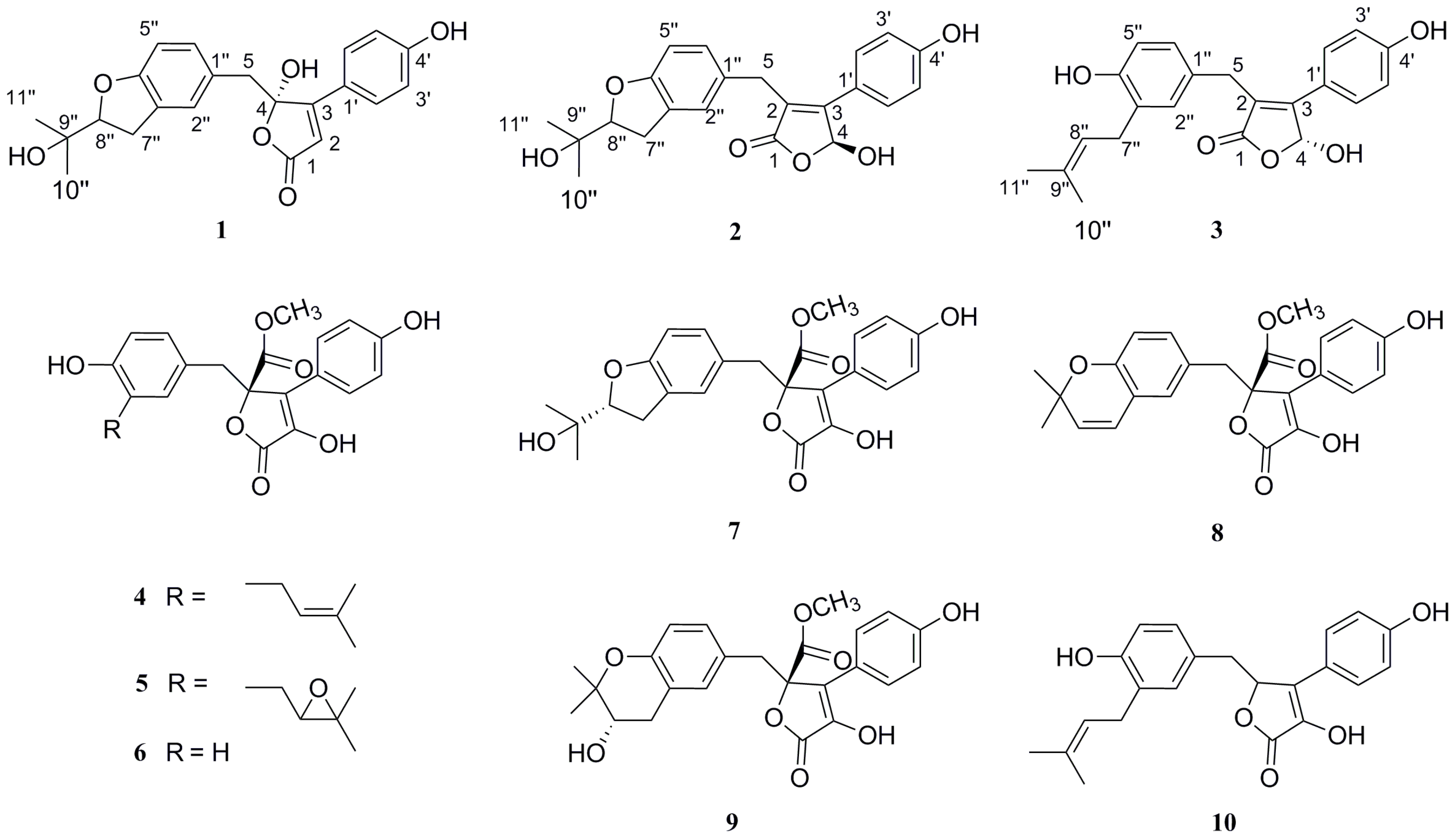
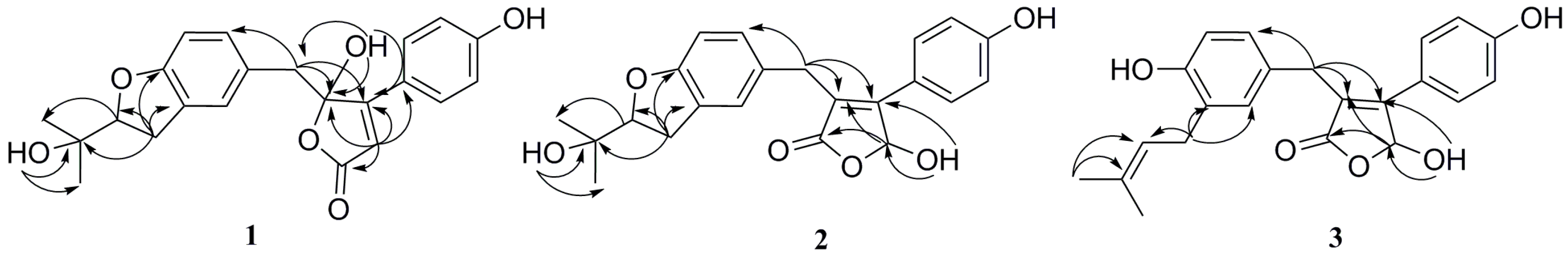
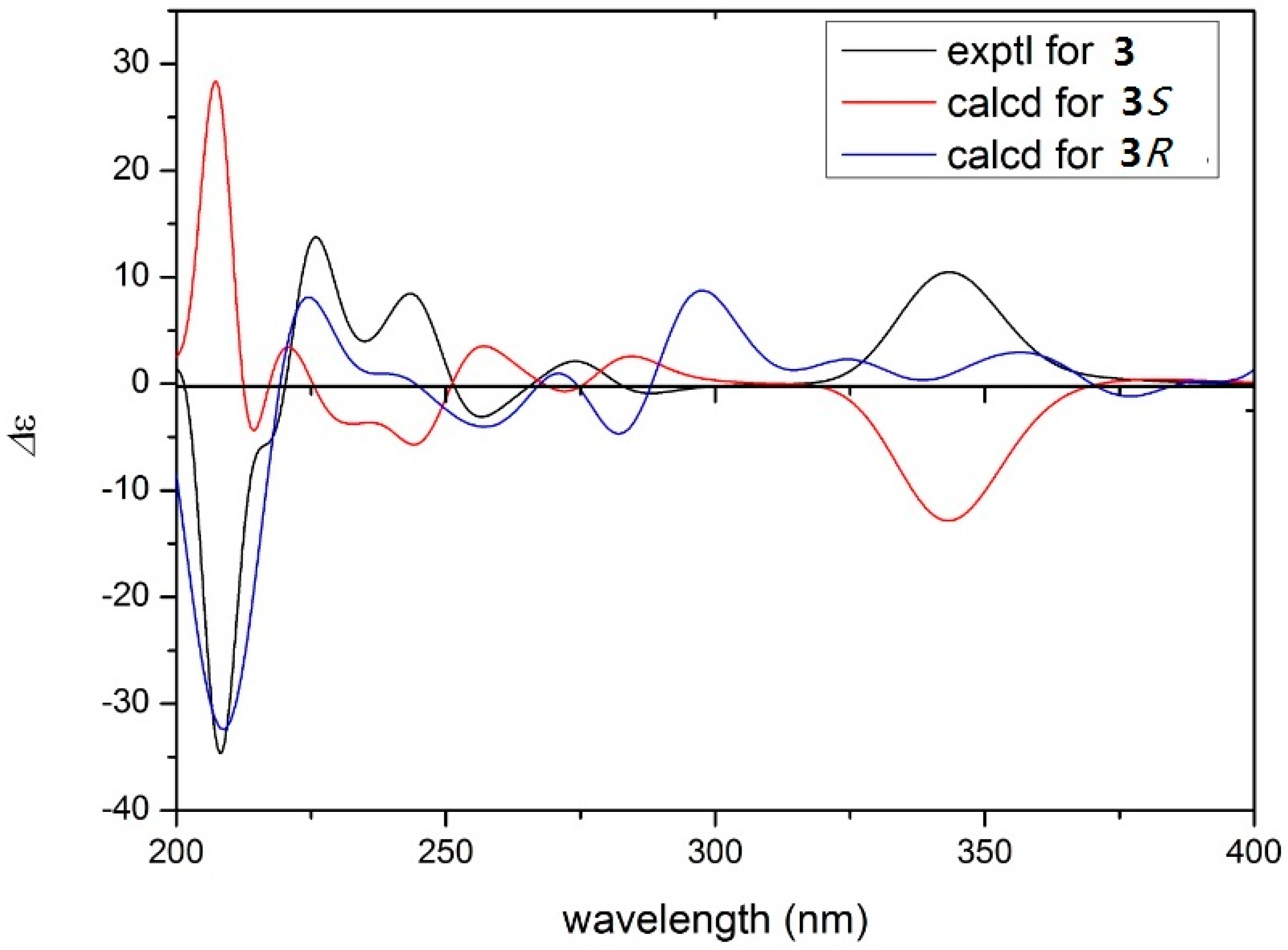
2. Results and Discussion

The fermentation broth of the fungal strain Aspergillus sp. CBS-P-2 was extracted with EtOAc and then concentrated under reduced pressure to give an extract. The EtOAc extract was subjected to various column chromatography protocols to afford compounds 110. These new structures were identified by spectroscopic analyses and physicochemical properties, while the known analogues were identified as butyrolactone I (4) [15], butyrolactone ΙΙΙ (5) [15], butyrolactone ΙΙ (6) [15], butyrolactone IV (7) [15], aspernolide E (8) [16], butyrolactone V (9) [8] and 3-hydroxy-5-[[4-hydroxy-3-(3-methyl-2-buten-1-yl)phenyl]methyl]-4-(4-hydroxyphenyl)-2(5H)-furanone (10) [17] by comparison of their spectroscopic data and specific rotations with those in the literature.
Compound 1 was isolated as a light yellowish gum. The molecular formula of 1 was determined to be C22H22O6 by positive mode HR-ESI-MS data at m/z 405.1300 [M + Na]+ (calcd. for C22H22O6Na, 405.1314), indicating 12 degrees of unsaturation. The 1H-NMR spectrum of 1 (Table 1) showed characteristic signals attributable to an olefinic proton singlet δH 6.23 (1H, s, H-2), a benzyl moiety with an 1, 2, 4-trisubstituted aromatic system [δH 6.48 (1H, brs, H-2′′), δH 6.46 (1H, d, J = 8.4 Hz, H-5′′), δH 6.54 (1H, brd, J = 8.4 Hz, H-6′′)] and a para-disubstituted benzene ring [δH 7.74 (2H, d, J = 8.4 Hz, H-2′/H-6′), 6.89 (2H, d, J = 8.4 Hz, H-3′/5′)]. At the lower frequency, the 1H-NMR spectrum displayed an oxygenated methine proton δH 4.44 (1H, t, J = 8.8 Hz, H-8′′), four methylene protons [δH 2.98 (1H, dd, J = 15.2, 8.8 Hz, H-7′′a), 2.92 (1H, dd, J = 15.2, 10.0 Hz, H-7′′b), 3.12 (1H, d, J = 13.5 Hz, H-5a) and 3.25 (1H, d, J = 13.5 Hz, H-5b)] and two methyl signals δH 1.06 (3H, s, H-10′′) and 1.07 (3H, s, H-11′′). The 13C-NMR spectrum showed 22 carbon signals corresponding to a carbonyl carbon, fourteen signals in the sp2 region of δC 116.1–164.3, three oxygenated carbons including a hemiacetal carbon δC 108.0 (C-4), two methylene groups and two methyl groups. Comparison of the NMR data of compound 1 with those of butyrolactone IV (7) suggested that they shared the same core frame unit except that the substitution of C-4 in 1 was a hydroxyl group and C-2 was unsubstituted. The structure was further determined by HMBC spectrum (Figure 2), the correlations from H-2 to C-1, C-3, C-4 and C-1′, from H2-5 to C-3, C-4 and C-6′′, as well as from H2-7′′ to C-3′′, C-4′′ and C-8′′ confirmed the assignment of the α,β-unsaturated-γ-lactone moiety. On the basis of these data, the planar structure of compound 1 was established.
The absolute configuration of the C-4 tertiary alcohol was deduced by the circular dichroism (CD) data of the in situ formed [Rh2(OCOCF3)4] complex, with the inherent contribution subtracted. A positive Cotton effect at 350 nm in the Rh2(OCOCF3)4-induced CD spectrum indicated the 4S absolute configuration on the basis of the bulkiness rule of 1 [18,19].
Compound 2 was obtained as a light brownish gum. Its positive HR-ESI-MS showed an ion peak at m/z 405.1283 [M + Na]+ (calcd. 405.1314), indicating the same molecular formula of C22H22O6 as 1. 1H- and 13C-NMR spectra of 2 were very similar to those of 1 except the signals of the lactone moiety. Combined with the HSQC spectrum, the 1H-NMR data of δH 6.54 (d, J = 8.6 Hz, H-4) in 2 was assigned to be a hemiacetal proton rather than an olefinic one. The 13C-NMR data of C-2 had shifted downfield (δC 124.1) and C-3 and C-4 shifted upfield (δC 156.4, 97.8), respectively, compared with those of 1C 112.9, 164.3 and 108.0). The above information illustrated the α,β-unsaturated-γ-lactone was isomerized, which was confirmed by the HMBC correlations from H-4 to C-1 and C-2, from 4-OH to C-4 and C-3, as well as from H2-5 to C-2, C-3 and C-6′′ as shown in Figure 2.
The determination of the absolute configuration of C-4 was based on the CD analysis. The CD of the α,β-unsaturated γ-lactone rings with a chiral γ-carbon shows Cotton effects associated with the π→π* transition in the region 200–235 nm [20] and the n→π* transition in the region 235–270 nm [20,21,22,23]. Compound 2 showed positive π→π* Cotton effect at 223 nm and negative n→π* Cotton effect at 275 nm, indicating the S absolute configuration at C-4 in 2 [20,21].
Compound 3 was also obtained as a light brownish gum. It has the molecular formula C22H22O5, as deduced from the HR-ESI-MS data [M + Na]+ at m/z 389.1367 (calcd. 389.1365). The formula differs from that of 2 by the loss of one oxygen atom, suggesting the absence of a hydroxyl group for 3. The NMR data of 3 were very similar to those of 2 and the difference was the side chain moiety was an isopentenyl instead of a furan ring. The 1H-NMR spectrum of 3 indicated the presence of two methylene protons at δH 3.14 (2H, d, J = 7.2 Hz, H2-7′′), an olefinic proton at δH 5.21 (1H, t, J = 7.2 Hz, H-8′′) and two methyl signals at δH 1.62 (3H, s, H-10′′) and 1.66 (3H, s, H-11′′), which suggested the existence of an isopentenyl group. The planar construction of 3 was determined by the HMBC correlations of H-4/C-1, C-2; H2-5/C-2, C-3, C-6′′; H-7′′/C-8′′, C-2′′, C-3′′ (Figure 2). The absolute configuration of 3 was determined as 4R by comparison of its experimental electronic CD curve with that calculated (Figure 3).
The antioxidant activities of 110 were evaluated by their ability to scavenge DPPH and ABTS radicals (Table 2). Compounds 410 showed significant DPPH radical scavenging activities with IC50 values of 15.9 to 34.3 μM, whereas compounds 13 (IC50 > 100 μM) did not display any DPPH radical scavenging activity (Table 2). In the ABTS assay, most of the isolated compounds exhibited significant antioxidant activity, with compounds 2 and 410 being stronger than that of the standard compound ascorbic acid (IC50 = 12.5 μM), whereas compounds 1 and 3 exhibited moderate ABTS radical-scavenging activities (IC50 = 33.1 and 29.5 μM). A comparison of the structures of 13 with those of 410 indicated that an enolic hydroxyl situated at the α,β-unsaturated-γ-lactone ring and the phenolic hydroxyl were crucial for the antioxidant activity, which was consistent with previous observations [24,25].
The isolated compounds were tested for their in vitro cytotoxicities using four tumor cell lines: HL-60, ASPC1, HCT-116 and PC-3. The resulting IC50 values are listed in Table 3. 5-Fluorouracil was used as a positive control and its IC50 values were 6.38, 2.70, 7.77 and 15.6 μM, respectively. Compounds 2, 4, 7 and 10 exhibited potent antitumor activity against HL-60 cell line with IC50 values ranging from 13.2 to 41.6 μM. Compounds 4 and 8 showed moderate cytotoxity against PC-3 cell line with IC50 values of 41.7 and 58.3 μM, respectively. Only compound 8 displayed weak cytotoxic activity against HCT-116 cell line with an IC50 of 41.7 μM and all tested isolates were inactive against the ASPC1 cell line. The antimicrobial activity of compounds 110 were assessed towards Bacillus subtilis, Staphylococcus aureus, Escherichia coli and Candida albicans. However, only compound 10 presented significant antimicrobial activity against Staphylococcus aureus with minimum inhibitory concentration (MIC) of 21.3 μM, and chloramphenicol was used as positive control with the MIC of 12.1 μM.

3. Experimental Section

3.1. General Experimental Procedures

Optical rotations were determined on a Anton Paar MCP200 automatic polarimeter (Anton Paar GmbH, Graz, Austria). UV spectra were recorded on a Shimadzu-2201 (Kyoto, Japan). The HR-ESI-MS data were obtained on a microTOF-Q Bruker mass instrument (Bruker Daltonics, Billerica, MA, USA). HR-ESI-MS data were measured on a Micro-mass Autospec-UntimaE TOF mass spectrophotometer (Waters, Milford, MA, USA). CD spectra were recorded with a BioLogic MOS-450 spectrometer (BioLogic Science, Grenoble, France). NMR spectra were run on a Bruker AVANCE-400/-600 spectrometer (Bruker BioSpin GmbH, Rheinstetten, Germany). Column chromatography was performed on Silic gel G (200–300 mesh; Qingdao Haiyang Chemical Factory, Qingdao, China) and Sephadex LH-20 (Pharmacia, Piscataway, NJ, USA) columns. Thin layer chromatography was carried out using Silic gel GF254 (Qingdao Haiyang Chemical Factory) plates. HPLC was performed using a Shimadzu LC-10AVP liquid chromatography using YMC-pack C18 (ODS) column (10 × 250 mm, 5 μm, Tokyo, Japan).

3.2. Microorganism

The fungus CBS-P-2 was obtained from the volcanic soil sample of Changbai Mountain in Jilin province, China. The fungus was identified as Aspergillus sp. on the basis of morphological and molecular taxonomic methods and was deposited at the School of Traditional Chinese Materia Medica, Shenyang Pharmaceutical University, China.

3.3. Extraction and Isolation

The fresh mycelia of Aspergillus sp. CBS-P-2 grew on potato dextrose agar medium at 28 °C for three days. Agar plugs were inoculated in a 500 mL Erlenmeyer flask containing 200 mL of media (20% potato, 2% maltose, 2% mannitol, 1% d-glucose, 0.5% monosodium glutamate, 0.5% peptone, 0.3% yeast extract) before sterilization, and incubated statically at 25 °C. After 30 days, the fermented broth (80 L) was filtered through cheesecloth to be separated into the supernatant and the mycelia.
The fermentation broth was concentrated and extracted three times with ethyl acetate. The ethyl acetate extract (70 g) was fractionated by silica gel column chromatography eluting with CH2Cl2–MeOH (100:0–0:100, v/v) to obtain nine fractions (Fr. 1–9) based on TLC analyses. Fr. 7 (10 g) was subjected to CC on silica gel columns using a stepwise solvent gradient method with petroleum ether (PE)/EtOAc (100:0–0:100) to give eight sub-fractions (Fr. 7.1–7.8). Fr. 7.6 (260 mg) was chromatographed over Sephadex LH-20 eluting with MeOH, followed by semi-preparative HPLC (56% CH3OH/H2O, a flow rate at 3 mL/min, 210 nm) to yield compounds 3 (8.9 mg, tR 61 min), 9 (34.6 mg, tR 40 min), 10 (4.5 mg, tR 67 min) and 4 (6.3 mg, tR 75 min). Fr. 7.7 (6.5 g) was purified by semi-preparative HPLC (41% CH3CN/H2O, a flow rate at 3 mL/min, 210 nm) to yield compounds 6 (2 g, tR 28 min), 7 (1 g, tR 58 min), 5 (1 g, tR 63 min) and 8 (12 mg, tR 150 min). Fr. 7.8 (400 mg) was subjected to Sephadex LH-20 (4 × 34 cm) eluting with CH3OH, then by the following semi-preparative HPLC (35% CH3CN/H2O and 0.1% CF3COOH, a flow rate at 3 mL/min, 210 nm) to yield compounds 1 (5.7 mg, tR 34 min) and 2 (8.8 mg, tR 36 min).

3.4. Spectroscopic Data

Aspernolides H (1): light yellowish gum, [α] D 20 = −18.7 (c 0.32, MeOH); UV (MeOH) λmax (log ε): 202 (4.42) nm (aromatic group); IR (KBr) νmax cm−1: 3407, 2948, 2843, 1640, 1454, 1384, 1113, 1053; 1H-NMR (DMSO-d6, 400 MHz) and 13C-NMR (DMSO-d6, 100 MHz) data see Table 1; HR-ESI-MS m/z: 405.1300 [M + Na]+ (calcd. for C22H22NaO6, 405.1314).
Aspernolides I (2): light brownish gum, [α] D 20 = −16.7 (c 0.33, MeOH); UV (MeOH) λmax (log ε): 300 (3.96) (carbonyl group), 204 (4.35) nm (aromatic group); IR (KBr) νmax cm−1: 3393, 2947, 2834, 1648, 1450, 1385, 1114, 1032; 1H-NMR (DMSO-d6, 400 MHz) and 13C-NMR (DMSO-d6, 100 MHz) data see Table 1; HR-ESI-MS m/z: 405.1283 [M + Na]+ (calcd. for C22H22NaO6, 405.1314).
Aspernolides J (3): light brownish gum, [α] D 20 = +2.57 (c 1.36, MeOH); UV (MeOH) λmax (log ε): 216 (4.08) nm (aromatic group); IR (KBr) νmax cm−1: 3407, 2948, 1640, 1454, 1385, 1114, 1019; 1H-NMR (DMSO-d6, 400 MHz) and 13C-NMR (DMSO-d6, 100 MHz) data see Table 1; HR-ESI-MS m/z: 389.1365 [M + Na]+ (calcd. for C24H24NaO4, 389.1367).
3-Hydroxy-5-[[4-hydroxy-3-(3-methyl-2-buten-1-yl)phenyl]methyl]-4-(4-hydroxyphenyl)-2(5H)-furanone (10): light brownish gum, [α] D 20 = −28.6 (c 0.28, MeOH); 1H-NMR (DMSO-d6, 400 MHz) δ 9.85 (1H, brs, H-4’), 9.15 (1H, brs, H-4’’), 7.54 (2H, d, J = 8.8 Hz, H-2’, 6’), 6.88 (2H, d, J = 8.8 Hz, H-3’, 5’), 6.63 (1H, dd, J = 8.0, 2.0 Hz, H-6’’), 6.59 (1H, d, J = 8.0 Hz, H-5’’), 6.55 (1H, brs, H-2’’), 5.63 (1H, dd, J = 5.6, 3.6 Hz, H-4), 5.12 (1H, t, J = 7.2 Hz, H-8’’), 3.14 (1H, dd, J = 14.8, 5.6 Hz, H-5a), 2.71 (1H, dd, J = 14.8, 3.6 Hz, H-5b), 1.65 (3H, s, H-10’’), 1.60 (3H, s, H-11’’); 13C-NMR (DMSO-d6, 100 MHz) δ 169.6 (C-1), 158.2 (C-4’), 153.9 (C-4’’), 136.7 (C-2), 131.4 (C-2’’), 130.9 (C-8’’), 130.1 (C-3), 129.4 (C-2’’), 128.3 (C-6’’), 127.1 (C-9’’), 125.6 (C-1’’), 123.2 (C-1’’), 122.2 (C-1’), 116.0 (C-3’, 5’), 114.7 (C-5’’), 78.3 (C-4), 38.6 (C-5), 28.3 (C-7’’), 25.9 (C-11’’), 18.1 (C-10’’).
The 1H- and 13C-NMR data of 110, HR-ESI-MS, 2D-NMR spectra of compounds 13 are available as Supplementary Materials.

3.5. Biological Activity

3.5.1. Cytotoxicity Assay

Cytotoxic activities of the isolates were evaluated by the trypan blue method [26,27] against the human leukaemia cell lines (HL-60), and the MTT assay [28] against the human colon cell lines (HCT-116), pancreatic cancer cell lines (ASPC1) and prostate cancer cell lines (PC-3). The cell lines were purchased from America Type Culture Collection, ATCC (Rockville, MD, USA) and cultured in Roswell Park Memorial Institute (RPMI)-1640 medium (Gibco, New York, NY, USA) supplemented with 100 U/mL penicillin, 100 µg/mL streptomycin, 1 mM glutamine and 10% heat-inactivated foetal bovine serum (Gibco) at 37 °C in humidified atmosphere with 5% CO2. Appropriate dilutions of the test metabolites were added to the cultures. The growth inhibition was calculated comparing with control cells (5-Fluorouracil was used as positive control) and a half growth inhibitory concentration (IC50) was obtained by regression analysis of the concentration response data. The cytotoxicity of 5-Fluorouracil against the HL-60, ASPC1, PC-3 and HCT-116 cell lines were estimated by their IC50 values of 6.38, 2.70, 7.77 and 15.6 μM, respectively.

3.5.2 Antioxidant Activity

1-Diphenyl-2-picrylhydrazyl Free Radical (DPPH·) Scavenging Assay

The DPPH· scavenging activity was assessed according to the method described with minor modifications with lower DPPH· concentration (0.2 mM) and different sample to DPPH ratio (1:1) [29]. To a 100 µL aliquot of the sample with different concentrations was added 100 µL of DPPH solution (0.2 mM) in a 96-well microplate. The mixture was shaken vigorously and incubated in darkness for 30 min. The absorbance of the reaction solution at 517 nm was recorded using a Varioskan flash multimode reader (Thermo, Waltham, MA, USA). Ascorbic acid was used as a positive control. The percentage of scavenging DPPH versus concentration was plotted using the following equation: DPPH· scavenging activity (%) = [1 − (S − SB)/(C − CB)] × 100%, where S, SB, C and CB are the absorption of the sample, the blank sample, the control and the blank control, respectively.

2,2′-Azinobis(3-ethylbenzothiazoline-6-sulfonate) Free Radical (ABTS•+) Scavenging

The total antioxidant capacity was evaluated using an improved ABTS radical cation (ABTS•+) decolorization assay with some modifications [30]. ABTS•+ was generated by reacting 7 mM aqueous solution of ABTS•+ with 2.45 mM K2S2O8 and putting the mixture in the dark at room temperature for 16 h before use. The ABTS•+ solution was diluted with ethanol, to an absorbance of 0.7 ± 0.02 at 734 nm. An ethanolic solution (100 μL) of different concentrations of samples (0.1−50 μM) was mixed with 150 μL of diluted ABTS•+ solution. After reacting at room temperature for 10 min, the absorbance was measured at 734 nm using a molecular devices versa max microplate reader (Thermo Scientific, Waltham, MA, USA). The ABTS•+ scavenging ability was calculated using the formula ABTS•+ scavenging activity (%) = [1 − (S − SB)/(C − CB)] × 100%, where S, S B, C, and CB are the absorption of the sample, the blank sample, the control, and the blank control, respectively. Tests were performed in triplicate.

3.5.3. Antimicrobial Bioassay

The MIC values for all metabolites were determined by the dilution method. For sample preparation, each of the test compounds was dissolved in DMSO and then diluted with sterile broth to a concentration of 500 μg/mL. Further dilutions of the compounds in the test medium were prepared at the required quantities of 250, 125, etc. down to 3.9 μg/mL. Chloramphenicol and fluconazole were used as positive controls for bacteria and fungus, respectively. The in vitro antimicrobial activity of the compounds was tested by tube-dilution technique using individually packaged, flat bottomed, 96-well microtitre plates (Corning Incorporated, Corning, NY, USA) (Clinical and Laboratory Standards Institute, NCCLS, 2000). Bacterial strains were maintained on luria bertani (LB) medium for 48 h at 37 °C and fungal strains were on Potato Destrose Agar (PDA) medium for 48 h at 28 °C. The cell density for bacteria was 2–4 × 10⁷ CFU/mL and 2–4 × 10⁵ CFU/mL for fungus. A serial dilution of compounds were performed in the microplates and incubated for 12 h. The last tube with no growth of microorganism was recorded to represent the MIC value expressed in μg/mL.

4. Conclusions

Chemical investigations of Aspergillus sp. CBS-P-2 afforded three new butenolides aspernolides H-J (13) and seven known ones 410. Chemical structures of the isolated compounds were identified on the basis of 1D-, 2D-NMR, HR-ESI-MS, CD, and IR spectroscopic data. All the compounds were evaluated for the antioxidant activity, the cytotoxity against four human tumor cell lines and antimicrobial activities. Most isolates showed significant capacity for scavenging DPPH and ABTS free radicals, which proved butenolides to be potent antioxidant agents. Furthermore, compounds 2, 4, 7 and 10 showed potent cytotoxic activities against HL-60 cell lines. Compound 10 showed strong antimicrobial activity against Staphylococcus aureus with MIC of 21.3 μM. These results provide further understanding about the chemistry and bioactivities of the butenolide derivatives.

Supplementary Materials

Supplementary materials can be accessed at: https://www.mdpi.com/1420-3049/21/10/1361/s1.

Acknowledgments

The work was supported by National Natural Science Foundation of China (NO. 81502951).

Author Contributions

Y.P. and H.W. designed the research; X.A. fractionated the extract, isolated the compounds, elucidated structures and wrote the paper. S.C. and X.H. performed the bioassays. S.L. and B.L. performed the CD data calculations. G.C. coordinated the study and reviewed the manuscript. All authors read and approved the final manuscript.

Conflicts of Interest

The authors declare no conflict of interest.

References

  1. Che, Y.S. Discovery of new bioactive natural products from fungi of unique ecological niches. J. Int. Pharm. Res. 2011, 38, 12–27. [Google Scholar]
  2. Schulz, B.; Boyle, C.; Draeger, S.; Römmert, A.K.; Krohn, K. Endophytic fungi: A source of novel biologically active secondary metabolites. Mycol. Res. 2002, 106, 996–1004. [Google Scholar] [CrossRef]
  3. Omura, S.; Oiwa, R. The Search for Bioactive Compounds from Microorganisms; Springer-Verlag: New York, NY, USA, 1992. [Google Scholar]
  4. An, X.; Feng, B.M.; Chen, G.; Chen, S.F.; Wang, H.F.; Pei, Y.H. Two new asterriquinols from Aspergillus sp. CBS-P-2 with anti-inflammatory activity. J. Asian Nat. Prod. Res. 2016, 18, 737–743. [Google Scholar] [CrossRef] [PubMed]
  5. Zhang, H.W.; Zhang, J.; Hu, S.; Zhang, Z.J.; Zhu, C.J.; Ng, S.W.; Tan, R.X. Ardeemins and cytochalasins from Aspergillus terreus residing in Artemisia annua. Planta Med. 2010, 76, 1616–1621. [Google Scholar] [CrossRef] [PubMed]
  6. Li, G.Y.; Li, B.G.; Yang, T.; Yin, J.H.; Qi, H.Y.; Liu, G.Y.; Zhang, G.L. Sesterterpenoids, terretonins A−D, and an alkaloid, asterrelenin, from Aspergillus terreus. J. Nat. Prod. 2005, 68, 1243–1246. [Google Scholar] [CrossRef] [PubMed]
  7. Qiao, K.J.; Zhou, H.; Xu, W.; Zhang, W.J.; Garg, N.; Tang, Y.A. fungal nonribosomal peptide synthetase module that can synthesize thiopyrazines. Org. Lett. 2011, 13, 1758–1761. [Google Scholar] [CrossRef] [PubMed]
  8. Haritakun, R.; Rachtawee, P.; Chanthaket, R.; Boonyuen, N.; Isaka, M. Butyrolactones from the fungus Aspergillus terreus BCC 4651. Chem. Pharm. Bull. 2010, 58, 1545–1548. [Google Scholar] [CrossRef] [PubMed]
  9. Parvatkar, R.R.; D’Souza, C.; Tripathi, A.; Naik, C.G. Aspernolides A and B, butenolides from a marine-derived fungus Aspergillus terreus. Phytochemistry 2009, 70, 128–132. [Google Scholar] [CrossRef] [PubMed]
  10. Suzuki, M.; Hosaka, Y.; Matsushima, H.; Goto, T.; Kitamura, T.; Kawabe, K. Butyrolactone I induces cyclin B1 and causes G2/M arrest and skipping of mitosis in human prostate cell lines. Cancer Lett. 1999, 138, 121–130. [Google Scholar] [CrossRef]
  11. Dewi, R.T.; Tachibana, S.; Darmawan, A. Effect on α-glucosidase inhibition and antioxidant activities of butyrolactone derivatives from Aspergillus terreus MC751. Med. Chem. Res. 2014, 23, 454–460. [Google Scholar] [CrossRef]
  12. Shen, Y.; Zou, J.H.; Xie, D.; Ge, H.L.; Cao, X.P.; Dai, J.G. Butyrolactone and cycloheptanetrione from mangrove-associated fungus Aspergillus terreus. Chem. Pharm. Bull. 2012, 60, 1437–1441. [Google Scholar] [CrossRef] [PubMed]
  13. Zhang, P.; Li, X.M.; Wang, J.N.; Li, X.; Wang, B.G. New butenolide derivatives from the marine-derived fungus Paecilomyces variotii with DPPH radical scavenging activity. Phytochem. Lett. 2015, 11, 85–88. [Google Scholar] [CrossRef]
  14. Gong, T.; Dong, S.H.; Zhu, P. Butyrolactone derivatives isolated from the marine fungus Aspergillus versicolor F62. Mycosystema 2014, 33, 706–712. [Google Scholar]
  15. Rao, K.V.; Sadhukhan, A.K.; Veerender, M.; Ravikumar, V.; Mohan, E.V.S.; Dhanvantri, S.D.; Sitaramkumar, M.; Moses Babu, J.; Vyas, K.; Reddy, O. Butyrolactones from Aspergillus terreus. Chem. Pharm. Bull. 2000, 48, 559–562. [Google Scholar] [CrossRef] [PubMed]
  16. He, F.; Bao, J.; Zhang, X.Y.; Tu, Z.C.; Shi, Y.M.; Qi, S.H. Asperterrestide A, a cytotoxic cyclic tetrapeptide from the marine-derived fungus Aspergillus terreus SCSGAF0162. J. Nat. Prod. 2013, 76, 1182–1186. [Google Scholar] [CrossRef] [PubMed]
  17. Morishima, H.; Fujita, K.; Nakano, M.; Atsumi, S.; Ookubo, M.; Kitagawa, M.; Matsumoto, H.; Okuyama, A.; Okabe, T. Preparation, Antitumor Activity, and Formulations of Dihydrofuran Compounds. Japanese Kokai Tokkyo Koho JP 06100445, 1994. [Google Scholar]
  18. Frelek, J.; Szczepek, W.J. [Rh2(OCOCF3)4] as an auxiliary chromophore in chiroptical studies on steroidal alcohols. Tetrahedron Asymmetry 1999, 10, 1507–1520. [Google Scholar] [CrossRef]
  19. Gerards, M.; Snatzke, G. Circular dichroism, XCIII1 determination of the absolute configuration of alcohols, olefins, epoxides, and ethers from the CD of their “in situ” complexes with [Rh2(O2CCF3)4]. Tetrahedron Asymmetry 1990, 1, 221–236. [Google Scholar] [CrossRef]
  20. Uchida, I.; Kuriyama, K. The π-π* circular dichroism of αβ-unsaturated γ-lactones. Tetrahedron Lett. 1974, 15, 3761–3764. [Google Scholar] [CrossRef]
  21. Gawronski, J.K.; Oeveren, A.V.; Deen, H.V.D.; Leung, C.W.; Feringa, B.L. Simple circular dichroic method for the determination of absolute configuration of 5-substituted 2(5H)-furanones. J. Org. Chem. 1996, 61, 1513–1515. [Google Scholar] [CrossRef]
  22. Beecham, A.F. The CD of αβ-unsaturated lactones. Tetrahedron 1972, 28, 5543–5554. [Google Scholar] [CrossRef]
  23. Lee, C.L.; Chang, F.R.; Hsieh, P.W.; Chiang, M.Y.; Wu, C.C.; Huang, Z.Y.; Lan, Y.H.; Chen, M.; Lee, K.H.; Yen, H.F. Cytotoxic ent-abietane diterpenes from Gelonium aequoreum. Phytochemistry 2008, 69, 276–287. [Google Scholar] [CrossRef] [PubMed]
  24. Hamerski, L.; Bomm, M.D.; Silva, D.H.S.; Young, M.C.M.; Furlan, M.; Eberlin, M.N.; Castro-Gamboa, I.; Cavalheiro, A.J.; Bolzani, V.D.S. Phenylpropanoid glucosides from leaves of Coussarea hydrangeifolia (Rubiaceae). Phytochemistry 2005, 66, 1927–1932. [Google Scholar] [CrossRef] [PubMed]
  25. Huang, X.X.; Bai, M.; Zhou, L.; Lou, L.L.; Liu, Q.B.; Zhang, Y.; Li, L.Z.; Song, S.J. Food Byproducts as a New and Cheap Source of Bioactive Compounds: Lignans with Antioxidant and Anti-infl amatory Properties from Crataegus pinnatifi da Seeds. J. Agric. Food. Chem. 2015, 63, 7252–7260. [Google Scholar] [CrossRef] [PubMed]
  26. Wang, F.; Hua, H.M.; Pei, Y.H.; Chen, D.; Jing, Y.K. Triterpenoids from the resin of Styrax tonkinensis and their antiproliferative and differentiation effects in human leukemia HL-60 Cells. J. Nat. Prod. 2006, 69, 807–810. [Google Scholar] [CrossRef] [PubMed]
  27. Hu, J.; Shi, X.D.; Chen, J.G.; Mao, X.; Zhu, L.; Yu, L.; Shi, J.Y. Alkaloids from Toddalia asiatica and their cytotoxic, antimicrobial and antifungal activities. Food Chem. 2014, 148, 437–444. [Google Scholar] [CrossRef] [PubMed]
  28. Wang, K.B.; Di, Y.T.; Bao, Y.; Yuan, C.M.; Chen, G.; Li, D.H.; Bai, J.; He, H.P.; Hao, X.J.; Pei, Y.H.; et al. Peganumine A, a β‑Carboline dimer with a new octacyclic scaffold from Peganum harmala. Org. Lett. 2014, 16, 4028–4031. [Google Scholar] [CrossRef] [PubMed]
  29. Smith, R.C.; Reeves, J.C.; Dage, R.C.; Schnettler, R.A. Antioxidant properties of 2-imidazolones and 2-imidazolthiones. Biochem. Pharmacol. 1987, 36, 1457–1460. [Google Scholar] [CrossRef]
  30. Wu, S.B.; Dastmalchi, K.; Long, C.L.; Kennelly, E.J. Metabolite profiling of Jaboticaba (Myrciaria cauliflora) and other dark-colored fruit juices. J. Agric. Org. Lett. 2012, 60, 7513–7525. [Google Scholar] [CrossRef] [PubMed]
  • Sample Availability: Samples of the compounds 27 and 910 are available from the authors.
Figure 1. The structures of compounds 110.
Molecules 21 01361 g001
Figure 2. The key HMBC correlations of compounds 110.
Molecules 21 01361 g002
Figure 3. Calculated and experimental circular dichroism (CD) spectra of compound 3.
Molecules 21 01361 g003
Table 1. 1H-NMR (400 MHz) and 13C-NMR (100 MHz) spectral data of compounds 1, 2 and 3 in DMSO-d6.
Position123
δH (J in Hz)δCδH (J in Hz)δCδH (J in Hz)δC
1 170.3 172.9 172.4
26.23 (s)112.9 124.1 124.3
3 164.3 156.4 156.6
4 1086.54 (d, 8.6)97.86.52 (d, 8.4)97.7
53.12 (d, 13.5)43.83.65 (d, 15.4)29.23.57 (d, 15.6)29
3.25 (d, 13.5) 3.74 (d, 15.4) 3.68 (d, 15.6)
1′ 121.2 122 122.1
2′7.74 (d, 8.4)130.87.45 (d, 8.8)130.97.44 (d, 8.8)130.8
3′6.89 (d, 8.4)116.36.84 (d, 8.8)116.26.82 (d, 8.8)116
4′ 160.6 158.9 159.8
5′6.89 (d, 8.4)116.36.84 (d, 8.8)116.26.82 (d, 8.8)116
6′7.74 (d, 8.4)130.87.45 (d, 8.8)130.97.44 (d, 8.8)130.8
1′′ 126.1 129.4 128.1
2′′6.48 (brs)129.67.02 (brs)124.86.88 (d, 2.0)129.5
3′′ 127.3 128.3 128.8
4′′ 159.1 159 154.1
5′′6.46 (d, 8.4)116.16.64 (d, 8.4)1096.67 (d, 8.0)115.3
6′′6.54 (brd, 8.4)129.76.89 (brd, 8.4)127.46.80 (dd, 8.8, 2.0)126.3
7′′2.98 (dd, 15.2, 8.8)30.23.07 (2H, m)30.43.14 (2H, d, 7.2)28.4
2.92 (dd, 15.2, 10.0)
8′′4.44 (t, 8.8)89.44.49 (t, 9.0)89.55.21 (t, 7.2)123.2
9′′ 70.4 70 131.7
10′′1.06 (3H,s)25.31.12 (3H, s)25.41.62 (3H, s)18
11′′1.07 (3H,s)26.51.12 (3H, s)26.61.66 (3H, s)25.9
4-OH7.91 (brs) 7.76 (d, 8.8) 7.77 (d, 8.4)
4′-OH10.26 (brs) 10.07 (brs) 10.06 (brs)
4′′-OH 9.15 (s)
Table 2. Antioxidant activities of compounds 110 (IC50, μM).
CompoundsAntioxidant Activities
DPPHABTS
1>10033.1 ± 1.12
2>1009.1 ± 1.21
3>10029.5 ± 1.11
434.1 ± 0.32.8 ± 0.11
525.6 ± 0.365.0 ± 0.7
630.0 ± 1.256.6 ± 0.6
715.9 ± 0.565.1 ± 0.3
825.6 ± 1.86.2 ± 0.3
920.7 ± 3.653.7 ± 0.08
1034.3 ± 0.035.3 ± 0.05
Ascorbic acid a25.1 ± 0.1812.5 ± 0.02
a Ascorbic acid was used as positive control in test of antioxidant activities.
Table 3. Cytotoxicity of compounds 110 against four tumor cell lines.
CompoundsIC50 (μM)
HL-60ASPC1PC-3HCT-116
1>80>80>80>80
239.4>80>80>80
3>80>80>80>80
413.2>8041.7>80
5>80>80>80>80
6>80>80>80>80
741.6>80>80>80
8>80>8058.366.9
9>80>80>80>80
1016.3>80>80>80
5-Fluorouracil a6.382.707.7715.6
a 5-Fluorouracil was used as positive control. Data expressed as IC50 values (μM). HL-60, human leukaemia cell; ASPC1, pancreatic cancer cell; HCT-116, human colon cell lines; PC-3, human prostate cancer cell.

