Article

Chemical Constituents and Bioactivities of Clinacanthus nutans Aerial Parts

1
Graduate Institute of Natural Products, College of Pharmacy, Kaohsiung Medical University, Kaohsiung 80708, Taiwan
2
School of Nutrition, College of Health Care and Management, Chung Shan Medical University, Taichung 40201, Taiwan
3
Research Center for Natural Product and New Drug, Kaohsiung Medical University, Kaohsiung 80708, Taiwan
4
Department of Pharmacognosy and Natural Products Chemistry, Faculty of Pharmacy, Ain-Shams University, Cairo 11566, Egypt
5
School of Pharmacy, College of Pharmacy, China Medical University, Taichung 404, Taiwan
6
Natural Medicinal Products Research Center, China Medical University Hospital, Taichung 404, Taiwan
7
Center for Molecular Medicine, China Medical University Hospital, Taichung 404, Taiwan
8
Cancer Center, Kaohsiung Medical University Hospital, Kaohsiung 80708, Taiwan
9
Research and Development Center of Chinese Herbal Medicines and New Drugs, College of Pharmacy, Kaohsiung Medical University, Kaohsiung 80708, Taiwan
10
Department of Marine Biotechnology and Resources, National Sun Yat-sen University, Kaohsiung 80708, Taiwan
*
Authors to whom correspondence should be addressed.
These authors contributed equally to this work.
Molecules 2014, 19(12), 20382-20390; https://doi.org/10.3390/molecules191220382
Submission received: 27 October 2014 / Revised: 25 November 2014 / Accepted: 27 November 2014 / Published: 5 December 2014
(This article belongs to the Section Natural Products Chemistry)

Abstract

:
Four new sulfur-containing compounds, named clinamides A-C (13), and 2-cis-entadamide A (4), were isolated together with three known compounds from the bioactive ethanol extract of the aerial parts of Clinacanthus nutans. These secondary metabolites possess sulfur atoms and acrylamide functionalities. The structures of the isolated components were established by interpretation of their spectroscopic data, especially 1D and 2D NMR.

1. Introduction

The genus Clinacanthus (family Acanthaceae) consists of two species. Clinacanthus nutans is a small shrub about one meter tall native to tropical Asia [1]. In Malaysia and Thailand, this plant has been claimed to be a useful folk medicine in the treatment of skin rashes, snake bites, herpes simplex virus (HSV), and varicella-zoster virus (VZV) lesions [2]. Actually, extracts from the leaves of C. nutans have been reported to possess analgesic, anti-inflammatory, and antiviral activities against VZV and HSV-2 [3]. In previous phytochemical studies, a series of flavonoids, steroids, triterpenoids, cerebrosides, glycoglycero-lipids, glycerides, sulfur-containing glycosides were isolated from this plant [1,2,4]. Among those natural products, glycoglycerolipids and digalactosyl diglycerides exhibited antiviral and anti-HSV activity, respectively [4]. In our bioactive screening, the 80% ethanol extract showed anti-inflammatory, anti-dengue virus, and immune-modulating activity. Those results implied that the active compounds were contained at the 80% ethanol extract. Thus, a phytochemical investigation of C. nutans was carried out. Herein, we describe the isolation and structural elucidation of four new sulfur-containing compounds, clinamides A-C (13) and 2-cis-entadamide A (4) (Figure 1).
Figure 1. The structures of clinamides A-C (13), 2-cis-entadamide A (4), and entadamide A (5).
Molecules 19 20382 g001

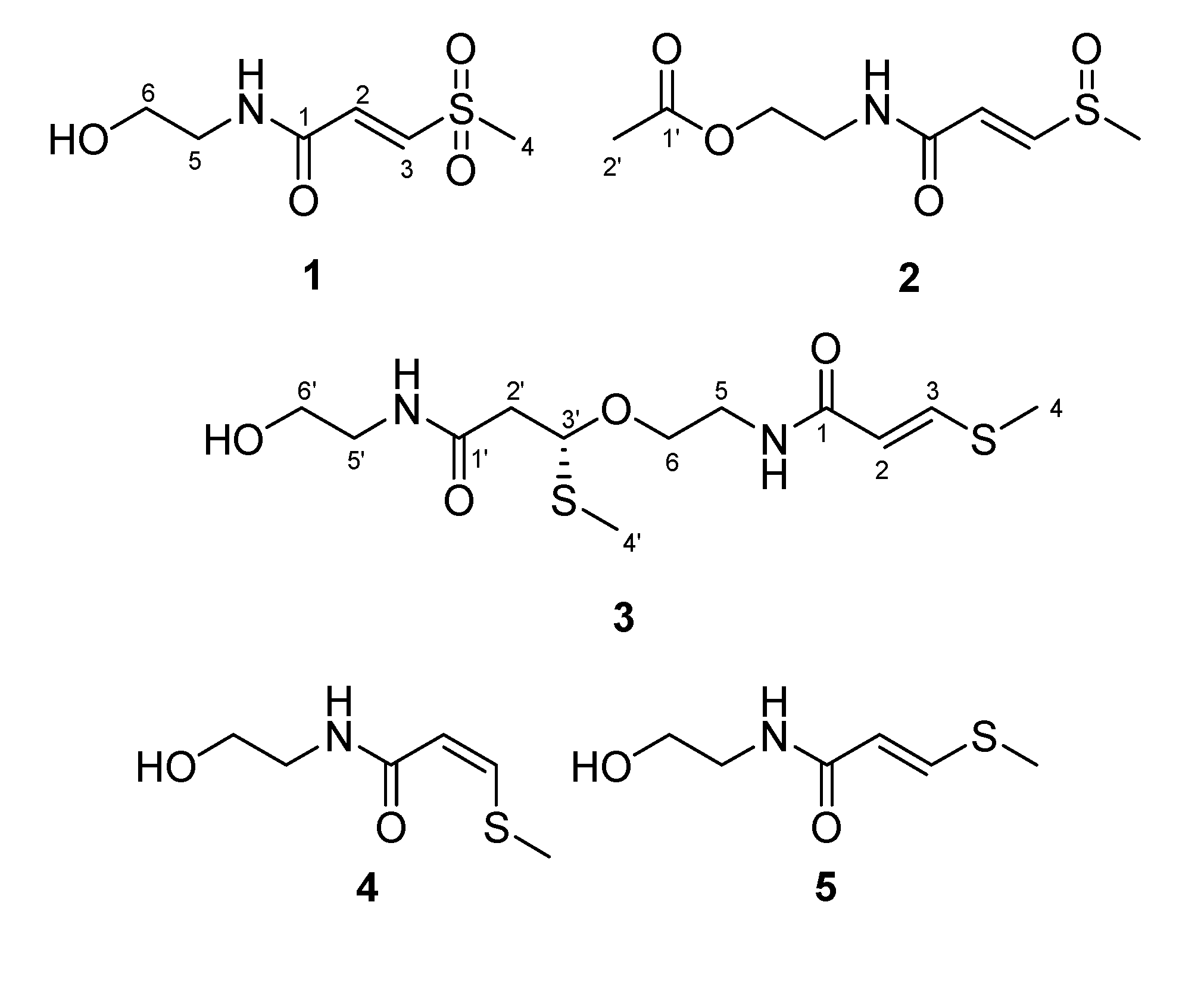
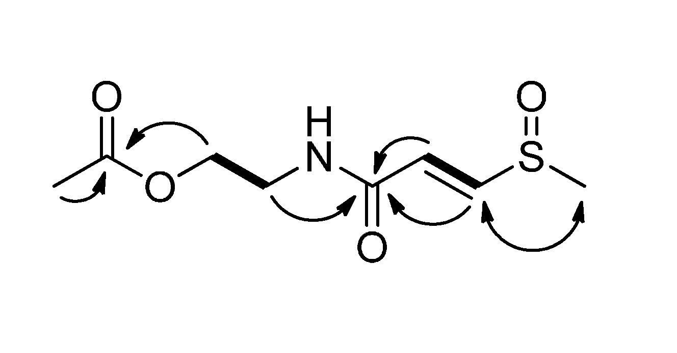
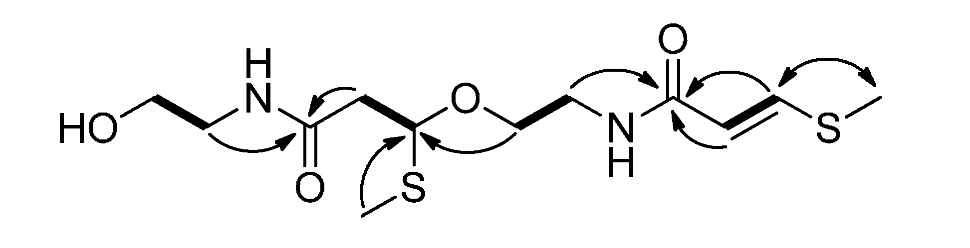
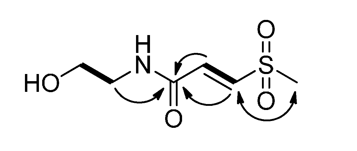
2. Results and Discussion

Four new compounds 14 and three known compounds, entadamide A (5) [5], entadamide C [6], and trans-3-methylsulfinyl-2-propenol [7] were isolated from the ethanolic extract of the aerial parts of C. nutans. The known compounds were identified by comparison of their physical and spectroscopic data to those reported in the literature. The structures of the new compounds were established by interpretation of their spectroscopic data, especially 2D NMR.
Clinamide A (1) was obtained as a pale yellow oil and was assigned the molecular formula, C6H11NO4S, as deduced from HRESIMS (m/z 216.0308 [M+Na]+). The IR spectrum showed strong absorption bands at 3380 cm−1 (OH), 1668 cm−1 (NH–C=O), 1555 cm−1 (C=C), and 1302 and 1135 cm−1 (O=S=O). The 1H-NMR data of 1 (Table 1) exhibited one methylsulfonyl signal [δH 3.08 (s)] [7], two methylene signals [δH 3.41 (t, J = 5.7 Hz) and 3.65 (t, J = 5.7 Hz)], and a trans-disubstituted double bond [δH 7.01 (d, J = 15.0 Hz)] and 7.43 (d, J = 15.0 Hz)]. The 13C-NMR (Table 2), DEPT and HSQC spectra of 1 showed six carbon signals, consisting of one methylsulfonyl group (δC 42.4), one oxymethylene group (δC 61.2), one methylene group (δC 43.4), one disubstituted olefin group (δC 136.3 and 140.1), and one amide carbonyl group (δC 164.6). These signature characteristics were used to identify 1 as an entadamide-type compound. The COSY spectrum of 1 (Figure 2) exhibited two proton spin systems of H-2 (δH 7.01)/H-3 (δH 7.43) and H-5 (δH 3.41)/H-6 (δH 3.65). In the HMBC spectrum, the H-2, H-3 and H-5 showed correlations to C-1 (δC 164.6), which indicated the presence of the double bond, the amide group and two methylene groups were connected. In addition, the key NOESY correlation between H-4 (δH 3.08) and H-3 indicated the methylsulfonyl group at C-3. These NMR data of 1 were closely similar to dambullin [8], except for the absence of an aromatic moiety. On the basis of the above discussion, the configuration of 1 was established as shown.
Table 1. 1H-NMR spectroscopic data of compounds 14 (1 and 2 in CD3OD, 3 and 4 in CDCl3, 400 MHz).
NO.1234
27.01 (d, 15.0)6.68 (d, 15.0)5.78 (d, 14.8)5.80 (d, 10.0)
37.43 (d, 15.0)7.63 (d, 15.0)7.62 (d, 14.8)6.83 (d, 10.0)
43.08 (s)2.76 (s)2.33 (s)2.35 (s)
53.41 (t, 5.7)3.54 (t, 5.4)3.65 (m)3.46 (t, 5.0)
3.38 (m)
63.65 (t, 5.7)4.17 (t, 5.4)3.88 (m)3.73 (t, 5.0)
3.52 (m)
2' 2.05 (s)2.80 (dd, 15.0, 9.8)
2.70 (dd, 15.0, 3.2)
3' 4.87 (dd, 9.8, 3.2)
4' 2.06 (s)
5' 3.52 (m)
3.38 (m)
6' 3.74 (m)
Table 2. 13C-NMR spectroscopic data of compounds 14 (1 and 2 in CD3OD, 3 and 4 in CDCl3, 100 MHz).
NO.1234
1164.6 (s)165.2 (s)165.1 (s)167.4 (s)
2136.3 (d)129.0 (d)115.9 (d)114.9 (d)
3140.1 (d)147.9 (d)142.6 (d)147.8 (d)
442.4 (q)39.9 (q)14.5 (q)19.4 (q)
543.4 (t)39.8 (t)38.9 (t)42.3 (t)
661.2 (t)63.8 (t)67.5 (t)62.4 (t)
1' 172.7 (s)171.0 (s)
2' 20.7 (q)43.3 (t)
3' 81.4 (d)
4' 10.2 (q)
5' 42.2 (t)
6' 62.1 (t)
Figure 2. COSY (bold lines), key HMBC (arrows), and NOESY (left right arrow) correlations of 1.
Molecules 19 20382 g002
Clinamide B (2) was assigned a molecular formula of C8H13NO4S by HRESIMS (m/z 242.0461 [M+Na]+). The UV spectrum exhibited absorption maximum at 247 nm. The IR spectrum showed bands at 1732 cm−1 (C=O), 1,660 cm−1 (NH–C=O), and 1,031 cm−1 (S=O). In the 1H-NMR spectrum (Table 1), a pair of trans-disubstituted olefinic protons [δH 6.68 (d, J = 15.0 Hz) and 7.63 (d, J = 15.0 Hz)] were found as similar to the case of 1. Besides, the proton spectrum showed a methylsulfinyl group [δH 2.76 (s)], a methyl group [δH 2.05 (s)], and two methylene groups [δH 3.54 (t, J = 5.4 Hz) and 4.17 (t, J = 5.4 Hz)]. The 13C-NMR (Table 2) spectrum revealed signals for an amide group as well as 1. In addition, the 13C-NMR and DEPT spectra exhibited one amide carbon group (δC 172.7), two methine groups (δC 129.0 and 147.9), two methylene groups (δC 39.8 and 63.8), and two methyl groups (δC 20.7 and 39.9). These signals were similar to entadamide C, suggesting that they possess the same skeleton. Compound 2 was found to have an additional acetyl group, when compared to entadamide C. This acetoxy group was assigned at C-6 by the HMBC correlations from both H-2' (δH 2.05) and H-6 (δH 4.17) to C-1' (δC 172.7) (Figure 3). Therefore, structure 2 was assigned to clinamide B.
Figure 3. COSY (bold lines), key HMBC (arrows), and NOESY (left right arrow) correlations of 2.
Molecules 19 20382 g003
Clinamide C (3) was isolated as pale yellow oil, and exhibited the molecular formula C12H22N2O4S2 by HRESIMS. The UV spectrum exhibited absorption maxima at 228 nm and 270 nm. The IR spectrum showed absorption bands at 3,296 cm−1 (OH), 1,644 cm−1 (NH–C=O), and 1,582 cm−1 (C=C). The 1H-NMR data (Table 1) showed two methylthio signals [δH 2.06 (s) and 2.33 (s)], and a trans-disubstituted double bond [δH 5.78 (d, J = 14.8 Hz) and 7.62 (d, J = 14.8 Hz)]. The 13C-NMR (Table 2), DEPT and HSQC spectra exhibited signals for 12 carbons, including two methylthio groups (δC 10.2 and 14.5), five methylene groups (δC 38.9, 42.2, 43.3, 62.1, and 67.5), one disubstituted olefin (δC 115.9 and 142.6), and two amide carbonyl groups (δC 165.1 and 171.0). The partial NMR spectroscopic data of 3 were close to those of entadamide A (5) [5]. The COSY spectrum (Figure 4) of 3 indicated the presence of fragments of H-2/H-3, H-5/H-6, H-2'/H-3', and H-5'/H-6'. The connectivity between two amide groups and above fragments were linked by HMBC correlations of H-2/C-1, H-3/C-1, H-5/C-1, H-4/C-3, H-2'/C-1', H-5'/C-1', H-3'/C-4', H-4'/C-3', and H-6/C-3'. The optical rotation value of 3 (−10.7) was opposite to the synthetic compound (S)-4-methoxy-4-(phenylthio)-2-butanone (+53.9). Thus, the relative configuration of C-3' was proposed to be R* on the basis of its optical rotation result. According to the above interpretation, the structure of 3 was established as clinamide C.
Figure 4. COSY (bold lines), key HMBC (arrows), and NOESY (left right arrow) correlations of 3.
Molecules 19 20382 g004
The HRESIMS indicated a molecular formula C6H11NO2S for compound 4, identical to that of 5. The 1H and 13C-NMR data of 4 were similar to those of 5, suggesting that they should be geometric isomers. The 1H-NMR spectrum of 4 exhibited the signals of a cis-double bond at [δH 5.80 (d, J = 10.0 Hz) and 6.83 (d, J = 10.0 Hz)]; however, a trans-one at [δH 5.85 (d, J = 14.8 Hz) and 7.56 (d, J = 14.8 Hz)] were found in that of 5. Thus, the structure of 2-cis-entadamide A (4) was determined as shown.
The results of the biological activity were discussed in the Conclusion section and the bioactivity tables were put in supporting information.

3. Experimental Section

3.1. General Procedures

Optical rotations were measured with a JASCO DIP-370 digital polarimeter. The IR spectra were measured on a Mattson Genesis II spectrometer. 1H and 13C-NMR spectra were recorded on Varian Unity Inova-600, Varian Unity Plus-400, or Varian Gemini 2000-200 NMR spectrometers. Chemical shifts are reported in parts per million (δ), and coupling constants (J) are expressed in Hertz. LRESIMS were measured on a VG Biotech Quattro 5022 mass spectrometer. HRESIMS were measured on a Bruker Daltonics APEX II mass spectrometer. Silica gel 60 (Merck, 230–400 mesh) and Sephadex LH-20 were used for column chromatography, while TLC analysis was carried out on silica gel GF254 pre-coated plates with detection using 50% H2SO4 followed by heating on a hot plate. HPLC was performed with Hitachi L-7100 series HPLC with a Bischoff RI detector and Shimadzu LC-10AT series HPLC with a SPD-10A UV-vis detector or a photodiode array detector. ODS (Hypersil®, 250 × 10 mm) columns were applied for HPLC separation.

3.2. Plant Material

The aerial parts of Clinacanthus nutans were collected in Taichung, Taiwan, in November 2010, and identified by Ming-Hong Yen. The samples were deposited at the Graduate Institute of Natural Products, Kaohsiung, Taiwan.

3.3. Extraction and Isolation

The aerial parts of C. nutans (2.3 kg) were collected freshly, cut into small pieces (ca. 0.5–1.0 cm thickness), and air-dried for three days. After drying, the materials were soaked and extracted with ethanol (10 L) at room temperature for three times, and then concentrated under reduced pressure to afford a crude extract (283.0 g). This crude extract was partitioned between EtOAc and H2O (1:1) to obtain an EtOAc-soluble layer. After evaporating the organic solvent, the EtOAc residue (51.1 g) was partitioned between n-hexane–EtOH–H2O (5:4:1) to afford an 80% ethanol extract. The 80% ethanol extract (14.6 g) was subjected to passage over a silica gel column using gradient elution of CH2Cl2–MeOH (1:0 to 5:1) to furnish fractions 1–20. Fraction 13 (618.5 mg) was separated on a Sephadex LH-20 column (5 × 55 cm) eluted with EtOAc–CH2Cl2–MeOH (1:1:6) to afford fraction 13-5 (298.0 mg), which was further chromatographied on a silica gel column (3 × 25 cm; CH2Cl2–MeOH, 20:1) to furnish subfractions 13-5-1 to 13-5-10. Compound 6 (16.0 mg) was obtained by separation of subfraction 13-5-8.9 (25.5 mg) on preparative TLC eluting with CH2Cl2–MeOH (6:1). Subfraction 13-5-4 (149.6 mg) was chromatographed by preparative TLC using CH2Cl2–MeOH (8:1) for elution, and further purified by reverse-phase HPLC (270 nm; Hypersil®, 250 × 10 mm) with MeOH–H2O (35:65) to yield 3 (3.8 mg; flow rate: 2 mL/min; Rt 11.5 min). Then the subfraction was eluted with MeOH–H2O (40:60) to yield 4 (6.8 mg; flow rate: 2 mL/min; Rt 8 min) as well as 5 (99.9 mg; flow rate: 2 mL/min; Rt 10 min). Fraction 15 (1035.1 mg) was purified on a Sephadex LH-20 column (5 × 55 cm) using EtOAc–CH2Cl2–MeOH (1:1:6) as eluting solvent to afford nine subfractions. Subfraction 15-5 (296.0 mg) was applied to a silica gel column (3 × 25 cm) eluted with CH2Cl2–MeOH (30:1) to give ten subfractions. Compound 2 was obtained by separation of subfraction 15-5-4 (40.1 mg) on preparative TLC eluting with CH2Cl2–MeOH (10:1) and further purification by reverse-phase HPLC (244 nm; Hypersil®, 250 × 10 mm) with MeOH–H2O (43:57) (0.8 mg; flow rate: 2 mL/min; Rt 7.5 min). Subfraction 15-5-7 (29.1 mg) was subjected to preparative TLC with CH2Cl2–MeOH (15:1) and further purified by reverse-phase HPLC (245 nm; Hypersil®, 250 × 10 mm) with MeOH–H2O (25:75) to yield 7 (14.2 mg; flow rate: 2 mL/min; Rt 9 min) and with MeOH–H2O (15:85) to yield 1 (2.2 mg; flow rate: 2 mL/min; Rt 13 min).
Clinamide A (1): Pale yellow oil; IR (neat): vmax 3380, 2927, 1668, 1555, 1302, 1135 cm−1; 1H-NMR (CD3OD, 400 MHz) and 13C-NMR (CD3OD, 100 MHz) spectroscopic data, see Table 1 and Table 2, respectively; HRESIMS: m/z 216.0308 [M+Na]+ (calcd. for C6H11NO4SNa, 216.0306).
Clinamide B (2): White amorphous powder; IR (neat): vmax 2918, 1732, 1031 cm−1; 1H-NMR (CD3OD, 400 MHz) and 13C-NMR (CD3OD, 100 MHz) spectroscopic data, see Table 1 and Table 2, respectively; HRESIMS: m/z 242.0461 [M+Na]+ (calcd. for C8H13NO4SNa, 242.0463).
Clinamide C (3): Pale yellow oil; [ α ] D 25 −10.7 (c 1.0, MeOH); IR (neat): vmax 3296, 2924, 1644, 1582 cm−1; 1H-NMR (CDCl3, 400 MHz) and 13C-NMR (CDCl3, 100 MHz) spectroscopic data, see Table 1 and Table 2, respectively; HRESIMS: m/z 345.0903 [M+Na]+ (calcd. for C12H22N2O4S2Na, 345.0905).
2-cis-Entadamide A (4): Pale yellow oil; IR (neat): vmax 3306, 2924, 1644, 1583 cm−1; 1H-NMR (CDCl3, 400 MHz) and 13C-NMR (CDCl3, 100 MHz) spectroscopic data, see Table 1 and Table 2, respectively; HRESIMS: m/z 184.0409 [M+Na]+ (calcd. for C6H11NO2SNa, 184.0408).

3.4. Anti-Inflammatory Assay

Human neutrophils were collected by means of Ficoll centrifugation and dextran sedimentation. Assessments of superoxide anion generation and elastase release were carried out according to known procedures [9,10]. In brief, superoxide anion production was assayed by monitoring the superoxide dismutase-inhibitable reduction of ferricytochrome c. Elastase release experiments were executed using MeO-Suc-Ala-Ala-Pro-Valp-nitroanilide as the elastase substrate. In the in vitro anti-inflammatory bioassay, the inhibitory effects on the generation of superoxide anion and the release of elastase by activated neutrophils were used as indicators. For significant activity of pure compounds, an inhibition rate ≥50% is required.

3.5. Anti-Dengue Virus Assay

Naïve Huh-7 cells were obtained from American type culture collection (ATCC) and cultured in Dulbecco’s modified Eagle’s medium (DMEM) containing 10% fetal bovine serum, 1% non-essential amino acids, and 1% antibiotic-antimycotic in a 5% CO2 in air atmosphere at 37 °C. Huh-7 cells were seeded at 24 wells plate at density 5 × 10⁴ cells/well for 12–16 h and then infected with DENV-2, strain 16681 at a multiplicity of infection (MOI) of 0.2 for 2 h at 37 °C. Cells were washed with PBS and then re-fed with DMEM-10% FBS medium containing test compounds. Cells were harvested at 72 h post-infection on a western blot assay with anti-DENV NS2B (1:2000; Abcam, Cambridge, MA, USA) or anti-GAPDH antibody (1:10000; GeneTex, Irvine, CA, USA), a loading control. The signal was detected using an ECL detection kit (PerkinElmer, Shelton, CT, USA). Ribavirin served as positive control [11].

3.6. Immune-Modulating Assay

To examine the inhibitory effect of Lactobacillus casei on IgE production, splenocyte obtained from ovalbumin (OVA)-primed BALB/c mice were re-stimulated in vitro with the same antigen in the presence of heat-killed L. casei. The effect of this bacterium on helper (Th) phenotype development was also examined with native T cells from T cell receptor-transgenic mice [12].

4. Conclusions

The plant material and extracts of C. nutans have now become popular commercial products in Thailand and Malaysia, even available through Internet markets worldwide, and dubitatively used in anticancer and anti-cardiovascular diseases, making the active components from this plant an important issue to be understood. Our current study enhances the bioactivity and chemical diversity of this plant material. In bioactive screening, the 80% ethanol extract showed anti-inflammatory, anti-dengue virus and immune-modulating activity. For the study of anti-inflammatory activity, the extract at 10 μg/mL had the strongest elastase release inhibitory effect at 68.33%. Moreover, this extract displayed moderate anti-dengue virus activity in the IC50 31.04 μg/mL. In an immune-modulating experiment, using 0.1 μg/mL of 80% ethanol extract led to up-regulation of IFN-γ. On the other hand, using high concentration of 80% ethanol extract (100 μg/mL) led to down-regulation of IFN-γ exhibiting immune-modulating activity. Unfortunately, the new compounds isolated so far did not showed any targeted activity.

Supplementary Materials

Supplementary materials can be found at https://www.mdpi.com/1420-3049/19/12/20382/s1.

Acknowledgments

This work was supported by grants from the National Science Council of Taiwan (103-2320-B-037-005-MY2, awarded to F.-R.C. and 102-2320-B-037-012-MY2, awarded to Y.-B.C.), and China Medical University under the Aim for Top University Plan of the Ministry of Education, Taiwan, awarded to Y.-C.W.

Author Contributions

Yang-Chang Wu and Fang-Rong Chang designed the whole experiment and contributed to manuscript preparation; Shu-Fen Tu analyzed data and wrote the manuscript; Rosa Huang Liu provided the plant materials; Yuan-Bin Cheng revised the manuscript and the format; Yu-Ming Hsu, Ying-Chi Du, and Mohamed El-Shazly analyzed the data and performed data acquisition.

Conflicts of Interest

The authors declare no conflict of interest.

References

  1. Sakdarat, S.; Shuyprom, A.; Pientong, C.; Ekalaksananan, T.; Thongchai, S. Bioactive constituents from the leaves of Clinacanthus nutans Lindau. Bioorg. Med. Chem. 2009, 17, 1857–1860. [Google Scholar] [CrossRef] [PubMed]
  2. Sakdarat, S.; Shuyprom, A.; Ayudhya, T.D.N.; Waterman, P.G.; Karagianis, G. Chemical composition investigation of the Clinacanthus nutans Lindau leaves. Thai J. Phytopharm. 2008, 15, 13–24. [Google Scholar]
  3. Kongkaew, C.; Chaiyakunapruk, N. Efficacy of Clinacanthus nutans extracts in patients with herpes infection: Systematic review and meta-analysis of randomized clinical trials. Complement. Ther. Med. 2011, 19, 47–53. [Google Scholar] [CrossRef] [PubMed]
  4. Janwitayanuchit, W.; Suwanborirux, K.; Patarapanich, C.; Pummangura, S.; Lipipun, V.; Vilaivan, T. Synthesis and anti-herpes simplex viral activity of monoglycosyl diglycerides. Phytochemistry 2003, 64, 1253–1264. [Google Scholar] [CrossRef] [PubMed]
  5. Ikegami, F.; Shibasaki, I.; Ohmiya, S.; Ruangrungsi, N.; Murakoshi, I. Entadamide A, a new sulphur-containing amide from Entada phaseoloides seeds. Chem. Pharm. Bull. 1985, 33, 5153–5154. [Google Scholar] [CrossRef]
  6. Ikegami, F.; Sekine, T.; Duangteraprecha, S.; Matsushita, N.; Matsuda, N.; Ruangrungsi, N.; Murakoshi, I. Entadamide C, a sulphur-containing amide from Entada phaseoloides. Phytochemistry 1989, 28, 881–882. [Google Scholar] [CrossRef]
  7. Tuntiwachwuttikul, P.; Pootaeng-on, Y.; Pansa, P.; Srisanpang, T.; Taylor, W.C. Sulfur-containing compounds from Clinacanthus siamensis. Chem. Pharm. Bull. 2003, 51, 1423–1425. [Google Scholar] [CrossRef] [PubMed]
  8. Greger, H.; Hofer, O.; Zechner, G.; Hadacek, F.; Wurz, G. Sulphones derived from methylthiopropenoic acid amides from Glycosmis angustifolia. Phytochemistry 1994, 37, 1305–1310. [Google Scholar] [CrossRef]
  9. Yu, H.-P.; Hsieh, P.-W.; Chang, Y.-J.; Chung, P.-J.; Kuo, L.-M.; Hwang, T.-L. 2-(2-Fluorobenzamido) benzoate ethyl ester (EFB-1) inhibits superoxide production by human neutrophils and attenuates hemorrhagic shock-induced organ dysfunction in rats. Free Radic. Biol. Med. 2011, 50, 1737–1748. [Google Scholar] [CrossRef]
  10. Yang, S.-C.; Chung, P.-J.; Ho, C.-M.; Kuo, C.-Y.; Hung, M.-F.; Huang, Y.-T.; Chang, W.-Y.; Chang, Y.-W.; Chan, K.-H.; Hwang, T.-L. Propofol inhibits superoxide production, elastase release, and chemotaxis in formyl peptide-activated human neutrophils by blocking formyl peptide receptor 1. J. Immunol. 2013, 190, 6511–6519. [Google Scholar] [CrossRef] [PubMed]
  11. Hsu, Y.-C.; Chen, N.-C.; Chen, P.-C.; Wang, C.-C.; Cheng, W.-C.; Wu, H.-N. Identification of a small-molecule inhibitor of dengue virus using a replicon system. Arch. Virol. 2012, 157, 681–688. [Google Scholar] [CrossRef] [PubMed]
  12. Shida, K.; Makino, K.; Morishita, A.; Takamizawa, K.; Hachimura, S.; Ametani, K.; Sato, T.; Kumagai, Y.; Habu, S.; Kaminogawa, S. Lactobacillus casei Inhibits Antigen-Induced IgE Secretion through Regulation of Cytokine Production in Murine Splenocyte Cultures. Int. Arch. Allergy Immunol. 1998, 115, 278–287. [Google Scholar] [CrossRef] [PubMed]
  • Sample Availability: Not available.

