Article

Methanolic Extracts of Solieria robusta Inhibits Proliferation of Oral Cancer Ca9-22 Cells via Apoptosis and Oxidative Stress

1
Department of Dentistry, Ten Chan General Hospital, Chung-Li 32043, Taiwan
2
Laboratory for Translational Oncology and Personalized Medicine, Rashid Latif Medical College, Lahore 54000, Pakistan
3
Department of Biomedical Science and Environmental Biology, Kaohsiung Medical University, Kaohsiung 80708, Taiwan
4
Department of Botany, Government College University, Lahore, Katchery Road Lahore 54000, Pakistan
5
Cancer Center, Kaohsiung Medical University Hospital, Kaohsiung Medical University, Kaohsiung 80708, Taiwan
6
Department of Radiation Oncology, Faculty of Medicine, College of Medicine, Kaohsiung Medical University, Kaohsiung 80708, Taiwan
7
Department of Radiation Oncology, Kaohsiung Medical University Hospital, Kaohsiung 80708, Taiwan
8
Department of Radiation Oncology, Kaohsiung Municipal Ta-Tung Hospital, Kaohsiung 80145, Taiwan
9
Department of Biological Sciences, National Sun Yat-Sen University, Kaohsiung 80424, Taiwan
10
Graduate Institute of Natural Products, College of Pharmacy, Kaohsiung Medical University, Kaohsiung 80708, Taiwan
11
Institute of Clinical Medicine, Kaohsiung Medical University, Kaohsiung 80708, Taiwan
12
Department of Surgery, Kaohsiung Municipal Ta-Tung Hospital, Kaohsiung 80145, Taiwan
13
Institute of Medical Science and Technology, National Sun Yat-sen University, Kaohsiung 80424, Taiwan
14
Research Center of Excellence for Environmental Medicine, Kaohsiung Medical University, Kaohsiung 80708, Taiwan
*
Authors to whom correspondence should be addressed.
Molecules 2014, 19(11), 18721-18732; https://doi.org/10.3390/molecules191118721
Submission received: 10 October 2014 / Revised: 7 November 2014 / Accepted: 10 November 2014 / Published: 14 November 2014
(This article belongs to the Section Natural Products Chemistry)

Abstract

:
Many red algae-derived natural products are known to have anticancer effects. The biological functions of the red alga Solieria robusta from the Karachi coast (Pakistan) remain unclear. Here, we prepared a methanolic extracts of S. robusta (MESR) to examine its possible anti-oral cancer effects and the corresponding mechanism of action. Cell viability of MESR-incubated oral cancer Ca9-22 cells was dose-responsively decreased (p < 0.001). According to a propidium iodide (PI)-based assay the cell cycle distribution was dramatically changed, especially for subG1 accumulation. Annexin V/PI assay of apoptosis using flow cytometry also showed that MESR-incubated Ca9-22 cells were dose-responsively increased (p < 0.001). For evaluation of oxidative stress in MESR-incubated Ca9-22 cells, we found that reactive oxygen species (ROS) were overexpressed dose- and time-responsively and mitochondrial depolarization was also increased (p < 0.001). Taken together, MESR showed inhibitory effects on oral cancer proliferation coupled with apoptosis and oxidative stress.

1. Introduction

The occurrence of oral cancers is high in Asian countries, including Taiwan. Although some tumor markers [1,2] of oral cancer have been reported for potential therapeutic targets, the main strategy for oral cancer therapy is still drug treatment. Recently, the strategy of combined treatment for cancer treatment is favored [3,4], making the identification of more potential drugs or natural products as candidates in drug combinations helpful. Mounting evidence shows that marine algae extracts and components have anti-cancer effects [5], for example green [6], brown [7], blue-green [8], and red algae [9]. Lycopene isolated from the green alga Chlorella marina displayed considerable anticancer activity against PC-3 and DU-145 prostate cancer cell lines [10]. The methanolic extracts of a brown alga Sargassum muticum reportedly inhibited the MCF-7 and MDA-MB-231 breast cancer cell lines [11]. A blue-green alga Spirulina platensis is a source of tetrapyrrolic components with notable activity against tumor growth in mice xenografted with pancreatic cancer cells [12]. Aqueous extracts of a red alga Gracilaria corticata [10] displayed the antiproliferative effect against human leukemic cells.
We have focused on the biological functions of red algae. Previously, we found that both ethanolic [13] and methanolic [14] extracts of the red alga Gracilaria tenuistipitata caused growth inhibition of oral cancer cells. Similarly, a methanolic extracts of Plocamium telfairiae displayed antiproliferative effects against colon cancer cells [15]. The red alga Agardhiella robusta, officially named Solieria robusta, can effectively reduce the lipid profile of diet-induced hyperlipidaemic rats [16]. According to the NCBI taxonomy browser [17], the species S. robusta, belonging to the family Solieriaceae contains 15 genera of red algae. Among them, S. robusta, Eucheuma serra, and Kappaphycus alvarezii are the main industrial resources of the polysaccharide carrageenan [18], a common food industry and medicinal supplement [19]. However, the possible anticancer function of the red alga S. robusta remains unclear. Because S. robusta is abundant along the Karachi coast of Pakistan, it is convenient to prepare methanolic extracts of S. robusta (namely for MESR). In this study, we aimed to explore the biological function of MESR toward oral cancer cells by analyzing their cell proliferation, cell cycle changes, apoptosis, and oxidative stress.

2. Results

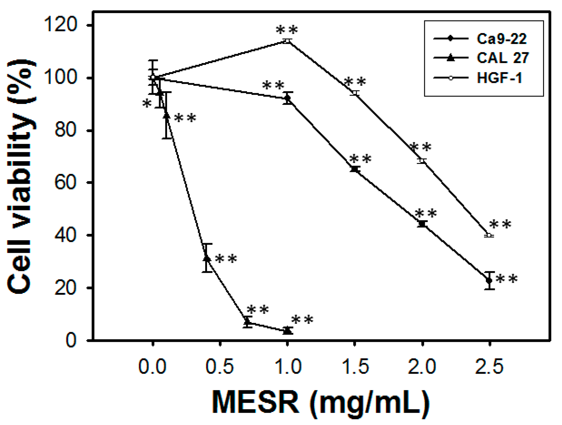
2.1. Cell Viability of MESR-Incubated Ca9-22 Cells

In oral cancer Ca9-22 (Figure 1), the relative cell viabilities (%) in terms of MTS assay for MESR treatments (0, 1, 1.5, 2, and 2.5 mg/mL) after 24 h were 100.0 ± 2.9, 92.1 ± 2.3, 65.2 ± 1.0, 44.3 ± 1.1, and 22.8 ± 3.2, respectively. In oral cancer CAL 27 cells, the relative cell viabilities (%) in terms of MTS assay for MESR treatments (0, 0.05, 0.1, 0.4, 0.7, and 1 mg/mL) after 24 h were 100.0 ± 6.3, 94.1 ± 5.6, 85.5 ± 8.8, 31.3 ± 5.3, 7.0 ± 1.9, and 3.7 ± 1.3, respectively. In oral normal HGF-1 cells, the relative cell viabilities (%) in terms of MTS assay for MESR treatments (0, 1, 1.5, 2, and 2.5 mg/mL) after 24 h were 100.0 ± 0.5, 113.9 ± 0.8, 94.1 ± 0.8, 68.4 ± 0.9, and 39.9 ± 0.3, respectively. Accordingly, cell viabilities of MESR-incubated oral cancer Ca9-22 and CAL 27 cells were dose-responsively decreased and treatment was less harmful to normal oral HGF-1 cells (p < 0.05–0.001 compared to control).
Figure 1. MTS-based cell viability of oral cancer Ca9-22 and CAL 27 cells as well as oral normal HGF-1 cells was differentially decreased by MESR. Cells were incubated with MESR (0–2.5 mg/mL) for 24 h. Data, means ± SDs (n = 11, 10, and 3, respectively). *p < 0.001 and ** p < 0.001 against control.
Molecules 19 18721 g001

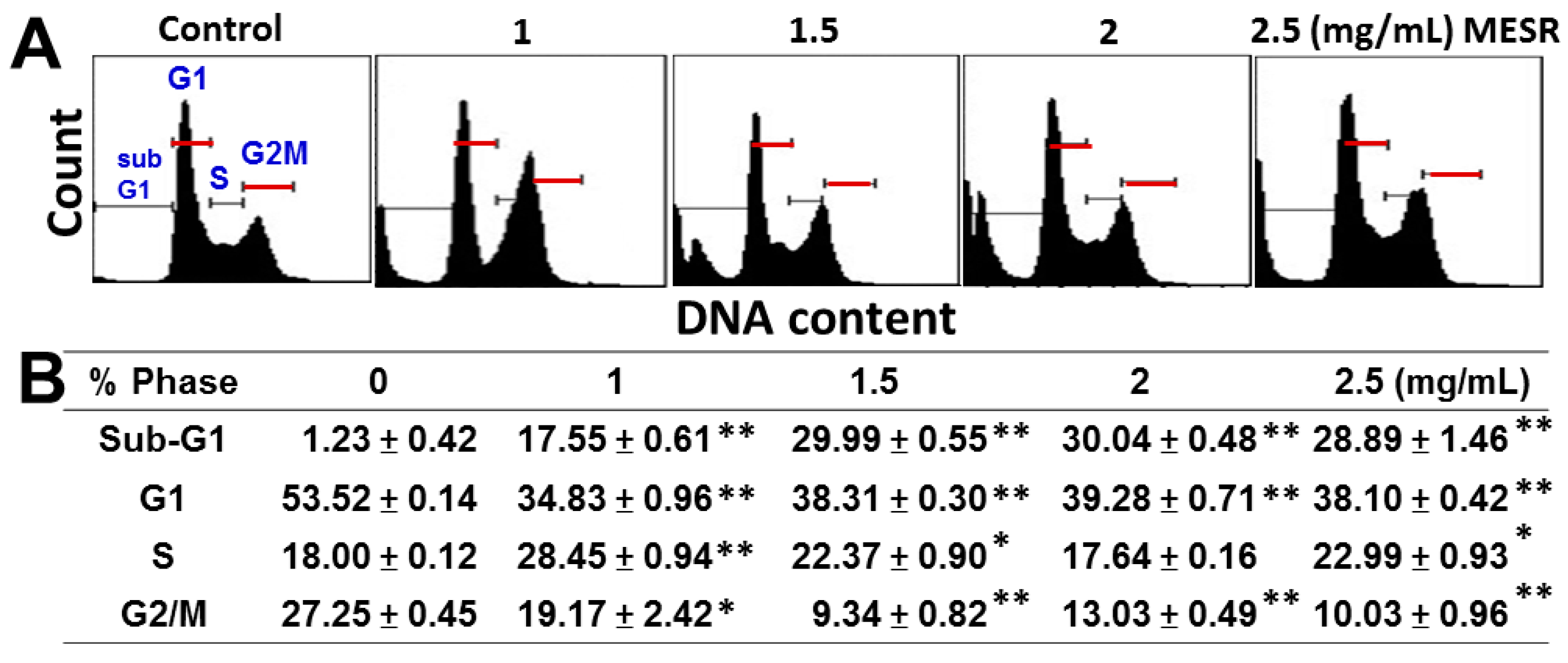
2.2. Cell Cycle Distribution of MESR-Incubated Ca9-22 Cells

The cell cycle patterns of flow cytometry of MESR-incubated cells are shown in Figure 2A. After MESR treatment (Figure 2B), the sub-G1 populations were dose-responsively increased in MESR-incubated oral cancer Ca9-22 cells (p < 0.001). G1 and G2/M phases were dramatically decreased and S phase was slightly increased after MESR treatments (p < 0.05–0.001).
Figure 2. Changes of the cell cycle distribution of MESR-incubated oral cancer Ca9-22 cells. Cells were incubated with MESR (0–2.5 mg/mL) for 24 h for flow cytometry analysis. (A) Typical cell cycle patterns of MESR-incubated Ca9-22 cells. (B) Statistics of cell cycle phases (%) for Figure 2A. Data, means ± SDs (n = 3). * p < 0.05; ** p < 0.001 against control.
Molecules 19 18721 g002

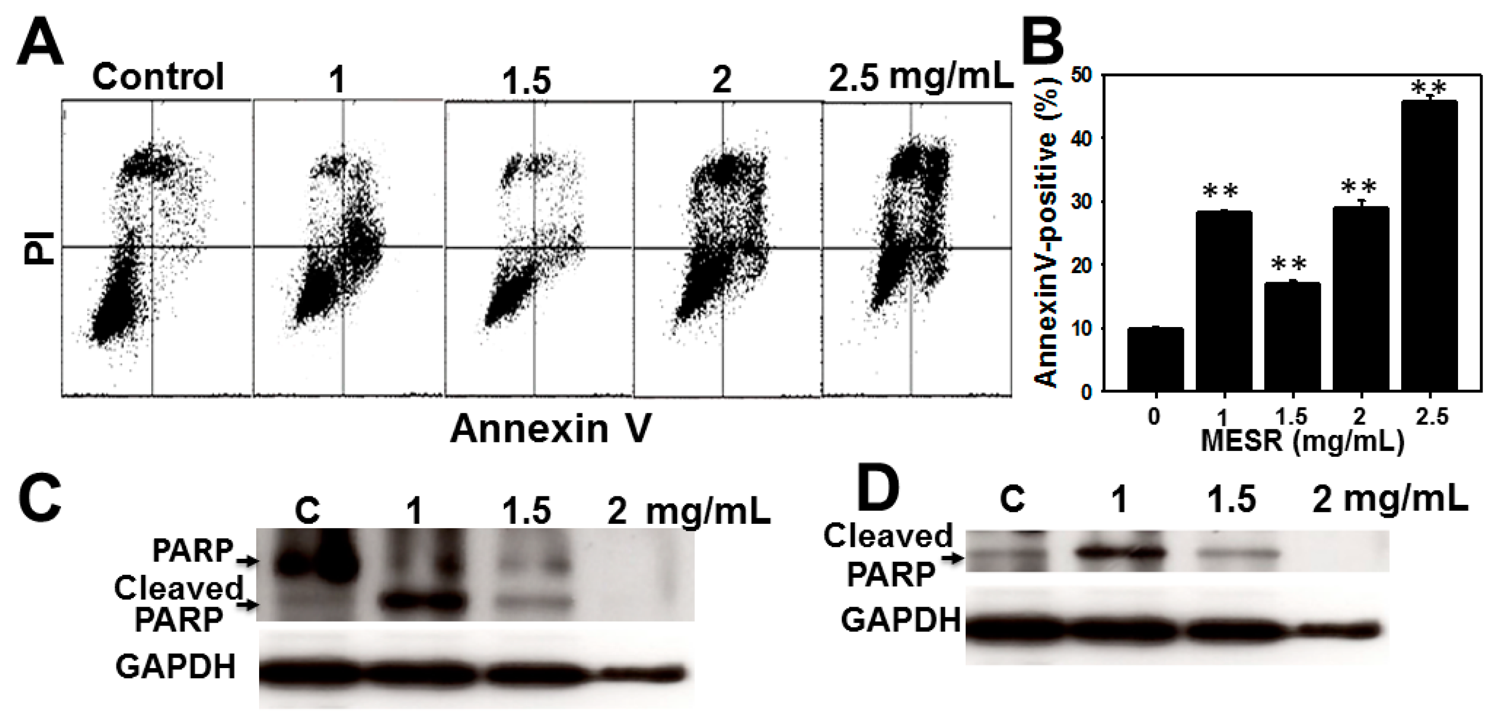
2.3. Apoptosis Analysis of MESR-Incubated Ca9-22 Cells

The flow cytometry annexin V/PI patterns of MESR-incubated cells are displayed in Figure 3A. In Figure 3B, the annexin V-positive expression (%) for MESR treatment of oral cancer Ca9-22 cells was dose-responsively increased (p < 0.001).
Figure 3. Changes of apoptosis of MESR-incubated oral cancer Ca9-22 cells. Cells were incubated with MESR (0–2.5 mg/mL) of for 24 h for flow cytometry and western blotting analyses. (A) Typical patterns of annexin V/PI method for MESR-incubated Ca9-22 cells. (B) Apoptosis statistics (%) in Figure 3A. Data, means ± SDs (n = 3). ** p < 0.001 against control. (C and D) western blotting of the uncut PARR and the apoptotic marker of cleaved PARP in MESR-treated Ca9-22 cells, respectively.
Molecules 19 18721 g003
For western blotting, the expressions of uncut poly ADP-ribose polymerase (PARP) were decreased and the apoptotic marker of the cleaved PARP [20] were increased by MESR treatment at 1 and 1.5 mg/mL compared to control (Figure 3C,D), respectively.

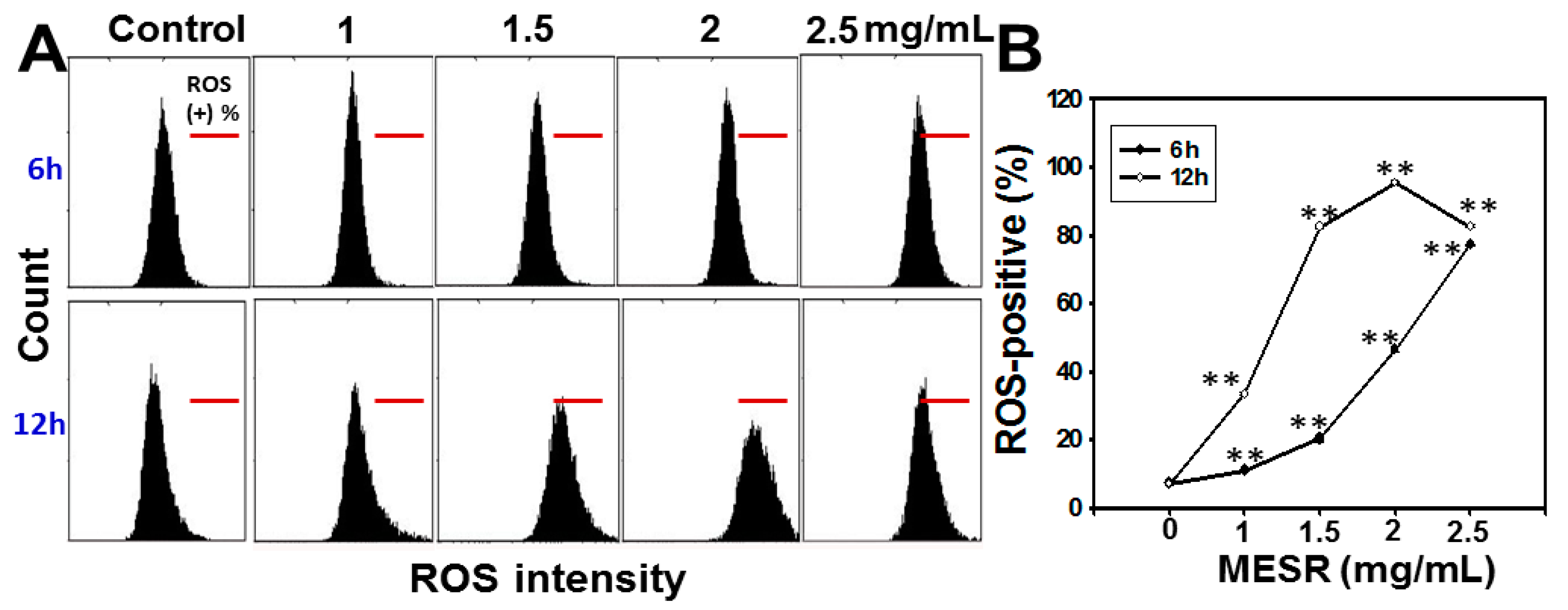
2.4. ROS Changes of MESR-Incubated Ca9-22 Cells

The ROS staining-positive patterns of MESR-incubated Ca9-22 cells are displayed in Figure 4A. After MESR treatments for 6 h and 12 h, the ROS staining-positive expression (%) of MESR-incubated oral cancer Ca9-22 cells was significantly accumulated in both dose- and time-responsive manners (p < 0.001) (Figure 4B).
Figure 4. Changes of ROS levels of MESR-incubated oral cancer Ca9-22 cells. Cells were incubated with MESR (0–2.5 mg/mL) for 6 h and 12 h for flow cytometry analysis. The horizontal red lines in each plot indicated ROS-positive (%). (A) Typical ROS patterns of MESR-incubated Ca9-22 cells. (B) Statistics of ROS-positive intensity (%) in Figure 4A. Data, means ± SDs (n = 3). ** p < 0.001 against control.
Molecules 19 18721 g004
Figure 5. Changes of MMP levels of MESR-incubated Ca9-22 oral cancer cells. Cells were incubated with MESR (0–2.5 mg/mL) for 24 h for flow cytometry analysis. The vertical lines in each plot dichotomised the MMP intensity scales into 50%/50% in reference to the left and right sides of control. The right side of MMP intensity was regarded as MMP-positive (%). (A) Typical MMP patterns of MESR-incubated Ca9-22 cells. (B) Statistics of relative MMP-positive intensity (%) in Figure 5A. All the MMP intensity data were normalized to control as 100% for the relative MMP (%) calculation. Data, means ± SDs (n = 3). ** p < 0.001 against control.
Molecules 19 18721 g005

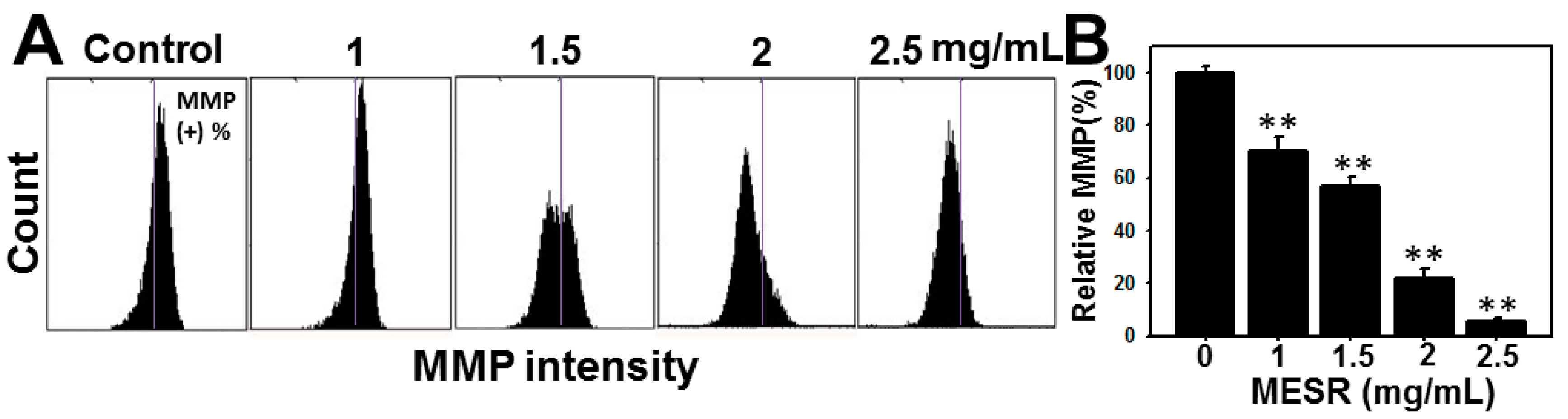
2.5. MMP Changes of MESR-Incubated Ca9-22 Cells

The MMP positive patterns of flow cytometer of MESR-incubated cells are displayed in Figure 5A. After MESR for 24 h, the MMP-positive expression (%) of oral cancer Ca9-22 cells was dose-responsively decreased (p < 0.001) (Figure 5B).

2.6. Discussion

Most marine algae are the common antioxidant-rich edible plants, especially their organic solvent extracts [21,22]. Although antioxidants were reported to have many benefits for chemoprevention [23,24], accumulating evidence shows that antioxidants may induce DNA damages, mutagenicity, and cell death [25,26]. Consistent with our results, the antiproliferative effect of MESR against oral cancer Ca9-22 cells was firstly demonstrated.
Many studies of methanol extracts of red algae also show the growth inhibitory effects against cancers. For example, the IC50 value of methanol extracts of red alga Plocamium telfairiae-treated colon cancer HT-29 cells after 24 h incubation was 2 µg/mL [15]. Methanol extracts of the red alga Halurus equisetifolius exhibited the IC50 values of 0.075, 0.060 and 0.175 mg/mL for human lung A549, colon HCT15 and breast MCF7 cancer cell lines, respectively [27]. The IC50 value of methanol [14] extracts of the red alga Gracilaria tenuistipitata-treated Ca9-22 cells after 24 h treatment was 0.326 mg/mL, respectively. In the present study, the IC50 values of the MESR-incubated Ca9-22 cells after 24 h were 1.89 mg/mL. For other oral cancer cell line CAL 27, its IC50 value was 0.296 mg/mL after 24 h treatment. Therefore, the dosages for growth inhibitory effects may differ for different species of red algae and cancer types. Although MESR showed higher a IC50 value than other red algae methanol extracts as previously described, it may be helpful for cancer treatment considering the possible synergy effects. Combination of different natural product extracts and bioactive constituents may generate synergistic multi-target effects [28,29].
Moreover, the IC50 value of MESR in normal oral HGF-1 cells is 2.32 mg/mL and of Ca9-22 cells is 1.89 mg/m, therefore MESR is more cytotoxic to Ca9-22 cells but less harmful to normal HGF-1 cells, especially for the concentrations ranging from 0 to 1.5 mg/mL. Under the IC50 value of MESR of Ca9-22 cells, the normal HGF-1 cells are still 80% viable. Therefore, the MESR has the selective killing potential for oral cancer therapy at suitable concentrations.
ROS induction is known as one of the causes for cancer cell death [30,31]. Induction of oxidative stress from drugs such as cisplatin and doxorubicin may have genotoxicity for cancer therapy [26,32]. Extracts of red alga Jania longifurca displayed significantly higher anticancer activity against MCF-7 breast cancer cells, primarily by enhancing oxidative stress and apoptosis in cancer cells [33]. During apoptosis, it was reported that discontinuous fragmentation of nuclear DNA may display discrete sub-G1 peaks using PI staining-based flow cytometer analysis [34]. Likewise, we found that MESR can induce subG1 accumulation and apoptosis in oral cancer Ca9-22 cells using flow cytometry analysis and western blotting. Based on the morphological hallmarks of apoptosis, using a microscope we also found that the MESR-induced cell death of OSCC Ca9-22 displays physiological biomarkers of apoptosis, including membrane blubbing, cell rounding and the formation of apoptotic bodies in a dose-response manner (data not shown). Moreover, MESR can induce ROS overproduction and mitochondrial dysfunction in OSCC Ca9-22. Therefore, oxidative stress may play a role in growth inhibition of oral cancer Ca9-22 cells after MESR treatment.

3. Experimental Section

3.1. Cell Cultures and Methanolic Extracts of S. robusta

A human oral gingival cancer cell line (Ca9-22), purchased from the Health Science Research Resources Bank (Osaka, Japan), was maintained in DMEM/F12 (3:2) medium (Gibco, Grand Island, NY, USA) containing 10% fetal bovine serum (Gibco), penicillin/streptomycin, and 0.03% glutamine under 37 °C with humidified 5% CO2.
S. robusta was collected from the Karachi coast, Pakistan. The species was identified from available literature and the specimen was confirmed by its Herbarium Sheet from Karachi University and authenticated by the phycologist Dr. Mustafa Shameel. After carefully washing in running water to avoid marine contaminants, the red alga were dried, ground, and stored in airtight containers before use. Powdered shade-dried red algal samples (500 g of S. robusta) were extracted at room temperature with methanol (1500 mL) for three weeks and then they were filtered through Whatman filter paper (nos. 1, 2, 41, and 42) to collect clarified filtrates (1 L). Finally, they was evaporated (65–70 °C) under vacuum to produce a dark green viscous oily mass (17.34 g) of methanolic extract of S. robusta (MESR). The NMR spectrum revealed signals in the δH 3.0–4.0 region representing the sugar moiety and signals in the δH 0.5–2.5 region representing fatty acids (data not shown). It was dissolved in 0.1% dimethyl sulfoxide (DMSO) before treatment.

3.2. Cell Viability

Cell viability was detected by a CellTiter 96® AQueous One Solution Cell Proliferation Assay (MTS, Promega Corporation, Madison, WI, USA) as described [35]. In brief, cells after seeding overnight were incubated with MESR (0, 1, 1.5, 2 and 2.5 mg/mL) or DMSO (0.025%) as a control for 24 h. Subsequently, the MTS assay was performed and results recorded by an ELISA plate reader.

3.3. Cell Cycle Distribution

Cell cycle analysis was based on DNA staining method as described [36]. In brief, cells were incubated with0, 1, 1.5, 2 and 2.5 mg/mL of MESR for 24 h. After harvest and PBS washing, cells were fixed with 70% ethanol and harvested by centrifugation. Subsequently, cells were resuspended in PBS solution containing 50 µg/mL of propidium iodide (PI, Sigma, St Louis, MO, USA) and waited for 30 min at 37 °C in darkness. Cell cycle phases were determined by a FACS Calibur flow cytometer (Becton-Dickinson, Mansfield, MA, USA) and FlowJo software (version 10) (Tree Star Inc., Ashland, OR, USA).

3.4. Apoptosis Analysis

Apoptosis was monitored by annexin V (Strong Biotect Corporation, Taipei, Taiwan)/PI (Sigma) method as described [37,38]. In brief, cells after seeding overnight were treated with or without MESR for 24 h. After harvest, cells were resuspended with the binding buffer containing 5 ng/µL of annexin-V-fluorescein isothiocyanate (FITC) and 20 µg/mL of PI for 30 min incubation. Subsequently, 400 µL of PBS was added and resuspended for flow cytometer analysis (BD Accuri™ C6; Becton-Dickinson) and a BD Accuri™ C6 Software (version 1.0.264).
Apoptosis was monitored by western blotting in terms of cleaved PARP expression, which was a apoptosis marker [20]. Western blotting was performed as described previously [35]. Western blotting was performed as described previously [34]. Briefly, the 20 µg protein lysates were separated by 10% SDS-polyacrylamide gel electrophoresis. After electrotransferring, PVDF membranes were probed with monoclonal antibody specific for uncut/cleaved forms of the poly ADP-ribose polymerase (PARP) (1:1000; #9542, Cell Signaling Technology; Beverly, MA, USA) and the cleaved PARP (Asp214) (D64E10) XP® Rabbit mAb (1:1000; #5625, Cell Signaling Technology), glyceraldehyde-3-phosphate dehydrogenase (GAPDH) (1:10,000; GeneTex inc.; San Antonio, TX, USA). Signals were detected by the Advansta WesternBright™ ECL Western blotting detection kit (Menlo Park, CA, USA).

3.5. Intracellular ROS Level

ROS detection was based on the fluorescence change of dye as described [14]. After seeding overnight, cells were incubated with or without MESR for 6 h or 12 h. After washing, cells were incubated with 100 nM of 2′,7′-dichlorodihydrofluorescein diacetate (DCFH-DA) in the culture incubator for 30 min. After harvest and washing, cells were resuspended in PBS for flow cytometer analysis (BD Accuri™ C6).

3.6. Mitochondrial Membrane Potential (MMP)

MMP was analyzed as described [39]. After seeding overnight, cells were incubated with MESR. Cells were incubated with A) in an incubator for 20 min. After washing, cells were resuspended in PBS for flow cytometer analysis (BD 50 nM of DiOC2 (3) provided in the MitoProbe™ DiOC2 (3) assay kit (Invitrogen, Eugene, OR, US Accuri™ C6).

3.7. Statistical Analysis

All statistics were based on the comparison between the drug-treated data with the controls using Student’s t-test.

4. Conclusions

MESR was proved to be antiproliferative against oral cancer Ca9-22 cells coupled with apoptosis, ROS overexpression, and mitochondrial dysfunction effects. MESR thus has potential to be a supplement for oral cancer therapy.

Acknowledgments

This work was supported by funds of the Ministry of Science and Technology (MOST 103-2320-B-037-008), the Kaohsiung Medical University “Aim for the Top Universities Grant, grant No. KMU-TP103A33”, the National Sun Yat-sen University-KMU Joint Research Project (#NSYSU-KMU 103-p014), the Kaohsiung Municipal Ta-Tung Hospital (kmtth-102-011), the Ten Chan General Hospital, Chung-Li and KMU Joint Research Project (ST102004), and the Health and welfare surcharge of tobacco products, the Ministry of Health and Welfare, Taiwan, Republic of China (MOHW103-TD-B-111-05).

Author Contributions

Y.-H.Y. designed the experiments. A.A.F. and G.B. prepared the algal extract. K.-T.L. performed the survival and Flow cytometry analyses. J.-Y.T. and C.-Y.W. performed statistics analyses. Y.-B.C. performed the NMR analysis. Y.-H.Y., M.-F.H. and H.-W.C. were involved in discussion and editing the manuscript. All authors read and approved the final manuscript.

Conflicts of Interest

The authors declare no conflict of interest.

References

  1. Yen, C.Y.; Chen, C.H.; Chang, C.H.; Tseng, H.F.; Liu, S.Y.; Chuang, L.Y.; Wen, C.H.; Chang, H.W. Matrix metalloproteinases (MMP) 1 and MMP10 but not MMP12 are potential oral cancer markers. Biomarkers 2009, 14, 244–249. [Google Scholar] [CrossRef]
  2. Yen, C.Y.; Huang, C.Y.; Hou, M.F.; Yang, Y.H.; Chang, C.H.; Huang, H.W.; Chen, C.H.; Chang, H.W. Evaluating the performance of fibronectin 1 (FN1), integrin alpha4beta1 (ITGA4), syndecan-2 (SDC2), and glycoprotein CD44 as the potential biomarkers of oral squamous cell carcinoma (OSCC). Biomarkers 2013, 18, 63–72. [Google Scholar] [CrossRef] [PubMed]
  3. Okui, T.; Shimo, T.; Fukazawa, T.; Hassan, N.M.; Honami, T.; Ibaragi, S.; Takaoka, M.; Naomoto, Y.; Sasaki, A. Novel HSP90 inhibitor NVP-AUY922 enhances the anti-tumor effect of temsirolimus against oral squamous cell carcinoma. Curr. Cancer Drug Targets 2012, 13, 289–299. [Google Scholar] [CrossRef]
  4. Hu, Y.; McIntosh, G.H.; le Leu, R.K.; Nyskohus, L.S.; Woodman, R.J.; Young, G.P. Combination of selenium and green tea improves the efficacy of chemoprevention in a rat colorectal cancer model by modulating genetic and epigenetic biomarkers. PLoS One 2013, 8, e64362. [Google Scholar] [CrossRef] [PubMed]
  5. Lee, J.C.; Hou, M.F.; Huang, H.W.; Chang, F.R.; Yeh, C.C.; Tang, J.Y.; Chang, H.W. Marine algal natural products with anti-oxidative, anti-inflammatory, and anti-cancer properties. Cancer Cell Int. 2013, 13, 55. [Google Scholar] [CrossRef] [PubMed]
  6. Park, H.Y.; Lim, C.W.; Kim, Y.K.; Yoon, H.D.; Lee, K.J. Immunostimulating and anticancer activities of hot water extract from Capsosiphon fulvescens. J. Kor. Soc. Appl. Biol. Chem. 2006, 49, 343–348. [Google Scholar]
  7. Costa, L.S.; Fidelis, G.P.; Telles, C.B.; Dantas-Santos, N.; Camara, R.B.; Cordeiro, S.L.; Costa, M.S.; Almeida-Lima, J.; Melo-Silveira, R.F.; Oliveira, R.M.; et al. Antioxidant and antiproliferative activities of heterofucans from the seaweed Sargassum filipendula. Mar. Drugs 2011, 9, 952–966. [Google Scholar] [CrossRef] [PubMed]
  8. Khan, Z.; Bhadouria, P.; Bisen, P.S. Nutritional and therapeutic potential of Spirulina. Curr. Pharm. Biotechnol. 2005, 6, 373–379. [Google Scholar] [CrossRef] [PubMed]
  9. Zandi, K.; Ahmadzadeh, S.; Tajbakhsh, S.; Rastian, Z.; Yousefi, F.; Farshadpour, F.; Sartavi, K. Anticancer activity of Sargassum oligocystum water extract against human cancer cell lines. Eur. Rev. Med. Pharmacol. Sci. 2010, 14, 669–673. [Google Scholar] [PubMed]
  10. Renju, G.L.; Muraleedhara Kurup, G.; Bandugula, V.R. Effect of lycopene isolated from Chlorella marina on proliferation and apoptosis in human prostate cancer cell line PC-3. Tumour Biol. 2014. [Google Scholar] [CrossRef]
  11. Namvar, F.; Mohamad, R.; Baharara, J.; Zafar-Balanejad, S.; Fargahi, F.; Rahman, H.S. Antioxidant, antiproliferative, and antiangiogenesis effects of polyphenol-rich seaweed (Sargassum muticum). Biomed. Res. Int. 2013, 2013. [Google Scholar] [CrossRef] [PubMed]
  12. Konickova, R.; Vankova, K.; Vanikova, J.; Vanova, K.; Muchova, L.; Subhanova, I.; Zadinova, M.; Zelenka, J.; Dvorak, A.; Kolar, M.; et al. Anti-cancer effects of blue-green alga Spirulina platensis, a natural source of bilirubin-like tetrapyrrolic compounds. Ann. Hepatol. 2014, 13, 273–283. [Google Scholar]
  13. Yeh, C.C.; Tseng, C.N.; Yang, J.I.; Huang, H.W.; Fang, Y.; Tang, J.Y.; Chang, F.R.; Chang, H.W. Antiproliferation and induction of apoptosis in Ca9-22 oral cancer cells by ethanolic extract of Gracilaria tenuistipitata. Molecules 2012, 17, 10916–10927. [Google Scholar] [CrossRef] [PubMed]
  14. Yeh, C.C.; Yang, J.I.; Lee, J.C.; Tseng, C.N.; Chan, Y.C.; Hseu, Y.C.; Tang, J.Y.; Chuang, L.Y.; Huang, H.W.; Chang, F.R.; et al. Anti-proliferative effect of methanolic extract of Gracilaria tenuistipitata on oral cancer cells involves apoptosis, DNA damage, and oxidative stress. BMC Complement. Altern. Med. 2012, 12, 142. [Google Scholar]
  15. Kim, J.Y.; Yoon, M.Y.; Cha, M.R.; Hwang, J.H.; Park, E.; Choi, S.U.; Park, H.R.; Hwang, Y.I. Methanolic extracts of Plocamium telfairiae induce cytotoxicity and caspase-dependent apoptosis in HT-29 human colon carcinoma cells. J. Med. Food 2007, 10, 587–593. [Google Scholar] [CrossRef] [PubMed]
  16. Ara, J.; Sultana, V.; Qasim, R.; Ahmad, V.U. Hypolipidaemic activity of seaweed from Karachi coast. Phytother. Res. 2002, 16, 479–483. [Google Scholar] [CrossRef] [PubMed]
  17. Sayers, E.W.; Barrett, T.; Benson, D.A.; Bryant, S.H.; Canese, K.; Chetvernin, V.; Church, D.M.; DiCuccio, M.; Edgar, R.; Federhen, S.; et al. Database resources of the National Center for Biotechnology Information. Nucleic Acids Res. 2009, 37, D5–D15. [Google Scholar]
  18. Kamenarska, Z.; Taniguchi, T.; Ohsawa, N.; Hiraoka, M.; Itoh, N. A vanadium-dependent bromoperoxidase in the marine red alga Kappaphycus alvarezii (Doty) Doty displays clear substrate specificity. Phytochemistry 2007, 68, 1358–1366. [Google Scholar] [CrossRef] [PubMed]
  19. Pooja, S. Algae used as medicine and food-a short review. J. Pharm. Sci. Res. 2014, 6, 33–35. [Google Scholar]
  20. O’Brien, M.A.; Moravec, R.A.; Riss, T.L. Poly (ADP-ribose) polymerase cleavage monitored in situ in apoptotic cells. Biotechniques 2001, 30, 886–891. [Google Scholar] [PubMed]
  21. Zakaria, N.A.; Ibrahim, D.; Sulaiman, S.F.; Supardy, N.A. Assessment of antioxidant activity, total phenol content and in vitro toxicity of Malaysian red seaweed. J. Chem. Pharm. Res. 2011, 3, 182–191. [Google Scholar]
  22. Wang, T.; Jónsdóttir, R.; Ólafsdóttir, G. Total phenolic compounds, radical scavenging and metal chelation of extract from Icelandic seaweed. Food Chem. 2009, 116, 240–248. [Google Scholar] [CrossRef]
  23. Khan, N.; Afaq, F.; Mukhtar, H. Cancer chemoprevention through dietary antioxidants: Progress and promise. Antioxid. Redox Signal. 2008, 10, 475–510. [Google Scholar] [CrossRef] [PubMed]
  24. Yang, J.I.; Yeh, C.C.; Lee, J.C.; Yi, S.C.; Huang, H.W.; Tseng, C.N.; Chang, H.W. Aqueous extracts of the edible Gracilaria tenuistipitata are protective against H2O2-induced DNA damage, growth inhibition, and cell cycle arrest. Molecules 2012, 17, 7241–7254. [Google Scholar] [CrossRef] [PubMed]
  25. Lu, L.Y.; Ou, N.; Lu, Q.B. Antioxidant induces DNA damage, cell death and mutagenicity in human lung and skin normal cells. Sci. Rep. 2013, 3, 3169. [Google Scholar] [PubMed]
  26. Fox, J.T.; Sakamuru, S.; Huang, R.; Teneva, N.; Simmons, S.O.; Xia, M.; Tice, R.R.; Austin, C.P.; Myung, K. High-throughput genotoxicity assay identifies antioxidants as inducers of DNA damage response and cell death. Proc. Natl. Acad. Sci. USA 2012, 109, 5423–5428. [Google Scholar] [CrossRef] [PubMed]
  27. Dellai, A.; Deghrigue, M.; Bouraoui, A. Evaluation of the antiproliferative activity of methanol extract and its fractions from the mediterranean seaweed, Halurus equisetifolius. Int. J. Pharm. Pharm. Sci. 2013, 5, 148–152. [Google Scholar]
  28. Yang, Y.; Zhang, Z.; Li, S.; Ye, X.; Li, X.; He, K. Synergy effects of herb extracts: Pharmacokinetics and pharmacodynamic basis. Fitoterapia 2014, 92, 133–147. [Google Scholar] [CrossRef] [PubMed]
  29. Wagner, H. Synergy research: Approaching a new generation of phytopharmaceuticals. Fitoterapia 2011, 82, 34–37. [Google Scholar] [CrossRef] [PubMed]
  30. Gorrini, C.; Harris, I.S.; Mak, T.W. Modulation of oxidative stress as an anticancer strategy. Nat. Rev. Drug Discov. 2013, 12, 931–947. [Google Scholar] [CrossRef] [PubMed]
  31. Trachootham, D.; Alexandre, J.; Huang, P. Targeting cancer cells by ROS-mediated mechanisms: A radical therapeutic approach? Nat. Rev. Drug Discov. 2009, 8, 579–591. [Google Scholar] [CrossRef] [PubMed]
  32. Deavall, D.G.; Martin, E.A.; Horner, J.M.; Roberts, R. Drug-induced oxidative stress and toxicity. J. Toxicol. 2012, 2012. [Google Scholar] [CrossRef]
  33. Kurt, O.; Ozdal-Kurt, F.; Tuglu, M.; Akcora, C. The cytotoxic, neurotoxic, apoptotic and antiproliferative activities of extracts of some marine algae on the MCF-7 cell line. Biotech. Histochem 2014, 89, 568–576. [Google Scholar] [CrossRef] [PubMed]
  34. Kajstura, M.; Halicka, H.D.; Pryjma, J.; Darzynkiewicz, Z. Discontinuous fragmentation of nuclear DNA during apoptosis revealed by discrete “sub-G1” peaks on DNA content histograms. Cytometry A 2007, 71, 125–131. [Google Scholar] [CrossRef] [PubMed]
  35. Chiu, C.C.; Haung, J.W.; Chang, F.R.; Huang, K.J.; Huang, H.M.; Huang, H.W.; Chou, C.K.; Wu, Y.C.; Chang, H.W. Golden berry-derived 4beta-hydroxywithanolide E for selectively killing oral cancer cells by generating ROS, DNA damage, and apoptotic pathways. PLoS One 2013, 8, e64739. [Google Scholar] [CrossRef] [PubMed]
  36. Chen, B.H.; Chang, H.W.; Huang, H.M.; Chong, I.W.; Chen, J.S.; Chen, C.Y.; Wang, H.M. (−)-Anonaine induces DNA damage and inhibits growth and migration of human lung carcinoma h1299 cells. J. Agric. Food Chem. 2011, 59, 2284–2290. [Google Scholar] [CrossRef] [PubMed]
  37. Chiu, C.C.; Liu, P.L.; Huang, K.J.; Wang, H.M.; Chang, K.F.; Chou, C.K.; Chang, F.R.; Chong, I.W.; Fang, K.; Chen, J.S.; et al. Goniothalamin inhibits growth of human lung cancer cells through DNA damage, apoptosis, and reduced migration ability. J. Agric. Food Chem. 2011, 59, 4288–4293. [Google Scholar]
  38. Huang, H.W.; Chung, Y.A.; Chang, H.S.; Tang, J.Y.; Chen, I.S.; Chang, H.W. Antiproliferative effects of methanolic extracts of Cryptocarya concinna Hance roots on oral cancer Ca9-22 and CAL 27 cell lines involving apoptosis, ROS induction, and mitochondrial depolarization. Sci. World J. 2014, 2014. [Google Scholar] [CrossRef]
  39. Yen, C.Y.; Chiu, C.C.; Haung, R.W.; Yeh, C.C.; Huang, K.J.; Chang, K.F.; Hseu, Y.C.; Chang, F.R.; Chang, H.W.; Wu, Y.C. Antiproliferative effects of goniothalamin on Ca9-22 oral cancer cells through apoptosis; DNA damage and ROS induction. Mutat. Res. 2012, 747, 253–258. [Google Scholar] [CrossRef] [PubMed]
  • Sample Availability: Not available.

