<?xml version="1.0" encoding="UTF-8"?>
<!DOCTYPE article PUBLIC "-//NLM//DTD Journal Publishing DTD v2.3 20070202//EN" "journalpublishing.dtd">
<article xmlns:mml="http://www.w3.org/1998/Math/MathML" xmlns:xlink="http://www.w3.org/1999/xlink" xml:lang="en" article-type="research-article">
  <front>
    <journal-meta>
      <journal-id journal-id-type="publisher-id">molecules</journal-id>
      <journal-title>Molecules</journal-title>
      <abbrev-journal-title abbrev-type="publisher">Molecules</abbrev-journal-title>
      <abbrev-journal-title abbrev-type="pubmed">Molecules</abbrev-journal-title>
      <issn pub-type="epub">1420-3049</issn>
      <publisher>
        <publisher-name>MDPI</publisher-name>
      </publisher>
    </journal-meta>
    <article-meta>
      <article-id pub-id-type="doi">10.3390/molecules170911242</article-id>
      <article-id pub-id-type="publisher-id">molecules-17-11242</article-id>
      <article-categories>
        <subj-group>
          <subject>Article</subject>
        </subj-group>
      </article-categories>
      <title-group>
        <article-title>A Phytoanticipin Derivative, Sodium Houttuyfonate, Induces <italic>in Vitro</italic> Synergistic Effects with Levofloxacin against Biofilm Formation by <italic>Pseudomonas aeruginosa</italic></article-title>
      </title-group>
      <contrib-group>
        <contrib contrib-type="author">
          <name>
            <surname>Shao</surname>
            <given-names>Jing</given-names>
          </name>
        </contrib>
        <contrib contrib-type="author">
          <name>
            <surname>Cheng</surname>
            <given-names>Huijuan</given-names>
          </name>
          <xref rid="c1-molecules-17-11242" ref-type="corresp">*</xref>
        </contrib>
        <contrib contrib-type="author">
          <name>
            <surname>Wang</surname>
            <given-names>Changzhong</given-names>
          </name>
        </contrib>
        <contrib contrib-type="author">
          <name>
            <surname>Wang</surname>
            <given-names>Yan</given-names>
          </name>
        </contrib>
      </contrib-group>
      <aff id="af1-molecules-17-11242">Laboratory of Microbiology and Immunology, School of Chinese and Western Integrative Medicine, Anhui University of Traditional Chinese Medicine, Hefei 230038, China; Email: <email>ustcsj@126.com</email> (J.S.); <email>ahwcz63@sina.com</email> (C.W.); <email>abudao2002@163.com</email> (Y.W.)</aff>
      <author-notes>
        <corresp id="c1-molecules-17-11242"><label>*</label> Author  to whom correspondence should be addressed; Email: <email>chenghuijuan53@126.com</email>; Tel.: +86-551-516-9204; Fax: +86-551-516-9192.</corresp>
      </author-notes>
      <pub-date pub-type="epub">
        <day>20</day>
        <month>09</month>
        <year>2012</year>
      </pub-date>
      <pub-date pub-type="collection"><month>09</month>
        <year>2012</year>
      </pub-date>
      <volume>17</volume>
      <issue>9</issue>
      <fpage>11242</fpage>
      <lpage>11254</lpage>
      <history>
        <date date-type="received">
          <day>09</day>
          <month>08</month>
          <year>2012</year>
        </date>
        <date date-type="rev-recd">
          <day>17</day>
          <month>09</month>
          <year>2012</year>
        </date>
        <date date-type="accepted">
          <day>18</day>
          <month>09</month>
          <year>2012</year>
        </date>
      </history>
      <permissions>
        <copyright-statement>©  2012 by the authors; licensee MDPI, Basel, Switzerland.</copyright-statement>
        <copyright-year>2012</copyright-year>
        <license xmlns:xlink="http://www.w3.org/1999/xlink" license-type="open-access" xlink:href="http://creativecommons.org/licenses/by/3.0/">
          <p>This article is an open-access article distributed under the terms and conditions of the Creative Commons Attribution license (http://creativecommons.org/licenses/by/3.0/).</p>
        </license>
      </permissions>
      <abstract>
        <p>Antibiotic resistance has become the main deadly factor in infections, as bacteria can protect themselves by hiding in a self-constructed biofilm. Consequently, more attention is being paid to the search for “non-antibiotic drugs” to solve this problem. Phytoanticipins, the natural antibiotics from plants, could be a suitable alternative, but few works on this aspect have been reported. In this study, a preliminary study on the synergy between sodium houttuyfonate (SH) and levofloxacin (LFX) against the biofilm formation of <italic>Pseudomonas aeruginosa</italic> was performed. The minimal inhibitory concentrations (MIC) of LFX and SH, anti-biofilm formation and synergistic effect on <italic>Pseudomonas aeruginosa</italic>, and quantification of alginate were determined by the microdilution method, crystal violet (CV) assay, checkerboard method, and hydroxybiphenyl colorimetry. The biofilm morphology of <italic>Pseudomonas aeruginosa</italic> was observed by fluorescence microscope and scanning electric microscope (SEM). The results showed that: (i) LFX and SH had an obvious synergistic effect against <italic>Pseudomonas aeruginosa </italic>with MIC values of 0.25 μg/mL and 128 μg/mL, respectively; (ii) ½ × MIC SH combined with 2 × MIC LFX could suppress the biofilm formation of <italic>Pseudomonas aeruginosa </italic>effectively, with up to 73% inhibition; (iii) the concentration of alginate decreased dramatically by a maximum of 92% after treatment with the combination of antibiotics; and (iv) more dead cells by fluorescence microscope and more removal of extracellular polymeric structure (EPS) by SEM were observed after the combined treatment of LFX and SH. Our experiments demonstrate the promising future of this potent antimicrobial agent against biofilm-associated infections.</p>
      </abstract>
      <kwd-group>
        <kwd>biofilm</kwd>
        <kwd>sodium houttuyfonate</kwd>
        <kwd>levofloxacin</kwd>
        <kwd>phytoanticipin</kwd>
        <kwd>
          <italic>Pseudomonas aeruginosa</italic>
        </kwd>
      </kwd-group>
    </article-meta>
  </front>
  <body>
    <sec sec-type="intro">
      <title>1. Introduction</title>
      <p>The biofilm theory was first proposed by Costerton [<xref ref-type="bibr" rid="B1-molecules-17-11242">1</xref>] in 1978, and since then, people have realized that when bacteria are in the biofilm mode it makes them less sensitive to antibacterial agents, leading to high mortality due to repetitious infections [<xref ref-type="bibr" rid="B2-molecules-17-11242">2</xref>]. Biofilmed bacteria are enveloped by polysaccharides, proteins, nuclear acids, <italic>etc</italic>. secreted by bacteria to protect them from antimicrobial agents; the biofilm is a self-defensive mechanism, and a structural and dynamical complex system [<xref ref-type="bibr" rid="B2-molecules-17-11242">2</xref>]. <italic>Pseudomonas aeruginosa</italic> is one of the most widely-studied opportunistic human pathogens, and its biofilm is the main cause of many clinical infections, including chronic skin wounds [<xref ref-type="bibr" rid="B3-molecules-17-11242">3</xref>], and the characteristic chronic lung infections of cystic fibrosis (CF) [<xref ref-type="bibr" rid="B4-molecules-17-11242">4</xref>]. The biofilm formation tends to hinder enclosed bacterium from being killed, which is possibly related to different properties such as antimicrobial diffusion, physiological activity of inner and outer bacteria, quorum sensing, <italic>etc.</italic> [<xref ref-type="bibr" rid="B5-molecules-17-11242">5</xref>]. </p>
      <p>To suppress the biofilm formation, many natural [<xref ref-type="bibr" rid="B6-molecules-17-11242">6</xref>,<xref ref-type="bibr" rid="B7-molecules-17-11242">7</xref>] and synthesized substances [<xref ref-type="bibr" rid="B8-molecules-17-11242">8</xref>,<xref ref-type="bibr" rid="B9-molecules-17-11242">9</xref>] have been explored. However, single antibiotic treatment is apt to induce more or less antibiotic tolerance, and cannot fully eradicate <italic>Pseudomonas aeruginosa</italic> biofilms. In terms of this problem, the combination of two antibiotics is emerging as an effective way to enhance the efficiency of biofilm inhibition and removal, and many antibiotics of microbial derivatives and metabolites have been applied in combination treatments [<xref ref-type="bibr" rid="B7-molecules-17-11242">7</xref>,<xref ref-type="bibr" rid="B10-molecules-17-11242">10</xref>].</p>
      <p>Different from the traditional microbial antibiotics, the phytoanticipins [<xref ref-type="bibr" rid="B11-molecules-17-11242">11</xref>], preexisting antimicrobial agents of medicinal plants that act against pathogen and insect invasions [<xref ref-type="bibr" rid="B12-molecules-17-11242">12</xref>], present enormous potential for biofilm eradication and anti-pathogenic activity as a strong complement to microbial antibiotics [<xref ref-type="bibr" rid="B13-molecules-17-11242">13</xref>]. As compared with antibiotics from microbes, the phytoanticipins have the advantages of lower toxicity, more broad-spectrum antibacterial activities, and more importantly, much lower bacterial resistance for a long-time dosage.</p>
      <p>Houttuynin [<italic>i.e.</italic>, decanoyl acetaldehyde, CH<sub>3</sub>(CH<sub>2</sub>)<sub>8</sub>COCH<sub>2</sub>CHO], first isolated by Kosuge in 1952 [<xref ref-type="bibr" rid="B14-molecules-17-11242">14</xref>], is one of the main and effective phytoanticipins extracted from <italic>Houttuynia cordata</italic> Thunb (Saururaceae family) with antimicrobial [<xref ref-type="bibr" rid="B15-molecules-17-11242">15</xref>], antiviral [<xref ref-type="bibr" rid="B16-molecules-17-11242">16</xref>], and anti-inflammatory activities [<xref ref-type="bibr" rid="B17-molecules-17-11242">17</xref>]. Sodium houttuyfonate [SH, CH<sub>3</sub>(CH<sub>2</sub>)<sub>8</sub>COCH<sub>2</sub>CHOHSO<sub>3</sub>Na] is a derivative of houttuynin made by compounding with sodium bisulfite, soluble in hot water and alkaline solutions, but insoluble in chloroform and benzene, which exhibits broad-spectrum antibacterial activities, inhibits myocardial hypertrophy, prevents cardiac fibrosis, <italic>etc.</italic> [<xref ref-type="bibr" rid="B15-molecules-17-11242">15</xref>,<xref ref-type="bibr" rid="B18-molecules-17-11242">18</xref>]. It has been reported that SH and its homologues were more effective in suppressing Gram-positive (G<sup>+</sup>) bacteria, e.g., <italic>Staphylococcus aureus</italic> and <italic>Bacillus subtilis</italic>, than Gram-negative (G<sup>−</sup>) bacteria, e.g., <italic>Escherichia coli</italic> and <italic>Pseudomonas aeruginosa</italic> [<xref ref-type="bibr" rid="B19-molecules-17-11242">19</xref>]. However, the limited reports were largely concerned with the inhibition of planktonic bacteria, and there have been no works reporting the control and removal of bacterial biofilms and the relevant encapsulated bacteria. </p>
      <p>In this work, we firstly used SH as a sole antimicrobial agent to investigate its suppression of <italic>Pseudomonas aeruginosa</italic> biofilm and alginate, one of the main component of the biofilm. Then, we employed SH as a synergistic agent for levofloxacin (LFX) to evaluate its anti-pathogenic activity on the formation of <italic>Pseudomonas aeruginosa</italic> biofilm.</p>
    </sec>
    <sec sec-type="results">
      <title>2. Results and Discussion</title>
      <sec>
        <title>2.1. MICs of LFX and SH on Pseudomonas aeruginosa</title>
        <p>The minimal inhibitory concentrations (MICs) of SH and LFX were determined by the microdilution method, and they were 512 μg/mL and 2 μg/mL, respectively. According to the definition of fractional inhibitory concentration index (FICI) [<xref ref-type="bibr" rid="B20-molecules-17-11242">20</xref>], the FICI of SH and LFX could be calculated by:</p>
        <p><disp-formula id="molecules-17-11242-i001"><inline-graphic xmlns:xlink="http://www.w3.org/1999/xlink" xlink:href="molecules-17-11242-i001.tif"/><label>(1)</label></disp-formula></p>
        <p>in which MICs of LFX and SH in combination were 0.25 μg/mL and 128 μg/mL, respectively. Thus, FICI was 0.375 here, <italic>i.e</italic>., less than 0.5, thus exhibiting a relative strong synergy of SH and LFX.</p>
        <p>In our experiments, we begin to administrate in the medium after 24 h incubation. This medication mode mainly takes clinical applications into account. As biofilm forms, the infected focus will induce inflammation, and relevant medications commence. In other words, the biofilm may just be in the state of attachment (see <xref ref-type="sec" rid="sec2dot2-molecules-17-11242">Section 2.2</xref>) when the antimicrobial agents are applied.</p>
      </sec>
      <sec id="sec2dot2-molecules-17-11242">
        <title>
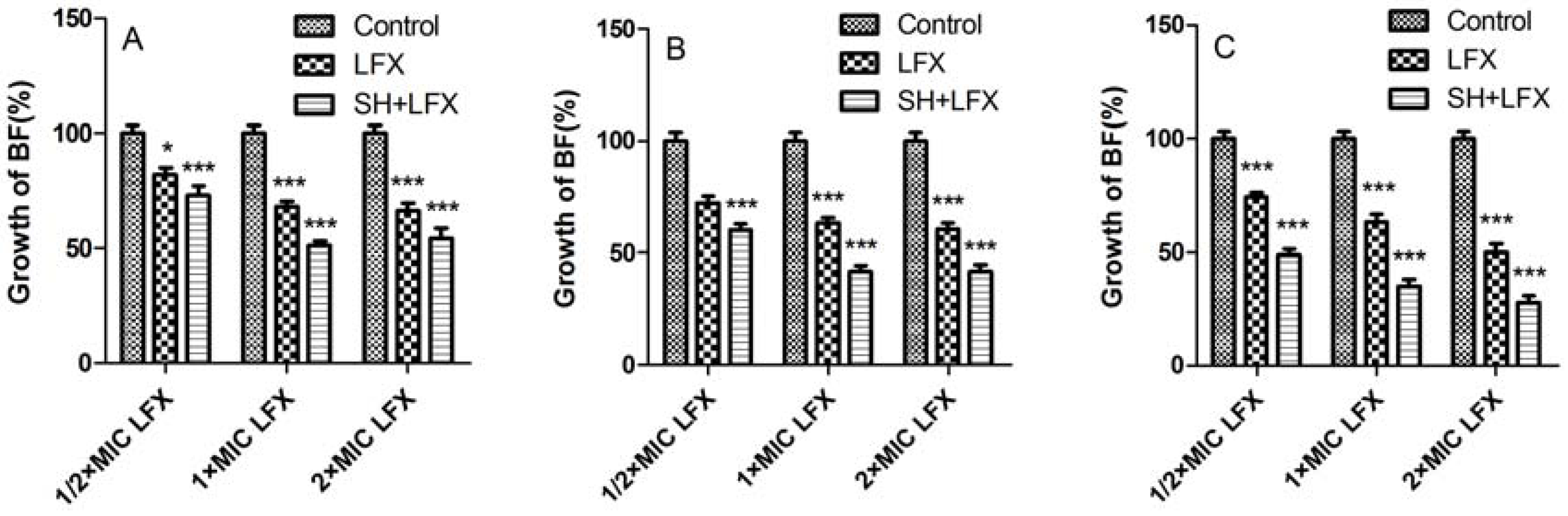
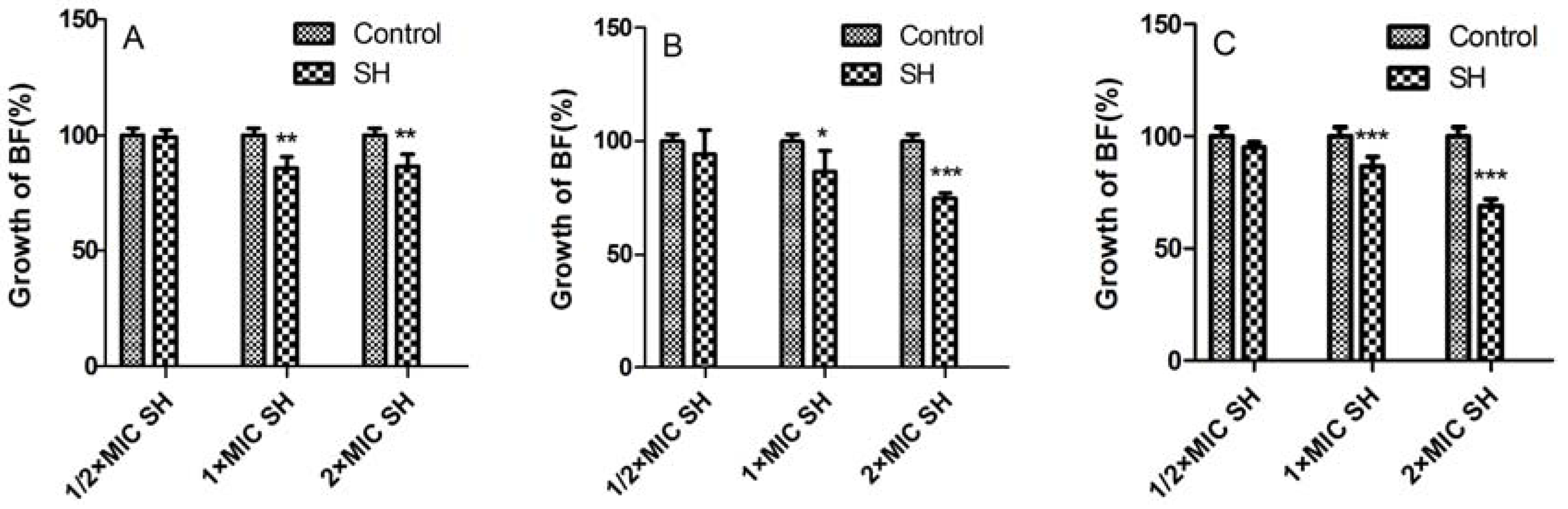
2.2. Biofilm Suppression by SH Alone and in Combination with LFX</title>
        <p>By numerous repeatable proteomic analyses, Sauer <italic>et al</italic>. put forward five stages of biofilm formation, namely: (i) reversible attachment (&gt;0 min); (ii) irreversible attachment (2 h); (iii) maturation-1 (3 days); (iv) maturation-2 (6 days); and (v) dispersion (9–12 days) [<xref ref-type="bibr" rid="B21-molecules-17-11242">21</xref>]. In terms of this rule, we chose 1 d, 3 d and 7 d as three time-points to observe the biofilm suppression and removal after medication. </p>
        <p><xref ref-type="fig" rid="molecules-17-11242-f001">Figure 1</xref> shows control of SH alone on biofilm formation of <italic>Pseudomonas aeruginosa</italic>. In the 1st day (<xref ref-type="fig" rid="molecules-17-11242-f001">Figure 1</xref>A, ½ × MIC SH has nearly no effect on biofilm growth as compared with that in the control group, while 1 × MIC and 2 × MIC SH have obvious suppressive effects on biofilm (<italic>p </italic>&lt; 0.01, n = 4). On the 3rd day (<xref ref-type="fig" rid="molecules-17-11242-f001">Figure 1</xref>B), there is no statistical significance of the impact by ½ × MIC SH treatment on biofilm, whereas, the growth of biofilm with 2 × MIC SH treatment (<italic>p </italic>= 0.000, n = 4) decreases even more than that with 1 × MIC SH treatment (<italic>p </italic>&lt; 0.05, n = 4) as compared with the control group. In the 7th day (<xref ref-type="fig" rid="molecules-17-11242-f001">Figure 1</xref>C × MIC and 2 × MIC SH have exceedingly significant differences (<italic>p </italic>= 0.000, n = 4).</p>
        <p><xref ref-type="fig" rid="molecules-17-11242-f002">Figure 2</xref> shows controls of LFX and SH + LFX on biofilm formation of <italic>Pseudomonas aeruginosa</italic>. In <xref ref-type="fig" rid="molecules-17-11242-f002">Figure 2</xref>A, LFX alone has obviously curbed biofilm formation, and except for the sample treated by ½ × MIC LFX (<italic>p </italic>&lt; 0.05, n = 4), the differences in other two cases by 1 × MIC and 2 × MIC LFX are extremely significant (<italic>p </italic>= 0.000, n = 4) as compared with control. After combination with SH, the biofilm growths are all lower than the corresponding ones treated by LFX alone (<italic>p </italic>= 0.000, n = 4). Similar results also appear in <xref ref-type="fig" rid="molecules-17-11242-f002">Figure 2</xref>B (day 3). With the extension of biofilm development, the synergistic effects in <xref ref-type="fig" rid="molecules-17-11242-f002">Figure 2</xref>C (day 7) are even more notable.</p>
        <fig id="molecules-17-11242-f001" position="float">
          <label>Figure 1</label>
          <caption>
            <p>The effect of SH alone on the biofilm growth of <italic>Pseudomonas aeruginosa</italic>. *, <italic>p </italic>&lt; 0.05; **, <italic>p </italic>&lt; 0.01; ***, <italic>p </italic>= 0.000; n = 4. SH: sodium houttuyfonate.</p>
          </caption>
          <graphic xmlns:xlink="http://www.w3.org/1999/xlink" xlink:href="molecules-17-11242-g001.tif"/>
        </fig>
        <fig id="molecules-17-11242-f002" position="float">
          <label>Figure 2</label>
          <caption>
            <p>The synergistic effect of SH and LFX on the biofilm formation of <italic>Pseudomonas aeruginosa</italic>. *, <italic>p </italic>&lt; 0.05; **, <italic>p </italic>&lt; 0.01; ***, <italic>p </italic>= 0.000; n = 4. SH: sodium houttuyfonate; LFX: levofloxacin.</p>
          </caption>
          <graphic xmlns:xlink="http://www.w3.org/1999/xlink" xlink:href="molecules-17-11242-g002.tif"/>
        </fig>
        <p>From the above results, it could be observed that the effect of SH alone on biofilm control is limited, as even the maximum dosage of SH (=2 × MIC) can merely restrain about 30% biofilm growth (<xref ref-type="fig" rid="molecules-17-11242-f001">Figure 1</xref>C). This may be related to the fact that SH is more active on Gram-positive (G<sup>+</sup>) bacteria than Gram-negative (G<sup>−</sup>) ones [<xref ref-type="bibr" rid="B19-molecules-17-11242">19</xref>]. However, the combination of SH with LFX exhibits strong (more than 50% biofilm growth in <xref ref-type="fig" rid="molecules-17-11242-f002">Figure 2</xref>C) and persistent (7 days) inhibition of biofilm. More importantly, SH increases the biofilm suppression in combination with LFX. That is to say, phytoanticipins may have a nice synergistic effect with microbial antibiotics. Next, it was of interest to probe into the synergistic effect of SH and LFX on alginate production.</p>
      </sec>
      <sec>
        <title>
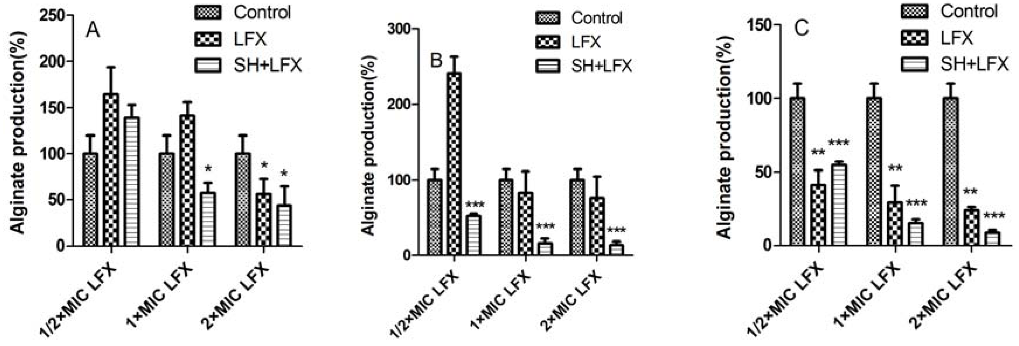
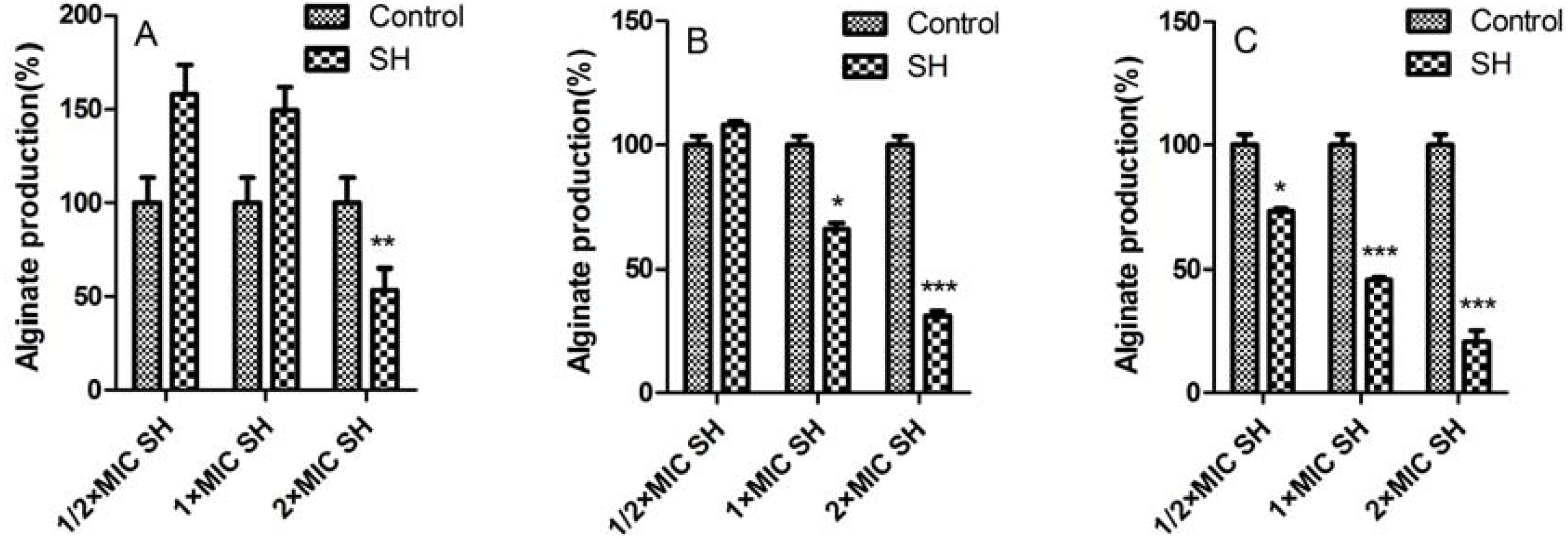
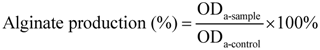
2.3. Alginate Removal by SH Alone and in Combination with LFX</title>
        <p>Alginate is an acetylated polymer, composed of non-repetitive monomers of β-1,4-linked L-guluronic and D-mannuronic acids [<xref ref-type="bibr" rid="B22-molecules-17-11242">22</xref>]. Until now, it has been assumed to be one of the most critical factors contributing to antibiotic tolerance and immune evasion, and not just playing a structural role [<xref ref-type="bibr" rid="B23-molecules-17-11242">23</xref>,<xref ref-type="bibr" rid="B24-molecules-17-11242">24</xref>,<xref ref-type="bibr" rid="B25-molecules-17-11242">25</xref>]. Recently, Mann and Wozniak published a comprehensive review on this aspect [<xref ref-type="bibr" rid="B26-molecules-17-11242">26</xref>]. Thus, it is necessary to investigate the effect of SH on alginate inhibition in combination of LFX.</p>
        <p><xref ref-type="fig" rid="molecules-17-11242-f003">Figure 3</xref> shows the effect of only SH on alginate production of <italic>Pseudomonas aeruginosa</italic> biofilm. It is interesting to observe that: (i) alginate is still produced on the first day after treatment of ½ × MIC SH and 1 × MIC SH, and its concentration decreased dramatically to about ⅓ (<italic>p </italic>&lt; 0.01, n = 4) after increasing the dosage of SH to 2 × MIC (<xref ref-type="fig" rid="molecules-17-11242-f003">Figure 3</xref>A); (ii) after three-day medication, the alginate concentration is still a little higher than the control concentration when using ½ × MIC SH, but then becomes lower and has statistically significant differences (<italic>p </italic>&lt; 0.05 and <italic>p </italic>= 0.000, n = 4) with the respective dosages of 1 × MIC SH and 2 × MIC SH (<xref ref-type="fig" rid="molecules-17-11242-f003">Figure 3</xref>B); and (iii) after administration until the seventh day, the inhibitions of alginate become more and more remarkable with the addition of SH from ½ × MIC to 2 × MIC, as the alginate productions are reducing continuously (<italic>p </italic>&lt; 0.05, <italic>p </italic>= 0.000 and <italic>p </italic>= 0.000, n = 4, <xref ref-type="fig" rid="molecules-17-11242-f003">Figure 3</xref>C).</p>
        <fig id="molecules-17-11242-f003" position="float">
          <label>Figure 3</label>
          <caption>
            <p>The effect of SH alone on alginate production of <italic>Pseudomonas aeruginosa</italic> biofilm. *, <italic>p </italic>&lt; 0.05; **, <italic>p </italic>&lt; 0.01; ***, <italic>p </italic>= 0.000; n = 4. SH: sodium houttuyfonate.</p>
          </caption>
          <graphic xmlns:xlink="http://www.w3.org/1999/xlink" xlink:href="molecules-17-11242-g003.tif"/>
        </fig>
        <p>In <xref ref-type="fig" rid="molecules-17-11242-f004">Figure 4</xref>, the synergistic effect of SH and LFX is evaluated. When treated by LFX alone, the situations are similar to those in <xref ref-type="fig" rid="molecules-17-11242-f003">Figure 3</xref>. The suppressions of alginate production basically have no statistical significance (<xref ref-type="fig" rid="molecules-17-11242-f004">Figure 4</xref>A,B) as compared with those in control group, which is changed until biofilm is treated for seven days (<italic>p </italic>&lt; 0.01, n = 4, <xref ref-type="fig" rid="molecules-17-11242-f004">Figure 4</xref>C × MIC and 2 × MIC SH in day 1 (<italic>p </italic>&lt; 0.05, n = 4, <xref ref-type="fig" rid="molecules-17-11242-f004">Figure 4</xref>A), and the effects become even more prominent in day 3 in all three dosage (<italic>p </italic>= 0.000, n = 4, <xref ref-type="fig" rid="molecules-17-11242-f004">Figure 4</xref>B). The trend is kept to the day 7 (<italic>p </italic>= 0.000, n = 4, <xref ref-type="fig" rid="molecules-17-11242-f004">Figure 4</xref>C</p>
        <p>Similar to the case in <xref ref-type="fig" rid="molecules-17-11242-f001">Figure 1</xref>, SH alone has a mild influence on alginate production (<xref ref-type="fig" rid="molecules-17-11242-f003">Figure 3</xref>). But with the addition of LFX, their synergistic effects gradually impose great limitations on alginate production (<xref ref-type="fig" rid="molecules-17-11242-f004">Figure 4</xref>). As known, alginate is secreted by bacteria to form an extracellular matrix, and finally encase the bacteria to help them hide from the toxic effects of antimicrobial agents. In <xref ref-type="fig" rid="molecules-17-11242-f002">Figure 2</xref>C and <xref ref-type="fig" rid="molecules-17-11242-f004">4</xref>C the  reduction  of  biofilm  growth  leads  to  the  stagnancy  or  loss  of  alginate  production  inferring  that  alginate  may  at  least  partly  participate  in  biofilm  formation. </p>
        <fig id="molecules-17-11242-f004" position="float">
          <label>Figure 4</label>
          <caption>
            <p>The synergistic effect of SH and LFX on the alginate production of <italic>Pseudomonas aeruginosa</italic> biofilm. *, <italic>p </italic>&lt; 0.05; **, <italic>p </italic>&lt; 0.01; ***, <italic>p </italic>= 0.000; n = 4. SH: sodium houttuyfonate; LFX: levofloxacin.</p>
          </caption>
          <graphic xmlns:xlink="http://www.w3.org/1999/xlink" xlink:href="molecules-17-11242-g004.tif"/>
        </fig>
      </sec>
      <sec>
        <title>2.4. In Vitro Antimicrobial Role of SH and Its Synergistic Activity with LFX</title>
        <p>It can be observed that the growth of biofilm decreases from 99 ± 2.77% after one-day administration of ½ × MIC SH to 69 ± 3.07% after seven-day administration of 2 × MIC SH in <xref ref-type="fig" rid="molecules-17-11242-f001">Figure 1</xref>, and the alginate production lowers from 158 ± 15.9% after one-day medication to 21 ± 4.61% after seven-day medication in <xref ref-type="fig" rid="molecules-17-11242-f003">Figure 3</xref>. Thus, SH exhibits a certain antimicrobial activity <italic>in vitro</italic>.</p>
        <p>It can be seen that the biofilm growth loses about 45% between one-day therapy of ½ × MIC SH + ½ × MIC LFX and seven-day therapy of ½ × MIC SH + 2 × MIC LFX in <xref ref-type="fig" rid="molecules-17-11242-f002">Figure 2</xref>, and the alginate production drops about 131% between one-day treatment of ½ × MIC SH + ½ × MIC LFX and seven-day treatment of ½ × MIC SH + 2 × MIC LFX in <xref ref-type="fig" rid="molecules-17-11242-f004">Figure 4</xref>. Therefore, SH has a very intense synergy with LFX in the restriction of biofilm growth and alginate production.</p>
        <p>From <xref ref-type="fig" rid="molecules-17-11242-f001">Figure 1</xref> to <xref ref-type="fig" rid="molecules-17-11242-f004">Figure 4</xref>, it can be manifested that: (i) both indexes, <italic>i.e.</italic>, growth of biofilm and alginate production, are dosage-dependent, because they display a decreasing tendency with the addition of LFX or SH; (ii) both items are time-dependent, as the suppression effects become better with the elongation of administration from day 1 to day 7. As for the synergistic effect of SH and LFX, it is also interesting to note that the alginate production effects are not synchronous with those of biofilm growth, which can be inferred from two aspects: (i) the alginate production still increases in comparison with control when employing a lower dosage of ½ × MIC LFX + ½ × MIC SH, which is more noteworthy in the first day of dosage (= 139 ± 13.52%, <xref ref-type="fig" rid="molecules-17-11242-f004">Figure 4</xref>A), suggesting no effective suppression of the combined antibiotics on alginate production; in the same period, the growth of biofilm is 73 ± 3.9% by SH in combination of LFX, showing a certain inhibition (<xref ref-type="fig" rid="molecules-17-11242-f002">Figure 2</xref>A); (ii) the alginate production is only 8 ± 1.71% when using a higher dosage of 2 × MIC LFX + ½ × MIC SH after seven days treatment (<xref ref-type="fig" rid="molecules-17-11242-f004">Figure 4</xref>C), while the growth of biofilm accounts for 28 ± 3.06% with the same time-span (<xref ref-type="fig" rid="molecules-17-11242-f002">Figure 2</xref>C), higher than that of alginate production. These discrepancies may be caused by following reasons: (i) in the stage of initial attachment (the first day), some planktonic bacteria are suppressed or killed by antibiotic alone or in combination, leading to biofilm reduction, while other irreversibly-attached bacteria begin to synthesize and secrete alginate in large amounts; (ii) the alginate is mainly produced by the above bacteria which have enough nutrients to continue their metabolism, but the inner bacteria tend to remain in a dormant mode due to limited nutrition, causing them to escape the destruction by combined antibiotics [<xref ref-type="bibr" rid="B5-molecules-17-11242">5</xref>]. As a result, when the alginate production is restrained, the encased bacteria mentioned above are exposed to be killed, while the continuous killing of the inner bacteria still needs time and re-addition of antibiotics in combination; and (iii) we cannot rule out other factors, e.g., quorum sensing (QS), that might influence biofilm formation except the production of alginate, which is still under investigation in our lab.</p>
      </sec>
      <sec>
        <title>
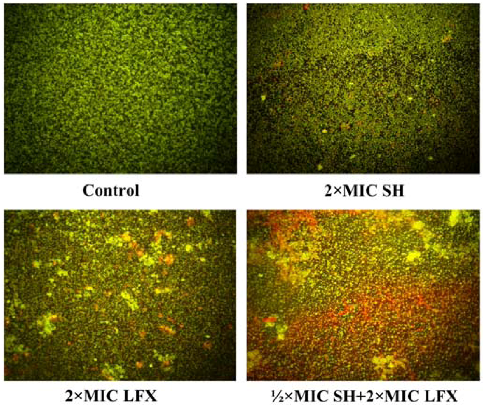
2.5. Observation of Pseudomonas aeruginosa by Fluorescence Microscope</title>
        <p><xref ref-type="fig" rid="molecules-17-11242-f005">Figure 5</xref> presents the fluorescence microscope images of the bacteria. The FAD-PI dyed bacteria are treated by SH alone, LFX alone, and SH + LFX. Living bacteria exhibit green and dead bacteria show red. </p>
        <fig id="molecules-17-11242-f005" position="float">
          <label>Figure 5</label>
          <caption>
            <p>Observation of bacterial viability by BX51 fluorescence microscope (×400) under the treatment of no drugs, 2 × MIC SH, 2 × MIC LFX, and ½ × MIC SH + 2 × MIC LFX, and the exposure of 100 μg/mL FAD for 5 min and 60 μg/mL PI for 5 min. Green: living cells stained by FAD; Red: dead cells stained by PI. Other conditions are given in <xref ref-type="sec" rid="sec3dot7-molecules-17-11242">Section 3.7</xref>.</p>
          </caption>
          <graphic xmlns:xlink="http://www.w3.org/1999/xlink" xlink:href="molecules-17-11242-g005.tif"/>
        </fig>
        <p>In <xref ref-type="fig" rid="molecules-17-11242-f005">Figure 5</xref>, it can be observed that: (i) living bacteria are scattered in control group as all cells are seen to be green; (ii) most bacteria are alive and less are dead as most cells are recorded to be green with less red in 2 × MIC SH group and 2 × MIC LFX group, however, the bacteria viability in the 2 × MIC LFX group is notably less than that in 2 × MIC SH group which has more red cells in view; and (iii) less bacteria in the ½ × MIC SH + 2 × MIC LFX group are alive than those in the SH alone group and LFX alone group as more red dots appear in the image. From the four images, the quantities of dead cell exhibit an increasing tendency, indicating the antimicrobial effects promoted by SH and LFX in combination. </p>
      </sec>
      <sec>
        <title>2.6. Observation of <italic>Pseudomonas aeruginosa</italic> Biofilm Morphology by SEM</title>
        <p><xref ref-type="fig" rid="molecules-17-11242-f006">Figure 6</xref> shows is the morphology of the bacterial biofilm by SEM. In the control group, the bacteria are largely covered by mucous substances which are mainly composed of alginate, and dense cells are seen to exist in the whole view, which is similar to that in the control group in <xref ref-type="fig" rid="molecules-17-11242-f005">Figure 5</xref>. In the 2 × MIC SH group, the mucous substances are largely removed, but the bacteria are not completely inhibited, indicating that the SH is mainly suppressing the production of mucuous substances. In terms of the results that the alginate production (%) is about 21% (<xref ref-type="fig" rid="molecules-17-11242-f003">Figure 3</xref>C) after the treatment of 2 × MIC SH, it is obvious that the alginate is the main substance in the mucuous substances and SH largely targets the alginate. In the 2 × MIC LFX group, both mucous substance and bacteria are dramatically decreased, however, there are still some matrix substances left, revealing the relative strong antipathogenic of LFX on alginate and encased bacteria. In the ½ × MIC SH + 2 × MIC LFX group, both mucous substance and bacteria are wiped out further compared with those in the 2 × MIC LFX group, reflecting that SH can enhance the suppressive effects of LFX on alginate to kill the bacteria under the mucus substances, <italic>i.e.</italic>, SH and LFX have better synergistic effects on biofilm removal.</p>
        <fig id="molecules-17-11242-f006" position="float">
          <label>Figure 6</label>
          <caption>
            <p>Observation of bacterial biofilm morphology by SEM.</p>
          </caption>
          <graphic xmlns:xlink="http://www.w3.org/1999/xlink" xlink:href="molecules-17-11242-g006.tif"/>
        </fig>
      </sec>
    </sec>
    <sec sec-type="methods">
      <title>3. Experimental</title>
      <sec>
        <title>3.1. Strains and Materials</title>
        <p><italic>Pseudomonas aeruginosa</italic> ATCC27853, sodium houttuyfonate (SH, <xref ref-type="fig" rid="molecules-17-11242-f007">Figure 7</xref>) and levofloxacin (LFX) were all obtained from the National Institute for the Control of Pharmaceutical and Biological Products (NICPBP, Beijing, China). The LB, MH and TSB media were purchased from Aoboxing Bio-tech Co., Ltd. (Beijing, China). Crystal violet solution was purchased from bioMérieux (Lyon, France). The alginate standards, fluorescein di-<italic>o</italic>-acetate (FDA) and propidium iodide (PI) were obtained from Sigma (St. Louis, MO, USA).</p>
        <fig id="molecules-17-11242-f007" position="float">
          <label>Figure 7</label>
          <caption>
            <p>The chemical structures of sodium houttuyfonate (SH) and houttuynin (decanoyl acetaldehyde).</p>
          </caption>
          <graphic xmlns:xlink="http://www.w3.org/1999/xlink" xlink:href="molecules-17-11242-g007.tif"/>
        </fig>
      </sec>
      <sec>
        <title>3.2. Cultivation of Pseudomonas aeruginosa</title>
        <p><italic>Pseudomonas aeruginosa</italic> ATCC27853 was inoculated in LB broth, cultivated in a constant-temperature shaker GLY (Fuma, Shanghai, China) of 220 rpm for 6 h at constant 37 °C. Then the bacteria were harvested by GL-20G-Ⅱ high-speed refrigerated centrifuge (Fuma) at 2,000 r/min for 10 min. The supernate was discarded, and the precipitate was resuspended with pH 7.2 phosphate-buffered saline (PBS), centrifuged again at 2,000 r/min for 10 min. The collection was mixed with PBS pH 7.2, and adjusted to 2 × 10<sup>5</sup> CFU/mL by the microdilution method. </p>
      </sec>
      <sec>
        <title>3.3. Preparation of SH and LFX Solutions</title>
        <p>Both stock solutions of SH and LFX were prepared freshly in MH broth and filtered through a 0.22 µm Millipore filter (Millipore, Billerica, MA, USA), and their concentrations were adjusted to 160 and 32 µg/mL, respectively. In the MIC determination experiments, SH was twofold-diluted into ten concentration gradients by MH medium at a final concentration from 0.03125 to 16 mg/mL, and LFX was diluted to a final concentration ranging from 0.0625 to 32 µg/mL.</p>
      </sec>
      <sec>
        <title>3.4. MIC Measurement</title>
        <p>Both SH and LFX were added into 96-well plates with eight wells in duplicate for each concentration gradient, and each well had both 100 μL either SH or LFX solution and 100 μL inoculum with the final concentration of 1 × 10<sup>5</sup> CFU/mL. The plates were incubated for 24 h at 37 °C, and then the bacteria were inoculated on nutrient agar medium and incubated for another 24 h at 37 °C. The MIC was determined as the minimum drug dilution with no growth of bacteria on plates. Each MIC assay was performed in triplicate. The checkerboard microdilution method was adopted for antimicrobial assay of SH and LFX in synergy. The concentrations of combined antibiotics were adjusted from ⅛ × MIC to 2 × MIC, and 100 μL drugs were added into each well. A volume of 100 μL/well inoculum was mixed with the drugs to the final concentration of 1 × 10<sup>5</sup> CFU/mL. Then, the setup was incubated at 37 °C for 24 h to observe the growth of bacteria, which was detected by a 318-microplate reader (Sanco Instruments, Shanghai, China). The control had medium and inoculum without any antimicrobial agents. Each test was performed in triplicate [<xref ref-type="bibr" rid="B27-molecules-17-11242">27</xref>].</p>
      </sec>
      <sec id="sec3dot5-molecules-17-11242">
        <title>3.5. Effects of SH Alone and SH + LFX in Combination on Anti-Biofilm Formation</title>
        <p>Ten groups were tested: control, ½ × MIC SH, 1 × MIC SH, 2 × MIC SH, ½ × MIC LFX, 1 × MIC LFX, 2 × MIC LFX, ½ × MIC SH + ½ × MIC LFX, ½ × MIC SH + 1 × MIC SH, ½ × MIC SH + 2 × MIC LFX (<xref ref-type="fig" rid="molecules-17-11242-f001">Figure 1</xref> and <xref ref-type="fig" rid="molecules-17-11242-f002">Figure 2</xref>). In a 96-well plate, each was added with 200 μL <italic>Pseudomonas aeruginosa</italic> suspension, incubated at 37 °C with no shaking. After 24 h, the strain suspension was discarded, and the planktonic microbes were washed by PBS, inoculated into TSB medium with the above concentrations of antibiotics. The control was cultivated in the medium with no antibiotics. According to the description of O’Toole [<xref ref-type="bibr" rid="B28-molecules-17-11242">28</xref>], the medium were exchanged for a new one with antibiotics every other day. At the end of the 1st, 3rd and 7th day, 4 °C PBS was employed to wash the planktonic bacteria on the 96-well plate. Then, each well was added into 200 μL 1% crystal violet solution for 20 min. After they were rinsed with de-ionized water till no crystal violet was visible, the wells were dried and 95% alcohol added to destain. The destained samples at the end of 1st, 3rd and 7th day of medications were transferred to a cuvette and diluted with 95% alcohol (3 mL) to determine the OD value (OD<sub>b-sample</sub>) at the wavelength of 570 nm. The corresponding OD values of negative control (OD<sub>b-control</sub>), consisting of biofilm without any medications, were treated as the above steps and set as 100% at the end of 1st, 3rd and 7th day of medications. Then, the growth of biofilm (%) could be calculated for each group as follows:</p>
        <p><disp-formula id="molecules-17-11242-i002"><inline-graphic xmlns:xlink="http://www.w3.org/1999/xlink" xlink:href="molecules-17-11242-i002.tif"/><label>(2)</label></disp-formula></p>
        <p>and each calculation was processed in triplicate.</p>
      </sec>
      <sec id="sec3dot6-molecules-17-11242">
        <title>
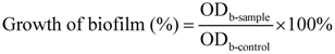
3.6. Effects of SH Alone and SH + LFX in Combination on Anti-Alginate Production</title>
        <p>There were also ten groups, the same as those in <xref ref-type="sec" rid="sec3dot5-molecules-17-11242">Section 3.5</xref>. The sterile cover-glass carrier was put into the 6-well culture plate, and then TSB medium (2 mL) and strain suspension (0.2 mL) were added. After incubating for 1 day at 37 °C, the medium was discarded, and the cover-glass was taken out to wash the planktonic bacteria out with sterile PBS. The cover-glass was then put into the well again. The antibiotic groups were given medium containing antibiotics, while the negative control used medium without antibiotics. All the above manipulations were repeated in each 24 h. At the end of the 1st, 3rd and 7th day of medication, the cover-glasses were fetched out to rinse the planktonic bacteria by PBS. Then, the cover-glasses were put into test tube with additions of PBS (6 mL), sulphuric acid and sodium borate (1.2 mL each), boiled for 5 min, and placed at 4 °C. 1% Hydroxybiphenyl (20 μL) was added to the glass for colorization, and vibrated for 30 min by ultrasound. The OD values at the end of 1st, 3rd and 7th day of medications were recorded at the wavelength of 570 nm (OD<sub>a-sample</sub>). According to the above treatments, the corresponding OD values of negative control (OD<sub>a-control</sub>), consisting of alginate without any medications, were set as 100%, and then the alginate production (%) could be calculated for each group as follows:</p>
        <p><disp-formula id="molecules-17-11242-i003"><inline-graphic xmlns:xlink="http://www.w3.org/1999/xlink" xlink:href="molecules-17-11242-i003.tif"/><label>(3)</label></disp-formula></p>
        <p>and each calculation was conducted in triplicate.</p>
      </sec>
      <sec id="sec3dot7-molecules-17-11242">
        <title>3.7. Fluorescence Microscopy of Bacteria</title>
        <p>The carrier disposition was similar to that described in <xref ref-type="sec" rid="sec3dot6-molecules-17-11242">Section 3.6</xref>. After the glass was removed and the planktonic bacteria rinsed with PBS at the end of the 7th day of medication, it was stained with fluorescein diacetate-propidium iodide (FDA-PI) fluorescent dyes to observe the vitality of dispersed bacteria, in which FDA concentration was 100 µg/mL, and PI concentration was 60 µg/mL. The bacteria were first stained by FDA for 5 min, and then with PI for 5 min. The dead bacteria were stained as red by PI, while the alive ones were as green by FDA. A BX51 fluorescence microscope was used (Olympus, Tokyo, Japan) to record the results.</p>
      </sec>
      <sec>
        <title>3.8. Scanning Electric Microscope of Biofilm Morphology</title>
        <p>The biofilm morphology was observed by silver-staining on a SEM (Sirion200, FEI, Columbia, MD, USA).</p>
      </sec>
    </sec>
    <sec sec-type="conclusions">
      <title>4. Conclusions</title>
      <p>A phytoanticipin derivative, sodium houttuyfonate, was adopted to investigate its synergistic effects with levofloxacin on the suppression of <italic>Pseudomonas aeruginosa</italic> biofilm. We separately evaluated the inhibitions by SH alone and LFX alone on biofilm formation and alginate production, and the synergistic effect of SH with LFX on biofilm formation and alginate production. The results demonstrate that SH has robust synergistic activity with LFX, and can dramatically decrease the amount of bacteria by effective inhibition of biofilm formation and alginate production. Consequently, phytoanticipins represent a promising alternative to coordinate with microbial antibiotics, playing an even greater role in anti-biofilm treatment.</p>
    </sec>
  </body>
  <back>
    <ack>
      <title>Acknowledgments</title>
      <p>This work was supported by the National Natural Science Foundation of China (No. 81173629). </p>
    </ack>
    <notes>
      <title>Conflict of interest</title>
      <p>The authors declare no conflict of interest.</p>
    </notes>
    <ref-list>
      <title>References</title>
      <ref id="B1-molecules-17-11242">
        <label>1.</label>
        <citation citation-type="journal">
          <person-group person-group-type="author">
            <name>
              <surname>Costerton</surname>
              <given-names>J.</given-names>
            </name>
          </person-group>
          <article-title>Introduction to biofilm</article-title>
          <source>Int. J. Antimicrob. Agents</source>
          <year>1999</year>
          <volume>11</volume>
          <fpage>217</fpage>
          <lpage>221</lpage>
          <pub-id pub-id-type="doi">10.1016/S0924-8579(99)00018-7</pub-id>
        </citation>
      </ref>
      <ref id="B2-molecules-17-11242">
        <label>2.</label>
        <citation citation-type="journal">
          <person-group person-group-type="author">
            <name>
              <surname>Hall-Stoodley</surname>
              <given-names>L.</given-names>
            </name>
            <name>
              <surname>Costerton</surname>
              <given-names>J.W.</given-names>
            </name>
            <name>
              <surname>Stoodley</surname>
              <given-names>P.</given-names>
            </name>
          </person-group>
          <article-title>Bacterial biofilms: From the natural environment to infectious diseases</article-title>
          <source>Nat. Rev. Microbiol.</source>
          <year>2004</year>
          <volume>2</volume>
          <fpage>95</fpage>
          <lpage>108</lpage>
          <pub-id pub-id-type="doi">10.1038/nrmicro821</pub-id>
        </citation>
      </ref>
      <ref id="B3-molecules-17-11242">
        <label>3.</label>
        <citation citation-type="journal">
          <person-group person-group-type="author">
            <name>
              <surname>Kirketerp-Møller</surname>
              <given-names>K.</given-names>
            </name>
            <name>
              <surname>Jensen</surname>
              <given-names>P.Ø.</given-names>
            </name>
            <name>
              <surname>Fazli</surname>
              <given-names>M.</given-names>
            </name>
            <name>
              <surname>Madsen</surname>
              <given-names>K.G.</given-names>
            </name>
            <name>
              <surname>Pedersen</surname>
              <given-names>J.</given-names>
            </name>
            <name>
              <surname>Moser</surname>
              <given-names>C.</given-names>
            </name>
            <name>
              <surname>Tolker-Nielsen</surname>
              <given-names>T.</given-names>
            </name>
            <name>
              <surname>Høiby</surname>
              <given-names>N.</given-names>
            </name>
            <name>
              <surname>Givskov</surname>
              <given-names>M.</given-names>
            </name>
            <name>
              <surname>Bjarnsholt</surname>
              <given-names>T.</given-names>
            </name>
          </person-group>
          <article-title>Distribution, organization, and ecology of bacteria in chronic wounds</article-title>
          <source>J. Clin. Microbiol.</source>
          <year>2008</year>
          <volume>46</volume>
          <fpage>2717</fpage>
          <lpage>2722</lpage>
          <pub-id pub-id-type="doi">10.1128/JCM.00501-08</pub-id>
        </citation>
      </ref>
      <ref id="B4-molecules-17-11242">
        <label>4.</label>
        <citation citation-type="journal">
          <person-group person-group-type="author">
            <name>
              <surname>Bjarnsholt</surname>
              <given-names>T.</given-names>
            </name>
            <name>
              <surname>Jensen</surname>
              <given-names>P.O.</given-names>
            </name>
            <name>
              <surname>Fiandaca</surname>
              <given-names>M.J.</given-names>
            </name>
            <name>
              <surname>Pedersen</surname>
              <given-names>J.</given-names>
            </name>
            <name>
              <surname>Hansen</surname>
              <given-names>C.R.</given-names>
            </name>
            <name>
              <surname>Andersen</surname>
              <given-names>C.B.</given-names>
            </name>
            <name>
              <surname>Pressler</surname>
              <given-names>T.</given-names>
            </name>
            <name>
              <surname>Givskov</surname>
              <given-names>M.</given-names>
            </name>
            <name>
              <surname>Hoiby</surname>
              <given-names>N.</given-names>
            </name>
          </person-group>
          <article-title>Pseudomonas aeruginosa biofilms in the respiratory tract of cystic fibrosis patients</article-title>
          <source>Pediatr. Pulmonol.</source>
          <year>2009</year>
          <volume>44</volume>
          <fpage>547</fpage>
          <lpage>558</lpage>
          <pub-id pub-id-type="doi">10.1002/ppul.21011</pub-id>
        </citation>
      </ref>
      <ref id="B5-molecules-17-11242">
        <label>5.</label>
        <citation citation-type="journal">
          <person-group person-group-type="author">
            <name>
              <surname>Harmsen</surname>
              <given-names>M.</given-names>
            </name>
            <name>
              <surname>Yang</surname>
              <given-names>L.</given-names>
            </name>
            <name>
              <surname>Pamp</surname>
              <given-names>S.J.</given-names>
            </name>
            <name>
              <surname>Tolker-Nielsen</surname>
              <given-names>T.</given-names>
            </name>
          </person-group>
          <article-title>An update on pseudomonas aeruginosa biofilm formation, tolerance, and dispersal</article-title>
          <source>FEMS Immunol. Med. Microbiol.</source>
          <year>2010</year>
          <volume>59</volume>
          <fpage>253</fpage>
          <lpage>268</lpage>
        <pub-id pub-id-type="pmid">20497222</pub-id></citation>
      </ref>
      <ref id="B6-molecules-17-11242">
        <label>6.</label>
        <citation citation-type="journal">
          <person-group person-group-type="author">
            <name>
              <surname>Carneiro</surname>
              <given-names>V.A.</given-names>
            </name>
            <name>
              <surname>Santos</surname>
              <given-names>H.S.</given-names>
            </name>
            <name>
              <surname>Arruda</surname>
              <given-names>F.V.</given-names>
            </name>
            <name>
              <surname>Bandeira</surname>
              <given-names>P.N.</given-names>
            </name>
            <name>
              <surname>Albuquerque</surname>
              <given-names>M.R.</given-names>
            </name>
            <name>
              <surname>Pereira</surname>
              <given-names>M.O.</given-names>
            </name>
            <name>
              <surname>Henriques</surname>
              <given-names>M.</given-names>
            </name>
            <name>
              <surname>Cavada</surname>
              <given-names>B.S.</given-names>
            </name>
            <name>
              <surname>Teixeira</surname>
              <given-names>E.H.</given-names>
            </name>
          </person-group>
          <article-title>Casbane diterpene as a promising natural antimicrobial agent against biofilm-associated infections</article-title>
          <source>Molecules</source>
          <year>2011</year>
          <volume>16</volume>
          <fpage>190</fpage>
          <lpage>201</lpage>
        </citation>
      </ref>
      <ref id="B7-molecules-17-11242">
        <label>7.</label>
        <citation citation-type="journal">
          <person-group person-group-type="author">
            <name>
              <surname>Hengzhuang</surname>
              <given-names>W.</given-names>
            </name>
            <name>
              <surname>Wu</surname>
              <given-names>H.</given-names>
            </name>
            <name>
              <surname>Ciofu</surname>
              <given-names>O.</given-names>
            </name>
            <name>
              <surname>Song</surname>
              <given-names>Z.</given-names>
            </name>
            <name>
              <surname>Hoiby</surname>
              <given-names>N.</given-names>
            </name>
          </person-group>
          <article-title><italic>In vivo</italic> pharmacokinetics/pharmacodynamics of colistin and imipenem in pseudomonas aeruginosa biofilm infection</article-title>
          <source>Antimicrob. Agents Chemother.</source>
          <year>2012</year>
          <volume>56</volume>
          <fpage>2683</fpage>
          <lpage>2690</lpage>
          <pub-id pub-id-type="doi">10.1128/AAC.06486-11</pub-id>
        </citation>
      </ref>
      <ref id="B8-molecules-17-11242">
        <label>8.</label>
        <citation citation-type="journal">
          <person-group person-group-type="author">
            <name>
              <surname>Saleh</surname>
              <given-names>S.</given-names>
            </name>
            <name>
              <surname>Taha</surname>
              <given-names>M.O.</given-names>
            </name>
            <name>
              <surname>Haddadin</surname>
              <given-names>R.N.</given-names>
            </name>
            <name>
              <surname>Marzooqa</surname>
              <given-names>D.</given-names>
            </name>
            <name>
              <surname>Hodali</surname>
              <given-names>H.</given-names>
            </name>
          </person-group>
          <article-title>Preparation of silver- and zinc-doped mullite-based ceramics showing anti-bacterial biofilm properties</article-title>
          <source>Molecules</source>
          <year>2011</year>
          <volume>16</volume>
          <fpage>2862</fpage>
          <lpage>2870</lpage>
          <pub-id pub-id-type="doi">10.3390/molecules16042862</pub-id>
        </citation>
      </ref>
      <ref id="B9-molecules-17-11242">
        <label>9.</label>
        <citation citation-type="journal">
          <person-group person-group-type="author">
            <name>
              <surname>Saleh</surname>
              <given-names>S.</given-names>
            </name>
            <name>
              <surname>Sweileh</surname>
              <given-names>B.</given-names>
            </name>
            <name>
              <surname>Taha</surname>
              <given-names>S.O.</given-names>
            </name>
            <name>
              <surname>Mahmoud</surname>
              <given-names>R.</given-names>
            </name>
            <name>
              <surname>Taha</surname>
              <given-names>M.O.</given-names>
            </name>
          </person-group>
          <article-title>Preparation of polyester-based metal-cross linked polymeric composites as novel materials resistant to bacterial adhesion and biofilm formation</article-title>
          <source>Molecules</source>
          <year>2011</year>
          <volume>16</volume>
          <fpage>933</fpage>
          <lpage>950</lpage>
          <pub-id pub-id-type="doi">10.3390/molecules16010933</pub-id>
        </citation>
      </ref>
      <ref id="B10-molecules-17-11242">
        <label>10.</label>
        <citation citation-type="journal">
          <person-group person-group-type="author">
            <name>
              <surname>Hengzhuang</surname>
              <given-names>W.</given-names>
            </name>
            <name>
              <surname>Wu</surname>
              <given-names>H.</given-names>
            </name>
            <name>
              <surname>Ciofu</surname>
              <given-names>O.</given-names>
            </name>
            <name>
              <surname>Song</surname>
              <given-names>Z.</given-names>
            </name>
            <name>
              <surname>Hoiby</surname>
              <given-names>N.</given-names>
            </name>
          </person-group>
          <article-title>Pharmacokinetics/pharmacodynamics of colistin and imipenem on mucoid and nonmucoid pseudomonas aeruginosa biofilms</article-title>
          <source>Antimicrob. Agents Chemother.</source>
          <year>2011</year>
          <volume>55</volume>
          <fpage>4469</fpage>
          <lpage>4474</lpage>
          <pub-id pub-id-type="doi">10.1128/AAC.00126-11</pub-id>
        </citation>
      </ref>
      <ref id="B11-molecules-17-11242">
        <label>11.</label>
        <citation citation-type="journal">
          <person-group person-group-type="author">
            <name>
              <surname>VanEtten</surname>
              <given-names>H.D.</given-names>
            </name>
            <name>
              <surname>Mansfield</surname>
              <given-names>J.W.</given-names>
            </name>
            <name>
              <surname>Bailey</surname>
              <given-names>J.A.</given-names>
            </name>
            <name>
              <surname>Farmer</surname>
              <given-names>E.E.</given-names>
            </name>
          </person-group>
          <article-title>Two classes of plant antibiotics: Phytoalexins <italic>versus</italic> “phytoanticipins”</article-title>
          <source>Plant Cell</source>
          <year>1994</year>
          <volume>6</volume>
          <fpage>1191</fpage>
          <lpage>1192</lpage>
        <pub-id pub-id-type="pmid">12244269</pub-id></citation>
      </ref>
      <ref id="B12-molecules-17-11242">
        <label>12.</label>
        <citation citation-type="journal">
          <person-group person-group-type="author">
            <name>
              <surname>Dixon</surname>
              <given-names>R.A.</given-names>
            </name>
          </person-group>
          <article-title>Natural products and plant disease resistance</article-title>
          <source>Nature</source>
          <year>2001</year>
          <volume>411</volume>
          <fpage>843</fpage>
          <lpage>847</lpage>
          <pub-id pub-id-type="doi">10.1038/35081178</pub-id>
        </citation>
      </ref>
      <ref id="B13-molecules-17-11242">
        <label>13.</label>
        <citation citation-type="journal">
          <person-group person-group-type="author">
            <name>
              <surname>Zeng</surname>
              <given-names>X.</given-names>
            </name>
            <name>
              <surname>Liu</surname>
              <given-names>X.</given-names>
            </name>
            <name>
              <surname>Bian</surname>
              <given-names>J.</given-names>
            </name>
            <name>
              <surname>Pei</surname>
              <given-names>G.</given-names>
            </name>
            <name>
              <surname>Dai</surname>
              <given-names>H.</given-names>
            </name>
            <name>
              <surname>Polyak</surname>
              <given-names>S.W.</given-names>
            </name>
            <name>
              <surname>Song</surname>
              <given-names>F.</given-names>
            </name>
            <name>
              <surname>Ma</surname>
              <given-names>L.</given-names>
            </name>
            <name>
              <surname>Wang</surname>
              <given-names>Y.</given-names>
            </name>
            <name>
              <surname>Zhang</surname>
              <given-names>L.</given-names>
            </name>
          </person-group>
          <article-title>Synergistic effect of 14-alpha-lipoyl andrographolide and various antibiotics on the formation of biofilms and production of exopolysaccharide and pyocyanin by pseudomonas aeruginosa</article-title>
          <source>Antimicrob. Agents Chemother.</source>
          <year>2011</year>
          <volume>55</volume>
          <fpage>3015</fpage>
          <lpage>3017</lpage>
          <pub-id pub-id-type="doi">10.1128/AAC.00575-10</pub-id>
        </citation>
      </ref>
      <ref id="B14-molecules-17-11242">
        <label>14.</label>
        <citation citation-type="journal">
          <person-group person-group-type="author">
            <name>
              <surname>Kosuge</surname>
              <given-names>T.</given-names>
            </name>
          </person-group>
          <article-title>Structure of an antimicrobial substance isolated from houttuynia cordata thunb</article-title>
          <source>J. Pharm. Sac. Jpn.</source>
          <year>1952</year>
          <volume>72</volume>
          <fpage>12271</fpage>
          <lpage>12275</lpage>
        </citation>
      </ref>
      <ref id="B15-molecules-17-11242">
        <label>15.</label>
        <citation citation-type="journal">
          <person-group person-group-type="author">
            <name>
              <surname>Liu</surname>
              <given-names>G.</given-names>
            </name>
            <name>
              <surname>Xiang</surname>
              <given-names>H.</given-names>
            </name>
            <name>
              <surname>Tang</surname>
              <given-names>X.</given-names>
            </name>
            <name>
              <surname>Zhang</surname>
              <given-names>K.</given-names>
            </name>
            <name>
              <surname>Wu</surname>
              <given-names>X.</given-names>
            </name>
            <name>
              <surname>Wang</surname>
              <given-names>X.</given-names>
            </name>
            <name>
              <surname>Guo</surname>
              <given-names>N.</given-names>
            </name>
            <name>
              <surname>Feng</surname>
              <given-names>H.</given-names>
            </name>
            <name>
              <surname>Wang</surname>
              <given-names>G.</given-names>
            </name>
            <name>
              <surname>Liu</surname>
              <given-names>L.</given-names>
            </name>
            <etal/>
          </person-group>
          <article-title>Transcriptional and functional analysis shows sodium houttuyfonate-mediated inhibition of autolysis in staphylococcus aureus</article-title>
          <source>Molecules</source>
          <year>2011</year>
          <volume>16</volume>
          <fpage>8848</fpage>
          <lpage>8865</lpage>
          <pub-id pub-id-type="doi">10.3390/molecules16108848</pub-id>
        </citation>
      </ref>
      <ref id="B16-molecules-17-11242">
        <label>16.</label>
        <citation citation-type="journal">
          <person-group person-group-type="author">
            <name>
              <surname>Zhou</surname>
              <given-names>Y.</given-names>
            </name>
            <name>
              <surname>Zhou</surname>
              <given-names>M.</given-names>
            </name>
            <name>
              <surname>Zhang</surname>
              <given-names>D.</given-names>
            </name>
            <name>
              <surname>Zhang</surname>
              <given-names>H.</given-names>
            </name>
            <name>
              <surname>Zhang</surname>
              <given-names>L.</given-names>
            </name>
          </person-group>
          <article-title>Immune response of aa broilers to ibv h120 vaccine and sodium new houttuyfonate</article-title>
          <source>Res. Vet. Sci.</source>
          <year>2010</year>
          <volume>89</volume>
          <fpage>373</fpage>
          <lpage>377</lpage>
          <pub-id pub-id-type="doi">10.1016/j.rvsc.2010.04.010</pub-id>
        </citation>
      </ref>
      <ref id="B17-molecules-17-11242">
        <label>17.</label>
        <citation citation-type="journal">
          <person-group person-group-type="author">
            <name>
              <surname>Pan</surname>
              <given-names>P.</given-names>
            </name>
            <name>
              <surname>Wang</surname>
              <given-names>Y.J.</given-names>
            </name>
            <name>
              <surname>Han</surname>
              <given-names>L.</given-names>
            </name>
            <name>
              <surname>Liu</surname>
              <given-names>X.</given-names>
            </name>
            <name>
              <surname>Zhao</surname>
              <given-names>M.</given-names>
            </name>
            <name>
              <surname>Yuan</surname>
              <given-names>Y.F.</given-names>
            </name>
          </person-group>
          <article-title>Effects of sodium houttuyfonate on expression of nf-kappab and mcp-1 in membranous glomerulonephritis</article-title>
          <source>J. Ethnopharmacol.</source>
          <year>2010</year>
          <volume>131</volume>
          <fpage>203</fpage>
          <lpage>209</lpage>
          <pub-id pub-id-type="doi">10.1016/j.jep.2010.06.020</pub-id>
        </citation>
      </ref>
      <ref id="B18-molecules-17-11242">
        <label>18.</label>
        <citation citation-type="journal">
          <person-group person-group-type="author">
            <name>
              <surname>Gao</surname>
              <given-names>J.P.</given-names>
            </name>
            <name>
              <surname>Chen</surname>
              <given-names>C.X.</given-names>
            </name>
            <name>
              <surname>Wang</surname>
              <given-names>Y.</given-names>
            </name>
            <name>
              <surname>Lu</surname>
              <given-names>J.</given-names>
            </name>
            <name>
              <surname>Gu</surname>
              <given-names>W.L.</given-names>
            </name>
          </person-group>
          <article-title>Effect of sodium houttuyfonate on myocardial hypertrophy in mice and rats</article-title>
          <source>J. Pharm. Pharmacol.</source>
          <year>2009</year>
          <volume>61</volume>
          <fpage>677</fpage>
          <lpage>683</lpage>
        <pub-id pub-id-type="doi">10.1211/jpp.61.05.0018</pub-id><pub-id pub-id-type="pmid">19406008</pub-id></citation>
      </ref>
      <ref id="B19-molecules-17-11242">
        <label>19.</label>
        <citation citation-type="journal">
          <person-group person-group-type="author">
            <name>
              <surname>Ye</surname>
              <given-names>X.</given-names>
            </name>
            <name>
              <surname>Li</surname>
              <given-names>X.</given-names>
            </name>
            <name>
              <surname>Yuan</surname>
              <given-names>L.</given-names>
            </name>
            <name>
              <surname>Ge</surname>
              <given-names>L.</given-names>
            </name>
            <name>
              <surname>Zhang</surname>
              <given-names>B.</given-names>
            </name>
            <name>
              <surname>Zhou</surname>
              <given-names>S.</given-names>
            </name>
          </person-group>
          <article-title>Interaction of houttuyfonate homologues with the cell membrane of gram-positive and gram-negative bacteria</article-title>
          <source>Colloids Surf. A Physicochem. Eng. Aspects</source>
          <year>2007</year>
          <volume>301</volume>
          <fpage>412</fpage>
          <lpage>418</lpage>
          <pub-id pub-id-type="doi">10.1016/j.colsurfa.2007.01.012</pub-id>
        </citation>
      </ref>
      <ref id="B20-molecules-17-11242">
        <label>20.</label>
        <citation citation-type="journal">
          <person-group person-group-type="author">
            <name>
              <surname>Johnson</surname>
              <given-names>D.</given-names>
            </name>
            <name>
              <surname>Jones</surname>
              <given-names>R.</given-names>
            </name>
          </person-group>
          <article-title><italic>In-vitro</italic> activity of a combination of two oral beta-lactams (cefpodoxime and amoxycillin) against streptococcus pneumoniae isolates with reduced susceptibilities to penicillin</article-title>
          <source>J. Antimicrob. Chemother.</source>
          <year>1998</year>
          <volume>42</volume>
          <fpage>555</fpage>
          <lpage>557</lpage>
          <pub-id pub-id-type="doi">10.1093/jac/42.4.555</pub-id>
        </citation>
      </ref>
      <ref id="B21-molecules-17-11242">
        <label>21.</label>
        <citation citation-type="journal">
          <person-group person-group-type="author">
            <name>
              <surname>Sauer</surname>
              <given-names>K.</given-names>
            </name>
            <name>
              <surname>Camper</surname>
              <given-names>A.K.</given-names>
            </name>
            <name>
              <surname>Ehrlich</surname>
              <given-names>G.D.</given-names>
            </name>
            <name>
              <surname>Costerton</surname>
              <given-names>J.W.</given-names>
            </name>
            <name>
              <surname>Davies</surname>
              <given-names>D.G.</given-names>
            </name>
          </person-group>
          <article-title>Pseudomonas aeruginosa displays multiple phenotypes during development as a biofilm</article-title>
          <source>J. Bacteriol.</source>
          <year>2002</year>
          <volume>184</volume>
          <fpage>1140</fpage>
          <lpage>1154</lpage>
          <pub-id pub-id-type="doi">10.1128/jb.184.4.1140-1154.2002</pub-id>
        </citation>
      </ref>
      <ref id="B22-molecules-17-11242">
        <label>22.</label>
        <citation citation-type="journal">
          <person-group person-group-type="author">
            <name>
              <surname>Evans</surname>
              <given-names>L.R.</given-names>
            </name>
            <name>
              <surname>Linker</surname>
              <given-names>A.</given-names>
            </name>
          </person-group>
          <article-title>Production and characterization of the slime polysaccharide of pseudomonas aeruginosa</article-title>
          <source>J. Bacteriol.</source>
          <year>1973</year>
          <volume>116</volume>
          <fpage>915</fpage>
          <lpage>924</lpage>
        <pub-id pub-id-type="pmid">4200860</pub-id></citation>
      </ref>
      <ref id="B23-molecules-17-11242">
        <label>23.</label>
        <citation citation-type="journal">
          <person-group person-group-type="author">
            <name>
              <surname>Simpson</surname>
              <given-names>J.A.</given-names>
            </name>
            <name>
              <surname>Smith</surname>
              <given-names>S.E.</given-names>
            </name>
            <name>
              <surname>Dean</surname>
              <given-names>R.T.</given-names>
            </name>
          </person-group>
          <article-title>Alginate inhibition of the uptake of pseudomonas aeruginosa by macrophages</article-title>
          <source>J. Gen. Microbiol.</source>
          <year>1988</year>
          <volume>134</volume>
          <fpage>29</fpage>
          <lpage>36</lpage>
        <pub-id pub-id-type="pmid">3141564</pub-id></citation>
      </ref>
      <ref id="B24-molecules-17-11242">
        <label>24.</label>
        <citation citation-type="journal">
          <person-group person-group-type="author">
            <name>
              <surname>Simpson</surname>
              <given-names>J.A.</given-names>
            </name>
            <name>
              <surname>Smith</surname>
              <given-names>S.E.</given-names>
            </name>
            <name>
              <surname>Dean</surname>
              <given-names>R.T.</given-names>
            </name>
          </person-group>
          <article-title>Scavenging by alginate of free radicals released by macrophages</article-title>
          <source>Free Radic. Biol. Med.</source>
          <year>1989</year>
          <volume>6</volume>
          <fpage>347</fpage>
          <lpage>353</lpage>
          <pub-id pub-id-type="doi">10.1016/0891-5849(89)90078-6</pub-id>
        </citation>
      </ref>
      <ref id="B25-molecules-17-11242">
        <label>25.</label>
        <citation citation-type="journal">
          <person-group person-group-type="author">
            <name>
              <surname>Simpson</surname>
              <given-names>J.</given-names>
            </name>
            <name>
              <surname>Smith</surname>
              <given-names>S.</given-names>
            </name>
            <name>
              <surname>Dean</surname>
              <given-names>R.</given-names>
            </name>
          </person-group>
          <article-title>Alginate may accumulate in cystic fibrosis lung because the enzymatic and free radical capacities of phagocytic cells are inadequate for its degradation</article-title>
          <source>Biochem. Mol. Biol. Int.</source>
          <year>1993</year>
          <volume>30</volume>
          <fpage>1021</fpage>
          <lpage>1034</lpage>
        <pub-id pub-id-type="pmid">8220249</pub-id></citation>
      </ref>
      <ref id="B26-molecules-17-11242">
        <label>26.</label>
        <citation citation-type="journal">
          <person-group person-group-type="author">
            <name>
              <surname>Mann</surname>
              <given-names>E.E.</given-names>
            </name>
            <name>
              <surname>Wozniak</surname>
              <given-names>D.J.</given-names>
            </name>
          </person-group>
          <article-title>Pseudomonas biofilm matrix composition and niche biology</article-title>
          <source>FEMS Microbiol. Rev.</source>
          <year>2012</year>
          <volume>36</volume>
          <fpage>893</fpage>
          <lpage>916</lpage>
          <pub-id pub-id-type="doi">10.1111/j.1574-6976.2011.00322.x</pub-id>
        </citation>
      </ref>
      <ref id="B27-molecules-17-11242">
        <label>27.</label>
        <citation citation-type="journal">
          <person-group person-group-type="author">
            <name>
              <surname>Zhou</surname>
              <given-names>Y.</given-names>
            </name>
            <name>
              <surname>Wang</surname>
              <given-names>G.</given-names>
            </name>
            <name>
              <surname>Li</surname>
              <given-names>Y.</given-names>
            </name>
            <name>
              <surname>Liu</surname>
              <given-names>Y.</given-names>
            </name>
            <name>
              <surname>Song</surname>
              <given-names>Y.</given-names>
            </name>
            <name>
              <surname>Zheng</surname>
              <given-names>W.</given-names>
            </name>
            <name>
              <surname>Zhang</surname>
              <given-names>N.</given-names>
            </name>
            <name>
              <surname>Hu</surname>
              <given-names>X.</given-names>
            </name>
            <name>
              <surname>Yan</surname>
              <given-names>S.</given-names>
            </name>
            <name>
              <surname>Jia</surname>
              <given-names>J.</given-names>
            </name>
          </person-group>
          <article-title><italic>In vitro</italic> interactions between aspirin and amphotericin b against planktonic cells and biofilm cells of candida albicans and c. Parapsilosis</article-title>
          <source>Antimicrob. Agents Chemother.</source>
          <year>2012</year>
          <volume>56</volume>
          <fpage>3250</fpage>
          <lpage>3260</lpage>
          <pub-id pub-id-type="doi">10.1128/AAC.06082-11</pub-id>
        </citation>
      </ref>
      <ref id="B28-molecules-17-11242">
        <label>28.</label>
        <citation citation-type="journal">
          <person-group person-group-type="author">
            <name>
              <surname>O’Toole</surname>
              <given-names>G.A.</given-names>
            </name>
          </person-group>
          <article-title>Microtiter dish biofilm formation assay</article-title>
          <source>J. Vis. Exp.</source>
          <year>2011</year>
          <volume>47</volume>
          <fpage>e2437</fpage>
        </citation>
      </ref>
    </ref-list>
 <fn-group>
  <fn>
    <p><italic>Sample Availability</italic>: Samples of Sodium Houttuyfonate are available from the authors.</p>
  </fn>
 </fn-group>	
	</back>
</article>
