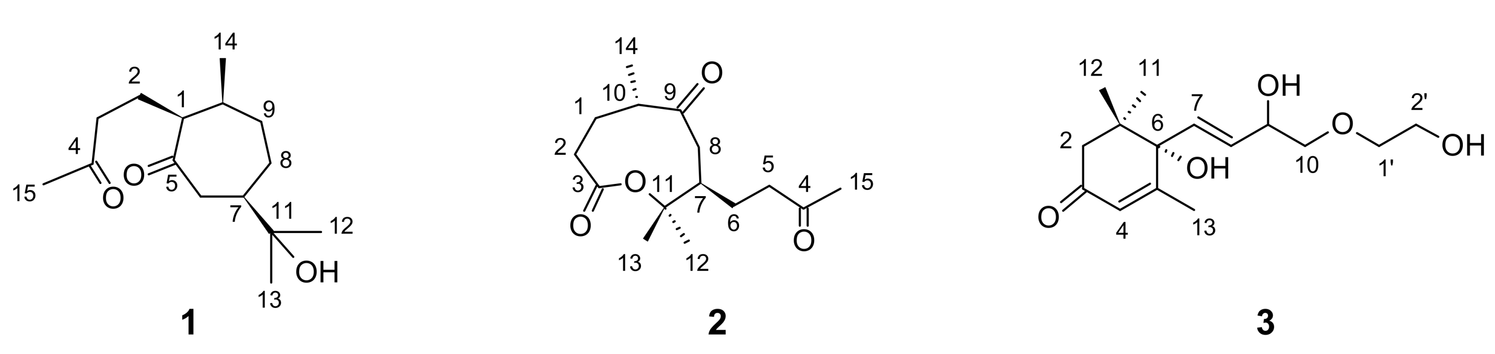
4,5-Seco-Guaiane and a Nine-Membered Sesquiterpene Lactone from Holostylis reniformis

Abstract
:1. Introduction
2. Results and Discussion
3. Experimental
3.1. General Experimental Procedures
3.2. Plant Materials
3.3. Extraction and Isolation of the Chemical Constituents
4. Conclusions
Supplementary Materials
Acknowledgments
References
- Hoehne, F.C. Aristolochiaceae. In Flora Brasílica; Lanzara, F., Ed.; Graphicards: São Paulo, Brasil, 1942; Volume 15, pp. 1–141. [Google Scholar]
- da Silva, T.; Krettli, A.U.; de Andrade-Neto, V.F.; Lopes, L.M.X. Lignans, and particularly aryltetralone lignans, Extracts containing them, Processes for obtaining the extracts and the lignans, and use of the lignans or the extracts in pharmaceutical compositions for treating or preventing malaria. Rev. Prop. Ind. 2005, 1795, 1774–1786. [Google Scholar]
- de Andrade-Neto, V.F.; da Silva, T.; Lopes, L.M.X.; do Rosario, V.E.; Varotti, F.P.; Krettli, A.U. Antiplasmodial activity of aryltetralone lignans from Holostylis reniformis. Antimicrob. Agents Chemother. 2007, 51, 2346–2350. [Google Scholar] [CrossRef] [PubMed]
- da Silva, T.; Lopes, L.M.X. Aryltetralone lignans and 7,8-seco-lignans from Holostylis reniformis. Phytochemistry 2004, 65, 751–759. [Google Scholar] [CrossRef] [PubMed]
- da Silva, T.; Lopes, L.M.X. Aryltetralol and aryltetralone lignans from Holostylis reniformis. Phytochemistry 2006, 67, 929–937. [Google Scholar] [CrossRef] [PubMed]
- Messiano, G.B.; da Silva, T.; Nascimento, I.R.; Lopes, L.M.X. Biosynthesis of antimalarial aryltetralone lignans from Holostylis reniformis. Planta Med. 2008, 74, 924–924. [Google Scholar] [CrossRef]
- Messiano, G.B.; da Silva, T.; Nascimento, I.R.; Lopes, L.M.X. Biosynthesis of antimalarial lignans from Holostylis reniformis. Phytochemistry 2009, 70, 590–596. [Google Scholar] [CrossRef] [PubMed]
- Messiano, G.B.; Wijeratne, E.M.K.; Lopes, L.M.X.; Gunatilaka, A.A.L. Microbial transformations of aryltetralone and aryltetralin lignans by Cunninghamella echinulata and Beauveria bassiana. J. Nat. Prod. 2010, 73, 1933–1937. [Google Scholar] [CrossRef] [PubMed]
- Raharivelomanana, P.; Bianchini, J.-P.; Cambon, A.; Azzaro, M.; Faure, R. Two-dimensional NMR of sesquiterpenes 8—Complete assignment of 1H and 13C-NMR spectra of seven sequiterpene alcohols from Neocallitropsis pancheri. Magn. Reson. Chem. 1995, 33, 233–235. [Google Scholar]
- Atta-ur-Rahman; Ahmad, V.U. 13C-NMR of Natural Products: Monoterpenes and Sesquiterpenes; Atta-ur-Rahman, Ahmad, V.U., Eds.; Plenum Press: New York, NY, USA, 1992; Volume 1, p. 337. [Google Scholar]
- Navickiene, H.M.D.; Lopes, L.M.X. Alkamides and phenethyl derivatives from Aristolochia gehrtii. J. Braz. Chem. Soc. 2001, 12, 467–472. [Google Scholar] [CrossRef]
- van der Gen, A.; van der Linde, L.M.; Witteveen, J.G. Synthesis of guaipyridine and some related sesquiterpene alkaloids. Recl. Trav. Chim. Pays-Bas 1972, 91, 1433–1440. [Google Scholar] [CrossRef]
- Su, J.-H.; Dai, C.-F.; Huang, H.-H.; Wu, Y.-C.; Sung, P.-J.; Hsu, C.-H.; Sheu, J.-H. Terpenoid-related metabolites from a Formosan soft coral Nephthea chabrolii. Chem. Pharm. Bull. 2007, 55, 594–597. [Google Scholar] [CrossRef] [PubMed]
- Su, J.-H.; Chiang, M.Y.; Wen, Z.-H.; Dai, C.-F.; Hsu, C.-H.; Sheu, J.-H. Sesquiterpenoids from the Formosan soft coral Sinularia leptoclados. Chem. Pharm. Bull. 2010, 58, 250–253. [Google Scholar] [CrossRef] [PubMed]
- Advanced Chemistry Development. ACD/C+H NMR predictors and DB: ChemSketch Window. Version 12.01. Toronto: ACD. 2010; 1 CD-ROM. [Google Scholar]
- Kai, H.; Baba, M.; Okuyama, T. Two New Megastigmanes from the leaves of Cucumis sativus. Chem. Pharm. Bull. 2007, 55, 133–136. [Google Scholar] [CrossRef] [PubMed]
- Weiss, G.; Koreeda, M.; Nakanishi, K. Stereochemistry of theaspirone and the blumenols. J. Chem. Soc. Chem. Commun. 1973, 565–566. [Google Scholar] [CrossRef]
- CSD—Cambridge Structural Database. Available online: http://www.ccdc.cam.ac.uk/products/csd/ (accessed on 31 November 2011).
Sample Availability: Samples of the compounds 1–3 are available from the authors. |


| Position | δC, type a | δH (J in Hz) b | gHMBC c |
|---|---|---|---|
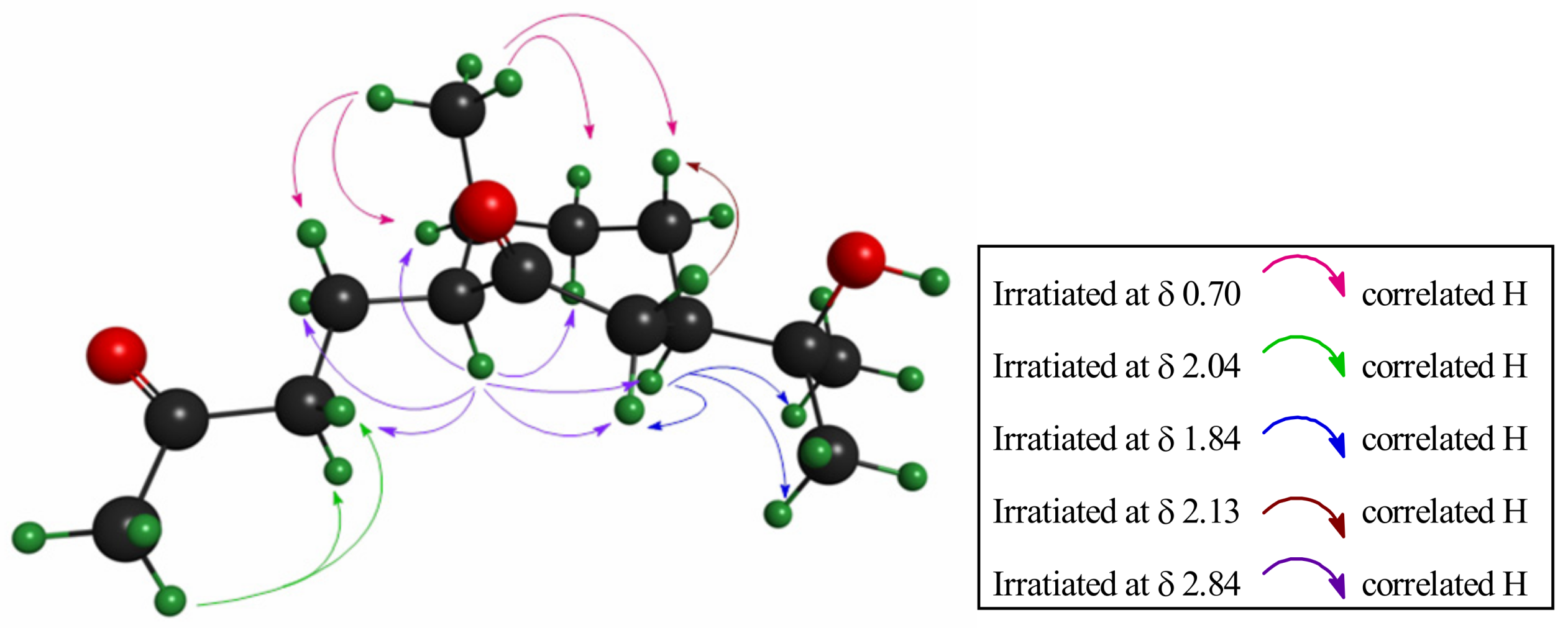
| 1α | 53.8, CH | 2.84, ddd (9.5, 4.5, 2.5) | 14 |
| 2a | 24.2, CH2 | 1.41, dddd (14.5, 8.5, 6.0, 4.5) | |
| 2b | 1.13, m | ||
| 3a | 41.7, CH2 | 2.37, ddd (17.0, 8.5, 6.0) | |
| 3b | 2.19, ddd (17.0, 9.0, 8.5) | ||
| 4 | 208.8, C | 3a, 3b, 15 | |
| 5 | 213.4, C | 6β | |
| 6 α | 47.0, CH2 | 2.52, ddd (17.5, 4.5, 1.7) | |
| 6β | 2.13, dd (17.5, 12.0) | ||
| 7 | 46.4, CH | 1.84, dddd (12.0, 10.0, 4.5, 1.5) | 12, 13 |
| 8α | 25.1, CH2 | 1.75, m | |
| 8β | 1.10, m | ||
| 9α | 37.0, CH2 | 1.73, m | 8β, 14 |
| 9β | 2.04, m | ||
| 10 | 34.9, CH | 1.93, m | 14, 2b and/or 8β |
| 11 | 73.0, C | 12, 13 and/or 8β | |
| 12 | 26.6, CH3 | 1.13, s | 13 |
| 13 | 26.5, CH3 | 1.10, s | 12 |
| 14 | 14.0, CH3 | 0.70, d (7.5) | |
| 15 | 29.8, CH3 | 2.04, s |
| H/C | δC, type a | δH (J in Hz) b, c | gHMBC d |
|---|---|---|---|
| 1α | 30.9, CH2 | 2.06, dddd c (14.5, 9.8, 9.3, 6.5) | 14 |
| 1β | 1.51, dddd c (14.5, 8.5, 2.5, 1.5) | ||
| 2α | 34.6, CH2 | 2.22, ddd c (15.0, 6.5, 1.5) | |
| 2β | 2.19, ddd c (15.0, 9.8, 8.5) | ||
| 3 | 171.0, C | ||
| 4 | 208.0, C | 5a, 5b, 15 | |
| 5a, 5b | 42.2, CH2 | 2.51 br, t (7.5) | 15 |
| 6a | 23.8, CH2 | 1.57, ddt (14.0, 2.0, 7.5) | 8β |
| 6b | 1.23, ddt (14.0, 11.5, 7.0) | ||
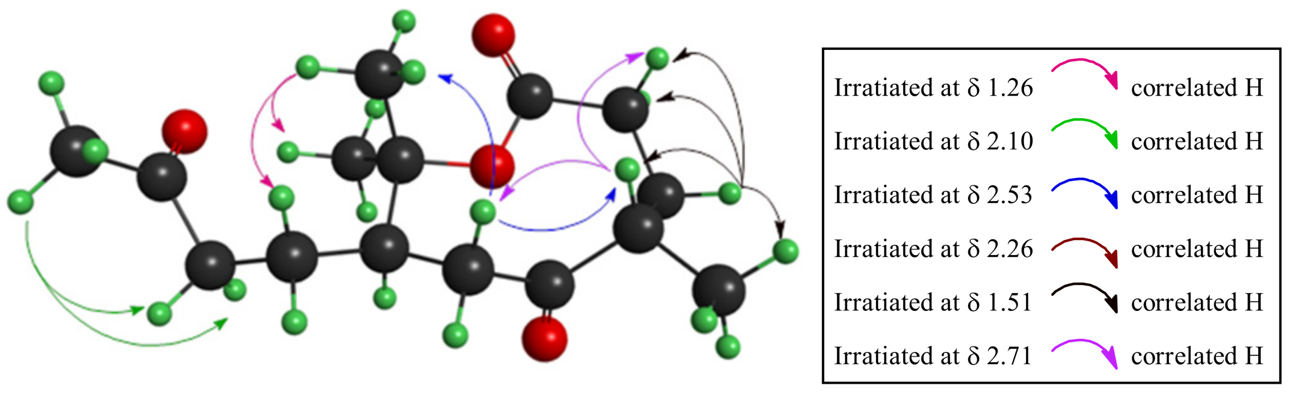
| 7 | 49.2, CH | 2.26, ddt (11.5, 11.0, 2.0) | 5a, 5b, 8α, 12, 13 |
| 8α | 43.0, CH2 | 1.96, dd (12.5, 2.0) | |
| 8β | 2.53, dd (12.5, 11.0) | ||
| 9 | 215.4, C | 8α, 14 | |
| 10 | 47.7, CH | 2.71, ddq (9.3, 2.5, 7.0) | 14 |
| 11 | 85.0, C | 8α, 13, 12 and/or 6a | |
| 12 | 25.7, CH3 | 1.57, s | 13 |
| 13 | 20.3, CH3 | 1.26, s | 12 |
| 14 | 18.8, CH3 | 0.97, d (7.0) | |
| 15 | 30.2, CH3 | 2.10, s |
| H/C | δC, type a | δH (J in Hz) b | gHMBC c |
|---|---|---|---|
| 1 | 40.9, C | 11, 12 | |
| 2 | 49.4, CH2 | 2.05, d (16.0) | 11, 12 |
| 2 | 2.39, d (16.0) | ||
| 3 | 197.4, C | ||
| 4 | 125.5, CH | 5.77, q (2.5) | 13 |
| 5 | 164.0, C | 13 | |
| 6 | 79.1, C | 11, 12, 13, 4, 7, OH | |
| 7 | 129.9, CH | 5.75, d (17.0) | |
| 8 | 131.8, CH | 5.69, dd (17.0, 5.0) | |
| 9 | 71.6, CH | 4.00, m | 7 |
| 10 | 72.2, CH2 | 3.30–3.40, m | 1' |
| 11 | 23.0, CH3 | 0.93, s | 12 |
| 12 | 23.9, CH3 | 0.91, s | 11 |
| 13 | 19.0, CH3 | 1.82, d (1.5) | 4 |
| 1' | 66.1, CH2 | 3.30–3.40, m | |
| 2' | 60.2, CH2 | 3.30–3.40, m | 1' |
| 7-membered ring | |||||
| Spatial Interaction | 71 | 72 | 73 | 74 | |
| ····–C1–H1…. H2a–C2–····· | 0.79 | 1.52 | 1.17 | 0.68 | |
| ····–C1–H1… H6α–C6–····· | 1.00 | 0.09 | ---- | 0.19 | |
| ····–C1–H1…. H7–C7–····· | ---- | ---- | 0.04 | 0.59 | |
| ····–C1–H1…. H9α–C9–····· | 0.65 | ---- | 0.67 | 0.43 | |
| ····–C1–H1..... H10–C10–···· | 1.81 | 1.74 | 2.07 | 0.99 | |
| ····–C2–H2b… CH3(14)–···· | 0.99 | ---- | 0.89 | 0.72 | |
| ····–C8–H8β… H6β–C6–···· | ---- | ---- | 0.63 | 0.27 | |
| ····–C8–H8β.... CH3(14)–···· | 1.39 | ---- | 0.78 | 0.54 | |
| ····–C8–H8β… O=C5–···· | ---- | 0.65 | ---- | 0.15 | |
| ····–C8–H8β….O-H···· | 2.10 | ---- | ---- | 0.12 | |
| ····–C9–H9β... .CH3(14)–···· | 0.30 | 0.76 | 0.40 | 0.75 | |
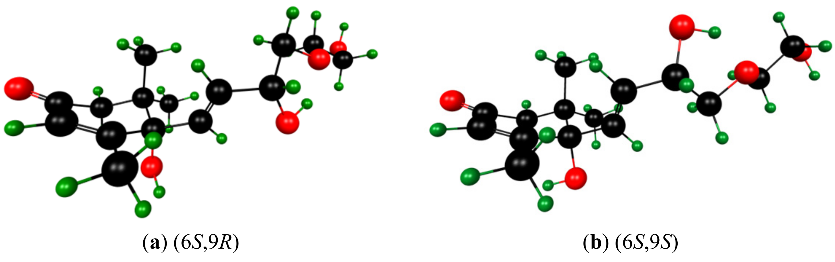
| 9-membered ring | |||||
| 91 | 92 | 93 | 94 | ||
| ····–C1–H1α… O=C9–····· | 1.21 | ---- | 0.02 | 0.01 | |
| ···–C7–H7…. O=C9–····· | 0.59 | ---- | ---- | 0.03 | |
| ····–C8–H8β….H10–C10–····· | 0.97 | 0.02 | ---- | 0.57 | |
| ····–C8–H8β….CH3(13)– ····· | 0.62 | 0.17 | 0.55 | 1.91 | |
| ····–CH3(12)….O=C4–····· | 11.04 | 9.24 | ---- | 8.18 | |
| ····–CH3(12)….O=C3–O–····· | 1.49 | 1.32 | 0.59 | 0.93 | |
© 2012 by the authors; licensee MDPI, Basel, Switzerland. This article is an open access article distributed under the terms and conditions of the Creative Commons Attribution license (http://creativecommons.org/licenses/by/3.0/).
Share and Cite
Pereira, M.D.P.; Da Silva, T.; Lopes, L.M.X.; Krettli, A.U.; Madureira, L.S.; Zukerman-Schpector, J. 4,5-Seco-Guaiane and a Nine-Membered Sesquiterpene Lactone from Holostylis reniformis. Molecules 2012, 17, 14046-14057. https://doi.org/10.3390/molecules171214046
Pereira MDP, Da Silva T, Lopes LMX, Krettli AU, Madureira LS, Zukerman-Schpector J. 4,5-Seco-Guaiane and a Nine-Membered Sesquiterpene Lactone from Holostylis reniformis. Molecules. 2012; 17(12):14046-14057. https://doi.org/10.3390/molecules171214046
Chicago/Turabian StylePereira, Marcos D. P., Tito Da Silva, Lucia M. X. Lopes, Antoniana U. Krettli, Lucas S. Madureira, and Julio Zukerman-Schpector. 2012. "4,5-Seco-Guaiane and a Nine-Membered Sesquiterpene Lactone from Holostylis reniformis" Molecules 17, no. 12: 14046-14057. https://doi.org/10.3390/molecules171214046