Article

Gombapyrones E and F, New α-Pyrone Polyenes Produced by Streptomyces sp. KMC-002

1
Natural Medicine Center, Korea Institute of Science and Technology (KIST), Gangneung, Gangwon-do 210-340, Korea
2
Natural Products Laboratory, School of Pharmacy, Sungkyunkwan University, Suwon 440-746, Korea
*
Author to whom correspondence should be addressed.
Molecules 2011, 16(5), 3519-3529; https://doi.org/10.3390/molecules16053519
Submission received: 29 March 2011 / Revised: 19 April 2011 / Accepted: 20 April 2011 / Published: 26 April 2011
(This article belongs to the Collection Bioactive Compounds)

Abstract

:
Microorganism-derived polyene polyketides have been shown to display a variety of biological activities and have attracted great interest due to their structurally intriguing chemical diversity. Two new polyenes were isolated from a culture broth of Streptomyces sp. KMC-002 obtained from a soil sample in an abandoned mine. The structures of these compounds were determined to be α-pyrone-containing polyene analogues through analyses of HRFABMS, UV and NMR data, and were named Gombapyrones E (1) and F (2). Gombapyrone E (1) showed antibacterial activity against Micrococcus luteus, Enterococcus hirae, Staphylococcus aureus and MRSA.

1. Introduction

Polyketides represent a highly diverse structural class of natural products found in bacteria, fungi, marine invertebrates, insect and higher plants [1,2,3,4,5,6,7]. From a biochemical point of view, they are thought to be involved in many different types of biological events acting as pheromones, offense and defense materials, pigments and biosynthetic intermediates [8,9,10,11]. Polyketide biosynthesis involves a series of enzyme-catalyzed processes taking place by successive decarboxylative Claisen condensations of activated acetyl-derived starter and malonyl-derived extender units [12,13]. Programmed events, modulated by variety of catalytic domains, endow them with structurally fascinating carbon backbones, ranging from simple pyrone groups to highly complex polyene macrolides [14,15,16,17]. In addition, polyketide natural products have provided many pharmaceutical lead compounds such as polyene-polyol macrocyclic lactones, nystatin and amphotericin B [18,19,20,21,22]. Because of their large structural diversity and varied biological functions, it is an undeniable fact that polyketide natural products are one of the attractive groups for developing useful chemicals as therapeutic approaches or as synthetic challenges [23,24,25]. In our preliminary screening for discovery of new polyketides produced by abandoned mine-derived actinomycetes, two new polyene-polyketides, gombapyrone E (1) and F (2), were isolated from organic extract of Streptomyces sp. KMC-002. The structures of these compounds (Figure 1), which represent new members of the gombapyrone family previously elucidated by Helaly et al. [26], were assigned by analyses of 2D-NMR, UV and HRFABMS (High-Resolution Fast Atom Bombardment Mass Spectrometry) data. Previously described gombapyrones possess triene chains attached to an aromatic ring, whereas gombapyrones E (1) and F (2) isolated from Streptomyces sp. KMC-002 are composed of α-pyrone and phenyl groups connected through a conjugated tetraene polyketide chain. Although the carbon skeleton of these compounds resembles the previously reported CRP-2504-1a, polyketide-derived tetraene metabolites with α-pyrone moieties are extremely rare in Nature [27]. We thus describe here the isolation, structure elucidation and antimicrobial activity of two new α-pyrone polyenes isolated from an abandoned mine-derived Streptomyces sp.
Figure 1. Structures of gombapyrone E (1) and F (2).
Molecules 16 03519 g001

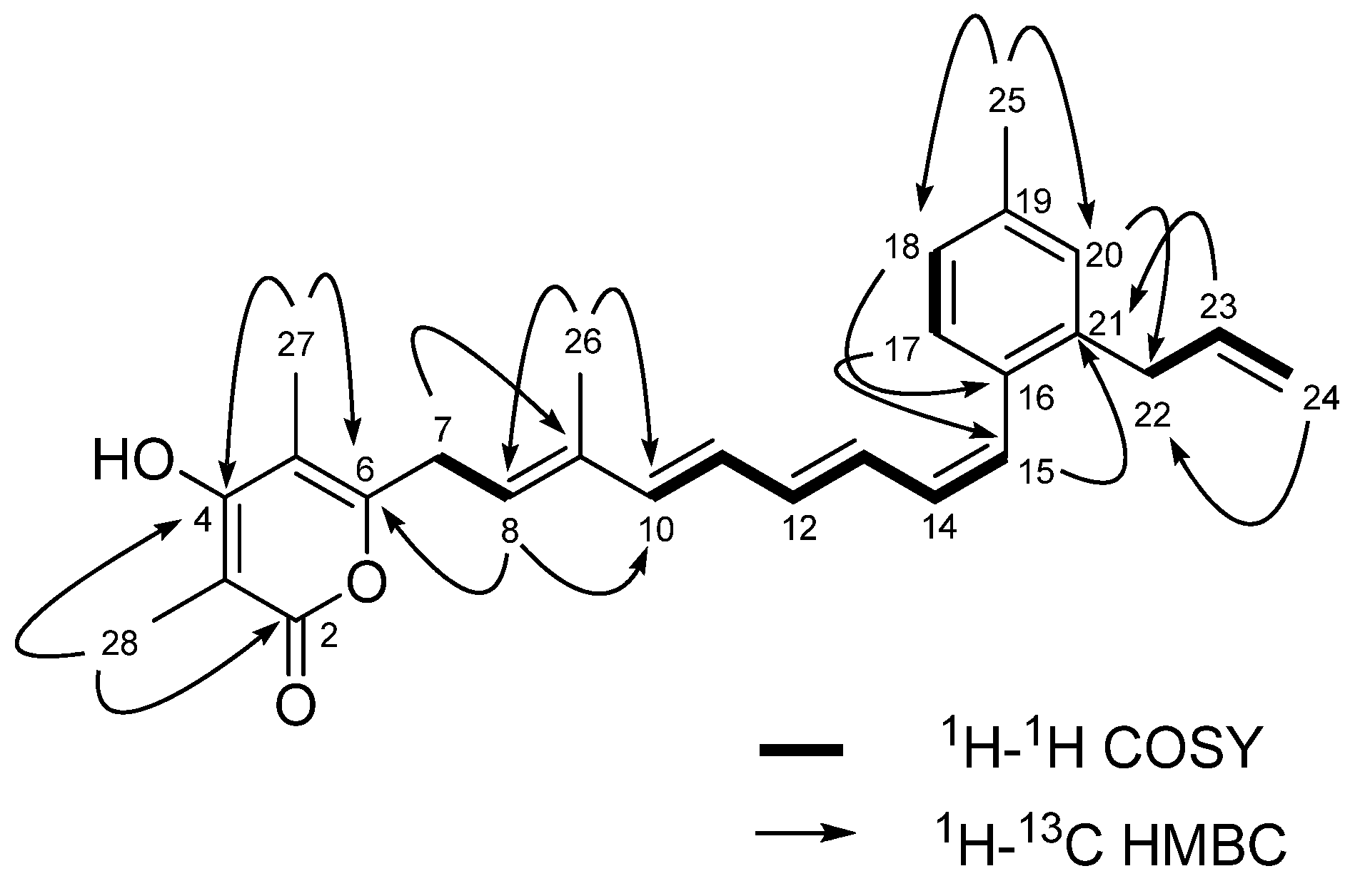
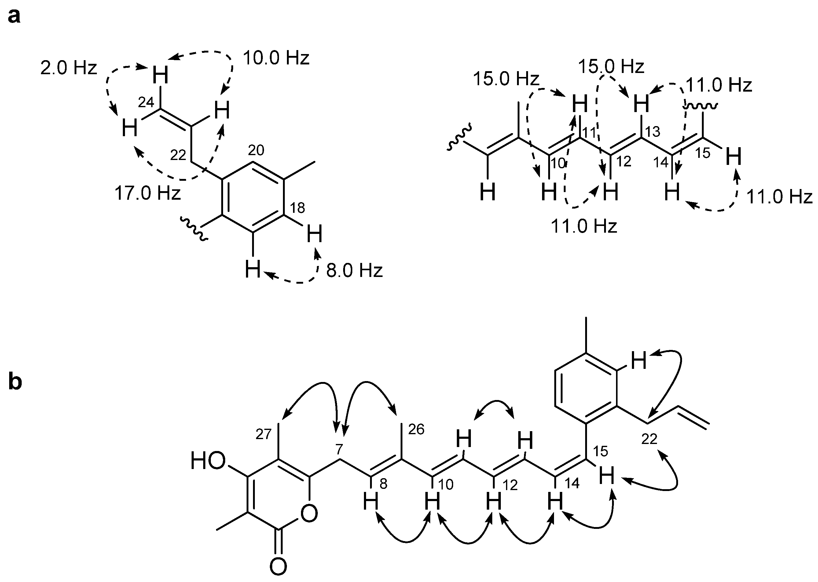
2. Results and Discussion
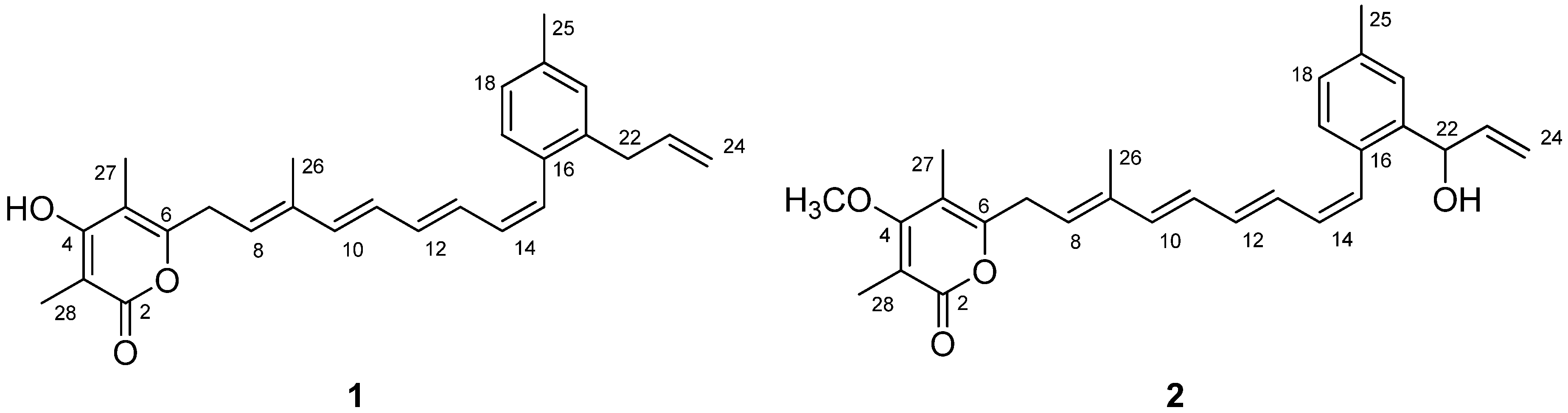
The sequence analysis of the 16S rRNA gene placed the strain KMC-002 within the genus Streptomyces on the basis of the 99.1% similarity with Streptomyces albireticuli strain JCM 4116 (AY999748). The cultured Streptomyces strain KMC-002 was deposited at the Korean Culture Center of Microorganisms (KCCM 90098).
The strain KMC-002 was isolated from a soil sample collected at the abandoned Yeonhwa zinc mine located at Gangneung, South Korea. In order to investigate the secondary metabolite production by KMC-002, the strain was cultured in thirty two 1 liter Erlenmeyer flasks each containing 500 mL of A1 liquid medium (total 16 L) [10 g of starch (Difco), 2 g of yeast (Difco) and 4 g of peptone (Difco) in 1 L sterilized distilled water]. Metabolite production in the culture broth was monitored daily by HPLC analysis using an Agilent 1100 LC-MS system with a Phenomenex Luna 5 μm C18(2) analytical column (4.6 × 150 mm, flow rate 0.7 mL/min) and a gradient elution of 10%–100% acetonitrile in water for 30 min. Two major peaks were observed at retention times 28 and 29 min on the fourth day of culture. At the end of the culture period (day 5), sterilized XAD-7 adsorbent resin (20 g/L) was added into the culture broth of the strain KMC-002, and the resin was collected 3 h later by filtration through cheesecloth, washed with deionized water and eluted twice with acetone. The acetone was removed under reduced pressure and the residue was partitioned between ethyl acetate and water. The dried ethyl acetate layer was subjected to a flash column chromatography using reversed phase C18 resin and step gradient elution of water and methanol. The 80% aqueous methanol elution fraction including the major peaks was purified by repeated HPLC using a reversed phase column and gradient elution of acetonitrile and water to afford gombapyrone E (1, 5 mg) and F (2, 3 mg).
Gombapyrone E (1) was isolated as a pale yellowish oil. Its molecular formula was deduced as C27H30O3 on the basis of the HRFABMS (obsd, [M + H]+ at m/z 403.2278, calcd, 403.2273) and 13C-NMR data. The IR spectrum of 1 displayed absorption bands at 3,376 and 1,673 cm−1, indicating the presence of hydroxyl and ester carbonyl functionalities, while a UV absorption band at 330 nm was suggestive of a conjugated tetraene-like chromophore [28]. 1H-NMR spectral data (Table 1), including correlations from 1H-1H COSY experiment, illustrated signals attributable to a conjugated tetraene [δH 5.56 (1H, t, J = 7.5 Hz), 6.28 (1H, dd, J = 15.0, 11.0 Hz), 6.32 (1H, d, J = 15.0 Hz), 6.33 (1H, dd, J = 11.0, 11.0 Hz), 6.40 (1H, dd, J = 15.0, 11.0 Hz), 6.49 (1H, d, J = 11.0 Hz) and 6.53 (1H, dd, J = 15.0, 11.0 Hz)]. Other characteristic features of the 1H-NMR spectrum of 1 were the presence of three additional nonconjugated olefinic protons [δH 4.94 (1H, ddd, J = 17.0, 3.5, 2.0 Hz), 4.99 (1H, ddd, J = 10.0, 3.5, 2.0 Hz) and 5.90 (1H, ddt, J = 17.0, 10.0, 6.5 Hz)], and two methylene protons at δH 3.33 and 3.44. In addition, the 1H-NMR spectrum showed additional signals attributed to a phenyl group [δH 7.02 (1H, s), 7.03 (1H, d, J = 8.0 Hz) and 7.14 (1H, d, J = 8.0 Hz)] and four methyl groups at δH 1.86, 1.91, 1.98 and 2.32. The 13C-NMR (Table 1) and HSQC spectra of 1 allowed all protons to be assigned to their respective carbons. The nonconjugated olefinic methylene protons at δH 4.94 and 4.99 correlated with olefinic carbon at δC 114.4, while the nonconjugated olefinic methine proton correlated with a carbon signal at δC 136.9. The four methyl protons at δH 1.86, 1.91, 1.98 and 2.32 correlated with δC 11.3, 7.5, 8.6 and 19.8, respectively, and the methylene protons to carbon signals at δC 37.3 and 29.9. The HSQC spectrum also showed that the phenyl protons at δH 7.02, 7.03 and 7.14 correlated to carbons at δC 129.8, 126.2 and 129.6, respectively. In addition, nine sp2 quaternary carbon signals were observed at δC 97.6, 107.9, 133.4, 136.6, 136.8, 138.0, 156.8, 166.5 and 167.0 in the 13C-NMR spectrum of 1.
Table 1. 1H and 13C-NMR spectral data for gombapyrone E (1) and F (2) in methanol-d4.
Gombapyrone E (1)Gombapyrone F (2)
PositionδH mult (J, Hz) aδC b δH mult (J, Hz) aδC b
2167.0C166.8C
3 97.6C 108.6C
4 166.5C 169.3C
5 107.9C 110.0C
6 156.8C 157.2C
73.44 d (7.5)29.9CH23.46 d (7.5)29.9CH2
85.56 t (7.5)125.4CH5.57 t (7.5)125.3CH
9 136.6C 136.7C
106.32 d (15.0)137.0CH6.33 d (15.0)137.1CH
116.40 dd (15.0, 11.0)134.8CH6.42 dd (15.0, 11.0)134.9CH
126.53 dd (15.0, 11.0)128.9CH6.49 dd (15.0, 11.0)128.9CH
136.28 dd (15.0, 11.0)128.0CH6.32 dd (15.0, 11.0)128.1CH
146.33 dd (11.0, 11.0)130.2CH6.36 dd (11.0, 11.0)130.6CH
156.49 d (11.0)128.3CH6.58 d (11.0)127.9CH
16 133.4C 132.5C
177.14 d (8.0)129.6CH7.11 d (8.0)129.6CH
187.03 d (8.0) c126.2CH7.12 d (8.0)127.3CH
19 136.8C 137.0C
207.02 s c129.8CH7.32 s126.6CH
21 138.0C 140.8C
223.33 d37.3CH25.32 d (5.5)71.2CH
235.90 ddt (17.0, 10.0, 6.5)136.9CH5.96 ddd (17.0, 10.0, 5.5)139.9CH
244.99 ddd (10.0, 3.5, 2.0)114.4CH25.20 dt (17.0, 1.5)113.3CH2
4.94 ddd (17.0, 3.5, 2.0)5.09 dt (10.0, 1.5)
252.32 s19.8CH32.36 s19.9CH3
261.86 s11.3CH31.86 s11.3CH3
271.98 s8.6CH31.98 s8.8CH3
281.91 s7.5CH32.00 s8.7CH3
4-OCH3 3.87 s59.7CH3
a 500 MHz; b 125 MHz; c-d Overlapping signals; Chemical shifts (δ) in ppm.
HMBC-NMR correlations from the methyl proton at δH 2.32 (H3-25) and the aromatic proton at δH 7.14 (H-17) to the aromatic quaternary carbon at δC 136.8 (C-19) allowed the methyl group to be positioned at C-19. HMBC correlations from the aromatic protons at δH 7.14 (H-17) to the olefinic carbon at δC 128.3 (C-15) allowed the polyene chain to be connected to the C-16 position of the phenyl group. HMBC correlations between the proton signal at δH 5.90 (H-23) and the aromatic quaternary carbon signal at δC 138.0 (C-21) also showed that the allyl functionality from C-22 to C-24 was connected to the C-21 position of the phenyl group (Figure 2). The HMBC correlations from H-27 (δH 1.98) to C-4 (δC 166.5), C-6 (δC 156.8) and C-5 (δC 107.9), and from H-28 (δH 1.91) to C-2 (δC 167.0), C-4 (δC 166.5) and C-3 (δC 97.6) indicated the presence of 3,5-dimethyl-α-pyrone. In addition, key HMBC correlations (Figure 2) allowed the polyene chain to be connected to the C-6 position of the α-pyrone ring. Comprehensive analysis of 2D-NMR data from 1H-1H COSY, 1H-1H TOCSY, NOESY, HSQC and HMBC experiments led to the construction of a 3,5-dimethyl-4-hydroxy-α-pyrone and a 2-allyl-4-methyl-benzene connected with the 7-methylnona-1,3,5,7-tetraene chain. The aromatic ring and the α-pyrone ring in this specific carbon framework were confirmed by comparison of the proton and carbon NMR chemical shifts of 1 with those of the previously reported gombapyrone B [26].
Figure 2. 1H-1H COSY and Key HMBC correlations of gombapyrone E (1).
Molecules 16 03519 g002
The full carbon chain from C-7 to C-15, including the presence of a vinyl methyl group at C-9, was readily assigned by the combined 2D-NMR data of 1. The configurations of the double bonds in the tetraene chain were assigned on the basis of 1D proton-proton NMR coupling constant data and upon analysis of NOESY-NMR information. The geometries of the Δ10,11, Δ12,13 and Δ14,15 olefins were assigned as E, E and Z, respectively, based upon proton coupling constants of JH-10,H-11 = 15.0 Hz, JH-12,H-13 = 15.0 Hz and JH-14,H-15 = 11.0 Hz (Figure 3). NOESY-NMR correlations between the vinyl methyl proton (H3-26) and the H2-7 proton, and between H-8 (δH 5.56) and H-10 (δH 6.32) established the configuration of the Δ8,9 olefin as E (Figure 3).
Figure 3. Core 1H-1H coupling constants (a) and Key NOE correlations (b) of gombapyrone E (1).
Molecules 16 03519 g003
Gombapyrone F (2) was isolated as a pale yellowish oil that analyzed for the molecular formula C28H32O4 by HRFABMS (obsd. [M + Na]+ at m/z 455.2206, calcd. 455.2198) and by comprehensive analysis of its NMR data (Table 1). The UV absorption spectrum of 2 showed bands at near 330 nm, which were suggestive of the presence of the conjugated polyene as in 1. The 1H-NMR spectrum of 2 was similar to that of 1, except for methoxy proton and oxygenated methine proton signals at δH 5.32 (1H, d, J = 5.5 Hz) and 3.87 (3H, s). The carbon signals observed at δ 71.2 and 59.7 supported the presence of these functionalities. The 1H-1H COSY correlation between the oxygenated methine proton at δH 5.32 (H-22) and H-23 (δH 5.96) and the HMBC correlation from H-23 to C-21 (δC 140.8) easily led to the construction of a 1-hydroxy-allyl group positioned at C-21 of the phenyl group. HMBC-NMR correlations from the methoxy proton to the aromatic quaternary carbon at δC 169.3 (C-4) allowed the methoxy group to be positioned at C-4. The chemical shifts of the α-pyrone ring in this specific constellation were comparable to those from gombapyrones as shown in the literature [26]. The double bond geometries of 2 were deduced from 1H-1H coupling constants and 2D NOE analyses, which were in good accordance with those of 1.
The gombapyrones E (1) and F (2) are polyketide secondary metabolites composed of three structural units—α-pyrone, tetraene chain and phenyl group. Naturally occurring α-pyrone-related compounds have been shown to display a variety of biological activities such as antibiotic, anticancer and neurotoxic effects [29,30,31]. α-Pyrones have also attracted great interest due to their effect on HIV protease inhibition and Alzheimer’s amyloid toxicity inhibition [15,32,33,34]. Although the basic carbon skeleton of 1 and 2 have previously been reported by Doi et al., a chemical structure with an α-pyrone and a phenyl group connected through a tetraene chain is unusual in Nature [27].
The structural composition of α-pyrone and phenyl group connected through a linear carbon chain is found in the gombapyrones and lehualides. Previously described gombapyrones, new members of the α-pyrone family isolated from Streptomyces spp. possess triene chains attached to an aromatic ring, while lehualide A, isolated from marine sponges, is composed of an α-pyrone and a benzyl group connected through an eleven carbon chain [4,26].
Compounds 1-2 were tested for antimicrobial activity towards six pathogenic microorganisms, including methicillin-resistant Staphylococcus aureus (Table 2). Interestingly, 1 showed selective antimicrobial activities against four pathogens (MIC = 3.13 μg/mL against M. luteus, 1.56 μg/mL against S. aureus, 3.13 μg/mL against E. hirae and 6.25 μg/mL against MRSA), whereas 2 did not show any activity at all against all six strains (Table 2).
Table 2. MIC test results for 1 and 2.
Pathogen listMIC (μg/mL)
12PC a
Escherichia coli KCTC 2593NANA6.25
Micrococcus luteus KCCM 115483.131000.78
Staphylococcus aureus KCTC 19161.56NA0.78
Bacillus subtilis KCTC1021NANA3.13
Enterococcus hirae KCCM 117683.131000.78
Staphylococcus aureus MRSA 26596.25NA12.5
a Positive control (PC) = Ampicillin; NA = Not Activity.

3. Experimental

3.1. General

A Waters 1525 system with a Phenomenex Luna C18 (2) 5 μm column (4.6 × 100 mm) and a PDA detector was used for HPLC analysis. Low resolution MS data was measured on an Agilent 1100 LC/MS system equipped with a Phenomenex Luna C18 (2) 5 μm column (4.6 × 150 mm, flow rate 0.7 mL/min). Lichroprep RP-18 (Merck, 40–63 μm) was used for reversed phase flash column chromatography. HPLC separation and purification were performed using a Gilson 321 HPLC system with a Phenomenex Luna C18 (2) 10 μm column (21.2 × 250 mm) at a flow rate 10 mL/min or Phenomenex Luna C18 (2) 10 μm column (10 × 250 mm) at a flow rate 4 mL/min. All NMR spectra were obtained in methanol-d4 (δH 3.32, δC 47.6) on a Varian UNITY Plus 500 MHz NMR system equipped with a cryogenic probe. FT-IR spectra were recorded on a Thermo Scientific Nicolet iS10 spectrometer. UV spectra were obtained on a PerkinElmer Lambda 35 UV/Vis spectrophotometer. Optical rotations were measured on a PerkinElmer model 343 polarimeter. HRFABMS data were obtained on a JEOL/JMS-AX505WA instrument.

3.2. Strain Collection, Isolation, and Identification

Soil samples were collected at the abandoned Yeonhwa zinc mine located at Gangneung, South Korea. The samples were diluted with autoclaved distilled water, heated to 55 °C for 6 min and the resulting suspension (100 μL) inoculated onto several kinds of agar plate. The plates were incubated 2 weeks at 25 °C under aerobic conditions. A pure Streptomyces strain KMC-002 was isolated from A1 agar medium (10 g of starch, 4 g of yeast, 2 g of peptone and 18 g of agar in 1 L of sterile distilled water). Stocks of the isolated bacterial strain were generated and stored at −80 °C in liquid culture medium containing 15% (v/v) glycerol. The strain KMC-002 was identified based on 16S rRNA gene sequencing analysis. The chromosomal DNA of strain KMC-002 was extracted using the G-spin genomic DNA extraction kit (iNtRON Biotechnology, Daejeon, Korea). The 16S rRNA gene of strain KMC-002 was amplified by PCR using universal primers 27f and 1492r corresponding to positions 27 in the forward direction and 1492 in the reverse direction of the Escherichia coli 16S rRNA gene [35]. The DNA sequencing reaction was carried out by using an ABI Prism Bigdye terminator cycle sequencing ready reaction kit V.3.1 (Applied Biosystems, Foster City, CA, USA). The PCR cycle-sequencing product was purified by using Montage dye remove kit (Millipore, Bedford, TX, USA) according to the manufacturer’s protocol. 16S rRNA gene sequence was determined on a Perkin-Elmer model ABI 3730XL capillary DNA sequencer (Applied Biosystems). The 16S rRNA gene sequence of strain KMC-002 was compared with primary sequence information within the GenBank/EMBL/DDBJ nucleotide sequence database using the BLAST algorithm [36].
The strain KMC-002 was classified as a Streptomyces sp. based on 16S rDNA sequence analysis which showed 99.1% similarity with Streptomyces albireticuli strain JCM 4116 (AY999748). The cultured Streptomyces strain KMC-002 was deposited with the Korean Culture Center of Microorganisms (KCCM 90098).

3.3. Cultivation and Extraction

The strain KMC-002 was cultured in 1 L Erlenmeyer flasks (32 × 500 mL, total 16 L), each containing 500 mL of A1 liquid medium (10 g of starch, 4 g of yeast, 2 g of peptone and 1 L of sterile distilled water) while shaking 200 rpm at 27 °C. After 5 days, sterilized XAD-7 adsorbent resin (20 g/L) was added into the culture broth of the strain KMC-002, and the resin was collected 3 h later by filtration through cheesecloth, washed with deionized water and eluted twice with acetone. The acetone was removed under reduced pressure and the residue was partitioned between ethyl acetate and water. The ethyl acetate layer was dried in vacuo to yield 800 mg of crude extract.

3.4. Isolation and Purification of Gombapyrones E (1) and F (2)

The crude extract was fractionated by Lichroprep RP-18 (40–63 μm, Merck, N.J., USA) flash column chromatography using a step gradient elution with H2O and CH3OH (20%, 40%, 60%, 80%, and 100% CH3OH in H2O) to give six subfractions. The 80% mathanol fraction contained gombapyrone E(1) and F(2), which were separated by reversed phase HPLC [Gilson 321; Phenomenex Luna C18 (2) 10 μm column (21.2 × 250 mm); 10 mL/min] using gradient elution from 10% to 100% aqueous CH3CN containing 0.02% trifluoroacetic acid (TFA) for 2 h. 1 (5 mg, tR 50 min) and 2 (3 mg, tR 40 min) were purified by reversed phase HPLC [Gilson 321; Phenomenex Luna C8 (2) 10 μm column (10 × 250 mm), 4 mL/min] with an CH3CN-H2O gradient elution system from 50% CH3CN to 100% CH3CN over 1 h.
Gombapyrone E (1): Pale yellowish oil; Molecules 16 03519 i001: −3.2 (c 0.10, MeOH); UV (MeCN) λmax (log ε) 200 (4.27), 260 (3.70), 329 (4.28); IR (film) νmax 3,376, 2,925, 1,672, 1,378, 1,202, 756 cm−1; 1H- and 13C-NMR spectra: see Table 1; HRFABMS [M + H]+ m/z 403.2278 (calcd. for C27H31O3, 403.2273).
Gombapyrone F (2): Pale yellowish oil; Molecules 16 03519 i001: −6.0 (c 0.05, MeOH); UV (MeCN) λmax (log ε) 200 (4.07), 260 (3.52), 332 (4.06); IR (film) νmax 3,356, 2,925, 1,683, 1,456, 1,026, 821 cm−1; 1H- and 13C-NMR spectra: see Table 1; HRFABMS [M + Na]+ m/z 455.2206 (calcd. for C28H32O4Na, 455.2198).

3.5. Antibacterial Activities Test

The antibacterial activities of 1 and 2 were evaluated by MIC assay against six pathogenic microorganisms, Escherichia coli (KCTC 2593), Bacillus subtilis (KCTC 1021), Staphylococcus aureus (KCTC 1916), Methicillin resistance Staphylococcus aureus MRSA 2659, Micrococcus luteus (KCCM 11548) and Enterococcus hirae (KCCM 11768). For MIC assays, all six strains were grown at 37 ºC in nutrient agar (Difco, USA). A loopful of all strains was resuspended in nutrient broth to give 105 colony forming units/mL and incubated at 37 ºC for 18 h and dispensed into 96 well plates (100 μL/well). The MIC evaluation was determined as the concentration of the compounds that completely inhibited pathogenic cell growth for 24 h incubation at 37 ºC. Ampicillin was used as a positive control.

4. Conclusions

Two new α-pyrone polyenes, gombapyrones E (1) and F (2), were isolated from the abandoned mine-derived Streptomyces sp. KMC-002. The key structural features of 1 and 2 were characterized by spectroscopic methods, including 2D-NMR experiments, as a 3,5-dimethyl-4-hydroxy-α-pyrone and a 2-allyl-4-methylbenzene connected by a 7-methylnona-1,3,5,7-tetraene chain. Gombapyrone E showed antibacterial activity against Micrococcus luteus, Enterococcus hirae, Staphylococcus aureus and MRSA. For decades, investigation at the chemical and biochemical levels to better understand polyketide diversity has been continuously making impressive progress in the natural products field. Isolation of gombapyrones E (1) and F (2) is expected to contribute towards providing a better understanding of the structural diversity of polyketide natural products.

Acknowledgements

This study was supported by the Korea Institute of Science and Technology institutional program, grant numbers 2Z03401.

References and Notes

  1. Oh, D.-C.; Gontang, E.A.; Kauffman, C.A.; Jensen, P.R.; Fenical, W. Salinipyrones and Pacificanones, mixed-precursor polyketides from the marine actinomycete Salinispora pacifica. J. Nat. Prod. 2008, 71, 570–575. [Google Scholar] [CrossRef]
  2. Igarashi, Y.; Kim, Y.; In, Y.; Ishida, T.; Kan, Y.; Fujita, T.; Iwashita, T.; Tabata, H.; Onaka, H.; Furumai, T. Alchivemycin A, a bioactive polycyclic polyketide with an unprecedented skeleton from Streptomyces sp. Org. Lett. 2010, 12, 3402–3405. [Google Scholar]
  3. Lin, Z.; Zhu, T.; Fang, Y.; Gu, Q.; Zhu, W. Polyketides from Penicillium sp. JP-1, an endophytic fungus associated with the mangrove plant Aegiceras corniculatum. Phytochemistry 2008, 69, 1273–1278. [Google Scholar]
  4. Sata, N.; Abinsay, H.; Yoshida, W.Y.; Horgen, F.D.; Sitachitta, N.; Kelly, M.; Scheuer, P.J. Lehualides A-D, metabolites from a Hawaiian sponge of the genus Plakortis. J. Nat. Prod. 2005, 68, 1400–1403. [Google Scholar] [CrossRef]
  5. Jiang, Z.-H.; Yang, Q.-X.; Tanaka, T.; Kouno, I. Bicyclic polyketide lactones from Chinese medicinal ants, Polyrhacis lamellidens. J. Nat. Prod. 2008, 71, 724–727. [Google Scholar] [CrossRef]
  6. Samappito, S.; Page, J.E.; Schmidt, J.; De-Eknamkul, W.; Kutchan, T.M. Aromatic and pyrone polyketides synthesized by a stilbene synthase from Rheum tataricum. Phytochemistry 2003, 62, 313–323. [Google Scholar]
  7. Cheng, M.-J.; Lee, S.-J.; Chang, Y.-Y.; Wu, S.-H.; Tsai, I.-L.; Jayaprakasam, B.; Chen, I.-S. Chemical and cytotoxic constituents from Peperomia sui. Phytochemistry 2003, 63, 603–608. [Google Scholar]
  8. Bartelt, R.J.; Weisleder, D. Polyketide origin of pheromones of Carpophilus davidsoni and C. mutilatus (Coleoptera: Nitidulidae). Bioorg. Med. Chem. 1996, 4, 429–438. [Google Scholar] [CrossRef]
  9. Scott, J.J.; Oh, D.-C.; Yuceer, M.C.; Klepzig, K.D.; Clardy, J.; Currie, C.R. Bacterial protection of beetle-fungus mutualism. Science 2008, 322, 63. [Google Scholar] [CrossRef]
  10. Gao, J.-M.; Qin, J.-C.; Pescitelli, G.; Di Pietro, S.; Ma, Y.-T.; Zhang, A.-L. Structure and absolute configuration of toxic polyketide pigments from the fruiting bodies of the fungus Cortinarius rufo-olivaceus. Org. Biomol. Chem. 2010, 8, 3543–3551. [Google Scholar]
  11. Wiemann, P.; Willmann, A.; Straeten, M.; Kleigrewe, K.; Beyer, M.; Humpf, H.-U.; Tudzynski, B. Biosynthesis of the red pigment bikaverin in Fusarium fujikuroi: Genes, their function and regulation. Mol. Microbiol. 2009, 72, 931–946. [Google Scholar] [CrossRef]
  12. Hopwood, D.A. Genetic contributions to understanding polyketide synthases. Chem. Rev. 1997, 97, 2465–2497. [Google Scholar] [CrossRef]
  13. Hertweck, C. The biosynthetic logic of polyketide diversity. Angew. Chem. Int. Ed. 2009, 48, 4688–4716. [Google Scholar] [CrossRef]
  14. O’Hagan, D. Biosynthesis of fatty acid and polyketide metabolites. Nat. Prod. Rep. 1995, 12, 1–32. [Google Scholar]
  15. McGlacken, G.P.; Fairlamb, I.J.S. 2-Pyrone natural products and mimetics: Isolation, characterisation and biological activity. Nat. Prod. Rep. 2005, 22, 369–385. [Google Scholar] [CrossRef]
  16. Busch, B.; Hertweck, C. Evolution of metabolic diversity in polyketide-derived pyrones: Using the non-colinear aureothin assembly line as a model system. Phytochemistry 2009, 70, 1833–1840. [Google Scholar]
  17. Kwon, H.C.; Kauffman, C.A.; Jensen, P.R.; Fenical, W. Marinomycins A-D, antitumor-antibiotics of a new structure class from a marine actinomycete of the recently discovered genus “Marinispora”. J. Am. Chem. Soc. 2006, 128, 1622–1632. [Google Scholar]
  18. Chopra, I.; Roberts, M. Tetracycline antibiotics: Mode of action, applications, molecular biology, and epidemiology of bacterial resistance. Microbiol. Mol. Biol. Rev. 2001, 65, 232–260. [Google Scholar] [CrossRef]
  19. Kotler-Brajtburg, J.; Medoff, G.; Kobayashi, G.S.; Boggs, S.; Schlessinger, D.; Pandey, R.C.; Rinehart, K.L., Jr. Classification of polyene antibiotics according to chemical structure and biological effects. Antimicrob. Agents Chemother. 1979, 15, 716–722. [Google Scholar] [CrossRef]
  20. Thomas, C.M.; Hothersall, J.; Willis, C.L.; Simpson, T.J. Resistance to and synthesis of the antibiotic mupirocin. Nat. Rev. Microbiol. 2010, 8, 281–289. [Google Scholar] [CrossRef]
  21. Tsukamoto, S.; Matsunaga, S.; Fusetani, N.; Toh-E, A. Theopederins F-J: five new antifungal and cytotoxic metabolites from the marine sponge, Theonella swinhoei. Tetrahedron 1999, 55, 13697–13702. [Google Scholar] [CrossRef]
  22. Huang, X.; He, J.; Niu, X.; Menzel, K.-D.; Dahse, H.-M.; Grabley, S.; Fiedler, H.-P.; Sattler, I.; Hertweck, C. Benzopyrenomycin, a cytotoxic bacterial polyketide metabolite with a benzo[a]pyrene-type carbocyclic ring system. Angew. Chem. Int. Ed. 2008, 47, 3995–3998. [Google Scholar]
  23. Tatsuta, K.; Hosokawa, S. Total syntheses of polyketide-derived bioactive natural products. Chem. Rec. 2006, 6, 217–233. [Google Scholar] [CrossRef]
  24. Dias, L.C.; Meira, P.R.R. Total synthesis of the potent antitumor polykeide (−)-callystatin A. J. Org. Chem. 2005, 70, 4762–4773. [Google Scholar] [CrossRef]
  25. Barth, R.; Mulzer, J. Total synthesis of efomycine M. Angew. Chem. Int. Ed. 2007, 46, 5791–5794. [Google Scholar] [CrossRef]
  26. Helaly, S.; Schneider, K.; Nachtigall, J.; Vikineswary, S.; Tan, G.Y.A.; Zinecker, H.; Imhoff, J.F.; Sussmuth, R.D.; Fiedler, H.-P. Gombapyrones, new α-pyrone metabolites produced by Streptomyces griseoruber Acta 3662. J. Antibiot. 2009, 62, 445–452. [Google Scholar] [CrossRef]
  27. Doi, S.; Katahira, R.; Uoshida, M.; Ochiai, K.; Ando, K.; Nakanishi, S.; Matsuda, J. Antibiotic CRP-2504-1 manufacture with Streptomyces. JP Patent 10287666, 27 October 1998. [Google Scholar]
  28. Bruno, T.J.; Svoronos, P.D.N. Handbook of Basic Tables for Chemical Analysis; CRC Press: Boca Raton, FL, USA, 2000; p. 222. [Google Scholar]
  29. Dickinson, J.M. Microbial pyran-2-ones and dihydropyran-2-ones. Nat. Prod. Rep. 1993, 10, 71–98. [Google Scholar] [CrossRef]
  30. Suzuki, K.; Kuwahara, A.; Yoshida, H.; Fujita, S.; Nishikiori, T.; Nakagawa, T. NF00659 A1,A2,A3,B1 and B2, novel antitumor antibiotics produced by Aspergillus sp. NF 00659. Ι. taxonomy, fermentation, isolation and biological activities. J. Antibiot. 1997, 50, 314–317. [Google Scholar] [CrossRef]
  31. Kondoh, M.; Usui, T.; Kobayashi, S.; Tsuchiya, K.; Nishikawa, K.; Nishikiori, T.; Mayumi, T.; Osada, H. Cell cycle arrest and antitumor activity of pironetin and its derivatives. Cancer Lett. 1998, 126, 29–32. [Google Scholar] [CrossRef]
  32. Thaisrivongs, S.; Romero, D.L.; Tommasi, R.A.; Janakiraman, M.N.; Strohbach, J.W.; Turner, S.R.; Biles, C.; Morge, R.R.; Johnson, P.D.; Aristoff, P.A.; et al. Structure-based design of HIV protease inhibitors: 5,6-Dihydro-4-hydroxy-2-pyrones as effective, nonpeptidic inhibitors. J. Med. Chem. 1996, 39, 4630–4642. [Google Scholar]
  33. Poppe, S.M.; Slade, D.E.; Chong, K.-T.; Hinshaw, R.R.; Pagano, P.J.; Markowitz, M.; Ho, D.D.; Mo, H.; Gorman, R.R., 3rd.; Dueweke, T.J.; et al. Antiviral activity of the dihydropyrone PNU-140690, a new nonpeptidic human immunodeficiency virus protease inhibitor. Antimicrob. Agents Chemother. 1997, 41, 1058–1063. [Google Scholar]
  34. Hong, H.-S.; Rana, S.; Barrigan, L.; Shi, A.; Zhang, Y.; Zhou, F.; Jin, L.-W.; Hua, D.H. Inhibition of alzheimer’s amyloid toxicity with a tricyclic pyrone molecule in vitro and in vivo. J. Neurochem. 2009, 108, 1097–1108. [Google Scholar] [CrossRef]
  35. Brosius, J.; Palmer, M.L.; Kennedy, P.J.; Noller, H.F. Complete nucleotide sequence of a 16S ribosomal RNA gene from Escherichia coli. Proc. Natl. Acad. Sci. USA 1978, 75, 4801–4805. [Google Scholar] [CrossRef]
  36. Altschul, S.R.; Gish, W.; Miller, W.; Myers, E.W.; Lipman, D.J. Basic local alignment search tool. J. Mol. Biol. 1990, 215, 403–410. [Google Scholar]
  • Sample Availability: Samples of the compounds are available from the authors.

