Review

Is Encephalopathy a Mechanism to Renew Sulfate in Autism? †

1
Computer Science and Artificial Intelligence Laboratory, MIT, Cambridge, MA 02139, USA
2
Independent Researcher, Houston, TX 77084, USA
3
Internal Medicine Group Practice, PhyNet, Inc., Longview, TX 75604, USA
4
Biochemistry Laboratory Director, Mount Holyoke College, South Hadley, MA 01075, USA
*
Author to whom correspondence should be addressed.
Note added by the Publisher: The editors of the journal have been alerted to concerns over potential bias in opinions and bias in the choice of citation sources used in this article. We note that the authors stand by the content as published. Since the nature of the claims against the paper concern speculation and opinion, and not fraud or academic misconduct, the editors would like to issue an Expression of Concern to make readers aware that the approach to collating literature citations for this article was likely not systematic and may not reflect the spectrum of opinions on the issues covered by the article. Please refer to our policy regarding <a href='https://www.mdpi.com/about/controversial-articles'>possibly controversial articles <a>.
Entropy 2013, 15(1), 372-406; https://doi.org/10.3390/e15010372
Submission received: 8 October 2012 / Revised: 14 January 2013 / Accepted: 15 January 2013 / Published: 22 January 2013
(This article belongs to the Special Issue Biosemiotic Entropy: Disorder, Disease, and Mortality)

Abstract

:
This paper makes two claims: (1) autism can be characterized as a chronic low-grade encephalopathy, associated with excess exposure to nitric oxide, ammonia and glutamate in the central nervous system, which leads to hippocampal pathologies and resulting cognitive impairment, and (2), encephalitis is provoked by a systemic deficiency in sulfate, but associated seizures and fever support sulfate restoration. We argue that impaired synthesis of cholesterol sulfate in the skin and red blood cells, catalyzed by sunlight and nitric oxide synthase enzymes, creates a state of colloidal instability in the blood manifested as a low zeta potential and increased interfacial stress. Encephalitis, while life-threatening, can result in partial renewal of sulfate supply, promoting neuronal survival. Research is cited showing how taurine may not only help protect neurons from hypochlorite exposure, but also provide a source for sulfate renewal. Several environmental factors can synergistically promote the encephalopathy of autism, including the herbicide, glyphosate, aluminum, mercury, lead, nutritional deficiencies in thiamine and zinc, and yeast overgrowth due to excess dietary sugar. Given these facts, dietary and lifestyle changes, including increased sulfur ingestion, organic whole foods, increased sun exposure, and avoidance of toxins such as aluminum, mercury, and lead, may help to alleviate symptoms or, in some instances, to prevent autism altogether.
PACS Codes:
87.19.xm; 87.19.xt; 87.19.xw; 87.18.Vf; 87.18.Sn; 87.19.lk; 87.19.lv; 87.19.um; 87.19.uj

Graphical Abstract

1. Introduction

Autism spectrum disorders (ASD), with early childhood autism at their core, are loosely defined by social, cognitive, and memory deficits leading to atypical neurodevelopment [1]. It is now well established that such disorders, especially the more severe types at the center of the spectrum, are associated with gastrointestinal problems in addition to the neurological impairment [2]. A compelling hypothesis for the etiology of the autism spectrum, based on the concept of “entero-colonic encephalopathy”, is developed in [3], where parallels are drawn with hepatic encephalopathy associated with liver failure. In ASD and hepatic disease, a leaky gut and/or impaired liver function and an impaired blood brain barrier result in the penetration of both allergenic peptides and toxins produced by gut bacteria into the blood stream and the brain, causing neurological effects [4,5]. It is argued that incomplete digestion of certain opiogenic peptides such as the exorphin, β-caseomorphine, followed by their penetration into the brain, can modulate the GABA-ergic, serotoninergic, dopaminergic, and noradrenergic systems [3]. However, others have been unable to detect an excess of such opiates in urinary analyses of children with ASD [6], and this may suggest that some other factor is involved.
Acute disseminated encephalomyelitis (ADEM) is an inflammatory demyelinating disorder of the central nervous system, which can also be provoked by vaccination, and occurs most often in children, especially infants [7]. While it is usually associated with a viral infection, some cases are characterized by an autoimmune response without obvious infection, as is the case for anti-N-methyl-D-aspartate (NMDA) receptor encephalitis [8,9]. ADEM develops in approximately one in 1,000 measles cases, but can also occur less commonly following other viral infections such as varicella zoster and rubella. It is estimated that up to 5% of the ADEM cases are post-vaccination encephalopathies. Characteristic symptoms include headache, fever, seizures and coma, distinguishing the condition from multiple sclerosis. Spontaneous full recovery is the norm, although a recurrence can be provoked by vaccination. Effective treatment programs include corticosteroids and plasma exchange, suggesting pathologies in the blood plasma.
It has been proposed that unhealthy diet and toxic substance exposure may be significant factors in the recent increased prevalence of ASD [10,11,12,13]. We have previously argued that key nutritional imbalances, aggravated by environmental toxins, lead to an inadequate supply of sulfate to the blood stream and the tissues, predisposing the susceptible child towards an increased sensitivity to environmental toxins [14,15]. Known offenders would include surfactants and the toxic metals mercury, lead, and aluminum, and suspects would include the herbicides glyphosate and atrazine. We have previously discussed how environmental toxins can become sources of interfacial water stress (IWS) by virtue of destructuring interfacial water, and how this can lead to the unfolded protein response and a destructive cascade, resulting in the extreme case in death [16].
Encephalitis is an inflammation of the brain, often associated with infection, which appears initially with symptoms such as headache, fever, confusion, drowsiness, and fatigue. It can progress to an acute phase with more serious symptoms such as seizures, tremors, coma, and, even death [17]. Characterized by edema and the release of inflammatory cytokines in the brain, encephalitis exemplifies the phenomenon of biosemiotic entropy as an abnormal, disrupted form of biological signaling that prevails in the diseased state. An ADEM-like reaction to an acute infection of the brain or a vaccine could lead to further neuronal damage via chronic inflammation in the brain in the ensuing months or years. Indeed, case reports exist of autism-like symptoms emerging in teenagers following herpes encephalitis [18,19]. Neonatal encephalopathy subsequent to perinatal asphyxia is a known risk factor for ASD, and long-term learning disabilities can develop in children who appear to fully recover [20].
In this paper, a biosemiotic signaling cascade associated with encephalitis is discussed showing how depleted sulfate (SO42−) might be partially restored in the context of an overactive immune response to environmental triggers, brought on by severe depletion of sulfate in the blood stream. The original hypothesis is that the inflammation, fever, and seizures associated with encephalitis catalyze the synthesis of sulfate from taurine, leading to a partial renewal of sulfated proteoglycans in the brain and in the vasculature. Aligning in part with the hypothesis proposed in [3] that ASD is characterized by a chronic low-grade pathology of biosulfate depletion, loss of barrier integrity, suppressed autophagy, and inflammation, the signaling cascade proposed here does not depend on penetration of opiogenic peptides into the brain.
We have shown previously that cholesterol sulfate deficiency seems to be a key factor in ASD [14,15,21]. Unlike cholesterol, cholesterol sulfate can travel freely through aqueous media, due to its amphiphilic nature, and it can therefore enable the export of cholesterol and sulfate from synthesis sites to all the tissues. This supplies a critical need, particularly for the endothelial glycocalyx layer (EGL), the highly-sulfated glycocalyx complex surrounding the lumen of blood vessels. The earlier review showed how insufficient sulfate can result in interfacial water stress (a pathological condition of excessive interfacial water tension that destabilizes enzymes, protein structure, and cell membranes) and a low zeta potential in the blood stream [16], increasing predisposition towards thrombohemorrhagic phenomena [THP] [22], as well as diabetes and cardiovascular disease [23].
The neuroinflammatory response associated with encephalopathies induces the release of free radicals of oxygen and nitrogen, as well as matrix metalloproteinases and cyclooxygenases, which attack the blood-brain barrier (BBB) and open up the tight junctions [24]. However angiogenesis is an important component of the recovery process, and the formation of new vessels depends upon the bioavailability of sulfate to populate the endothelial glycocalyx with sulfated proteoglycans [25]. Sulfate synthesis from the reduced-sulfur source, homocysteine [26], requires free-radical sources of oxygen such as superoxide to oxidize the sulfur. Taurine, an unusual sulfonated amino acid, is one of the most abundant free amino acids in the brain [27]. Its sulfur atom is present in a nearly fully oxidized state, at a +5 valence level, one short of the +6 needed for sulfate. However, the production of sulfate from taurine is not easily catalyzed, and the accepted dogma today is that taurine is metabolically inert in humans with an extremely slow turnover rate [28]. However, the paper just cited reported that 25% of the traced sulfur in taurine turned up as sulfate in the urine, a transformation attributed to gut bacteria.
Multiple stressors such as hypoxia [29], ammonia [30], and endotoxin [31] have been demonstrated to cause the opening up of tight junctions in the BBB [32], allowing not only pathogens but also small molecules such as glutamate, as well as neutrophils and water, to penetrate the barrier. Exposure to vasoactive cytokines, such as tumor necrosis factor-α (TNF-α), interleukin-1β (INF-1β), interferon-γ (INF-γ), histamines, and vascular endothelial growth factor (VEGF) induces a marked increase in membrane permeability in the BBB [33]. Seizures are very likely to follow [34]. The ensuing inflammatory response achieves the goal of killing the pathogens, but there is a high risk of collateral damage to the neighboring neuronal tissues.
The signaling cascade under consideration here suggests that metabolic and biophysical changes associated with encephalitis can activate an innate ability to produce sulfate that would ordinarily require sunlight exposure. This is where high fever and seizures play an important role, in providing the necessary energy to catalyze the production of sulfate from sulfur-containing precursor molecules such as homocysteine, 3-mercaptopyruvate, and, especially, taurine. We argue that this transformation is facilitated by endothelial nitric oxide synthase (eNOS), operating in red blood cells and platelets, as well as the endothelial cells lining the capillary walls, and by neuronal nitric oxide synthase (nNOS) operating in neurons. Taurine’s high concentration in the brain suggests that it may help in maintaining a reserve supply of sulfate, made available mainly during emergency conditions.
As argued elsewhere, eNOS and nNOS are dual-purpose enzymes, with their main objective being the synthesis of cholesterol sulfate, and a secondary one being the synthesis of nitric oxide (nitrate) [23]. In the absence of L-arginine substrate, eNOS produces superoxide [35], but the purpose of this superoxide production had not been adequately probed. Sound theory and empirical research suggest that eNOS uses sunlight to catalyze production of cholesterol sulfate in the skin [21]. Several cell types that are known producers of cholesterol sulfate also contain eNOS, including epithelial cells, endothelial cells, red blood cells and platelets [23]. Therefore, it is plausible that insufficient cholesterol sulfate supply to the fetus in utero predisposes the child to develop ASD, a problem that is then magnified by the child’s later dietary deficiencies in sulfur and inadequate sun exposure to the skin.
The section below provides evidence of impaired sulfur metabolism and excess nitric oxide production in ASD and shows that excess ammonia synthesis is a key sensitizing factor for encephalitis. Then, Section 3 shows that heparan sulfate, particularly in the lysosome, enables the killing and breakdown of invasive pathogens. Section 4 draws analogies between ASD and hepatic encephalopathy. Section 5 points out the significance of glutamate in the brain, both as a key neurotransmitter and as a metabolite for the renewal of ATP during impaired glucose uptake. Astrocytes respond to swelling by releasing taurine, glutamate, glutamine and aspartate, which can supply neurons with alternative fuel to protect them from mitochondrial complex I damage during an immune assault. Section 6 shows that the sulfonic amino acid, taurine, enables detoxifying of hypochlorite, a key weapon used by neutrophils in their attack on invasive pathogens. This section also describes how taurine chloramine, the product of the reaction of taurine with hypochlorite, can plausibly be metabolized to resupply sulfate. Section 7 develops the idea that seizures and high fever provide the necessary energy to fuel the synthesis of cholesterol sulfate by RBCs, endothelial cells, platelets, and neurons. Section 8 discusses environmental factors known, or plausibly believed, to be synergistically involved in ammonia exposure and/or encephalopathies. Section 9 shows how impairments in immune response and serotonin function associated with ASD may represent a method for replenishing depleted cellular sulfate supplies. Section 10, then, presents the hypothesized signaling cascade invoked whenever the blood stream reaches dangerous instability due to depletion of sulfate and/or over-production of nitric oxide. Following a discussion section, we conclude with a brief summary of our findings.

2. ASD, Sulfur Metabolism, Glutathione and Ammonia

ASD, which may be viewed as a chronic, low-grade encephalitis, is linked to abnormalities in sulfur metabolism. Plasma levels of glutathione (GSH) are reduced in ASD [36], and an observed increased ratio of GSSG (glutathione disulfide, oxidized form) to GSH (reduced) implies excess oxidative stress [37]. ASD is also associated with a significantly reduced level of plasma sulfate and sulfur metabolites [38] and with the presence of abnormally high levels of Clostridia and Desulfovibrio bacteria in the gut [39,40,41]. Clostridia microbes can produce noxious phenolic compounds such as p-cresol which require sulfation to be detoxified, while Desulfovibrio species metabolize sulfate to hydrogen sulfide. Hence, both of these types of bacteria could deplete the body’s sulfate supplies [38,39,40].
Since methionine, an essential sulfur-containing amino acid, sits at the crossroads between the transsulfuration pathway and the methylation pathway, pulling methionine towards the transsulfuration pathway due to excess sulfate consumption results in insufficient methylation capacity. In utero, DNA methylation alters expression of multiple proteins through epigenetics [42] and as a result DNA hypomethylation can lead to an epigenetic reprogramming of the fetal brain to adjust to sulfur deficiency [13,14,43]. It has also been demonstrated that parents of children with ASD have the same impairment in sulfur metabolism observed in ASD [44]. The reasonable expectation is that the child would experience the sulfur deficiency in utero, potentially leading to reprogramming of neural development strategies.
Another phenomenon seen in connection with ASD is an increase in plasma levels of nitric oxide (NO) [45], which can be induced by the epigenetic reprogramming to compensate for insufficient sulfate supplies [14]. NO has been shown to impair intestinal barrier function [46]. At the same time, NO plays a crucial role in defenses against an increased rate of infection through microbial penetration of defective barriers (both in the gut and in the skin), which are further aggravated by deficiencies in cholesterol sulfate bioavailability [21].
A significant scavenger of NO is glutathione, which forms the relatively stable nitrosylated molecule, S-nitrosylglutathione (GSNO) [47]. It has recently been demonstrated that the enzyme, glutathione-dependent formaldehyde dehydrogenase, which is widely present in both mammalian and bacterial cells, can metabolize GSNO, producing GSSG, ammonia and water, while oxidizing NADH to NAD+ [48]. Hence, a likely consequence of excess NO synthesis is the production of excess ammonia, with consequences in the brain, as shown in Section 4, also explaining why ASD is associated with overproduction of GSSG.

3. The Crucial Roles of Heparan Sulfate Proteoglycans

All cells in the body are decorated around their exterior membrane with an abundant supply of glycosaminoglycans (GAGs), complex molecules consisting of sugars (polysaccharides) and proteins, which are typically highly sulfated [49]. Recently, there has been increased awareness of the importance to blood stability of the glycocalyx, the sulfated GAG complex decorating the luminal wall of blood vessels [25]. One of the key components of these GAGs is heparan sulfate (HS) proteoglycan (HSPG), a linear polysaccharide in which two or three HS chains are attached in close proximity to the surface of the cell or to its extracellular matrix proteins. It has been shown that hyperglycemia is associated with a decrease specifically in heparan sulfate produced by aortic endothelial cells [50], and an association between ASD and impaired glucose metabolism has been recognized [51].
A recent study of genetically engineered mice offers strong support for the hypothesis that defects in heparan sulfate synthesis in neurons are associated with an autistic phenotype [52]. In the experiment, heparan sulfate was eliminated from postnatal neurons by inactivating the gene encoding an essential enzyme for its synthesis. Remarkably, these mice recapitulated “almost the full range of autistic symptoms, including impairments in social interaction, expression of stereotyped, repetitive behavior, and impairments in ultrasonic vocalization” ([52], p. 5052).
In this section, it is shown that HS in particular plays a crucial role in the lysosomes, the organelles residing in the cell cytoplasm which are responsible for degrading and recycling much of the debris that accumulates from dead cells, both native and invasive. Section 8 discusses the importance of the sulfated GAGs in the artery wall both for preventing blood clots and for enabling the renewal of sulfate supply by endothelial cells and red blood cells, energized by the excitation of neighboring water molecules through sunlight exposure.
Autophagy is a catabolic process by which cells degrade damaged proteins and other metabolites for recycling [53,54]. Intriguingly, autophagy also plays an important role in degrading foreign invaders, through a process called xenophagy. In many cases, the cell uses the exact same machinery, involving the autophagosome and subsequently the lysosome, to degrade and recycle materials, whether derived as waste products of its own activities or acquired by trapping an invasive microbe. Xenophagy and endocytosis are both highly-stereotyped biophysical phenomena which oppose biosemiotic entropy.
A clue to the role for HS in the lysosome comes from observations of the pathology that develops in a collection of diseases known as lysosomal storage diseases, associated with specific known genetic mutations often involved with impairment in sulfate metabolism [55]. A relevant example is Sanfilippo syndrome [56], also known as mucopolysaccharidosis (MPS) III. MPS III has four subgroups characterized by different enzyme impairments in the breakdown of heparan sulfate, with the most common and most severe form being MPS IIIA, caused by defects in the enzyme, heparan N-sulfatase. The results are developmental delay, slow acquisition of speech, sleep disturbances, severe hyperactivity, seizures, vision and hearing impairment, and early death [57].
In Sanfilippo syndrome, in addition to the accumulation of heparan sulfate in the lysosome, glycolipids such as gangliosides are also stored, even though there are no defects in the enzymes associated with their breakdown. Gangliosides are molecules consisting of ceramide attached to polysaccharides, and they are found in abundance in the nervous system. Ceramide-enriched membrane platforms emerging from lipid raft domains have been shown to mediate the internalization of bacteria, viruses and parasites into the host cell, prior to their digestion in the phagolysosome [58]. It is plausible that impaired ability to break down gangliosides will lead to their accumulation in the lysosome, causing a bottleneck stalling further capture and digestion of invasive pathogens. Thus, the release of sulfate from HS may be a rate-limiting step to enable the continued breakdown of bacterial metabolites.
The amoeba, Dictyostellum discoldeum, is an effective model of mammalian neutrophils, given its ability to kill and metabolize bacteria as a source of fuel [59]. Experiments with a genetically engineered mutant defective in the protein kil1, which is nearly homologous to a human sulfotransferase, have shown that kil1 is required for efficient killing of Klebsiella pneumoniae. The homologous human protein is involved in the addition of sulfate to sugars, as well as the synthesis of sulfated proteins and proteoglycans [60]. All of this suggests that sulfate plays a crucial role in the endocytosis and destruction of invasive bacteria.
In an investigation of GAG degradation by reactive oxygen species (ROS) derived from neutrophils [61], it was suggested that the non-enzymatic decomposition route involving ROS may be necessary to enhance existing lysosomal carbohydrase enzyme capabilities. A plausible basis for a rate-enhancing effect of HS desulfation on bacterial glycolipid breakdown may be inferred from a report of a protective effect of a highly sulfated polysaccharide, heparin, against Fe2+-catalyzed lipid peroxidation in vitro [62]. In this study, fully-sulfated heparin exhibited much greater antioxidant activity than modified heparin from which the N- and O-sulfate groups had been removed. Sequestration and/or oxidation of Fe2+ by sulfated heparin, preventing Fe2+-catalyzed formation of ROS, were invoked to explain the observed results. It was proposed that the acidic environment induced by sulfate allowed iron oxidation to produce H2O rather than H2O2 as the other reaction product, thus safeguarding the cell from H2O2 exposure while restoring iron to its oxidized state.

4. Insights from Hepatic Encephalopathy

As discussed in the introduction, there may be a shared etiology between ASD and hepatic encephalopathy [3,63], which develops following liver failure. Both conditions involve the gut-brain axis. Indeed, gut-brain interactions may be central to the abnormal neural development that leads to behaviorial impairment in ASD [4,5]. Derangements in the γ-aminobutyric acidergic (GABA-ergic) and serotoninergic systems have been found in association with both ASD and hepatic encephalopathy.
A recent review of hepatic encephalopathy revealed an important role for ammonia in inducing astrocyte swelling and triggering a reaction cascade [64]. Interestingly, exposure of astrocytes to ammonia leads to excess production of nitric oxide, which can induce further production of ammonia in a feedback loop involving GSNO, as previously discussed. In in vivo experiments, an ammonia infusion led to increased brain production of nitric oxide in rats [65]. Furthermore, the NOS inhibitor L-nitroarginine methyl ester (L-NAME) significantly reduced swelling in ammonia-treated cultured astrocytes [66], showing that nitric oxide produced by NOS was a major factor in the swelling.
By inducing the synthesis of nitric oxide and free radicals in the brain, ammonia exposure would lead to the production of peroxynitrite (ONOO), which impairs cell protein function, mainly due to tyrosine nitration [64]. Peroxynitrite, formed as a result of a rapid reaction between superoxide and nitric oxide, has a relatively long half-life, and can diffuse over several cell diameters to cause damage to cell proteins [67]. Damage to the iron-sulfur clusters in mitochondrial complex I [68] can induce the mitochondrial permeability transition (MPT), which results in a collapse of the inner mitochondrial membrane potential. Such a collapse often leads to cell death by necrosis or apoptosis. In fact, disruption of iron-sulfur clusters by ONOO is suspected to be the main method by which nitric oxide kills microbes [69]. Mitochondrial dysfunction has been identified in association with ASD [70].
Hyponatremia, inflammatory cytokines, and infection have all been shown to influence brain edema as well in association with acute liver failure. Hyponatremia has also been identified as a factor in ASD [71]. Furthermore, studies on immune activities in the brains of patients with ASD have shown increased levels of inflammatory cytokines such as TNF-α, IL-6, and IFN-γ [72].

5. Glutamate as a Neurotransmitter and an Energy Source

Glutamate is one of the most abundant amino acids in the liver, kidneys, skeletal muscles and brain [73], and it plays many important roles in all tissues [74]. In addition to its significance as a neurotransmitter [75], its role in the glutamate/aspartate shuttle helps regulate cytoplasmic NADH oxidation to NAD+. Conversion of glutamate to α-ketoglutarate via glutamate dehydrogenase allows it to feed into the citric acid cycle, supplying an alternative fuel to glucose. Under conditions of excess exposure to hypochlorite, which impairs glucose uptake [76], glutamate could become an important alternative energy source to maintain neuron viability.
Glutamate is the main excitatory neurotransmitter in the central nervous system [75]. Glutamate release from neuronal synaptic vesicles into the synapse triggers post-synaptic receptor response leading to rapid depolarization, calcium uptake, and signal transduction. The glutamate must be rapidly cleared from the synapse in order to reduce background noise, which would impair transmission effectiveness. This task is assumed mainly by astrocytes. An elegant system involves the conversion in astrocytes of glutamate to glutamine, consuming ammonia. Glutamine is then released into the extracellular fluid and taken up by neurons, which internally convert it back to glutamate, releasing ammonia. Because glutamine is neuroinactive, it enables safe transport of glutamate back to the neuron for recycling.
Glutamate can also be metabolized by both neurons and astrocytes. Both cell types are able to buffer glutamate supplies, such that, under adverse conditions such as hypoglycemia or oxidative/nitrosative stress, glutamate can substitute for glucose as a fuel source. The importance of this feature is seen when glutamate enters the citric acid cycle beyond mitochondrial complex I [77], thus protecting complex I from oxidative/nitrosative damage. Astrocytes respond to excess serum ammonia by swelling, and then subsequently releasing glutamate into the extracellular fluid [78]. A proposal to explain this phenomenon offered in [64] suggests that astrocytes initially convert glutamate to glutamine in the cytoplasm, thus consuming ammonia supplied by the blood stream. However, the enzyme that converts glutamine back to glutamate, glutaminase, is localized to the mitochondria [64]. The ammonia is thus transported into the mitochondria through this process. The mitochondria can utilize ammonia as a buffering agent to help maintain their basic pH (typically over 8.0), while simultaneously removing it from the blood stream, where it can have toxic effects.
Taurine is one of the few other biologically common molecules that have a sufficiently high pKa (9.0 at 25 °C as against 9.25 for ammonia) to be effective in this pH buffering role [79]. Thus, by supplying ammonia to the mitochondria, the astrocyte can free up taurine and release it alongside the glutamate into the extracellular space, without suffering from a drop in the pH within the mitochondria, which would interfere with their energy production capabilities. Indeed, it has been demonstrated that ammonia stimulates the release of taurine from cultured astrocytes [80], as well as inducing excess glutamate release with associated neurotoxicity [81].
The astrocyte-provided glutamate thus augments the earlier supply of glutamate brought through the blood brain barrier, protecting neurons from energy depletion and from oxidative stress to mitochondrial complex I. However, the flooding of the interstitium with glutamate can lead to excitotoxity and subsequent neuronal damage, a problem that is implicated in neurodegenerative diseases like amyotrophic lateral sclerosis (ALS) [82]. Prolonged exposure to high concentrations of glutamate can lead to mitochondrial failure and neuronal cell death [83].
It has been proposed that a key component of the neuronal dysfunction in ASD involves a dysregulation of glutamatergic neurotransmission in the brain [84]. A similar impairment has been identified for the hepatic encephalopathy associated with liver failure [85]. Excess ammonia induces calcium uptake by neurons through stimulation of N-methyl-D-aspartate (NMDA) receptors, thus inducing production of nitric oxide via calmodulin signaling. It was proposed that chronic exposure to low doses of ammonia leads to a selective loss of NMDA binding sites for glutamate, essentially suppressing the neuron’s ability to respond to glutamate signaling.
Since the glutamate-NMDA response is associated with long term potentiation [86], learning ability [87] and the sleep-wake cycle [88], impairment in these areas associated with both ASD and hepatic encephalopathy could be explained via reduced NMDA receptor sensitivity to glutamate. A new form of encephalitis has recently been identified which occurs primarily among young adults [8,9]. It is believed to be caused by antibodies against NMDA receptor proteins, which result in an inhibition of NMDA response. Antibody reactivity predominantly involved the hippocampus. The acute stage was manifested by seizures, echolalia, poor eye contact, and loss of consciousness progressing to a catatonic-like state. Following recovery, the patients exhibited neuronal deficits in the brain, manifested as poor attention and planning, impulsivity, and behavioral disinhibition. Thus, this condition has significant overlap with features known to be associated with ASD. In [89], it is proposed that childhood disintegrative disorder, early onset schizophrenia, late onset ASD, and all stages of anti-NMDA-receptor encephalitis may share a common etiology caused by anti-NMDA-receptor encephalitis.
Valproic acid is a drug used in the treatment of a wide range of disorders, including seizures, bipolar disorder, migraine headache, and social anxiety. It has been found that valproic acid exposure in the womb is a risk factor for environmentally induced ASD [90,91]. It is interesting to note that valproic acid also induces an excess of ammonia in the blood serum, and causes ammonia-induced encephalitis [92,93]. Thus, valproic acid links ammonia toxicity with ASD. Furthermore, in experiments with mice, valproic acid has been shown to induce overexpression of NMDA receptors that are involved in long-term potentiation [94].

6. Taurine’s Dual Roles in Detoxification and Sulfate Renewal

This section first discusses the role of hypochlorite produced by neutrophils as a potent antibacterial agent, but also as a key instigator of collateral damage to nearby neurons. It also shows that taurine protects neurons from damage by hypochlorite, by reacting with it to form taurine chloramine. Finally, it shows that taurine chloramine can potentially be further broken down to sulfoacetaldehyde, and, eventually, to acetyl coenzyme A and sulfate. Thus, taurine provides both protection from damage due to exposure to hypochlorite and a way to renew sulfate and energy supplies.
Neutrophils readily endocytose bacteria and enclose them in a phagosome where an acidic pH catalyzes reactions that yield hypochlorite (HOCl) from hydrogen peroxide via the enzyme myeloperoxidase, effectively killing the bacteria [95]. Hypochlorite is an extremely toxic molecule, achieving its effects mainly by oxidizing sulfhydryl units in membrane proteins [76]. Potential collateral damage to neighboring cells, such as neurons and astrocytes, can damage membrane proteins, causing cell swelling due to excess potassium leaks. Furthermore, glucose transport, amino acid transport, and plasma membrane ATPases are all inhibited by HOCl, due to disruption of membrane transport mechanisms following the formation of disulfide bridges. In high dosages, HOCl-induced loss of ATP eventually results in cell lysis and death.
Given these considerations, it is reasonable to suppose that a mechanism exists to protect neighboring cells from damage due to HOCl exposure. Taurine can serve effectively in such a role, by reacting with HOCl to produce taurine chloramine, a much less toxic molecule [96]. While it has often been maintained that taurine is inert, taurine chloramine is much more reactive, and therefore it is conceivable that it could be broken down to yield sulfate. Taurine’s sulfur molecule is unique among the sulfur-containing amino acids in that it is oxidized at a +5 oxidation state, just one level short of the +6 needed for sulfate. Thus, the conversion of taurine to taurine chloramine is a step towards the release of sulfate, which appears to be the ultimate goal of the entire encephalitis reaction cascade. The implication is that taurine buffering of the heart and brain provide a system for sulfate renewal under pathological conditions.
In [97], several potential pathways were discussed, both enzymatic and non-enzymatic, which could contribute to sulfoacetaldehyde formation from taurine chloramine and its derivatives at sites of inflammation. In [98], it was demonstrated that sulfoacetaldehyde can be synthesized from taurine chloramine at 37 °C and pH 7.4. The synthesis rate went up by a factor of 5 at pH 5, and by a factor of 40 when liver homogenates were added. Preliminary results indicated that brain homogenates also contained this unidentified enhancing factor. The authors proposed the likely existence of a complete route for taurine catabolism, which would however only be available under the conditions of oxidative stress associated with microbial infection.
It has been shown that the anaerobic bacterium, Alcaligenes defragrans NKNTU, can oxidize taurine to sulfoacetaldehyde and then to sulfite (SO32−) and acetyl coenzyme A [99]. Such a reaction would fit the expectations for the signaling cascade under consideration, because sulfite readily oxidizes to sulfate, and acetyl coenzyme A is the substrate for the citric acid cycle to generate ATP. Thus, taurine could offer cells an alternative fuel other than glucose, something that would be valuable under conditions of exposure to hypochlorite, which impairs glucose uptake. Whether humans can utilize a similar pathway remains to be demonstrated.
The enzyme, sulfoacetaldehyde acetyltransferase, requires magnesium as a cofactor, and magnesium deficiency has been implicated in ASD [100,101]. Magnesium deficiency has also been shown to be a factor in attention deficit hyperactivity disorder (ADHD), and use of magnesium supplements along with vitamin B6 resulted in improvements in ADHD symptoms [102]. Indeed, magnesium sulfate readily crosses the blood-brain barrier and raises the threshold for seizures in rats [103] and in humans [104]. Perhaps both the magnesium and the sulfate are significant factors in observed improvements, as placebo-controlled studies failed to show a benefit with magnesium oxide supplements for ASD [105].
Further evidence of a role for magnesium comes from research on non-human models. In experiments on the amoeba, D. discoldeum, a mutant with a defective form of the protein kil2 exhibited an impaired ability to break down proteins in the cell walls of certain bacteria following phagocytosis [106]. The experiments demonstrated conclusively that kil2 enables magnesium to be pumped into the lysosome, and that this magnesium enrichment is essential for the proper function of a protease that breaks down the bacterial cell walls. Thus, there appears to be a direct link between magnesium deficiency and defective clearance of bacteria. In another experiment, dietary magnesium deficiency in rats induced enhanced NO production, resulting in a depletion of glutathione in red blood cells [107]. Neutrophils from the deficient rats exhibited 3–5 fold increases in superoxide generation.

7. Seizures, Electromagnetic Fields, and Sulfate Synthesis by RBCs

It has always been recognized that water is essential to life. The human body is made up of 90% water by molecule count. Water has many unique physical properties that have made it the subject of intense studies, but most biochemists are unaware of the remarkable discoveries that are being made in the field of physical chemistry, and, especially, of how these unique properties of water might impact biological systems. Particularly relevant to our discussion here is the seminal work by Gerald Pollack and his colleagues [108,109], who have demonstrated that water near hydrophilic charged surfaces behaves very differently from the bulk water. It is easy to see the connection with the glycocalyx [25], the polyanionic field of sulfated GAGs attached to the endothelial membranes of the arteries and the microvasculature. The sulfate headgroups provide hydrophilicity, negative charge density, and high polarizability. Through experimental studies with tubes and dyes, Pollack’s team has been able to show that a layer of substantially more viscous and impenetrable water accumulates near charged hydrophilic interfacial surfaces, water that is in a semi-crystalline state and resistant to flow. He has coined the term “exclusion zone” to characterize this special layer. Furthermore, exposure to light stimuli, particularly infrared light, causes this exclusion zone to grow dramatically, up to four times its original size [110].
A recent review paper on the topic of water’s special properties can help the non-specialist grasp the relevant concepts [111]. Central to this discussion are “coherence domains,” [CDs] which are likely to comprise Pollack’s exclusion zones [EZs], large regions of semi-crystalline water almost completely without solutes. Remarkably, the interfacial water adjacent to polyanionic biomembranes excludes red blood cells, bacteria, colloidal gold and molecules like serum albumin [112]. The water molecules in these special EZs/CDs can enter an excited state upon exposure to electromagnetic radiation such as light stimuli, providing the energy to fuel redox reactions which form the basis of metabolism in biology. The electron conductivity of the water layer next to a negatively charged hydrophilic surface, such as that provided by the sulfate moiety, is increased by up to five orders of magnitude compared to the bulk water [113]. Hydrogen ions are easily transported through highly structured water by the Grotthuss mechanism [114], in which hydrogen bonds and covalent bonds are sequentially broken and reformed [115], greatly enhancing the reactivity of acid-base reactions.
Elsewhere [16,21,23], evidence has been presented showing that eNOS in cells near the surface of the skin plausibly utilizes superoxide to synthesize sulfate in response to sunlight. It is well established that eNOS synthesizes both superoxide and nitric oxide [34]. Indeed, synthesis of superoxide requires significantly fewer constraints, and we believe that this is eNOS’ main function. Nitric oxide is only synthesized after a complex cascade involving calcium influx, calmodulin binding, detachment from the membrane, and VEGF-stimulated phosphorylation, as well as a dependence on the bioavailability of L-arginine as substrate and tetrahydrobiopterin as cofactor.
Red blood cells contain abundant eNOS, but they maintain very low bioavailability of L-arginine [116], suggesting that their eNOS serves some other purpose besides nitric oxide synthesis. The eNOS dimer forms a central cavity containing a zinc ion, bound to four sulfurs from four highly-conserved cysteine residues in the eNOS molecule [117]. The positive charge of the zinc ion could draw superoxide into the cavity, allowing sulfate to form when the surrounding water is energized by sunlight. Given that Zinc is deficient in ASD [118], its absence would impair the function of eNOS in producing cholesterol sulfate. From the finding that persulfides serve as substrates for bacterial sulfur-oxidizing enzymes [119], it is reasonable to hypothesize that glutathione persulfide (GSSH) and/or several other sulfur species may act as the proximal sulfur donor for eNOS-catalyzed sulfate synthesis. Details of the proposed basis for eNOS-facilitated sulfate production are found in [23].
eNOS binds to caveolin, the protein that is necessary for the formation of caveolae, which are small dimples or invaginations in the membrane surface. In fact, when attached to the membrane at caveolae, eNOS binds to a complex of caveolin with gluthathione-S-transferase [120]. Caveolin also binds cholesterol and plays an essential role in trafficking cholesterol from the endoplasmic reticulum through the Golgi complex to the plasma membrane [121]. eNOS bound to caveolin is disabled from producing nitric oxide. Yet, binding to caveolin would enable eNOS to provide sulfate for cholesterol sulfate production in the caveolae. It has been demonstrated that red blood cells are able to store electromagnetic energy through conversion of their shape from stomatocytic to echinocytic [122], and this shape change may be important for activating eNOS in the membrane at caveolae.
It is conceivable, although speculative, that the fever and seizures associated with encephalitis provide energy to allow the metabolism of taurine to sulfate. It has been observed anecdotally that fever often induces dramatic improvements in social interactions and speech in ASD children [123]. Experiments on rabbits have demonstrated that pyrogen IL-1, a known fever-inducing cytokine, induces increased levels of taurine in the cerebral spinal fluid (CSF), along with increased levels of GABA (γ-aminobutyric acid), an inhibitory neurotransmitter [124]. Applying heat shock alone induced the taurine increase but not the GABA increase. In [123], it was proposed that those children with ASD who failed to respond positively to fever may have had severe deficiencies in taurine. It seems possible that the extra energy supply provided by fever and seizures is sufficient, along with HOCl and other ROS released in response to cytokines, to oxidize the sulfur in taurine (from +5 to +6, sulfate), and therefore to renew sulfate supply.
A group of Japanese researchers have discovered a remarkable ability of human neutrophils to respond to a 6Hz magnetic field in the presence of visible light and at an optimal temperature of 40 °C (104 °F) [125]. Six Hz is the characteristic frequency of therapy-resistant limbic seizures [126], and the optimal temperature matches a high fever. The neutrophils respond by enhancing calcium uptake and initiating microbe phagocytosis, a response that is also characteristic of neutrophils in the presence of external ATP. A similar phenomenon can induce firefly luminescence [127]. Plausibly, fever combined with seizures could.induce usable energy.
If these ideas are valid, then the high fever and seizures associated with encephalitis may be vital for the regeneration of sulfate supplies. The fever produces an effect similar to that of the heat produced by infrared light, and seizures may induce an electromagnetic field to energize the water, for example inside the eNOS cavity, replacing the role of UV light. Such changes would support synthesis of sulfate, by both eNOS in endothelial cells, red blood cells, and platelets suspended in the blood stream, in addition to sulfate coming from the breakdown of taurine described in Section 6 above.
It is likely that nNOS (neuronal nitric oxide synthase) in neurons also produces cholesterol sulfate in response to seizures. In vitro experiments have demonstrated that nitric oxide produced by nNOS initiates seizure-like events in the hippocampus and entorhinal cortex [128]. Furthermore, an increase in superoxide synthesis follows immediately in response to the seizure. This implies that the seizure induces superoxide production, which supports the reasonable supposition that sulfate synthesis requires superoxide [23].
Low zinc levels in the hippocampus are associated with increased risk of epilepsy in mice [129], suggesting a role for zinc in the nNOS cavity. Zinc deficiency also exacerbates loss in blood-brain-barrier integrity [130], which may result from impaired cholesterol sulfate supply. Zinc deficiency also leads to an increased risk of infection [131], likely due to impaired barrier function in the gut and skin following sulfate depletion [132].

8. Environmental Factors

There are several environmental factors that could work synergistically to induce a low-grade encephalitic state, often involving ammonia synthesis and/or sulfate depletion. Glyphosate, the active ingredient in Roundup, is a likely primary factor in gut dysbiosis. This would work synergistically with dietary factors such as thiamine deficiency and excess sugar and processed carbohydrates, leading to yeast overgrowth and blood sugar instabilities, as well as exposure to toxic metals such as lead (through ingestion of lead paint) and aluminum (through vaccination and aluminum-containing sunscreens and antiperspirants). Such environmental toxins along with mercury from various sources can impact the mother during pregnancy to induce sulfate deficiencies in the developing fetus. Continued nutritional inadequacies and exposure to environmental toxins postnatally will have synergistic effects. In this section a few factors are singled out. The ones discussed, however, are meant to be representative rather than exhaustive.
Glyphosate: In [133], it was proposed that impaired metabolism of aromatic amino acids might play a role in ASD. Glyphosate, the active ingredient in the popular herbicide, Roundup is known to disrupt aromatic amino acid metabolism in plants [134]. Glyphosate exposure to gut bacteria could elicit a similar response, deflecting aromatic amino acids towards toxic phenolic compounds such as p-cresol [135]. It has recently been shown that glyphosate exposure shifts the distribution of gut bacteria from beneficial towards pathogenic forms [136]. In [133], it was proposed that excess synthesis of p-cresol from the aromatic amino acid precursor, phenylalanine, by the pathogenic gut bacterium Clostridium difficile might explain impaired sulfation capacity in association with ASD. Sulfation of p-cresol to detoxify it would result in a depletion of available sulfation capacity. C. difficile is the most frequent form of colitis in hospitals, and it is a growing problem today, resulting in severe diarrhea following antibiotic treatments. C difficile toxins are known to destroy epithelial cells, opening up the tight junctions [137]. In [138], it was reported that individuals with high urinary levels of p-cresol sulfate had low urinary ratios of acetaminophen sulfate to acetaminophen glucuronide following acetaminophen administration, directly illustrating impaired sulfation capacity and therefore an impaired ability to detoxify acetaminophen.
Glyphosate could also play a role in ammonia synthesis by gut bacteria, as it has been shown to enhance the activity of phenylalanine ammonia lyase (PAL), an enzyme found in plants, microbes, and mammals that converts phenylalanine to trans-cinnamate, releasing ammonia [139,140]. This would provide an important source of ammonia to compromise blood brain barrier, initiating the signaling cascade presented in this paper.
Thiamin Deficiency: Wernicke’s encephalopathy, a condition characterized by ataxia, confusion, and impaired short-term memory, is usually caused by excess alcohol consumption [141], but infantile cases can arise due to excess carbohydrate consumption in conjunction with thiamine deficiency, and this can induce epilepsy [142]. A case study of a 3-year-old boy with infantile autism and a severe eating disorder resulted in seizures and loss of consciousness, which was effectively treated with high-dose thiamine administration [143]. Even in the presence of adequate thiamine ingestion, impaired thiamine absorption leading to Wernicke’s encephalopathy can occur in association with ulcerative colitis [144]. There may be a connection with glyphosate here as well, since a species of Pseudomonas, a common gut pathogen, fully degrades glyphosate, but absolutely depends upon thiamine [145]. Thiamine uptake by these bacteria could deplete its supply to the body. These studies, collectively, suggest a link between ASD, inflammatory gut, and encephalopathies.
Sunscreen and Aluminum: Because sunlight catalyzes eNOS’ synthesis of sulfate [23], it is expected that insufficient sunlight penetration due to excess use of sunscreen would interfere. Furthermore, many high-SPF sunscreens contain aluminum hydroxide as an emulsifier, and aluminum binds with calmodulin with an affinity that is ten-fold that of calcium [146]. This would force eNOS to switch from sulfate synthesis to NO synthesis in the epidermis, the endothelium and in RBCs. Since impaired cholesterol sulfate synthesis leads to increased skin permeability, aluminum is likely to penetrate into cells in the skin and in the blood stream, resulting in a positive-feedback effect.
Aluminum is also added to several vaccines as an adjuvant. Epidemiological studies comparing ASD rates for several countries have revealed a strong correlation between the cumulative aluminum exposure from mandatory vaccines and the incidence of ASD [147]. Children with ASD, due to their impaired serum sulfate levels, would be impaired in the ability to dispose of aluminum. In a recent paper, Blaylock has proposed that aluminum’s toxicity to neurons results from a combination of the induction of inflammatory cytokines and chemokines and the induction of excitotoxic glutamate [148]. Both in vitro and in vivo evidence demonstrate that aluminum increases both glutamate levels and pro-inflammatory cytokines like TNF-alpha in the brain [149,150,151,152,153,154], thus promoting an encephalitic reaction cascade. Aluminum has been shown to facilitate increases in intracellular calcium and ROS that are induced by other neurotoxicants, such as glutamate and ferrous iron [155]. It likely achieves this effect by enhancing the effect of ferrous-iron-induced peroxidation of fatty acids in the plasma membrane [156]. Our own studies have shown a strong association between aluminum-containing vaccines and seizures as an adverse reaction [157]. Hence, aluminum, if not the primary cause, must nevertheless promote the encephalitic state associated with ASD.
Mercury and Lead: Both mercury and lead have toxic effects, and they can work synergistically to enhance toxicity [158]. Elevated mercury body-burden in subjects diagnosed with ASD was associated with transsulfuration abnormalities, likely arising from increased oxidative stress and decreased detoxification capacity [36]. Mercury in the yearly influenza vaccine adds to the mercury burden in other required childhood vaccines, such as Hepatitis-B. Acute cases of childhood lead poisoning can lead to an encephalopathy characterized by pallor, vomiting, and loss of consciousness [159]. Milder forms of lead poisoning are known to lead to learning disabilities, but whether, in the extreme, lead poisoning might induce ASD is not clear. Children with acute lead poisoning can develop mental deficits, seizures, optic atrophy, and sensory-motor deficits long after the acute poisoning experience has passed [160]. In an experiment exposing rats to lead intraperitoneally, it was determined that hippocampal cells were susceptible to excess apoptosis, after the rats were treated for just 7 days with 15 mg/kg of lead acetate [161]. This experiment confirms that, at least in rats, lead can penetrate the BBB and damage the hippocampus.
Dietary Carbohydrates and Phytates: Excess dietary processed high-glycemic index carbohydrates can lead to erratic insulin control, with hyperglycemia postprandially followed by hypoglycemia a few hours later due to over-stimulation of insulin production [162]. Insulin-induced hypoglycemia can itself disrupt the BBB [163]. In these experiments, intraperitoneal injection of magnesium sulfate protected rats from insulin-induced BBB dysfunction. We hypothesize that sulfate may have played a greater role than magnesium in ameliorating the effects of insulin on the BBB, directly through replenishment of sulfate in the barrier. If eNOS requires its zinc cofactor to catalyze sulfate synthesis, then zinc deficiency could lead to sulfate deficiency. Excess dietary phytates from nuts, seeds and grains can result in zinc deficiency due to tight binding of phytates to zinc, particularly in the context of impaired zinc intake in reduced animal-based food sources [164]. Glyphosate may play a role here by shifting the balance in gut biota away from the beneficial lactobacilli, which produce the enzyme phytase that can break down phytates and improve mineral absorption [165]. Furthermore, both glyphosate and glyphosate-resistance gene modification have been shown to reduce the levels of zinc and sulfur in soy [166].
Sugar and Yeast Overgrowth: It has been recently demonstrated that different forms of enterobacteria have varying degrees of efficiency in processing different sugars. In particular, short-chain fructoligosaccharides, found, for example, in wheat, favor growth of more pathogenic forms of E. coli [167]. Furthermore, these pathogenic forms encourage growth of yeast, which can produce excess ammonia under stressful conditions [168], thus inducing the blood-brain barrier dysfunction leading to chronic inflammation and ASD. In fact, many varieties of yeast have a functional PAL enzyme, which could be influenced by glyphosate to over-produce ammonia [169]. A pathology linking yeast ammonia production and ASD has been recently proposed [170]. A study of peripheral blood mononuclear cells taken from children with ASD showed elevated TNF-α production in the gastrointestinal tract, indicative of inflammation, and nearly 1/3 of the children had overgrowth of Candida albicans in their colon [171].

9. Anergy and Serotonin Impairment

Another alternative approach to renewing sulfate that may exist in ASD is through exploitation of invasive bacteria that are not efficiently killed due to the impaired immune response found in association with ASD [172,173]. Thus, anergy allows the host to potentially take advantage of the bacteria’s innate ability to synthesize sulfate or sulfate-containing polysaccharides, which, in turn, are essential for restoring immune function. Utilizing nutrients provided by live bacteria is not without precedent: gut bacteria provide important nutrients to the body, such as cobalamin and vitamin K (menaquinone), and they metabolize fructose and indigestible fiber into short-chain fatty acids that are then absorbed through the intestinal wall [174].
A statistically significant increased presence of multiple infective agents in the blood has been identified in subjects diagnosed with ASD, compared to normal controls, including Chlamydia pneumoniae, Mycoplasma ssp., and Human Herpes Virus-6 [175]. Chlamydia is especially significant for our arguments, as it has been demonstrated in in vitro studies that Chlamydia is capable of producing a type of polysaccharide that is almost indistinguishable from heparan sulfate [176]. High concentrations of this polysaccharide were detected specifically in intracellular vacuoles harboring Chlamydia, in three strains of eukaryotic cells that were impaired in different ways in their ability to produce heparan sulfate. This result supports the hypothesis that Chlamydia can provide heparan sulfate, and this may ironically be beneficial to the host. The product produced by the bacterium includes glucosamine as well as sulfate, and these are the two key components of the extracellular matrix that have been shown to be reduced in the context of hyperglycemia [62]. A similar role for Chlamydia in producing heparan sulfate may allow it to serendipitously restore the endothelial glycocalyx in cardiovascular disease [23]. Chlamydia is not unique in this ability to synthesize heparan sulfate, as this capability has also been shown to exist in a respiratory syncytial virus [177]. These viruses are a very common source of respiratory tract infection, and nearly all children have been infected with this class of virus by the age of 3.
Many bacteria, including E. coli [178,179], produce the enzyme taurine α-ketoglutarate dioxygenase under conditions of sulfate starvation, which catalyzes the hydroxylation of taurine, in the presence of oxygen and α-ketoglutarate (αKG), to yield carbon dioxide, succinate, sulfite, and aminoacetaldehyde [180]. Since sulfite readily oxidizes to sulfate via sulfite oxidase, allowing bacteria to carry out this reaction would be very effective in renewing sulfate supplies. The observed release of glutamate, αKG and succinate from astrocytes under stressful conditions [181] would supply bacteria infiltrating the brain with raw materials to carry out this reaction, thus supplying sulfate to the brain.
An interesting corollary to this idea is that the reaction could potentially build up an excess of carbon dioxide in the brain, which might explain the abnormalities in serotonin response observed in association with ASD [182,183]. In [184], a unified theory for the serotonin system is proposed, which argues that the sensing of excess CO2 by the serotonergic neurons in the brain stem nuclei leads to diverse responses besides controlling breathing, which could explain the anxiety and sleep disorders (arousal from sleep) associated with ASD, all tied to a common goal of maintaining serum pH homeostasis in the presence of an excess of CO2.
Another way in which bacteria may contribute is through the remarkable ability of some bacterial DNA to produce electromagnetic fields, which could conceivably play a role in seizure initiation. In recent work by Montagnier et al. [185] electromagnetic signals (EMS) were detected from various cell-free bacterial DNA sequences, at high dilutions, thought to possibly originate from aqueous nanostructures. This suggests to us that highly structured, quantum coherent water, e.g., that found in an ensemble of water CDs, is involved in the formation of these nanostructures. A Grotthuss-style proton-hopping mechanism could be envisioned to provide the charge separation and current [114], while the energy for the EMS emission might be provided by sunlight and noise energy captured from the environment by the water CDs.

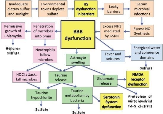
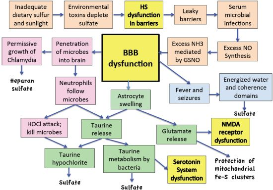
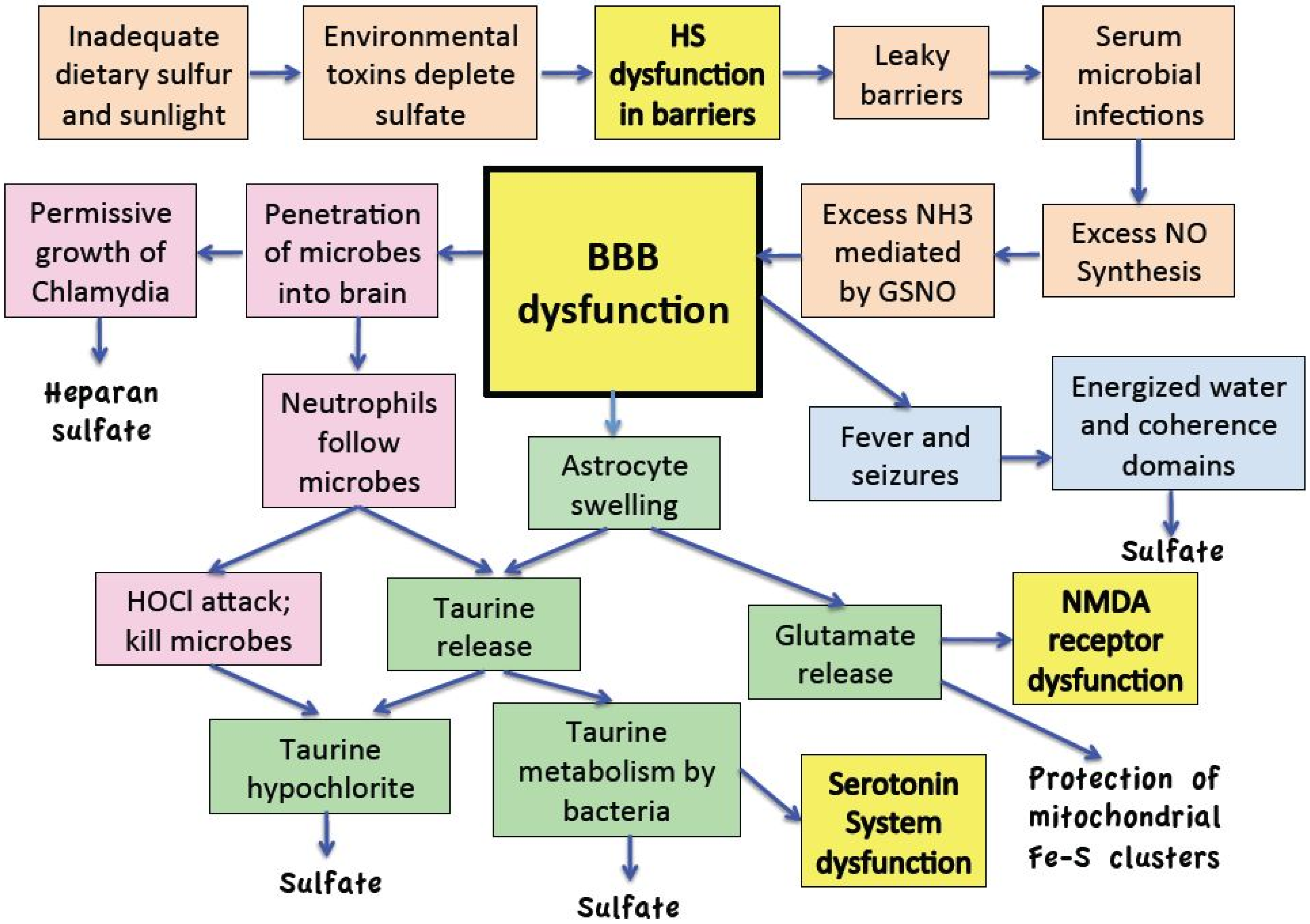
10. The Reaction Cascade

This section describes the complete signaling cascade that characterizes the encephalitic response, schematized in Figure 1. It seems that a sufficient precondition is severe depletion of free sulfate in the blood as well as sulfate in the GAGs in the glycocalyx and the extracellular matrix proteins of suspended red blood cells and platelets. Excess ammonia induced by environmental toxins will result in an enhanced production of nitric oxide by inducible NOS (iNOS) in activated macrophages, triggering the cascade. An aluminum-containing vaccine has the potential to initiate an acute response in the child who is predisposed due to widespread sulfate deficiencies. Aluminum’s strong calmodulin-binding tendencies induce eNOS in the artery wall to synthesize excess NO [186], beyond the immune response induced by the antigen in the vaccine. A child who is already over-producing nitric oxide, for example in response to endotoxins being released by gram-negative bacteria in a leaky gut environment, would be especially vulnerable. The end result is a profusion of NO released into the blood stream, which results in the excess production of ammonia, mediated by GSH. GSH reacts with NO to produce GSNO, which is metabolized to release ammonia as already described above. The result is an additional burden beyond what is already being produced by microbes in the impaired gut, for example due to excess glyphosate exposure.
The ammonia and the NO provoke, after Selye-type sensitization [187], a cascade in the brain which begins with the opening up of the blood-brain barrier, allowing entry of water, glutamate, neutrophils, and bacteria. A concurrent increased permeability of the gut barrier allows microbes from the gut to gain entry into the blood, and hence into the brain. Neutrophils, also entering through the leaky barrier, will launch an attack, releasing cytokines and reactive oxygen and nitrogen species such as H2O2, NO, O2, ONOO and HOCl. Bacteria like Chlamydia pneumoniae and viruses like respiratory syncytial virus, that can produce heparan sulfate in vacuoles, may flourish inside the immune cells long enough to yield an abundant product, as a direct consequence of impaired lysosomal function, thus inadvertently assisting in the recovery process.
Figure 1. Schematic of cascade that we believe leads to ASD, which can best be characterized as low-grade chronic encephalitis, brought on by impaired sulfate synthesis. The activities in the brain are corrective in that they produce sulfate to resupply the blood and the tissues, leading to healing. However, the damaging effects of chronic ammonia and glutamate exposure lead over time to brain impairment, which is especially apparent in the hippocampus. The four boxes indicating dysfunction (in yellow) identify phenomena that are closely associated with or directly give rise to ASD/encephalitis symptoms. HS: heparan sulfate; NMDA: N-methyl-D-aspartate.
Entropy 15 00372 g001
Astrocytes react to the presence of excess ammonia and water initially by swelling, followed by the release of several osmolytes [188], but most important are glutamate and taurine. Glutamate plays an important role in supplying an alternative fuel to the neurons, allowing them to bypass mitochondrial stage I, thus reducing the need for release of superoxide in complex I, which could react with NO to produce the toxic agent, peroxynitrite. Glutamate is also a precursor for α-ketoglutarate (αKG), which can supply bacteria the necessary metabolite to support sulfate synthesis from taurine [178,179,180,181].
The taurine released by astrocytes plays two very important roles. The first is to neutralize the HOCl that escapes from the phagolysosomes of the neutrophils before it can do damage to the cell membranes of neighboring cells [96]. Such damage would lead to excess ion leaks and impaired membrane transport, ultimately resulting in cell lysis [76]. The second role is to replenish depleted sulfate, both in the blood stream and in the brain. Conversion to taurine chloramine is an important first step in activating taurine so that it can be fully metabolized [97]. However, taurine can also be metabolized directly to sulfate by any bacteria that remain viable [98,99]. The mental confusion or even coma associated with acute encephalitis may result from excess CO2 exposure in the brain stem nuclei due to taurine metabolism, and reflects the need to minimize the metabolic requirements of the neurons during a time when glucose supply is short, due to the suppression of glucose transport mechanisms by excess HOCl. Minimizing the activities in Complex I helps prevent damage to the iron-sulfur clusters there by peroxynitrite [68].
ASD is associated with deficiencies in magnesium [100], zinc [118], and various sulfur metabolites [38]. A dangerous condition can arise when these nutrients are severely depleted, as it has been established that both magnesium and heparan sulfate play important roles in xenophagy, the processes in the phagolysosome leading to capture, killing and digestion of the invasive microbes [106]. As previously shown the zinc ion in eNOS may play a catalytic role in the synthesis of sulfate from eNOS [23].
Impairments in phagocytosis would likely lead to the accumulation of debris from dead bacteria in the blood stream. Thus, the possibility of developing specific antibodies to the proteins and DNA in the debris and mimicry could lead to autoimmune reactions to similar native proteins and DNA [189]. Such a basis has been proposed as a possible cause of multiple sclerosis [190], involving an autoimmune attack on myelin as a consequence of exposure to otherwise harmless gut bacteria in the brain. In [191], it was shown that ASD is associated with an increased risk of autoantibody response to a specific but unidentified protein of molecular weight 52 kDa found in cells in the cerebellum. Thus, a similar situation might explain some of the neuronal damage in ASD.
Mono-anionic sulfates, i.e. the sulfated GAGs and sterol sulfates, are essential in stabilizing cell membranes of suspended cells. They support the EZs necessary to keep red blood cells and platelets dispersed, thereby preventing them from aggregating, agglutinating, and coagulating, and they promote barrier function [16]. Furthermore, cholesterol sulfate synthesis is essential to replenish cholesterol supply, as well as sulfate supply, to cell membranes. The seizures and fever, we propose, are necessary to support the synthesis of sulfate by eNOS and nNOS in red blood cells, platelets, endothelial cells, and neurons. Recent advances in our understanding of the special properties of interfacial water show how seizures may supply electric currents that would enable water to form nanomolecular clusters of quantum coherent water: coherence domains (CDs). These would enhance proton transport, Grotthuss-style “proton-hopping”, to support the reactions, localized to caveolae, that oxidize sulfur to sulfate and combine it with cholesterol to produce cholesterol sulfate, presumably although not necessarily involving eNOS and nNOS. A non-enzymatic process has not been excluded. On-water heterogeneous catalysis is likely involved in both enzymatic and non-enzymatic mechanisms.
Absence epilepsy is a relatively common condition that appears in young children, characterized by frequent short intervals of loss of consciousness in association with seizures. Glutamatergic receptors are involved in maintaining a state of consciousness [192]. In a mouse model of absence epilepsy, it has been demonstrated that the condition is associated with impaired metabolism in the cerebellum and cortex, reflected in increased rates of glycolysis (cytoplasmic metabolism of glucose to pyruvate) and increased use of glutamate as an energy source in the mitochondria [193]. This is easily explained as a mechanism to spare complex I of the mitochondrial electron transport chain. The excess bioavailability of glutamate in the synapse leads to increased glutamatergic activity in the thalamus, resulting in a suppression of thalamic input to the cortex and impaired conscious awareness. Interestingly, the mice with genetically-induced absence epilepsy exhibited improved memory, suggesting that the reaction cascade associated with seizures may have benefitted their memory system.

11. Discussion

This paper shows how a finely choreographed biosemiotic cascade associated with encephalitis may help restore depleted sulfate supplies to the brain and blood stream. Because ASD involves a severe deficiency in sulfate, it follows that ASD can be characterized as low-grade encephalitis, with compromised immune system involvement [194]. Inflammation in the brain is a characteristic feature of ASD [195], and recent reviews have discussed the role of “neuroimmune interactions” [172,173] Such features in ASD, and in various other disorders and disease conditions, are explained by the proposed inflammation cascade. Zinc and magnesium deficiency are associated with reduced levels of serum sulfate in patients with many chronic diseases, including myalgic encephalomyelitis, irritable bowel syndrome, migraine, arthritis, multiple chemical sensitivity and depression [196]. Furthermore, extremely low blood serum ratios of sulfate to cysteine are found in association with Alzheimer's disease, Parkinson's disease and amyotrophic lateral sclerosis (ALS) [197]. Sulfate deficiency is also associated with all these other neurological conditions suggesting wide relevance of the proposed signaling cascade beyond its application to ASD.
A recent study using metabolomics to detect abnormal amounts of a large number of metabolites in urinary specimens from subjects with ASD compared to controls has revealed a number of significant differences [198], many of which are in alignment with the signaling cascade presented here. First of all, researchers found that subjects with both ASD and digestive disturbances indicative of inflammatory bowel disease had increased levels of bacterial co-metabolites in their urine, suggesting bacterial invasion and impaired endocytosis. Secondly, they observed a very low level of taurine in urine specimens from subjects with ASD, indicative of taurine depletion. Thirdly, they observed a marked increase in the concentration of urinary gamma glutamyl transferase, an enzyme which can supply glutamate by breaking down glutathione. This would predict both excess glutamate and depleted glutathione, both of which are found in association with ASD. Finally, products indicative of oxidative stress were more highly concentrated in the urine of subjects with ASD.
Insufficient sulfate supply to the blood has potentially catastrophic consequences. To explain this requires consideration of the anomalous properties of water, especially the ability of water to create EZs surrounding negatively charged hydrophilic regions, i.e., polyanionic biomembranes. When there is insufficient sulfate in the extracellular matrix proteins, i.e., the heparan sulfate glycosaminoglycans, of cells suspended in the blood, these cells are predisposed to aggregate and agglutinate, resulting in a coagulation cascade that could lead to thrombosis and death if left unchecked. Impaired cholesterol sulfate delivery to the fetus during pregnancy followed by impaired cholesterol sulfate synthesis in the skin postnatally leads to a global deficiency in sulfate supply to the extracellular matrix proteins in association with ASD [21]. X-linked ichthyosis, a genetic disease affecting the enzyme steroid sulfatase, and thus impairing the ability to break down cholesterol sulfate into cholesterol and sulfate, is associated with increased risk to both ADHD and ASD and with an accumulation of cholesterol sulfate in the outer epidermis [199].
Deficiencies in magnesium, zinc, glutathione, sulfate, and, particularly, heparan sulfate are associated with pathologies related to ASD. Displacement in the diet of animal protein, fat, and cholesterol by grains, whose components can actively bind and flush out or leak minerals and other compounds from the gut, may contribute to the deficits. Food-based toxic chemicals such as glyphosate, the most widely used herbicide in agricultural practices, are also implicated.
Impairment in heparan N-sulfatase can lead to ASD-like behaviors in humans, as exemplified by the lysosomal storage disease, Sanfilippo syndrome [55]. Thus, lysosomal dysfunction is likely a factor in ASD. However, heparan sulfate also plays an important role in neural transmission. The authors of the paper that showed ASD-like behaviors in mice with impaired heparan sulfate supply stated that “removal of HS (heparan sulfate) compromises glutamatergic synaptic transmission by affecting the synaptic localization of AMPA receptors” ([52], pp. 5052–5053). Thus, there is a direct link between heparan sulfate deficiencies, glutamate, and neuronal dysfunction. The syndecans in heparan sulfate proteoglycans play a supporting role in cell migration, neurite extension, and plasticity, important aspects of neural development [200]. Heparan sulfate is enriched in synapses, and it is involved in the morphological maturation of dendritic spines in rat hippocampal neurons [201]. It is also involved in long-term potentiation in the hippocampus [202], which has been shown to be impaired in ASD [94].
Neural synaptic transmission defects in the hippocampus are implicated in the pathology of ASD. By comparing the patterns of cognitive dysfunction in ASD and adult amnesia with the deficits associated with hippocampal lesions in animals, DeLong has made a case for hippocampal dysfunction as a main contributor to the cognitive and motivational deficits observed in ASD [203]. Epilepsy is a strong feature associated with ASD [204,205], and it has been demonstrated that epilepsy is associated with reduced complex I activity in the hippocampus [206]. This fits well with the model that glutamate is entering the citric acid cycle beyond mitochondrial complex I.
The flooding of the extracellular fluid with glutamate could be a key contributor to the observed impaired glutamatergic signaling in ASD. Serum glutamate levels are abnormally high in ASD [207]. In the hippocampus, a large percentage of the glutamate receptors are located outside of the synapse. This extrasynaptic pool triggers a signaling cascade that inactivates NMDA receptors in the synapse [208]. Mutations in NMDA glutamate receptors have been found to be causative in certain rare cases of ASD [209]. The presence of large amounts of glutamate outside the cell in non-synaptic regions is a signal of metabolic stress, thus resulting in the shutting down of receptors in the synapse in order to conserve energy. However, the net result is impaired glutamatergic signaling.
While the signaling cascade proposed in this paper involves plausible inferences at many points, it provides evidence that conditions that appear at first to be pathological, such as encephalopathies, may be part of a larger and more coherent biosemiotic process that is protective and salubrious. To the extent that the ideas suggested are on the right track, they provide hope for a child diagnosed with ASD. Biological cascades evidently exist in part to maintain a supply of taurine and glutamate to protect neurons from collateral damage during a necessary and productive inflammatory response. Such systems may protect neurons against permanent damage. The neurons are being maintained in a chronic state of suppressed glutamatergic signaling, due to sustained exposure to excess NO and derivative oxidative and nitrosative products.
There is evidence in the research literature that dietary supplements related to the deficiencies discussed here can improve symptoms and/or biomarkers in ASD. Parents of children with ASD have found empirically that a gluten-free diet helps improve the behavior of their children, and placebo-controlled studies have shown modest benefits [210]. Since gluten contains phytates, which deplete minerals such as zinc and magnesium, this could be a factor in the observed improvements. Evidence for a role for thiamine deficiency comes from a study where eight out of 10 children with ASD improved clinically following thiamine tetrahydrofurfuryl disulfide treatment [211]. A placebo-controlled study involving 141 children with ASD demonstrated improvements in several sulfur-related biomarkers, including total sulfate, adenosylmethionine, GSH, GSSG:GSH ratio, and taurine, following a vitamin/mineral supplement treatment program that specifically included methylsulfonyl-methane (MSM) as a sulfur source [212], supporting the concept of sulfur deficiency in association with ASD. Cholesterol supplementation has been shown to improve symptoms such as irritability, hyperactivity, and sleep disorders in some children with ASD [213]. This is particularly true for those diagnosed with Smith-Lemli-Opitz syndrome (SLOS), a condition caused by genetic mutations in a gene involved in cholesterol synthesis associated with the ASD phenotype [214,215].
The analysis presented here suggests certain simple adjustments in lifestyle and nutrition to reduce the risk of ASD in a child, or to improve the prognosis. The first step is to make sure that the diet is adequate in supplying zinc, magnesium, thiamine and sulfur, for both the pregnant mother and the child. A conscious effort to consume organic non-genetically modified vegetables is encouraged, along with a reduction in dietary sugars and processed carbohydrates. Since taurine is absent from plant-based foods, it is important to include sufficient animal proteins in the diet, for adequate supply of taurine. Grains should be avoided, as phytates bind minerals such as magnesium and zinc and prevent their uptake through the gut, a concept that has been popularized by the cardiologist William Davis [216]. Epsom salt baths may be of therapeutic benefit, as they could provide magnesium sulfate, which is readily absorbed through the skin [196].
Another key lifestyle change is to assure adequate sun exposure to the skin. A recent study has confirmed a strong inverse correlation between ASD prevalence in the individual states within the United States and the calculated mean annual solar UVB availability, suggesting that ASD risk is increased by inadequate UVB exposure [217]. This is further supported by evidence that inflammatory bowel disease incidence is higher in geographical regions with less sunlight availability [218]. High SPF sunscreens often contain aluminum hydroxide as an emulsifier, which could provide synergistic effects, along with the aluminum burden from vaccines [219].

12. Conclusions

Encephalitis can be viewed as a response of the body to the need to augment the supply of sulfate to the brain and to the blood stream, under conditions of extreme deficits. Autism spectrum disorder can be framed as a condition associated with chronic low-grade encephalitis, which maintains the brain in a hypometabolic state in order to protect it from oxidation and nitration damage. The pathology arises from impaired cholesterol sulfate synthesis in the skin, due to insufficient sunlight exposure and excessive use of sunscreens. Exposure to toxic metals such as aluminum, mercury and lead, and to toxic chemicals such as those found in commonly used herbicides, further deplete the supply of sulfate and other micronutrients to the blood stream and the tissues, potentially leading to a life-threatening state of blood instability. The reaction cascade associated with encephalitis, involving fever, seizures, neutrophil activation, and the release of glutamate and taurine from astrocytes, can be protective and salubrious in partially regenerating sulfate supplies to stabilize the blood and improve neuronal synaptic transmission. While the proposed biosemiotic cascade involves inferences, they are plausible and well supported by existing research and theory, and the entire proposal can help to guide further research. The upside is to encourage research seeking to prevent ASD and many other disorders and disease conditions. Proposed remedies involve simple changes such as abundant sun exposure, avoidance of environmental toxins and chelating agents, avoidance of dietary processed foods, grains and sugars, and greater intake of sulfur-containing foods.

Acknowledgements

This work was funded in part by Quanta Computers, Taipei, Taiwan, under the auspices of the Qmulus Project. We would like to thank Anthony Samsel for alerting us to a role for glyphosate in disruption of gut bacteria. We are indebted to two anonymous reviewers whose thoughtful comments led to a much-improved version of the paper.

References

  1. Caronna, E.B.; Milunsky, J.M.; Tager-Flusberg, H. Autism spectrum disorders: Clinical and research frontiers. Arch. Dis. Child. 2008, 93, 518–523. [Google Scholar] [CrossRef] [PubMed]
  2. Horvath, K.; Papadimitriou, J.C.; Rabsztyn, A.; Drachenberg, C.; Tildon, J.T. Gastrointestinal abnormalities in children with autistic disorder. J. Pediatr. 1999, 135, 559–563. [Google Scholar] [CrossRef]
  3. Wakefield, A.J.; Puleston, J.M.; Montgomery, S.M.; Anthony, A.; O’Leary, J.J.; Murch, S.H. Review article: The concept of entero-colonic encephalopathy, autism and opioid receptor ligands. Aliment. Pharmacol. Ther. 2002, 16, 663–674. [Google Scholar] [CrossRef] [PubMed]
  4. Theoharides, T.C.; Doyle, R. Autism, gut-blood-brain barrier, and mast cells. J. Clin. Psychopharm. 2008, 2, 479–483. [Google Scholar] [CrossRef] [PubMed]
  5. Theoharides, T.C.; Zhang, B. Hypothesis: Neuro-inflammation, blood-brain barrier, seizures and autism. J. Neuroinflam. 2011, 8, 168. [Google Scholar] [CrossRef] [PubMed]
  6. Dettmer, K.; Hanna, D.; Whetstone, P.; Hansen, R.; Hammock, B.D. Autism and urinary exogenous neuropeptides: development of an on-line SPE-HPLC-tandem mass spectrometry method to test the opioid excess theory. Anal. Bioanal. Chem. 2007, 388, 1643–1651. [Google Scholar] [CrossRef] [PubMed]
  7. Bennetto, L.; Scolding, N. Inflammatory/post-infectious encephalomyelitis. J. Neurol. Neurosurg. Psychiatry 2004, 75, i22–i28. [Google Scholar] [CrossRef] [PubMed]
  8. Dalmau, J.; Gleichman, A.J.; Hughes, E.G.; Rossi, J.E.; Peng, X.; Lai, M.; Dessain, S.K.; Rosenfeld, M.R.; Balice-Gordon, R.; Lynch, D.R. Anti-NMDA-receptor encephalitis: case series and analysis of the effects of antibodies. Lancet. Neurol. 2008, 7, 1091–1098. [Google Scholar] [CrossRef]
  9. Gable, M.S.; Gavali, S.; Radner, A.; Tilley, D.H.; Lee, B.; Dyner, L.; Collins, A.; Dengel, A.; Dalmau, J.; Glaser, C.A. Anti-NMDA receptor encephalitis: Report of ten cases and comparison with viral encephalitis. Eur. J. Clin. Microbiol. Infect. Dis. 2009, 28, 1421–1429. [Google Scholar] [CrossRef] [PubMed]
  10. Oller, J.W., Jr.; Oller, S.D.; Oller, S.N. Autism: The Diagnosis, Treatment, & Etiology of the Undeniable Epidemic; Jones & Bartlett Learning: Sudbury, MA, USA, 2010. [Google Scholar]
  11. Vojdani, A.; Pangborn, J.B.; Vojdani, E.; Cooper, E.L. Infections, toxic chemicals and dietary peptides binding to lymphocyte receptors and tissue enzymes are major instigators of autoimmunity in autism. Int. J. Immunopath. Ph. 2003, 16, 189–199. [Google Scholar]
  12. Dufault, R.; Schnoll, R.; Lukiw, W.J.; LeBlanc, B.; Cornett, C.; Patrick, L.; Wallinga, D.; Gilbert, S.G.; Crider, R. Mercury exposure, nutritional deficiencies and metabolic disruptions may affect learning in children. Behav. Brain Funct. 2009, 5, 44. [Google Scholar] [CrossRef] [PubMed]
  13. Dufault, R.; Lukiw, W.J.; Crider, R.; Schnoll, R.; Wallinga, D.; Deth, R.A. Macroepigenetic approach to identify factors responsible for the autism epidemic in the United States. Clin. Epigenet. 2012, 4, 6. [Google Scholar] [CrossRef] [PubMed]
  14. Hartzell, S.; Seneff, S. Impaired sulfate metabolism and epigenetics: Is there a link in autism? Entropy 2012, 14, 1953–1977. [Google Scholar] [CrossRef]
  15. Seneff, S.; Davidson, R.M.; Liu, J. Is cholesterol sulfate deficiency a common factor in preeclampsia, autism, and pernicious anemia? Entropy 2012, 14, 2265–2290. [Google Scholar] [CrossRef] [Green Version]
  16. Davidson, R.M.; Seneff, S. The initial common pathway of inflammation, disease, and sudden death. Entropy 2012, 14, 1399–1442. [Google Scholar] [CrossRef] [Green Version]
  17. Beckham, J.D.; Tyler, K.L. Encephalitis. In Principles and Practice of Infectious Diseases, 7th ed.; Mandell, G.L., Bennett, J.E., Dolin, R., Eds.; Elsevier Churchill Livingstone: Philadelphia, PA, USA, 2009; Chapter 87. [Google Scholar]
  18. Ghaziuddin, M.; Al-Khouri, I.; Ghaziuddin, N. Autistic symptoms following herpes encephalitis. Eur. Child Adolesc Psy. 2002, 11, 142–146. [Google Scholar] [CrossRef]
  19. Gilberg, I.C. Onset at age 14 of a typical autistic syndrome: A case report of a girl with herpes simplex encephalitis. J. Autism Dev. Disord. 1986, 16, 369–375. [Google Scholar] [CrossRef]
  20. Long-term cognitive and behavioral consequences of neonatal encephalopathy following perinatal asphyxia: A review. Eur J Pediatr. 2007, 166, 645–654.
  21. Seneff, S.; Davidson, R.; Mascitelli, L. Might cholesterol sulfate deficiency contribute to the development of autistic spectrum disorder? Med. Hypotheses 2012, 8, 213–217. [Google Scholar] [CrossRef] [PubMed]
  22. Horan, F.E.; Hirsch, F.G.; Wood, L.A.; Wright, L.S. Surface effects on blood-clotting components as determined by Zeta-potentials. J. Clin. Invest. 1950, 29, 202–211. [Google Scholar] [CrossRef] [PubMed]
  23. Seneff, S.; Lauritzen, A.; Davidson, R.; Lentz-Marino, L. Is endothelial nitric oxide synthase a moonlighting protein whose day job is cholesterol sulfate synthesis? Implications for cholesterol transport, diabetes and cardiovascular disease. Entropy 2012, 2492, 2492–2530. [Google Scholar] [CrossRef] [Green Version]
  24. Rosenberg, G.A. Neurological diseases in relation to the blood-brain barrier. J. Cerebr. Blood F. Met. 2012, 32, 1139–1151. [Google Scholar] [CrossRef] [PubMed]
  25. Reitsma, S.; Slaaf, D.W.; Vink, H.; van Zandvoort, M.A.M.J.; oude Egbrink, M.G.A. The endothelial glycocalyx: composition, functions, and visualization. Pflugers Arch. Eur. J. Physiol. 2007, 454, 345–359. [Google Scholar] [CrossRef] [PubMed]
  26. McCully, K.S. Chemical pathology of homocysteine V: Thioretinamide, thioretinaco, and cystathionine synthase function in degenerative diseases. Ann. Clin. Lab. Sci. 2011, 41, 300–313. [Google Scholar]
  27. Oja, S.S.; Saransaari, P. Taurine as osmoregulator and neuromodulator in the brain. Metab. Brain Dis. 1996, 11, 153–164. [Google Scholar] [CrossRef] [PubMed]
  28. Sturman, J.A.; Hepner, G.W.; Hofmann, A.F.; Thomas, P.J. Metabolism of [35S] taurine in man. J. Nutr. 1975, 105, 1206–1214. [Google Scholar] [PubMed]
  29. Schoch, H.J.; Fischer, S.; Marti, H.H. Hypoxia-induced vascular endothelial growth factor expression causes vascular leakage in the brain. Brain 2002, 125, 2549–2557. [Google Scholar] [CrossRef] [PubMed]
  30. Skowrońska, M.; Albrecht, J. Alterations of blood brain barrier function in hyper-ammonemia: An overview. Neurotox. Res. 2012, 21, 236–244. [Google Scholar] [CrossRef] [PubMed]
  31. Xaio, H.; Banks, W.A.; Niehoff, M.L.; Morley, J.E. Effect of LPS on the permeability of the blood brain barrier to insulin. Brain Res. 2001, 896, 36–42. [Google Scholar] [CrossRef]
  32. Stamatovic, S.M.; Keep, R.F.; Andjelkovic, A.V. Brain endothelial cell-cell junctions: How to open the blood brain barrier. Curr. Neuropharmacol. 2008, 6, 179–192. [Google Scholar] [CrossRef] [PubMed]
  33. Huber, J.D.; Egleton, R.D.; Davis, T.P. Molecular physiology and pathophysiology of tight junctions in the bloodbrain barrier. Trends Neurosci. 2001, 24, 719–725. [Google Scholar] [CrossRef]
  34. Misra, U.K.; Tan, C.T.; Kalita, J. Seizures in encephalitis. Neurol. Asia 2008, 13, 1–13. [Google Scholar]
  35. Chen, C.-A.; Wang, T.-Y.; Varadharaj, S.; Reyes, L.A.; Hemann, C.; Talukder, M.A.H.; Chen, Y.-R.; Druhan, J.; Zweier, J.L. S-glutathionylation uncouples eNOS and regulates its cellular and vascular function. Nature 2010, 468, 1115–1120. [Google Scholar] [CrossRef] [PubMed]
  36. Geier, D.A.; Kern, J.K.; Garver, C.R.; Adams, J.B.; Audhya, T.; Nataf, R.; Geier, M.R. Biomarkers of environmental toxicity and susceptibility in autism. J. Neurolog. Sci. 2009, 280, 101–108. [Google Scholar] [CrossRef] [PubMed]
  37. James, S.J.; Cutler, P.; Melnyk, S.; Jernigan, S.; Janak, L.; Gaylor, D.W.; Neubrander, J.A. Metabolic biomarkers of increased oxidative stress and impaired methylation capacity in children with autism. Am. J. Clin. Nutr. 2004, 80, 1611–1617. [Google Scholar] [PubMed]
  38. Waring, R.H.; Klovrza, L.V. Sulphur metabolism in autism. J. Nutr. Environ. Med. 2000, 10, 25–32. [Google Scholar] [CrossRef]
  39. Finegold, S.M.; Molitoris, D.; Song, Y.; Liu, C.; Vaisanen, M.-L.; Bolte, E.; McTeague, M.; Sandler, R.; Wexler, H.; Marlowe, E.M.; et al. Gastrointestinal microflora studies in late-onset autism. Clin. Infect. Dis. 2002. [Google Scholar] [CrossRef] [PubMed]
  40. Finegold, S.M.; Downes, J.; Summanen, P.H. Microbiology of regressive autism. Anaerobe 2012, 18, 260–262. [Google Scholar] [CrossRef] [PubMed]
  41. Finegold, S.M. Desulfovibrio species are potentially important in regressive autism. Med. Hypotheses 2011, 77, 270–274. [Google Scholar] [CrossRef] [PubMed]
  42. LaSalle, J.M. A genomic point-of-view on environmental factors influencing the human brain methylome. Epigenetics 2001, 6, 862–869. [Google Scholar] [CrossRef]
  43. Deth, R.; Muratore, C.; Benzecry, J.; Power-Charnitsky, V.-A.; Waly, M. How environmental and genetic factors combine to cause autism: A redox/ methylation hypothesis. Neurotoxicology 2008, 29, 190–201. [Google Scholar] [CrossRef] [PubMed]
  44. James, S.J.; Melnyk, S.; Jernigan, S.; Hubanks, A.; Rose, S.; Gaylor, D.W. Abnormal transmethylation/transsulfuration metabolism and DNA hypomethylation among parents of children with autism. Autism Dev. Disord. 2008, 38, 1966–1975. [Google Scholar] [CrossRef] [PubMed]
  45. Sweeten, T.L.; Posey, D.J.; Shankar, S.S.; McDougle, C.J. High nitric oxide production in autistic disorder: A possible role for interferon-γ. Biol. Psychiat. 2004, 55, 434–437. [Google Scholar] [CrossRef] [PubMed]
  46. Xu, D.-Z.; Lu, Q.; Deitch, E.A. Nitric oxide directly impairs intestinal barrier function. Shock 2002, 17, 139–145. [Google Scholar] [CrossRef] [PubMed]
  47. Mayer, B.; Pfeiffer, S.; Schrammel, A.; Koesling, D.; Schmidt, K.; Brunner, F. A new pathway of nitric oxide/cyclic GMP signaling involving S-nitrosoglutathione. J. Biol. Chem. 1998, 273, 3264–3270. [Google Scholar] [CrossRef] [PubMed]
  48. Liu, L.; Hausladen, A.; Zeng, M.; Que, L.; Heitman, J.; Stamler, J.S. A metabolic enzyme for S-nitrosothiol conserved from bacteria to humans. Nature 2001, 410, 490–494. [Google Scholar] [CrossRef] [PubMed]
  49. Esko, J.D.; Kimata, K.; Lindahl, U. Proteoglycans and sulfated glycosaminoglycans. In Essentials of Glycobiology, 2nd ed.; Varki, A., Cummings, R.D., Esko, J.D., Freeze, H.H., Stanley, P., Bertozzi, C.R., Hart, G.W., Etzler, M.E., Eds.; Cold Spring Harbor Laboratory Press: Cold Spring Harbor, NY, USA, 2009; Chapter 16. [Google Scholar]
  50. Vogl-Willis, C.A.; Edwards, I.J. High-glucose-induced structural changes in the heparan sulfate protegolycan, perlecan, of cultured human aortic endothelial cells. Biochim. Biophys. Acta 2004, 1672, 36–45. [Google Scholar] [CrossRef] [PubMed]
  51. Stern, M. Insulin signaling and autism. Front. Endocrinol. 2011, 2, 54. [Google Scholar] [CrossRef] [PubMed]
  52. Fumitoshi Irie, F.; Hedieh Badie-Mahdavi, H.; Yamaguchi, Y. Autism-like sociocommunicative deficits and stereotypies in mice lacking heparan sulfate. Proc. Natl. Acad. Sci. USA 2012, 109, 5052–5056. [Google Scholar] [CrossRef] [PubMed]
  53. Levine, B.; Mizushima, N.; Virgin, H.W. Autophagy in immunity and inflammation. Nature 2011, 469, 323–335. [Google Scholar] [CrossRef] [PubMed]
  54. Mizushima, N. Autophagy: Process and function. Gene. Dev. 2007, 21, 2861–2873. [Google Scholar] [CrossRef] [PubMed]
  55. Neufeld, E.F. Lysosomal storage diseases. Annu. Rev. Biochem. 1991, 60, 257–280. [Google Scholar] [CrossRef] [PubMed]
  56. Valstar, M.J.; Bruggenwirth, H.T.; Olmer, R.; Wevers, R.A.; Verheijen, F.W.; Poorthuis, B.J.; Halley, D.J.; Wijburg, F.A. Mucopolysaccharidosis type IIIB may predominantly present with an attenuated clinical phenotype. Inherit. Metab. Dis. 2010, 33, 759–767. [Google Scholar] [CrossRef] [PubMed]
  57. Mucopolysaccharidoses Fact Sheet. National Institute of Neurological Disorders and Stroke Website. Available online: http://www.ninds.nih.gov/disorders/mucopolysaccharidoses/detail/mucopolysacchari-doses.htm/ (accessed 2 April 2012).
  58. Gulbins, E.; Dreschers, S.; Wilker, B.; Grassme, H. Ceramide, membrane rafts and infections. J. Mol. Med. 2004, 82, 357–363. [Google Scholar] [CrossRef] [PubMed]
  59. Bengheza, M.; Fauvarque, M.O.; Tournebize, R.; Froquet, R.; Marchetti, A.; Bergeret, E.; Lardy, B.; Klein, G.; Sansonetti, P.; Charette, S.J.; et al. Specific host genes required for the killing of Klebsiella bacteria by phagocytes. Cell Microbiol. 2006, 8, 139–148. [Google Scholar] [CrossRef] [PubMed]
  60. Hashimoto, Y.; Orellana, A.; Gil, G.; Hirschberg, C.B. Molecular cloning and expression of rat liver N-heparan sulfate sulfotransferase. J. Biol. Chem. 1992, 267, 15744–15750. [Google Scholar] [PubMed]
  61. Moseley, R.; Waddington, R.J.; Embery, G. Degradation of glycosaminoglycans by reactive oxygen species derived from stimulated polymorphonuclear leukocytes. BBA-Mol Basis. Dis. 1997, 1362, 221–231. [Google Scholar] [CrossRef]
  62. Ross, M.A.; Long, W.F.; Williamson, F.B. Inhibition by heparin of Fe(II)-catalysed free-radical peroxidation of linolenic acid. Biochem. J. 1992, 286, 717–720. [Google Scholar] [PubMed]
  63. Wakefield, A.J. The gut brain axis in childhood developmental disorders. J. Pediatr. Gastr. Nutr. 2002, 34, S14–S17. [Google Scholar] [CrossRef]
  64. Norenberg, M.D.; Jayakumar, A.R.; Rama Rao, K.V.; Panickar, K.S. New concepts in the mechanism of ammonia-induced astrocyte swelling. Metab. Brain Dis. 2007, 22, 219–234. [Google Scholar] [CrossRef] [PubMed]
  65. Master, S.; Gottstein, J.; Blei, A.T. Cerebral blood flow and the development of ammonia-induced brain edema in rats after portacaval anastomosis. Hepatology 1999, 30, 876–880. [Google Scholar] [CrossRef] [PubMed]
  66. Jayakumar, A.R.; Panickar, K.S.; Murthy, C.R.K.; Norenberg, M.D. Oxidative stress and MAPK phosphorylation mediate ammonia-induced cell swelling and glutamate uptake inhibition in cultured astrocytes. J. Neurosci. 2006, 26, 4774–4784. [Google Scholar] [CrossRef] [PubMed]
  67. Oury, T.D.; Piantadosi, C.A.; Crapo, J.D. Cold-induced brain edema in mice: Involvement of extracellular superoxide dismutase and nitric oxide. J. Biol. Chem. 1993, 268, 15394–15398. [Google Scholar] [PubMed]
  68. Brown, G.C.; Borutaite, V. Inhibition of mitochondrial respiratory complex I by nitric oxide, peroxynitrite and S-nitrosothiols. BBA-Bioenergetics 2004, 1658, 44–49. [Google Scholar] [CrossRef] [PubMed]
  69. Alvarez, M.N.; Peluffo, G.; Piacenza, L.; Radi, R. Intraphagosomal peroxynitrite as a macrophage-derived cytotoxin against internalized Trypanosoma cruzi: Consequences for oxidative killing and role of microbial peroxiredoxins in infectivity. J. Biol. Chem. 2011, 286, 6627–6640. [Google Scholar] [CrossRef] [PubMed]
  70. Poling, J.S.; Frye, R.E.; Shoffner, J.; Zimmerman, A.W. Developmental regression and mitochondrial dysfunction in a child with autism. J. Child Neurol. 2006, 21, 170–172. [Google Scholar] [CrossRef] [PubMed]
  71. Good, P. Do salt cravings in children with autistic disorders reveal low blood sodium depleting brain taurine and glutamine? Med. Hypotheses 2011, 77, 1015–1021. [Google Scholar] [CrossRef] [PubMed]
  72. Lia, X.; Chauhana, A.; Sheikha, A.M.; Patilb, S.; Chauhana, V.; Lib, X.-M.; Jia, L.; Browna, T.; Malika, M. Elevated immune response in the brain of autistic patients. J. Neuroimmunol. 2009, 201, 111–116. [Google Scholar] [CrossRef] [PubMed]
  73. Brosnan, J.T.; Man, K.-C.; Hall, D.E.; Colbourne, S.A.; Brosnan, M.E. Interorgan metabolism of amino acids in the streptozotocin-diabetic ketoacidotic rat. Am. J. Physiol. 1983, 244, E151–E158. [Google Scholar] [PubMed]
  74. Brosnan, J.T. Glutamate, at the interface between amino acid and carbohydrate metabolism. In Proc. International Symposium on Glutamate, Bergamo, Italy, 12–14 October 1998.
  75. Daikhin, Y.; Yudkoff, M. Compartmentation of brain glutamate metabolism in neurons and glia. In Proc. International Symposium on Glutamate, Bergamo, Italy, 12–14 October 1998.
  76. Schraufstitter, I.U.; Browne, K.; Harris, A.; Hyslop, P.A.; Jackson, J.H.; Quehenberger, O.; Cochrane, C.G. Mechanisms of hypochlorite injury of target cells. J. Clin. Invest. 1990, 85, 554–562. [Google Scholar] [CrossRef] [PubMed]
  77. Sutherland, G.R.; Tyson, R.L.; Auer, R.N. Truncation of the Krebs cycle during hypoglycemic coma. Med. Chem. 2008, 4, 379–385. [Google Scholar] [CrossRef] [PubMed]
  78. Massieu, L.; Montiel, T.; Robles, G.; Quesada, O. Brain amino acids during hyponatremia in vivo: Clinical observations and experimental studies. Neurochem. Res. 2004, 29, 73–81. [Google Scholar] [CrossRef] [PubMed]
  79. Hansen, S.H.; Andersen, M.L.; Cornett, C.; Gradinaru, R.; Grunnet, N. A role for taurine in mitochondrial function. J. Biomed. Sci. 2010, 17, S23–530. [Google Scholar] [CrossRef] [PubMed]
  80. Albrecht, J.; Bender, A.S.; Norenberg, M.D. Ammonia stimulates the release of taurine from cultured astrocytes. Brain Res. 1994, 660, 228–232. [Google Scholar] [CrossRef]
  81. Albrecht, J. Roles of neuroactive amino acids in ammonia neurotoxicity. J. Neurosci. Res. 1993, 51, 133–138. [Google Scholar] [CrossRef]
  82. Foran, E.; Trotti, D. Glutamate transporters and the excitotoxic path to motor neuron degeneration in amyotrophic lateral sclerosis. Antioxid. Redox Sign. 2009, 11, 1587–1602. [Google Scholar] [CrossRef] [PubMed]
  83. Choi, D.W. Glutamate receptors and the induction of excitotoxic neuronal death. Prog. Brain Res. 1994, 100, 47–51. [Google Scholar] [PubMed]
  84. Blaylock, R.L.; Strunecka, A. Immune-glutamatergic dysfunction as a central mechanism of the autism spectrum disorders. Curr. Med. Chem. 2009, 16, 157–170. [Google Scholar] [CrossRef] [PubMed]
  85. Monfort, P.; Muñoz, M.-D.; El Ayadi, A.; Kosenko, E.; Felipo, V. Effects of hyperammonemia and liver failure on glutamatergic neurotransmission. Metab. Brain Dis. 2002, 17, 237–250. [Google Scholar] [CrossRef] [PubMed]
  86. Muñoz, M.D.; Monfort, P.; Gaztelu, J.M.; Felipo, V. Hyperammonemia impairs NMDA receptor-dependent long-term potentiation in the CA1 of rat hippocampus in vitro. Neurochem. Res. 2000, 25, 437–441. [Google Scholar] [CrossRef] [PubMed]
  87. Aguilar, M.A.; Miñarro, J.; Felipo, V. Chronic moderate hyperammonemia impairs active and passive avoidance behavior and conditional discrimination learning in rats. Exp. Neurol. 2000, 161, 704–713. [Google Scholar] [CrossRef] [PubMed]
  88. Romero-Vives, M.; Barrenechea, C.; Insausti, R.; Felipo, V.; Gaztelu, J.M. Sleep alterations in hepatic encephalopathy could be due to chronic hyperammonemia. J. Sleep Res. 1998, 7, 228. [Google Scholar]
  89. Creten, C.; van der Zwaan, S.; Blankenspoor, R.J.; Maatkamp, A.; van Os, J.; Schieveld, J.N.M. Late onset autism and anti-NMDA-receptor encephalitis. Lancet 2011, 378, 98–99. [Google Scholar] [CrossRef]
  90. Rasalam, A.D.; Hailey, H.; Williams, J.H.G.; Moore, S.J.; Turnpenny, P.D.; MooDeanre, J.C.S. Characteristics of fetal anticonvulsant syndrome associated autistic disorder. Dev. Med. Child Neurol. 2005, 47, 551–555. [Google Scholar] [CrossRef] [PubMed]
  91. Williams, G.; King, J.; Cunningham, M.; Stephan, M.; Kerr, B.; Hersh, J.H. Fetal valproate syndrome and autism: additional evidence of an association. Dev. Med. Child Neurol. 2001, 43, 202–206. [Google Scholar] [CrossRef] [PubMed]
  92. Wadzinski, J.; Franks, R.; Roane, D.; Bayard, M. Valproate-associated hyperammonemic encephalopathy. JABFM 2007, 20, 499–502. [Google Scholar] [CrossRef] [PubMed]
  93. Yehya, N.; Saldarini, C.T.; Koski, M.E.; Davanzo, P. Valproate-induced hyperammonemic encephalopathy. Metab. Brain Dis. 2004, 43, 926–927. [Google Scholar] [CrossRef] [PubMed]
  94. Rinaldi, T.; Kulangara, K.; Antoniello, K.; Markram, H. Elevated NMDA receptor levels and enhanced postsynaptic long-term potentiation induced by prenatal exposure to valproic acid. Proc. Natl. Acad. Sci. USA 2007, 104, 13501–13506. [Google Scholar] [CrossRef] [PubMed]
  95. Hampton, M.B.; Kettle, A.J.; Winterbourn, C.C. Inside the neutrophil phagosome: Oxidants, myeloperoxidase and bacterial killing. Blood 1998, 92, 3007–3017. [Google Scholar] [PubMed]
  96. Schuller-Levis, G.B.; Park, E. Taurine and Its Chloramine: Modulators of Immunity. Neurochem. Res. 2004, 29, 117–126. [Google Scholar] [CrossRef] [PubMed]
  97. Olszowski, S.; Olszowska, E.; Kusior, D.; Szneler, E. Sulphoacetaldehyde as a product of taurine chloramine peroxidation at site of inflammation. Amino Acids 2002, 22, 145–153. [Google Scholar] [CrossRef] [PubMed]
  98. Cunningham, C.; Tipton, K.F.; Dixon, H.B.F. Conversion of taurine into N-chlorotaurine (taurine chloramine) and sulphoacetaldehyde in response to oxidative stress. Biochem. J. 1998, 330, 939–945. [Google Scholar] [PubMed]
  99. Ruff, J.; Denger, K.; Cook, A.M. Sulphoacetaldehyde acetyltransferase yields acid phosphate: Purification from Alcaligenes defragrans and gene clusters in taurine degradation. Biochem. J. 2003, 369, 275–285. [Google Scholar] [CrossRef] [PubMed]
  100. Johnson, S. The multifaceted and widespread pathology of magnesium deficiency. Med. Hypotheses 2001, 56, 163–170. [Google Scholar] [CrossRef] [PubMed]
  101. Strambi, M.; Longini, M.; Hayek, J.; Berni, S.; Macucci, F.; Scalacci, E.; Vezzosi, P. Magnesium profile in autism. Biol. Trace Elem. Res. 2006, 109, 97–104. [Google Scholar] [CrossRef]
  102. Mousain-Bosc, M.; Roche, M.; Polge, A.; Pradal-Prat, D.; Rapin, J.; Bali, J.P. Improvement of neurobehavioural disorders in children supplemented with magnesium-B6. Magnesium Res. 2006, 19, 53–62. [Google Scholar]
  103. Hallak, M.; Berman, R.F.; Irtenkauf, S.M.; Evans, M.I.; Cotton, D.B. Peripheral magnesium sulfate enters the brain and increases the threshold for hippocampal seizures in rats. Am. J. Obstet. Gynecol. 1992, 167, 1605–1610. [Google Scholar] [CrossRef]
  104. Cotton, D.B.; Janusz, C.A.; Berman, R.F. Anticonvulsant effects of magnesium sulfate on hippocampal seizures: Therapeutic implications in preeclampsia-eclampsia. Am. J. Obstet. Gynecol. 1992, 166, 1127–1134. [Google Scholar] [CrossRef]
  105. Findling, R.L.; Maxwell, K.; Scotese-Wojtila, L.; Huang, J.; Yamashita, T.; Wiznitzer, M. High-dose pyridoxine and magnesium administration in children with autistic disorder: An absence of salutary effects in a double-blind, placebo-controlled study. J. Autism Dev. Disord. 1997, 27, 467–478. [Google Scholar] [CrossRef] [PubMed]
  106. Lelong, E.; Marchetti, A.; Gueho, A.; Lima, W.C.; Sattler, N.; Molmeret, M.; Hagedorn, M.; Soldati, T.; Cosson, P. Role of magnesium and a phagosomal P-type ATPase in intracellular bacterial killing. Cell. Microbiol. 2011, 13, 246–258. [Google Scholar] [CrossRef] [PubMed]
  107. Mak, I.T.; Komarov, A.M.; Wagner, T.L.; Stafford, R.E.; Dickens, B.F.; Weglicki, W.B. Enhanced NO production during Mg deficiency and its role in mediating red blood cell glutathione loss. Am. J. Physiol. 1996, 271, C385–C390. [Google Scholar] [PubMed]
  108. Chai, B.-H.; Zheng, J.-M.; Zhao, Q.; Pollack, G.H. Spectroscopic studies of solutes in aqueous solution. J. Phys. Chem. A 2008, 112, 2242–2247. [Google Scholar] [CrossRef] [PubMed]
  109. Pollack, G.H.; Figueroa, X.; Zhao, Q. Review: Molecules, water, and radiant energy: New clues for the origin of life. Int. J. Mol. Sci. 2009, 10, 1419–1429. [Google Scholar] [CrossRef] [PubMed]
  110. Chai, B.; Yoo, H.; Pollack, G.H. Effect of Radiant Energy on Near-Surface Water. J. Phys. Chem. B 2009, 113, 13953–13958. [Google Scholar] [CrossRef] [PubMed]
  111. Del Giudice, E.; Spinetti, P.R.; Tedeschi, A. Water dynamics at the root of metamorphosis in living organisms. Water 2010, 2, 566–586. [Google Scholar] [CrossRef]
  112. Zheng, J.-M.; Chin, W.-C.; Khijniak, E.; Khijniak, E., Jr.; Pollack, G.H. Surfaces and interfacial water: Evidence that hydrophilic surfaces have long-range impact. Adv. Colloid Interface Sci. 2006, 127, 19–27. [Google Scholar] [CrossRef] [PubMed]
  113. Guckenberger, R.; Heim, M.; Cevc, G.; Knapp, H.F.; Wiegrbe, W.; Hillebrand, A. Scanning tunneling microscopy of insulators and biological specimens based on lateral conductivity of ultrathin water films. Science 1994, 266, 1538–1540. [Google Scholar] [CrossRef] [PubMed]
  114. Markovitch, O.; Chen, H.; Izvekov, S.; Paesani, S.; Voth, G.A.; Agmon, N. Special Pair Dance and Partner Selection: Elementary Steps in Proton Transport in Liquid Water. J. Phys. Chem. B 2008, 112, 9456–9466. [Google Scholar] [CrossRef] [PubMed]
  115. Verdel, N.; Jerman, I.; Bukovec, P. The autothixotropic phenomenon of water and its role in proton transfer. Int. J. Mol. Sci. 2011, 12, 7481–7494. [Google Scholar] [CrossRef] [PubMed]
  116. Kleinbongard, P.; Schulz, R.; Rassaf, T.; Lauer, T.; Dejam, A.; Jax, T.; Kumara, I.; Gharini, P.; Kabanova, S.; zuyaman, B.O.; et al. Red blood cells express a functional endothelial nitric oxide synthase. Blood 2006, 107, 2943–2951. [Google Scholar] [CrossRef] [PubMed]
  117. Li, H.; Raman, C.S.; Glaser, C.B.; Blasko, E.; Young, A.; Parkinson, J.F.; Whitlow, M.; Poulos, T.L. Crystal structures of zinc-free and -bound heme domain of human inducible nitric-oxide synthase. Implications for dimer stability and comparison with endothelial nitric-oxide synthase. J. Biol. Chem. 1999, 274, 21276–21284. [Google Scholar] [CrossRef] [PubMed]
  118. Faber, S.; Zinn, G.M.; Kern, J.C., II; Kingston, H.M.S. The plasma zinc/serum copper ratio as a biomarker in children with autism spectrum disorders. Biomarkers 2009, 14, 171–180. [Google Scholar] [CrossRef] [PubMed]
  119. Rohwerder, T.; Sand, W. The sulfane sulfur of persulfides is the actual substrate of the sulfur-oxidizing enzymes from Acidithiobacillus and Acidiphilium spp. Microbiology 2003, 149, 1699–1709. [Google Scholar] [CrossRef] [PubMed]
  120. Gratton, J.-P.; Fontana, J.; O’Connor, D.S.; García-Cardeña, G.; McCabe, T.J.; Sessa, W.C. Reconstitution of an endothelial nitric-oxide synthase (eNOS), hsp90, and caveolin-1 complex in vitro. JBC 2000, 275, 22268–22272. [Google Scholar] [CrossRef] [PubMed]
  121. Fulton, D.; Gratton, J.-P.; Sessa, W.C. Post-translational control of endothelial nitric oxide synthase: Why isn’t calcium/calmodulin enough? J. Pharmacol. Exp. Ther. 2001, 299, 818–824. [Google Scholar] [PubMed]
  122. Muñoz, S.; Sebastían, J.L.; Sancho, M.; Martínez, G. Analysis of radiofrequency energy stored in the altered shapes: Stomatocyteechinocyte of human erythrocytes. Bioelectrochemistry 2010, 77, 158–161. [Google Scholar] [CrossRef] [PubMed]
  123. Helt, M.; Kelley, E.; Kinsbourne, M.; Pandey, J.; Boorstein, H.; Herbert, M.; Fein, D. Can children with autism recover? If so, how? Neuropsych. Rev. 2008, 18, 339–366. [Google Scholar] [CrossRef] [PubMed]
  124. Frosini, M. Changes in CSF composition during heat stress and fever in conscious rabbits. Prog. Brain Res. 2007, 162, 449–457. [Google Scholar] [PubMed]
  125. Fukushima, M.; Mohri, K.; Kataoka, T.; Matsumoto, M. Milli gauss pulsed magnetic field applied phosphate buffered saline solution elevates intracellular Ca2+ level and stimulates phagocytic activity of human neutrophils. Trans. Magn. Soc. Jpn. 2002, 2, 15–18. [Google Scholar] [CrossRef]
  126. Barton, M.E.; Klein, B.D.; Wolf, H.H.; White, H.S. Pharmacological characterization of the 6 Hz psychomotor seizure model of partial epilepsy. Epilepsy Res. 2001, 47, 217–227. [Google Scholar] [CrossRef]
  127. Fukushima, M.; Kataoka, T.; Sugiyama, N.; Mohri, K. Milligauss Magnetic Field Applied Pure Water Exert Firefly Luciferin-Luciferase Luminescence and Induce Intracellular Calcium Elevation of CHO Cells Without ATP. IEEE Trans. Magn. 2005, 41, 4188–4190. [Google Scholar] [CrossRef]
  128. Kovács, R.; Rabanus, A.; Otáhal; Patzak, A.; Kardos, J.; Albus, K.; Heinemann, U.; Kann, O. Endogenous nitric oxide is a key promoting factor for initiation of seizure-like events in hippocampal and entorhinal cortex slices. J. Neurosci. 2009, 29, 8565–8577. [Google Scholar] [CrossRef] [PubMed]
  129. Fukahori, M.; Itoh, M. Effects of dietary zinc status on seizure susceptibility and hippocampal zinc content in the El (epilepsy) mouse. Brain Res. 1990, 529, 16–22. [Google Scholar] [CrossRef]
  130. Noseworthy, M.D.; Bray, T.M. Zinc deficiency exacerbates loss in blood-brain barrier integrity induced by hyperoxia measured by dynamic MRI. Proc. Soc. Exp. Biol. Med. 2000, 223, 175–182. [Google Scholar] [CrossRef] [PubMed]
  131. Rink, L.; Gabriel, P. Extracellular and immunological actions of zinc. BioMetals 2001, 14, 367–383. [Google Scholar] [CrossRef] [PubMed]
  132. Murch, S.H.; MacDonald, T.T.; Walker-Smith, J.A.; Levin, M.; Lionetti, P.; Klein, N.J. Disruption of sulphated glycosaminoglycans in intestinal inflammation. Lancet 1993, 341, 711–714. [Google Scholar] [CrossRef]
  133. Clayton, T.A. Metabolic differences underlying two distinct rat urinary phenotypes, a suggested role for gut microbial metabolism of phenylalanine and a possible connection to autism. FEBS Lett. 2012, 586, 956–961. [Google Scholar] [CrossRef] [PubMed]
  134. Herrmann, K.M.; Weaver, L.M. The shikimate pathway. Annu. Rev. Plant Physiol. Plant Mol. Biol. 1999, 50, 473–503. [Google Scholar] [CrossRef] [PubMed]
  135. Michalowicz, J.; Duda, W. Phenols sources and toxicity. Polish J. Environ. Stud. 2007, 16, 347–362. [Google Scholar]
  136. Shehata, A.A.; Schrödl, W.; Aldin, A.A.; Hafez, H.M.; Kruger, M. The effect of glyphosate on potential pathogens and beneficial members of poultry microbiota in vitro. Curr. Microbiol 2012, in press. [Google Scholar] [CrossRef] [PubMed]
  137. Pothoulakis, C. Effects of Clostridium difficile toxins on epithelial cell barrier. Ann. New York Acad. Sci. 2000, 915, 347–356. [Google Scholar] [CrossRef]
  138. Clayton, T.A.; Baker, D.; Lindon, J.C.; Everett, J.R.; Nicholson, J.K. Pharmacometabonomic identification of a significant hostmicrobiome metabolic interaction affecting human drug metabolism. Proc. Natl. Acad. Sci. USA 2009, 106, 14728–14733. [Google Scholar] [CrossRef] [PubMed]
  139. Hoagland, R.E. Effects of glyphosate on metabolism of phenolic compounds: VI. Effects of glyphosine and glyphosate metabolites on phenylalanine ammonia-lyase activity, growth, and protein, chlorophyll, and anthocyanin levels in soybean (Glycine max) seedlings. Weed Sci. 1980, 28, 393–400. [Google Scholar]
  140. Duke, S.O.; Hoagland, R.E. Effects of glyphosate on metabolism of phenolic compounds I. Induction of phenylalanine ammonia-lyase activity in dark-grown maize roots. Plant Sci. Lett. 1978, 11, 185–190. [Google Scholar] [CrossRef]
  141. Hoyumpa, A.M., Jr. Mechanisms of thiamin deficiency in chronic alcoholism. Am. J. Clin. Nutr. 1980, 33, 2750–2761. [Google Scholar] [PubMed]
  142. Fattal-Valevski, A.; Bloch-Mimouni, A.; Kivity, S.; Heyman, E.; Brezner, A.; Strausberg, R.; Inbar, D.; Kramer, U.; Goldberg-Stern, H. Epilepsy in children with infantile thiamine deficiency. Neurology 2009, 73, 828–833. [Google Scholar] [CrossRef] [PubMed]
  143. Watanabe, S.; Yamakura, S.; Hirano, K.; Okumura, Y.; Aiba, H. Case of infantile autism with pediatric Wernicke’s encephalopathy due to severe eating disorder (in Japanese). No To Hattatsu. 2009, 41, 43–46. [Google Scholar] [PubMed]
  144. Mattioli, S.; Miglioli, M.; Montagna, P.; Lerro, M.F.; Pilotti, V.; Gozzetti, G. Wernicke’s encephalopathy during total parenteral nutrition: observation in one case. J. Parenter. Enteral. Nutr. 1988, 12, 626–627. [Google Scholar] [CrossRef]
  145. Moore, J.K.; Braymer, H.D.; Larson, A.D. Isolation of a Pseudomonas sp. which utilizes the phosphonate herbicide glyphosate. Appl. Environ. Microbiol. 1983, 46, 316–320. [Google Scholar] [PubMed]
  146. Siegel, N.; Haug, A. Aluminum interaction with calmodulin. Evidence for altered structure and function from optical and enzymatic studies. Biochim. Biophys. Acta 1983, 744, 36–45. [Google Scholar] [CrossRef]
  147. Tomljenovic, L.; Christopher A. Shaw, C.A. Do aluminum vaccine adjuvants contribute to the rising prevalence of autism? J. Inorg. Biochem. 2011, 105, 1489–1499. [Google Scholar] [CrossRef] [PubMed]
  148. Blaylock, R.L. Aluminum induced immunoexcitotoxicity in neurodevelopmental and neurodegenerative disorders. Curr. Inorg. Chem. 2012, 2, 46–53. [Google Scholar] [CrossRef]
  149. Johnson, V.J.; Sharma, R.P. Aluminum disrupts the proinflammatory cytokine/neurotrophin balance in primary brain rotation-mediated aggregate cultures: Possible role in neurodegeneration. Neurotoxicology 2003, 24, 261–268. [Google Scholar] [CrossRef]
  150. Nayak, P.; Chatterjee, A.K. Effects of aluminum exposure on brain glutamate and GABA systems: An experimental study in rats. Food Chem. Toxicol. 2001, 39, 1285–1289. [Google Scholar] [CrossRef]
  151. Tsunoda, M.; Sharma, R.P. Modulation of tumor necrosis factor alpha expression in mouse brain after exposure to aluminum in drinking water. Arch. Toxicol. 1999, 73, 419–426. [Google Scholar] [CrossRef] [PubMed]
  152. El-Rahman, S.S. Neuropathology of aluminum toxicity in rats (glutamate and GABA impairment). Pharmacol. Res. 2003, 47, 189–194. [Google Scholar] [CrossRef]
  153. Bondy, S.C. The neurotoxicity of environmental aluminum is still an issue. Neurotoxicology 2010, 31, 575–581. [Google Scholar] [CrossRef] [PubMed]
  154. Campbell, A.; Becaria, A.; Lahiri, D.K.; Sharman, K.; Bondy, S.C. Chronic exposure to aluminum in drinking water increases inflammatory parameters selectively in the brain. J. Neurosci. Res. 2004, 75, 565–572. [Google Scholar] [CrossRef] [PubMed]
  155. Mundy, W.R.; Freudenrich, T.M.; Kodavanti, P.R.S. Aluminum potentiates glutamate-induced calcium accumulation and iron-induced oxygen free radical formation in primary neuronal cultures. Mol. Chem. Neuropathol. 1997, 32, 41–57. [Google Scholar] [CrossRef] [PubMed]
  156. Oteiza, P.I. A mechanism for the stimulatory effect of aluminum on iron-induced lipid peroxidation. Arch. Biochem. Biophys. 1994, 308, 374–379. [Google Scholar] [CrossRef] [PubMed]
  157. Seneff, S.; Davidson, R.M.; Liu, J. Empirical Data Confirm Autism Symptoms Related to Aluminum and Acetaminophen Exposure. Entropy 2012, 14, 2227–2253. [Google Scholar] [CrossRef] [Green Version]
  158. Schubert, J.; Riley, E.J.; Tyler, S.A. Combined effects in toxicology--a rapid systematic testing procedure: cadmium, mercury, and lead. J. Toxicol. Environ. Health 1978, 4, 763–776. [Google Scholar] [CrossRef] [PubMed]
  159. Heeney, G.M.; Woolf, M.M. Encephalopathy from lead poisoning masquerading as a flu-like syndrome in an autistic child. Pediatr. Emerg. Care 2010, 26, 370–373. [Google Scholar]
  160. Mushak, P.; Davis, J.M.; Crocetti, A.F.; Grant, L.D. Prenatal and postnatal effects of low-level lead exposure: Integrated summary of a report to the U.S. congress on childhood lead poisoning. Environ. Res. 1989, 50, 11–36. [Google Scholar] [CrossRef]
  161. Sharifi, A.M.; Baniasadi, S.; Jorjani, M.; Rahimi, F.; Bakhshayesh, M. Investigation of acute lead poisoning on apoptosis in rat hippocampus. Neurosci. Lett. 2002, 329, 45–48. [Google Scholar] [CrossRef]
  162. Björck, I.; Granfeldt, Y.; Liljeberg, H.; Tovar, J.; Asp, N.-G. Food properties affecting the of carbohydrates, digestion and absorption. Am. J. Clin. Nutr. 1994, 59, 699S–705S. [Google Scholar] [PubMed]
  163. Kaya, M.; Küçük, M.; Bulut Kalayci, R.; Cimen, V.; Gürses, C.; Elmas, I.; Arican, N. Magnesium sulfate attenuates increased blood-brain barrier permeability during insulin-induced hypoglycemia in rats. Can. J. Physiol. Pharm. 2001, 79, 793–798. [Google Scholar] [CrossRef]
  164. Bindra, G.S.; Gibson, R.S.; Thompson, L.U. [Phytate][calcium]/[zinc] ratios in Asian immigrant lacto-ovo vegetarian diets and their relationship to zinc nutriture. Nutr. Res. 1986, 6, 475–483. [Google Scholar] [CrossRef]
  165. Famularo, G.; De Simone, C.; Pandey, V.; Sahu, A.R.; Minisola, G. Probiotic lactobacilli: an innovative tool to correct the malabsorption syndrome of vegetarians? Med. Hypotheses 2005, 65, 1132–1135. [Google Scholar] [CrossRef] [PubMed]
  166. Saes Zobiole, L.H.; de Oliveira, R.S., Jr.; Huber, D.M.; Constantin, J.; de Castro, C.; de Oliveira, F.A.; de Oliveira, A., Jr. Glyphosate reduces shoot concentrations of mineral nutrients in glyphosate-resistant soybeans. Plant Soil 2010, 328, 57–69. [Google Scholar] [CrossRef]
  167. Le Bougu ́enec, C.; Schouler, C. Sugar metabolism, an additional virulence factor in enterobacteria. Int. J. Med. Microbiol. 2011, 301, 1–6. [Google Scholar] [CrossRef] [PubMed]
  168. Palková, Z.; Vachova, L. Ammonia signaling in yeast colony formation. Int. Rev. Cytol. 2003, 225, 229–272. [Google Scholar] [PubMed]
  169. Nlu, M.; Kupletskaia, M.B.; Bab'eva, I.P.; Egorov, N.S. Phenylalanine ammonia-lyase of pigmented yeasts (in Russian). Mikrobiologiia 1980, 49, 269–273. [Google Scholar]
  170. Burrus, C.J. A biochemical rationale for the interaction between gastrointestinal yeast and autism. Med. Hypotheses 2012, 79, 784–785. [Google Scholar] [CrossRef] [PubMed]
  171. Jyonouchi, H.; Geng, L.; Ruby, A.; Reddy, C.; Zimmerman-Bier, B. Evaluation of an association between gastrointestinal symptoms and cytokine production against common dietary proteins in children with autism spectrum disorders. J. Pediatr. 2005, 146, 605–610. [Google Scholar] [CrossRef] [PubMed]
  172. Ashwood, P.; Wills, S.; Van de Water, J. The immune response in autism: A new frontier for autism research. J. Leukoc. Biol. 2006, 80, 1–15. [Google Scholar] [CrossRef] [PubMed]
  173. Careaga, M.; van de Water, J.; Paul Ashwood, P. Immune dysfunction in autism: A pathway to treatment. Neurotherapeutics 2010, 7, 283–292. [Google Scholar] [CrossRef] [PubMed]
  174. Cummings, J.H.; Macfarlane, G.T. Role of intestinal bacteria in nutrient metabolism. Clin. Nutr. 1997, 16, 3–11. [Google Scholar] [CrossRef]
  175. Nicolson, G.L.; Gan, R.; Nicolson, N.L.; Haier, J. Evidence for Mycoplasma ssp., Chlamydia pneunomiae, and human Herpes Virus-6 coinfections in the blood of patients with autistic spectrum disorders. J. Neurosci. Res. 2007, 85, 1143–1148. [Google Scholar] [CrossRef] [PubMed]
  176. Rasmussen-Lathrop, S.J.; Koshiyama, K.; Phillips, N.; Stephens, R.S. Chlamydia-dependent biosynthesis of a heparan sulphate-like compound in eukaryotic cells. Cell. Microbiol. 2000, 2, 137–144. [Google Scholar] [CrossRef] [PubMed]
  177. Bourgeois, C.; Bour, J.B.; Lidholt, K.; Gauthray, C.; Pothier, P. Heparin-like structures on respiratory syncytial virus are involved in its infectivity in vitro. J. Virol. 1998, 72, 7221–7227. [Google Scholar] [PubMed]
  178. Eichhorn, E.; van der Ploeg, J.R.; Kertesz, M.A.; Leisinger, T. Characterization of α-ketoglutarate-dependent taurine dioxygenase from Escherichia coli. J. Biol. Chem. 1997, 272, 23031–23036. [Google Scholar] [CrossRef] [PubMed]
  179. Van der Ploeg, J.R.; Eichhorn, E.; Leisinger, T. Sulfonate-sulfur metabolism and its regulation in Escherichia coli. Arch Microbiol. 2001, 176, 1–8. [Google Scholar] [CrossRef] [PubMed]
  180. McCusker, K.P.; Klinman, J.P. Facile synthesis of 1,1-[2h2]-2-methylaminoethane-1-sulfonic acid as a substrate for taurine α-ketoglutarate dioxygenase (TauD). Tetrahedron Lett. 2009, 50, 611–613. [Google Scholar] [CrossRef]
  181. Westergaard, N.; Sonnewald, U.; Schousboea, A. Release of -ketoglutarate, malate and succinate from cultured astrocytes: Possible role in amino acid neurotransmitter homeostasis. Neurosci. Lett. 1994, 176, e105–e109. [Google Scholar] [CrossRef]
  182. Cook, E.H., Jr.; Leventhal, B.L. The serotonin system in autism. Curr. Opin. Pediatr. 1996, 8, 348–354. [Google Scholar] [CrossRef] [PubMed]
  183. Tordjman, S.; Gutknecht, L.; Carlier, M.; Spitz, E.; Antoine, C.; Slama, F.; Carsalade, V.; Cohen, D.J.; Ferrari, P.; Roubertoux, P.L.; et al. Role of the serotonin transporter gene in the behavioral expression of autism. Mol. Psychiatr. 2001, 6, 434–439. [Google Scholar] [CrossRef] [PubMed]
  184. Richerson, G.B. Serotonergic neurons as carbon dioxide sensors that maintain pH homeostasis. Nat. Rev. Neurosci. 2004, 5, 449–461. [Google Scholar] [CrossRef] [PubMed]
  185. Montagnier, L.; Jamal Aïssa, J.; Ferris, S.; Montagnier, J.-L.; Lavallée, C. Electromagnetic Signals Are Produced by Aqueous Nanostructures Derived from Bacterial DNA Sequences. Interdiscip. Sci. Comput. Life Sci. 2009, 1, 89–91. [Google Scholar] [CrossRef] [PubMed]
  186. Cauwels, A.; Janssen, B.; Buys, E.; Sips, P.; Brouckaert, P. Anaphylactic shock depends on PI3K and eNOS-derived NO. J. Clin. Invest. 2006, 116, 2244–2251. [Google Scholar] [CrossRef] [PubMed]
  187. Selye, H. Stress and disease. The Laryngoscope 1955, 65, 500–514. [Google Scholar] [CrossRef] [PubMed]
  188. Kimelberg, H.K.; Goderie, S.K.; Higman, S.; Pang, S.; Waniewski, R.A. Swelling-induced release of glutamate, aspartate, and taurine from astrocyte cultures. J. Neurosci. 1990, 10, 1583–1591. [Google Scholar] [PubMed]
  189. Viorritto, I.C.B.; Nikolov, N.P.; Siegel, R.M. Autoimmunity versus tolerance: Can dying cells tip the balance? Clin. Immunol. 2007, 122, 125–134. [Google Scholar] [CrossRef] [PubMed]
  190. Westall, F.C. Molecular mimicry revisited: Gut bacteria and multiple sclerosis. J. Clin. Microbiol. 2006, 44, 2099–2104. [Google Scholar] [CrossRef] [PubMed]
  191. Wills, S.; Cabanlit, M.; Bennett, J.; Ashwood, P.; Amaral, D.G.; van de Water, J. Detection of autoantibodies to neural cells of the cerebellum in the plasma of subjects with autism spectrum disorders. Brain Behav. Immun. 2009, 23, 64–74. [Google Scholar] [CrossRef] [PubMed]
  192. McEntee, W.J.; Crook, T.H. Glutamate: Its role in learning, memory, and the aging brain. Psychopharmacol 1993, 111, 391–401. [Google Scholar] [CrossRef]
  193. Meló, T.M.; Sonnewald, U.; Touret, M.; Nehlig, A. Cortical glutamate metabolism is enhanced in a genetic model of absence epilepsy. J. Cererbr. Blood F. Met. 2006, 26, 1496–1506. [Google Scholar] [CrossRef] [PubMed]
  194. Farrell, R.; Ciaran, P.K. Celiac Sprue. N. Engl. J. Med. 2002, 346, 180–188. [Google Scholar] [CrossRef] [PubMed]
  195. Vargas, D.L.; Nascimbene, C.; Krishnan, C.; Zimmerman, A.W.; Pardo, C.A. Neuroglial activation and neuroinflammation in the brain of patients with autism. Ann. Neurol. 2005, 57, 67–81. [Google Scholar] [CrossRef] [PubMed]
  196. Moss, M.; Waring, R.H. The plasma cysteine/sulphate ratio: A possible clinical biomarker. J. Nutr. Environ. Med. 2003, 13, 215–229. [Google Scholar] [CrossRef]
  197. Heafield, M.T.; Fearn, S.; Steventon, G.B.; Waring, R.H.; Williams, A.C.; Sturman, S.G. Plasma cysteine and sulphate levels in patients with motor neurone, Parkinson’s and Alzheimer’s disease. Neurosci. Lett. 1990, 110, 216–220. [Google Scholar] [CrossRef]
  198. Ming, X.; Stein, P.; Barnes, V.; Rhodes, N.; Guo, L. Metabolic perturbance in autism spectrum disorders: A metabolomics study. J. Proteome Res. 2012. [Google Scholar] [CrossRef] [PubMed]
  199. Kent, L.; Emerton, J.; Bhadravathi, V.; Weisblatt, E.; Pasco, G.; Willatt, L.R.; McMahon, R.; Yates, J.R. X-linked ichthyosis (steroid sulfatase deficiency) is associated with increased risk of attention deficit hyperactivity disorder, autism and social communication deficits. J. Med. Genet. 2008, 45, 519–524. [Google Scholar] [CrossRef] [PubMed]
  200. Hsueh, Y.P.; Sheng, M. Regulated expression and subcellular localization of syndecan heparan sulfate proteoglycans and the syndecan-binding protein CASK/LIN-2 during rat brain development. J. Neurosci. 1999, 19, 7415–7425. [Google Scholar] [PubMed]
  201. Ethell, I.M.; Yamaguchi, Y. Cell surface heparan sulfate proteoglycan syndecan-2 induces the maturation of dendritic spines in rat hippocampal neurons. J. Cell. Biol. 1999, 144, 575–586. [Google Scholar] [CrossRef] [PubMed]
  202. Lauri, S.E.; Kaukinen, S.; Kinnunen, T.; Ylinen, A.; Imai, S.; Kaila, K.; Taira, T.; Rauvala, H. Regulatory role and molecular interactions of a cell-surface heparan sulfate proteoglycan (N-syndecan) in hippocampal long-term potentiation. J. Neurosci. 1999, 19, 1226–1235. [Google Scholar] [PubMed]
  203. DeLong, G.R. Autism, amnesia, hippocampus, and learning. Neurosci. Biobehav. R. 1992, 16, 63–70. [Google Scholar] [CrossRef]
  204. Bolton, P.J.; Carcani-Rathwell, I.; Hutton, J.; Sue Goode, S.; Howlin, P.; Rutter, M. Epilepsy in autism: Features and correlates. BJP 2011, 198, 289–294. [Google Scholar] [CrossRef] [PubMed]
  205. Gabisa, L.; Pomeroyb, J.; Andriolac, M.R. Autism and epilepsy: Cause, consequence, comorbidity, or coincidence? Epilepsy Behav. Dec. 2005, 7, 652–656. [Google Scholar] [CrossRef] [PubMed]
  206. Kunz, W.S.; Kudin, A.P.; Vielhaber, S.; Blmcke, I.; Zuschratter, W.; Schramm, J.; Beck, H.; Elger, C.E. Mitochondrial complex I deficiency in the epileptic focus of patients with temporal lobe epilepsy. Ann. Neurol. 2000, 48, 766–773. [Google Scholar] [CrossRef]
  207. Shinohea, A.; Hashimotob, K.; Nakamuraa, K.; Tsujiic, M.; Iwataa, Y.; Tsuchiyaa, K.J.; Sekinea, Y.; Sudaa, S.; Suzukia, K.; Sugiharaa, G.I.; et al. Increased serum levels of glutamate in adult patients with autism. Prog. Neuro. Psychoph. 2006, 30, 1472–1477. [Google Scholar] [CrossRef] [PubMed]
  208. Ivanov, A.; Pellegrino, C.; Rama, S.; Dumalska, I.; Salyha, Y.; Ben-Ari, Y.; Medina, I. Opposing role of synaptic and extrasynaptic NMDA receptors in regulation of the extracellular signal-regulated kinases (ERK) activity in cultured rat hippocampal neurons. J. Physiol. 2006, 572, 789–798. [Google Scholar] [CrossRef] [PubMed]
  209. Tarabeux, J.; Kebir, O.; Gauthier, J.; Hamdan, F.F.; Xiong, L.; Piton, A.; Spiegelman, D.; Henrion, EÉ.; Millet, B.; S2D team; et al. Rare mutations in N-methyl-D-aspartate glutamate receptors in autism spectrum disorders and schizophrenia. Transl. Psychiatry 2011, 1, e55. [Google Scholar] [CrossRef] [PubMed]
  210. Elder, J.H.; Shankar, M.; Shuster, J.; Theriaque, D.; Burns, S.; Sherrill, L. The gluten-free, casein-free diet in autism: Results of a preliminary double blind clinical trial. J. Autism Dev. Disord. 2006, 36, 413–420. [Google Scholar] [CrossRef] [PubMed]
  211. Lonsdale, D.; Shamberger, R.J.; Audhya, T. Treatment of autism spectrum children with thiamine tetrahydrofurfuryl disulfide: a pilot study. Neuro. Endocrinol. Lett. 2002, 23, 303–308. [Google Scholar] [PubMed]
  212. Adams, J.B.; Audhya, T.; McDonough-Means, S.; Rubin, R.A.; Quig, D.; Geis, E.; Gehn, E.; Loresto, M.; Mitchell, J.; Atwood, S.; Barnhouse, S.; Lee, W. Effect of a vitamin/mineral supplement on children and adults with autism. BMC Pediatr. 2011, 11, 111. [Google Scholar] [CrossRef] [PubMed]
  213. Aneja, A.; Tierney, E. Autism: The role of cholesterol in treatment. Int. Rev. Psychiatr. 2008, 20, 165–170. [Google Scholar] [CrossRef] [PubMed]
  214. Bukelis, I.; Porter, F.D.; Zimmerman, A.W.; Tierney, E. Smith-Lemli-Opitz syndrome and autism spectrum disorder. Am. J. Psychiatry 2007, 164, 1655–1661. [Google Scholar] [CrossRef] [PubMed]
  215. Sikora, D.M.; Pettit-Kekel, K.; Penfield, J.; Merkens, L.S.; Steiner, R.D. The near universal presence of autism spectrum disorders in children with Smith-Lemli-Opitz syndrome. Am. J. Med. Genet. Part A 2006, 140A, 1511–1518. [Google Scholar] [CrossRef] [PubMed]
  216. Davis, W. Wheat Belly: Lose the Wheat, Lose the Weight, and Find Your Path Back to Health; Rodale Books: Emmaus, PA, USA, 2011. [Google Scholar]
  217. Grant, W.B.; Cannell, J.J. Autism prevalence in the United States with respect to solar UV-B doses: An ecological study. Dermatoendocrinology 2012, 4. in press. [Google Scholar] [CrossRef] [PubMed]
  218. Khalili, H.; Huang, E.S.; Ananthakrishnan, A.N.; Higuchi, L.; Richter, J.M.; Fuchs, C.S.; Chan, A.T. Geographical variation and incidence of inflammatory bowel disease among us women. Gut 2012, 61, 1686–1692. [Google Scholar] [CrossRef] [PubMed]
  219. Tomljenovic, L; Shaw, C.A. Aluminum vaccine adjuvants: Are they safe? Curr. Med. Chem. 2011, 18, 2630–2637. [Google Scholar] [CrossRef] [PubMed]

