Diagnostics 2022, 12(11), 2715; https://doi.org/10.3390/diagnostics12112715 - 7 Nov 2022
Cited by 33 | Viewed by 2769
Abstract
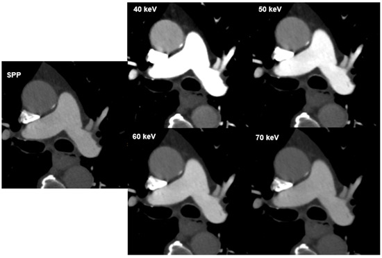
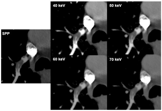
Purpose: To assess the impact of virtual-monoenergetic-image (VMI) energies on the diagnosis of pulmonary embolism (PE) in photon-counting-detector computed-tomography (PCD-CT). Methods: Eighty patients (median age 60.4 years) with suspected PE were retrospectively included. Scans were performed on PCD-CT in the multi-energy mode at
[...] Read more.
Purpose: To assess the impact of virtual-monoenergetic-image (VMI) energies on the diagnosis of pulmonary embolism (PE) in photon-counting-detector computed-tomography (PCD-CT). Methods: Eighty patients (median age 60.4 years) with suspected PE were retrospectively included. Scans were performed on PCD-CT in the multi-energy mode at 120 kV. VMIs from 40–70 keV in 10 keV intervals were reconstructed. CT-attenuation was measured in the pulmonary trunk and the main branches of the pulmonary artery. Signal-to-noise (SNR) ratio was calculated. Two radiologists evaluated subjective-image-quality (noise, vessel-attenuation and sharpness; five-point-Likert-scale, non-diagnostic–excellent), the presence of hardening artefacts and presence/visibility of PE. Results: Signal was highest at the lowest evaluated VMI (40 keV; 1053.50 HU); image noise was lowest at the highest VMI (70 keV; 15.60 HU). Highest SNR was achieved at the lowest VMI (p < 0.05). Inter-reader-agreement for subjective analysis was fair to excellent (k = 0.373–1.000; p < 0.001). Scores for vessel-attenuation and sharpness were highest at 40 keV (both:5, range 4/3–5; k = 1.000); scores for image-noise were highest at 70 keV (4, range 3–5). The highest number of hardening artifacts were reported at 40 keV (n = 22; 28%). PE-visualization was rated best at 50 keV (4.7; range 4–5) and decreased with increasing VMI-energy (r = −0.558; p < 0.001). Conclusions: While SNR was best at 40 keV, subjective PE visibility was rated highest at 50 keV, potentially owing to the lower image noise and hardening artefacts.
Full article
(This article belongs to the Special Issue Imaging of Pulmonary Vascular Disease)
►
Show Figures
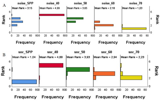
Figure 1


