Life 2022, 12(4), 516; https://doi.org/10.3390/life12040516 - 31 Mar 2022
Cited by 7 | Viewed by 4157
Abstract
Protein kinases (PKs) are established gameplayers in biological signalling pathways, and a large body of evidence points to their dysregulation in diseases, in particular cancer, where rewiring of PK networks occurs frequently. Fluorescent biosensors constitute attractive tools for probing biomolecules and monitoring dynamic
[...] Read more.
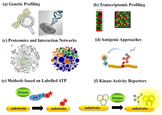
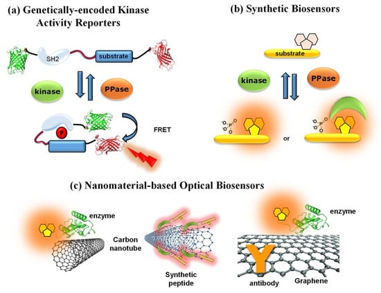
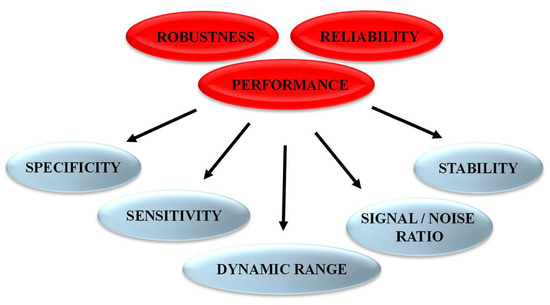
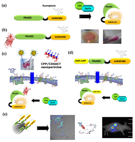
Protein kinases (PKs) are established gameplayers in biological signalling pathways, and a large body of evidence points to their dysregulation in diseases, in particular cancer, where rewiring of PK networks occurs frequently. Fluorescent biosensors constitute attractive tools for probing biomolecules and monitoring dynamic processes in complex samples. A wide variety of genetically encoded and synthetic biosensors have been tailored to report on PK activities over the last decade, enabling interrogation of their function and insight into their behaviour in physiopathological settings. These optical tools can further be used to highlight enzymatic alterations associated with the disease, thereby providing precious functional information which cannot be obtained through conventional genetic, transcriptomic or proteomic approaches. This review focuses on fluorescent peptide biosensors, recent developments and strategies that make them attractive tools to profile PK activities for biomedical and diagnostic purposes, as well as insights into the challenges and opportunities brought by this unique toolbox of chemical probes.
Full article
(This article belongs to the Collection Feature Review Papers for Life)
►
Show Figures
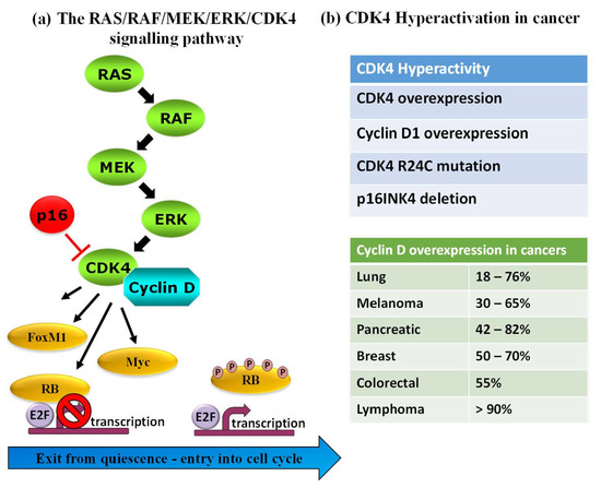
Figure 1


