Cells 2023, 12(4), 555; https://doi.org/10.3390/cells12040555 - 9 Feb 2023
Cited by 23 | Viewed by 8611
Abstract
The number of people suffering from hair loss is increasing, and hair loss occurs not only in older men but also in women and young people. Prostaglandin D2 (PGD2) is a well-known alopecia inducer. However, the mechanism by which PGD
[...] Read more.
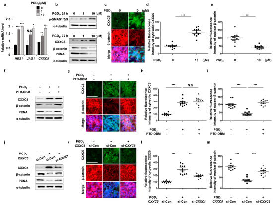
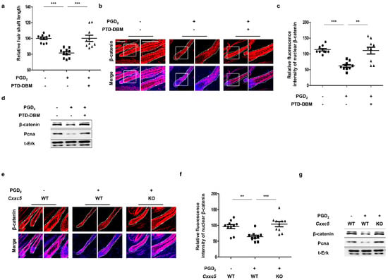
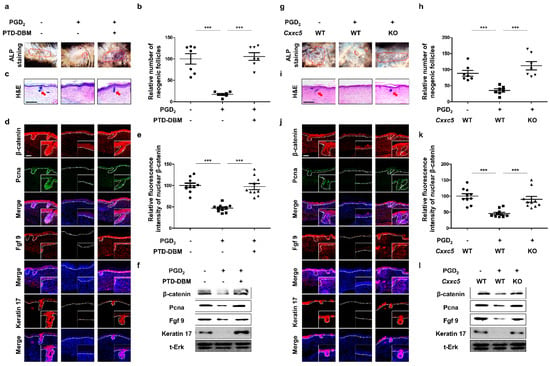
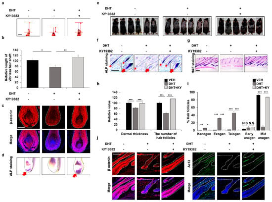
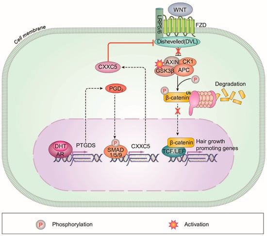
The number of people suffering from hair loss is increasing, and hair loss occurs not only in older men but also in women and young people. Prostaglandin D2 (PGD2) is a well-known alopecia inducer. However, the mechanism by which PGD2 induces alopecia is poorly understood. In this study, we characterized CXXC5, a negative regulator of the Wnt/β-catenin pathway, as a mediator for hair loss by PGD2. The hair loss by PGD2 was restored by Cxxc5 knock-out or treatment of protein transduction domain–Dishevelled binding motif (PTD-DBM), a peptide activating the Wnt/β-catenin pathway via interference with the Dishevelled (Dvl) binding function of CXXC5. In addition, suppression of neogenic hair growth by PGD2 was also overcome by PTD-DBM treatment or Cxxc5 knock-out as shown by the wound-induced hair neogenesis (WIHN) model. Moreover, we found that CXXC5 also mediates DHT-induced hair loss via PGD2. DHT-induced hair loss was alleviated by inhibition of both GSK-3β and CXXC5 functions. Overall, CXXC5 mediates the hair loss by the DHT-PGD2 axis through suppression of Wnt/β-catenin signaling.
Full article
(This article belongs to the Topic Cell Signaling Pathways)
►
Show Figures
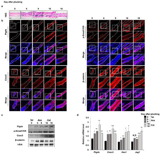
Figure 1


