by
Thalita Marcolan Valverde 1, Bruna Nayane Goncalves de Souza Soares 2, Andréa Mendes do Nascimento 2 , Ângela Leão Andrade 2, Lucas Resende Dutra Sousa 3, Paula Melo de Abreu Vieira 3, Vagner Rodrigues Santos 4, Janaína Brandão Seibert 5, Tatiane Cristine Silva de Almeida 6, Caio Fabrini Rodrigues 1, Samantha Roberta Machado de Oliveira 7, Flaviano dos Santos Martins 7, Jeronimo Geraldo Ferreira Júnior 2 and Viviane Martins Rebello dos Santos 2,*
, Ângela Leão Andrade 2, Lucas Resende Dutra Sousa 3, Paula Melo de Abreu Vieira 3, Vagner Rodrigues Santos 4, Janaína Brandão Seibert 5, Tatiane Cristine Silva de Almeida 6, Caio Fabrini Rodrigues 1, Samantha Roberta Machado de Oliveira 7, Flaviano dos Santos Martins 7, Jeronimo Geraldo Ferreira Júnior 2 and Viviane Martins Rebello dos Santos 2,*
1
Department of Morphology, Institute of Biological Sciences, Federal University of Minas Gerais (UFMG), Belo Horizonte 31270-901, MG, Brazil
2
Department of Chemistry, Federal University of Ouro Preto (UFOP), Ouro Preto 35400-000, MG, Brazil
3
Laboratory of Morphopathology, Center for Research in Biological Sciences, Federal University of Ouro Preto (UFOP), Ouro Preto 35400-000, MG, Brazil
4
Laboratory of Microbiology and Biomaterials, School of Dentistry, Federal University of Minas Gerais (UFMG), Belo Horizonte 31270-901, MG, Brazil
5
Natural Products Laboratory, Department of Chemistry, Federal University of São Carlos (UFSCAR), São Carlos 13565-905, SP, Brazil
6
Nuclear Technology Development Center, Belo Horizonte 6627, MG, Brazil
7
Department of Microbiology, Institute of Biological Sciences, Federal University of Minas Gerais (UFMG), Belo Horizonte 31270-901, MG, Brazil
Int. J. Mol. Sci. 2023, 24(6), 5112; https://doi.org/10.3390/ijms24065112 - 7 Mar 2023
Cited by 16 | Viewed by 4978
Abstract
Many activities have been described for propolis, including, antiviral, antibacterial, antifungal, anti-inflammatory, immunoregulatory, antioxidant and wound healing properties. Recently, propolis has been highlighted due to its potential application in the pharmaceutical and cosmetic industries, motivating a better understanding of its antioxidant and anti-inflammatory
[...] Read more.
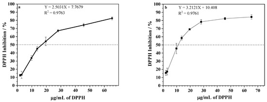
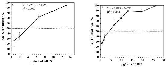
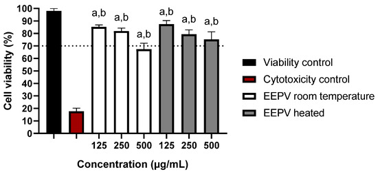
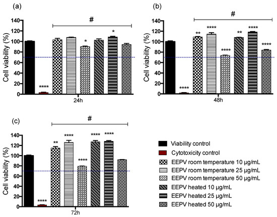
Many activities have been described for propolis, including, antiviral, antibacterial, antifungal, anti-inflammatory, immunoregulatory, antioxidant and wound healing properties. Recently, propolis has been highlighted due to its potential application in the pharmaceutical and cosmetic industries, motivating a better understanding of its antioxidant and anti-inflammatory activities. Propolis and its main polyphenolic compounds presented high antioxidant activity, and effectiveness as broad spectrum UVB and UVA photoprotection sunscreens. Through a qualitative phytochemical screening, the ethanolic red propolis extracts (EEPV) (70% at room temperature and 70% at a hot temperature) presented a positive result for flavonoids and terpenoids. It presented an antioxidant activity for reducing 50% of DPPH of 17 and 12 μg/mL for extraction at room temperature and at a hot temperature, respectively. The UPLC-QTOF-MS/MS analysis allowed the annotation of 40 substances for EEPV-Heated and 42 substances for EEPV-Room Temperature. The IC50 results of the ABTS scavenging activity was 4.7 μg/mL for both extractions, at room temperature and at a hot temperature. Additionally, we also evaluated the cytotoxic profile of propolis extracts against macrophage (RAW 264.7 cells) and keratinocytes (HaCaT cells), which showed non-cytotoxic doses in cell viability assays even after a long period of exposure. In addition, propolis extracts showed antibacterial activity for Gram-positive bacteria (Staphylococcus aureus and Staphylococcus epidermidis), demonstrating potential biological activity for the creation of formulations aimed at disease control and prevention.
Full article
(This article belongs to the Special Issue Flavonoids and Their Impact on Human Health)
▼
Show Figures

Figure 1


