12 pages, 4143 KiB  
Article
Reduction of Osteoclastic Differentiation of Raw 264.7 Cells by EMF Exposure through TRPV4 and p-CREB Pathway
by Myeong-Hyun Nam, Hee-Jung Park and Young-Kwon Seo *
Department of Medical Biotechnology, Dongguk University, Goyang-si 10326, Republic of Korea
Int. J. Mol. Sci. 2023, 24(4), 3058; https://doi.org/10.3390/ijms24043058 - 4 Feb 2023
Cited by 7 | Viewed by 4256
Abstract
In this study, we investigated the effect of EMF exposure on the regulation of RANKL-induced osteoclast differentiation in Raw 264.7 cells. In the EMF-exposed group, the cell volume did not increase despite RANKL treatment, and the expression levels of Caspase-3 remained much lower [...] Read more.
In this study, we investigated the effect of EMF exposure on the regulation of RANKL-induced osteoclast differentiation in Raw 264.7 cells. In the EMF-exposed group, the cell volume did not increase despite RANKL treatment, and the expression levels of Caspase-3 remained much lower than those in the RANKL-treated group. TRAP and F-actin staining revealed smaller actin rings in cells exposed to EMF during RANKL-induced differentiation, indicating that EMF inhibited osteoclast differentiation. EMF-irradiated cells exhibited reduced mRNA levels of osteoclastic differentiation markers cathepsin K (CTSK), tartrate-resistant acid phosphatase (TRAP), and matrix metalloproteinase 9 (MMP-9). Furthermore, as measured by RT-qPCR and Western blot, EMF induced no changes in the levels of p-ERK and p-38; however, it reduced the levels of TRPV4 and p-CREB. Overall, our findings indicate that EMF irradiation inhibits osteoclast differentiation through the TRPV4 and p-CREB pathway. Full article
(This article belongs to the Special Issue Advances in the Molecular Biological Effects of Magnetic Fields)
Show Figures

Figure 1

22 pages, 1609 KiB  
Review
Mechanistic Insights into the Biological Effects of Engineered Nanomaterials: A Focus on Gold Nanoparticles
by Nhung H. A. Nguyen 1 and Priscila Falagan-Lotsch 2,*
1 Institute for Nanomaterials, Advanced Technologies and Innovation, Technical University of Liberec (TUL), Studentsk. 2, 46117 Liberec, Czech Republic
2 Department of Biological Sciences, College of Sciences and Mathematics, Auburn University, Auburn, AL 36849, USA
Int. J. Mol. Sci. 2023, 24(4), 4109; https://doi.org/10.3390/ijms24044109 - 18 Feb 2023
Cited by 32 | Viewed by 4252
Abstract
Nanotechnology has great potential to significantly advance the biomedical field for the benefit of human health. However, the limited understanding of nano–bio interactions leading to unknowns about the potential adverse health effects of engineered nanomaterials and to the poor efficacy of nanomedicines has [...] Read more.
Nanotechnology has great potential to significantly advance the biomedical field for the benefit of human health. However, the limited understanding of nano–bio interactions leading to unknowns about the potential adverse health effects of engineered nanomaterials and to the poor efficacy of nanomedicines has hindered their use and commercialization. This is well evidenced considering gold nanoparticles, one of the most promising nanomaterials for biomedical applications. Thus, a fundamental understanding of nano–bio interactions is of interest to nanotoxicology and nanomedicine, enabling the development of safe-by-design nanomaterials and improving the efficacy of nanomedicines. In this review, we introduce the advanced approaches currently applied in nano–bio interaction studies—omics and systems toxicology—to provide insights into the biological effects of nanomaterials at the molecular level. We highlight the use of omics and systems toxicology studies focusing on the assessment of the mechanisms underlying the in vitro biological responses to gold nanoparticles. First, the great potential of gold-based nanoplatforms to improve healthcare along with the main challenges for their clinical translation are presented. We then discuss the current limitations in the translation of omics data to support risk assessment of engineered nanomaterials. Full article
(This article belongs to the Special Issue Recent Advances in Nanomaterials Science)
Show Figures

Figure 1

20 pages, 4876 KiB  
Review
Biomass-Derived Carbon Materials for the Electrode of Metal–Air Batteries
by Xiaodong Lv, Ming Chen, Hideo Kimura, Wei Du * and Xiaoyang Yang *
1 School of Environmental and Material Engineering, Yantai University, No. 30 Qingquan Road, Yantai 264005, China
These authors contributed equally to this work.
Int. J. Mol. Sci. 2023, 24(4), 3713; https://doi.org/10.3390/ijms24043713 - 13 Feb 2023
Cited by 19 | Viewed by 4237
Abstract
Facing the challenges of energy crisis and global warming, the development of renewable energy has received more and more attention. To offset the discontinuity of renewable energy, such as wind and solar energy, it is urgent to search for an excellent performance energy [...] Read more.
Facing the challenges of energy crisis and global warming, the development of renewable energy has received more and more attention. To offset the discontinuity of renewable energy, such as wind and solar energy, it is urgent to search for an excellent performance energy storage system to match them. Metal–air batteries (typical representative: Li–air battery and Zn–air battery) have broad prospects in the field of energy storage due to their high specific capacity and environmental friendliness. The drawbacks preventing the massive application of metal–air batteries are the poor reaction kinetics and high overpotential during the charging–discharging process, which can be alleviated by the application of an electrochemical catalyst and porous cathode. Biomass, also, as a renewable resource, plays a critical role in the preparation of carbon-based catalysts and porous cathode with excellent performance for metal–air batteries due to the inherent rich heteroatom and pore structure of biomass. In this paper, we have reviewed the latest progress in the creative preparation of porous cathode for the Li–air battery and Zn–air battery from biomass and summarized the effects of various biomass sources precursors on the composition, morphology and structure-activity relationship of cathode. This review will help us understand the relevant applications of biomass carbon in the field of metal–air batteries. Full article
(This article belongs to the Special Issue Material Design and Mechanism Research of Metal-Air Batteries)
Show Figures

Figure 1

16 pages, 857 KiB  
Review
Pathogenicity of Type I Interferons in Mycobacterium tuberculosis
by Akaash Mundra 1, Aram Yegiazaryan 1, Haig Karsian 1, Dijla Alsaigh 1, Victor Bonavida 1, Mitchell Frame 1, Nicole May 1, Areg Gargaloyan 1, Arbi Abnousian 1 and Vishwanath Venketaraman 1,2,*
1 College of Osteopathic Medicine of the Pacific, Western University of Health Sciences, Pomona, CA 91766, USA
2 Graduate College of Biomedical Sciences, Western University of Health Sciences, Pomona, CA 91768, USA
Int. J. Mol. Sci. 2023, 24(4), 3919; https://doi.org/10.3390/ijms24043919 - 15 Feb 2023
Cited by 15 | Viewed by 4236
Abstract
Tuberculosis (TB) is a leading cause of mortality due to infectious disease and rates have increased during the emergence of COVID-19, but many of the factors determining disease severity and progression remain unclear. Type I Interferons (IFNs) have diverse effector functions that regulate [...] Read more.
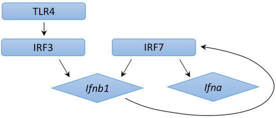
Tuberculosis (TB) is a leading cause of mortality due to infectious disease and rates have increased during the emergence of COVID-19, but many of the factors determining disease severity and progression remain unclear. Type I Interferons (IFNs) have diverse effector functions that regulate innate and adaptive immunity during infection with microorganisms. There is well-documented literature on type I IFNs providing host defense against viruses; however, in this review, we explore the growing body of work that indicates high levels of type I IFNs can have detrimental effects to a host fighting TB infection. We report findings that increased type I IFNs can affect alveolar macrophage and myeloid function, promote pathological neutrophil extracellular trap responses, inhibit production of protective prostaglandin 2, and promote cytosolic cyclic GMP synthase inflammation pathways, and discuss many other relevant findings. Full article
(This article belongs to the Special Issue Genomics: Infectious Disease and Host-Pathogen Interaction 2.0)
Show Figures

Figure 1

14 pages, 1802 KiB  
Article
Detection of Breast Cancer-Specific Extracellular Vesicles with Fiber-Optic SPR Biosensor
by Yagmur Yildizhan 1, Kaat Driessens 1, Hong Shen Kevin Tsao 1, Robin Boiy 2, Debby Thomas 3, Nick Geukens 3, An Hendrix 2, Jeroen Lammertyn 1,* and Dragana Spasic 1
1 Department of Biosystems, Biosensors Group, Katholieke Universiteit Leuven, 3001 Leuven, Belgium
2 Laboratory of Experimental Cancer Research, Cancer Research Institute Ghent, Department of Human Structure and Repair, Ghent University, 9000 Ghent, Belgium
3 PharmAbs, The KU Leuven Antibody Center, University of Leuven, 3000 Leuven, Belgium
Int. J. Mol. Sci. 2023, 24(4), 3764; https://doi.org/10.3390/ijms24043764 - 13 Feb 2023
Cited by 16 | Viewed by 4236
Abstract
Extracellular vesicles (EVs) have attracted great attention as potential biomarkers for cancer diagnostics. Although several technologies have been developed for EV detection, many of them are still not applicable to clinical settings as they rely on complex EV isolation processes, while lacking sensitivity, [...] Read more.
Extracellular vesicles (EVs) have attracted great attention as potential biomarkers for cancer diagnostics. Although several technologies have been developed for EV detection, many of them are still not applicable to clinical settings as they rely on complex EV isolation processes, while lacking sensitivity, specificity or standardization. To solve this problem, we have developed a sensitive breast cancer-specific EV detection bioassay directly in blood plasma using a fiber-optic surface plasmon resonance (FO-SPR) biosensor, previously calibrated with recombinant EVs. First, we established a sandwich bioassay to detect SK-BR-3 EVs by functionalizing the FO-SPR probes with anti-HER2 antibodies. A calibration curve was built using an anti-HER2/Banti-CD9 combination, resulting in an LOD of 2.1 × 10⁷ particles/mL in buffer and 7 × 10⁸ particles/mL in blood plasma. Next, we investigated the potential of the bioassay to detect MCF7 EVs in blood plasma using an anti-EpCAM/Banti-mix combination, obtaining an LOD of 1.1 × 10 8 particles/mL. Finally, the specificity of the bioassay was proven by the absence of signal when testing plasma samples from 10 healthy people unknown to be diagnosed with breast cancer. The remarkable sensitivity and specificity of the developed sandwich bioassay together with the advantages of the standardized FO-SPR biosensor highlight outstanding potential for the future of EV analysis. Full article
(This article belongs to the Special Issue Molecular Biosensor)
Show Figures

Figure 1

25 pages, 1589 KiB  
Review
Comprehensive Analysis of Mitochondrial Dynamics Alterations in Heart Diseases
by Giampaolo Morciano 1,2,*, Caterina Boncompagni 1, Daniela Ramaccini 2, Gaia Pedriali 2, Esmaa Bouhamida 2, Elena Tremoli 2, Carlotta Giorgi 1 and Paolo Pinton 1,2,*
1 Department of Medical Sciences, University of Ferrara, 44121 Ferrara, Italy
2 GVM Care & Research, Maria Cecilia Hospital, 48033 Cotignola, Italy
Int. J. Mol. Sci. 2023, 24(4), 3414; https://doi.org/10.3390/ijms24043414 - 8 Feb 2023
Cited by 18 | Viewed by 4222
Abstract
The most common alterations affecting mitochondria, and associated with cardiac pathological conditions, implicate a long list of defects. They include impairments of the mitochondrial electron transport chain activity, which is a crucial element for energy formation, and that determines the depletion of ATP [...] Read more.
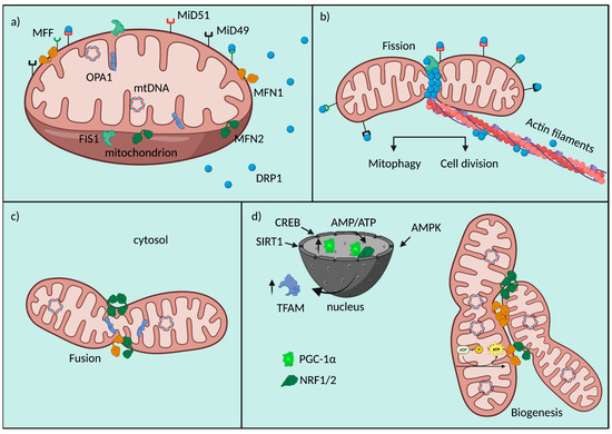
The most common alterations affecting mitochondria, and associated with cardiac pathological conditions, implicate a long list of defects. They include impairments of the mitochondrial electron transport chain activity, which is a crucial element for energy formation, and that determines the depletion of ATP generation and supply to metabolic switches, enhanced ROS generation, inflammation, as well as the dysregulation of the intracellular calcium homeostasis. All these signatures significantly concur in the impairment of cardiac electrical characteristics, loss of myocyte contractility and cardiomyocyte damage found in cardiac diseases. Mitochondrial dynamics, one of the quality control mechanisms at the basis of mitochondrial fitness, also result in being dysregulated, but the use of this knowledge for translational and therapeutic purposes is still in its infancy. In this review we tried to understand why this is, by summarizing methods, current opinions and molecular details underlying mitochondrial dynamics in cardiac diseases. Full article
Show Figures

Figure 1

12 pages, 2087 KiB  
Article
Isosinensetin Stimulates Glucagon-like Peptide-1 Secretion via Activation of hTAS2R50 and the Gβγ-Mediated Signaling Pathway
by Seung-Hyeon Lee 1,2, Hyun Min Ko 1,2, Wona Jee 1,2, Hyungsuk Kim 1, Won-Seok Chung 1 and Hyeung-Jin Jang 1,2,*
1 College of Korean Medicine, Kyung Hee University, 26, Kyungheedae-ro, Dongdaemun-gu, Seoul 02447, Republic of Korea
2 Department of Science in Korean Medicine, Graduate School, Kyung Hee University, Seoul 02447, Republic of Korea
Int. J. Mol. Sci. 2023, 24(4), 3682; https://doi.org/10.3390/ijms24043682 - 12 Feb 2023
Cited by 10 | Viewed by 4217
Abstract
Bitter taste receptors (TAS2Rs) are G protein-coupled receptors localized in the taste buds of the tongue. They may also be present in non-lingual organs, including the brain, lung, kidney, and gastrointestinal (GI) tract. Recent studies on bitter taste receptor functions have suggested TAS2Rs [...] Read more.
Bitter taste receptors (TAS2Rs) are G protein-coupled receptors localized in the taste buds of the tongue. They may also be present in non-lingual organs, including the brain, lung, kidney, and gastrointestinal (GI) tract. Recent studies on bitter taste receptor functions have suggested TAS2Rs as potential therapeutic targets. The human bitter taste receptor subtype hTAS2R50 responds to its agonist isosinensetin (ISS). Here, we demonstrated that, unlike other TAS2R agonists, isosinensetin activated hTAS2R50 as well as increased Glucagon-like peptide 1 (GLP-1) secretion through the Gβγ-mediated pathway in NCI-H716 cells. To confirm this mechanism, we showed that ISS increased intracellular Ca2+ and was suppressed by the IP3R inhibitor 2-APB as well as the PLC inhibitor U73122, suggesting that TAS2Rs alters the physiological state of enteroendocrine L cells in a PLC-dependent manner. Furthermore, we demonstrated that ISS upregulated proglucagon mRNA and stimulated GLP-1 secretion. ISS-mediated GLP-1 secretion was suppressed in response to small interfering RNA-mediated silencing of Gα-gust and hTAS2R50 as well as 2-APB and U73122. Our findings improved the understanding of how ISS modulates GLP-1 secretion and indicates the possibility of using ISS as a therapeutic agent in the treatment of diabetes mellitus. Full article
(This article belongs to the Special Issue Anti-inflammatory Effects of Glucagon-Like Peptide-1)
Show Figures

Graphical abstract

15 pages, 1950 KiB  
Article
A Novel Preclinical In Vitro 3D Model of Oral Carcinogenesis for Biomarker Discovery and Drug Testing
by Ravi Teja Chitturi Suryaprakash 1, Kate Shearston 1, Camile S. Farah 2, Simon A. Fox 1,3, Muhammad Munir Iqbal 4, Ulrich Kadolsky 4, Xiao Zhong 4, Alka Saxena 4 and Omar Kujan 1,*
1 UWA Dental School, The University of Western Australia, Nedlands, WA 6009, Australia
2 Australian Centre for Oral Oncology Research and Education, Nedlands, WA 6009, Australia
3 Harry Perkins Institute of Medical Research, QEII Medical Centre and Centre for Medical Research, The University of Western Australia, Nedlands, WA 6009, Australia
4 Genomics WA, Harry Perkins Institute of Medical Research, Telethon Kids Institute, The University of Western Australia, Nedlands, WA 6009, Australia
Int. J. Mol. Sci. 2023, 24(4), 4096; https://doi.org/10.3390/ijms24044096 - 17 Feb 2023
Cited by 10 | Viewed by 4203
Abstract
This study aimed to develop an in vitro three-dimensional (3D) cell culture model of oral carcinogenesis for the rapid, scalable testing of chemotherapeutic agents. Spheroids of normal (HOK) and dysplastic (DOK) human oral keratinocytes were cultured and treated with 4-nitroquinoline-1-oxide (4NQO). A 3D [...] Read more.
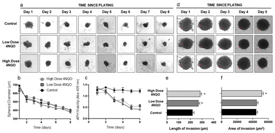
This study aimed to develop an in vitro three-dimensional (3D) cell culture model of oral carcinogenesis for the rapid, scalable testing of chemotherapeutic agents. Spheroids of normal (HOK) and dysplastic (DOK) human oral keratinocytes were cultured and treated with 4-nitroquinoline-1-oxide (4NQO). A 3D invasion assay using Matrigel was performed to validate the model. RNA was extracted and subjected to transcriptomic analysis to validate the model and assess carcinogen-induced changes. The VEGF inhibitors pazopanib and lenvatinib were tested in the model and were validated by a 3D invasion assay, which demonstrated that changes induced by the carcinogen in spheroids were consistent with a malignant phenotype. Further validation was obtained by bioinformatic analyses, which showed the enrichment of pathways associated with hallmarks of cancer and VEGF signalling. Overexpression of common genes associated with tobacco-induced oral squamous cell carcinoma (OSCC), such as MMP1, MMP3, MMP9, YAP1, CYP1A1, and CYP1B1, was also observed. Pazopanib and lenvatinib inhibited the invasion of transformed spheroids. In summary, we successfully established a 3D spheroid model of oral carcinogenesis for biomarker discovery and drug testing. This model is a validated preclinical model for OSCC development and would be suitable for testing a range of chemotherapeutic agents. Full article
(This article belongs to the Special Issue Organoids: The New 3D-Frontier to Model Different Diseases In Vitro)
Show Figures

Figure 1

17 pages, 2348 KiB  
Article
N-Acetylcysteine Suppresses Microglial Inflammation and Induces Mortality Dose-Dependently via Tumor Necrosis Factor-α Signaling
by Mai Sakai 1, Zhiqian Yu 2,*, Masayuki Taniguchi 3, Rosanne Picotin 4, Nanami Oyama 1, David Stellwagen 5, Chiaki Ono 2, Yoshie Kikuchi 2, Ko Matsui 6, Miharu Nakanishi 1, Hatsumi Yoshii 1, Tomoyuki Furuyashiki 3, Takaaki Abe 7 and Hiroaki Tomita 2,8
1 Department of Psychiatric Nursing, Graduate School of Medicine, Tohoku University, Sendai 980-8575, Japan
2 Department of Psychiatry, Graduate School of Medicine, Tohoku University, Sendai 980-8575, Japan
3 Division of Pharmacology, Kobe University Graduate School of Medicine, Kobe 650-0017, Japan
4 Medical Faculty, Heinrich Heine University Düsseldorf, 40225 Düsseldorf, Germany
5 Department of Neurology and Neurosurgery, The Research Institute of the McGill University Health Center, Montreal, QC H3G 1A4, Canada
6 Super-network Brain Physiology, Graduate School of Life Sciences, Tohoku University, Sendai 980-8577, Japan
7 Department of Biomedical Engineering Regenerative and Biomedical Engineering Medical Science, Graduate School of Biomedical Engineering, Tohoku University, Sendai 980-8575, Japan
8 Department of Disaster Psychiatry, International Research Institute for Disaster Science, Tohoku University, Sendai 980-8573, Japan
Int. J. Mol. Sci. 2023, 24(4), 3798; https://doi.org/10.3390/ijms24043798 - 14 Feb 2023
Cited by 10 | Viewed by 4203
Abstract
N-acetylcysteine (NAC) is an antioxidant that prevents tumor necrosis factor (TNF)-α-induced cell death, but it also acts as a pro-oxidant, promoting reactive oxygen species independent apoptosis. Although there is plausible preclinical evidence for the use of NAC in the treatment of psychiatric disorders, [...] Read more.
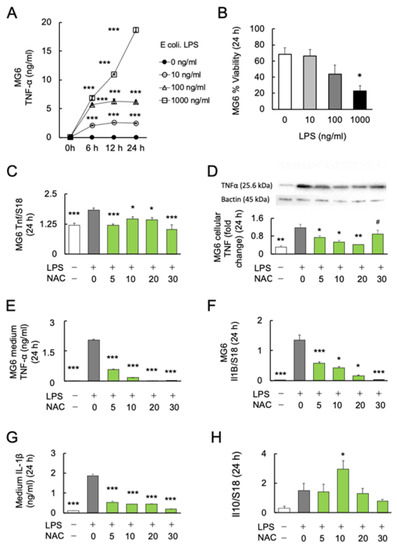
N-acetylcysteine (NAC) is an antioxidant that prevents tumor necrosis factor (TNF)-α-induced cell death, but it also acts as a pro-oxidant, promoting reactive oxygen species independent apoptosis. Although there is plausible preclinical evidence for the use of NAC in the treatment of psychiatric disorders, deleterious side effects are still of concern. Microglia, key innate immune cells in the brain, play an important role in inflammation in psychiatric disorders. This study aimed to investigate the beneficial and deleterious effects of NAC on microglia and stress-induced behavior abnormalities in mice, and its association with microglial TNF-α and nitric oxide (NO) production. The microglial cell line MG6 was stimulated by Escherichia coli lipopolysaccharide (LPS) using NAC at varying concentrations for 24 h. NAC inhibited LPS-induced TNF-α and NO synthesis, whereas high concentrations (≥30 mM) caused MG6 mortality. Intraperitoneal injections of NAC did not ameliorate stress-induced behavioral abnormalities in mice, but high-doses induced microglial mortality. Furthermore, NAC-induced mortality was alleviated in microglial TNF-α-deficient mice and human primary M2 microglia. Our findings provide ample evidence for the use of NAC as a modulating agent of inflammation in the brain. The risk of side effects from NAC on TNF-α remains unclear and merits further mechanistic investigations. Full article
Show Figures

Figure 1

17 pages, 2682 KiB  
Review
‘Toxic Masculinity’: What Is Known about the Role of Androgen Receptors in Head and Neck Squamous Cell Carcinoma
by Josipa Čonkaš, Maja Sabol and Petar Ozretić *
Laboratory for Hereditary Cancer, Division of Molecular Medicine, Ruđer Bošković Institute, HR-10000 Zagreb, Croatia
Int. J. Mol. Sci. 2023, 24(4), 3766; https://doi.org/10.3390/ijms24043766 - 13 Feb 2023
Cited by 6 | Viewed by 4201
Abstract
Head and neck squamous cell carcinoma (HNSCC), the most prevalent cancer in the head and neck region, develops from the mucosal epithelium of the upper aerodigestive tract. Its development directly correlates with alcohol and/or tobacco consumption and infection with human papillomavirus. Interestingly, the [...] Read more.
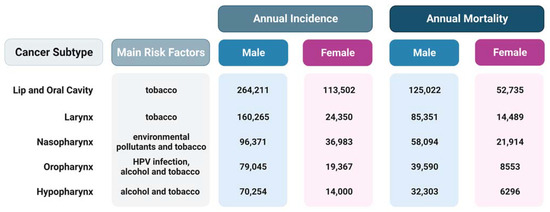
Head and neck squamous cell carcinoma (HNSCC), the most prevalent cancer in the head and neck region, develops from the mucosal epithelium of the upper aerodigestive tract. Its development directly correlates with alcohol and/or tobacco consumption and infection with human papillomavirus. Interestingly, the relative risk for HNSCC is up to five times higher in males, so it is considered that the endocrine microenvironment is another risk factor. A gender-specific risk for HNSCC suggests either the existence of specific risk factors that affect only males or that females have defensive hormonal and metabolic features. In this review, we summarized the current knowledge about the role of both nuclear and membrane androgen receptors (nAR and mARs, respectively) in HNSCC. As expected, the significance of nAR is much better known; it was shown that increased nAR expression was observed in HNSCC, while treatment with dihydrotestosterone increased proliferation, migration, and invasion of HNSCC cells. For only three out of five currently known mARs—TRPM8, CaV1.2, and OXER1—it was shown either their increased expression in various types of HNSCC or that their increased activity enhanced the migration and invasion of HNSCC cells. The primary treatments for HNSCC are surgery and radiotherapy, but targeted immunotherapies are on the rise. On the other hand, given the evidence of elevated nAR expression in HNSCC, this receptor represents a potential target for antiandrogen therapy. Moreover, there is still plenty of room for further examination of mARs’ role in HNSCC diagnosis, prognosis, and treatment. Full article
(This article belongs to the Special Issue Molecular Mechanisms and Pathophysiology of Head and Neck Diseases)
Show Figures

Figure 1

13 pages, 1930 KiB  
Review
Thinking on the Construction of Antimicrobial Peptide Databases: Powerful Tools for the Molecular Design and Screening
by Kun Zhang 1,2,3, Da Teng 1,2,3, Ruoyu Mao 1,2,3, Na Yang 1,2,3, Ya Hao 1,2,3 and Jianhua Wang 1,2,3,*
1 Gene Engineering Laboratory, Feed Research Institute, Chinese Academy of Agricultural Sciences, Beijing 100081, China
2 Innovative Team of Antimicrobial Peptides and Alternatives to Antibiotics, Feed Research Institute, Chinese Academy of Agricultural Sciences, Beijing 100081, China
3 Key Laboratory of Feed Biotechnology, Ministry of Agriculture and Rural Affairs, Beijing 100081, China
Int. J. Mol. Sci. 2023, 24(4), 3134; https://doi.org/10.3390/ijms24043134 - 5 Feb 2023
Cited by 17 | Viewed by 4198
Abstract
With the accelerating growth of antimicrobial resistance (AMR), there is an urgent need for new antimicrobial agents with low or no AMR. Antimicrobial peptides (AMPs) have been extensively studied as alternatives to antibiotics (ATAs). Coupled with the new generation of high-throughput technology for [...] Read more.
With the accelerating growth of antimicrobial resistance (AMR), there is an urgent need for new antimicrobial agents with low or no AMR. Antimicrobial peptides (AMPs) have been extensively studied as alternatives to antibiotics (ATAs). Coupled with the new generation of high-throughput technology for AMP mining, the number of derivatives has increased dramatically, but manual running is time-consuming and laborious. Therefore, it is necessary to establish databases that combine computer algorithms to summarize, analyze, and design new AMPs. A number of AMP databases have already been established, such as the Antimicrobial Peptides Database (APD), the Collection of Antimicrobial Peptides (CAMP), the Database of Antimicrobial Activity and Structure of Peptides (DBAASP), and the Database of Antimicrobial Peptides (dbAMPs). These four AMP databases are comprehensive and are widely used. This review aims to cover the construction, evolution, characteristic function, prediction, and design of these four AMP databases. It also offers ideas for the improvement and application of these databases based on merging the various advantages of these four peptide libraries. This review promotes research and development into new AMPs and lays their foundation in the fields of druggability and clinical precision treatment. Full article
(This article belongs to the Section Molecular Microbiology)
Show Figures

Figure 1

15 pages, 3431 KiB  
Article
Efficient Synthesis of Chlorin e6 and Its Potential Photodynamic Immunotherapy in Mouse Melanoma by the Abscopal Effect
by Rajeev Shrestha, Shyam Kumar Mallik, Junmo Lim, Pallavi Gurung, Til Bahadur Thapa Magar and Yong-Wan Kim *
1 Dongsung Cancer Center, Dongsung Biopharmaceutical, Daegu 41061, Republic of Korea
These authors contributed equally to this work.
Int. J. Mol. Sci. 2023, 24(4), 3901; https://doi.org/10.3390/ijms24043901 - 15 Feb 2023
Cited by 15 | Viewed by 4197
Abstract
Photodynamic therapy (PDT) can eradicate not only cancer cells but also stimulate an antitumor immune response. Herein, we describe two efficient synthetic methodologies for the preparation of Chlorin e6 (Ce6) from Spirulina platensis and address the phototoxic effect of Ce6 in vitro along [...] Read more.
Photodynamic therapy (PDT) can eradicate not only cancer cells but also stimulate an antitumor immune response. Herein, we describe two efficient synthetic methodologies for the preparation of Chlorin e6 (Ce6) from Spirulina platensis and address the phototoxic effect of Ce6 in vitro along with antitumor activity in vivo. Melanoma B16F10 cells were seeded and phototoxicity was monitored by the MTT assay. The C57BL/6 mice were subcutaneously inoculated on the left and right flank with B16F10 cells. The mice were intravenously injected with Ce6 of 2.5 mg/kg and then exposed to red light (660 nm) on the left flank tumors 3 h after the injection. The immune response was studied by analyzing Interferon-gamma (IFN-γ), tumor necrosis factor-alpha (TNF-α), and Interleukin-2 (IL-2) of the right flank tumors through qPCR. Our results revealed that the tumor was suppressed not only in the left flank but also in the right flank, where no PDT was given. The upregulated gene and protein expression of IFN-γ, TNF-α, and IL-2 revealed antitumor immunity due to Ce6-PDT. The findings of this study suggest an efficient methodology of Ce6 preparation and the efficacy of Ce6-PDT as a promising antitumor immune response. Full article
(This article belongs to the Section Molecular Biology)
Show Figures

Figure 1

12 pages, 2039 KiB  
Review
D-Amino Acids and Cancer: Friends or Foes?
by Giulia Murtas and Loredano Pollegioni *
Department of Biotechnology and Life Sciences, University of Insubria, 21100 Varese, Italy
Int. J. Mol. Sci. 2023, 24(4), 3274; https://doi.org/10.3390/ijms24043274 - 7 Feb 2023
Cited by 20 | Viewed by 4191
Abstract
α-amino acids exist in two configurations, named D-(dextro) and L-(levo) enantiomers. L-amino acids are used in protein synthesis and play a central role in cell metabolism. The effects of the L-amino acid composition of foods and the dietary modifications [...] Read more.
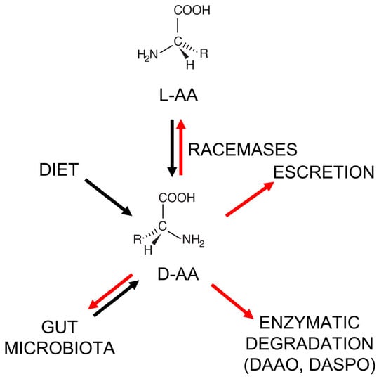
α-amino acids exist in two configurations, named D-(dextro) and L-(levo) enantiomers. L-amino acids are used in protein synthesis and play a central role in cell metabolism. The effects of the L-amino acid composition of foods and the dietary modifications of this composition on the efficacy of cancer therapies have been widely investigated in relation to the growth and reproduction of cancerous cells. However, less is known about the involvement of D-amino acids. In recent decades, D-amino acids have been identified as natural biomolecules that play interesting and specific roles as common components of the human diet. Here, we focus on recent investigations showing altered D-amino acid levels in specific cancer types and on the various roles proposed for these biomolecules related to cancer cell proliferation, cell protection during therapy, and as putative, innovative biomarkers. Notwithstanding recent progress, the relationship between the presence of D-amino acids, their nutritional value, and cancer cell proliferation and survival represents an underrated scientific issue. Few studies on human samples have been reported to date, suggesting a need for routine analysis of D-amino acid content and an evaluation of the enzymes involved in regulating their levels in clinical samples in the near future. Full article
(This article belongs to the Collection Feature Papers in Molecular Oncology)
Show Figures

Figure 1

25 pages, 12683 KiB  
Article
Nicotine-Mediated Recruitment of GABAergic Neurons to a Dopaminergic Phenotype Attenuates Motor Deficits in an Alpha-Synuclein Parkinson’s Model
by Jessica IChi Lai 1, Alessandra Porcu 1,2, Benedetto Romoli 1, Maria Keisler 1, Fredric P. Manfredsson 3, Susan B. Powell 1 and Davide Dulcis 1,*
1 Department of Psychiatry, University of California San Diego, La Jolla, CA 92093, USA
2 Department of Drug Discovery and Biomedical Sciences, University of South Carolina, Columbia, SC 29208, USA
3 Department of Neurobiology, Barrow Neurological Institute, Phoenix, AZ 85013, USA
Int. J. Mol. Sci. 2023, 24(4), 4204; https://doi.org/10.3390/ijms24044204 - 20 Feb 2023
Cited by 13 | Viewed by 4188
Abstract
Previous work revealed an inverse correlation between tobacco smoking and Parkinson’s disease (PD) that is associated with nicotine-induced neuroprotection of dopaminergic (DA) neurons against nigrostriatal damage in PD primates and rodent models. Nicotine, a neuroactive component of tobacco, can directly alter the activity [...] Read more.
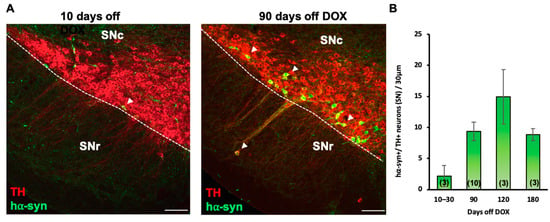
Previous work revealed an inverse correlation between tobacco smoking and Parkinson’s disease (PD) that is associated with nicotine-induced neuroprotection of dopaminergic (DA) neurons against nigrostriatal damage in PD primates and rodent models. Nicotine, a neuroactive component of tobacco, can directly alter the activity of midbrain DA neurons and induce non-DA neurons in the substantia nigra (SN) to acquire a DA phenotype. Here, we investigated the recruitment mechanism of nigrostriatal GABAergic neurons to express DA phenotypes, such as transcription factor Nurr1 and DA-synthesizing enzyme tyrosine hydroxylase (TH), and the concomitant effects on motor function. Wild-type and α-syn-overexpressing (PD) mice treated with chronic nicotine were assessed by behavioral pattern monitor (BPM) and immunohistochemistry/in situ hybridization to measure behavior and the translational/transcriptional regulation of neurotransmitter phenotype following selective Nurr1 overexpression or DREADD-mediated chemogenetic activation. We found that nicotine treatment led to a transcriptional TH and translational Nurr1 upregulation within a pool of SN GABAergic neurons in wild-type animals. In PD mice, nicotine increased Nurr1 expression, reduced the number of α-syn-expressing neurons, and simultaneously rescued motor deficits. Hyperactivation of GABA neurons alone was sufficient to elicit de novo translational upregulation of Nurr1. Retrograde labeling revealed that a fraction of these GABAergic neurons projects to the dorsal striatum. Finally, concomitant depolarization and Nurr1 overexpression within GABA neurons were sufficient to mimic nicotine-mediated dopamine plasticity. Revealing the mechanism of nicotine-induced DA plasticity protecting SN neurons against nigrostriatal damage could contribute to developing new strategies for neurotransmitter replacement in PD. Full article
(This article belongs to the Special Issue Development of Dopaminergic Neurons 2.0)
Show Figures

Figure 1

16 pages, 2982 KiB  
Article
Development of a Novel Anti-CD44 Variant 6 Monoclonal Antibody C44Mab-9 for Multiple Applications against Colorectal Carcinomas
by Ryo Ejima 1,†, Hiroyuki Suzuki 1,*,†, Tomohiro Tanaka 1, Teizo Asano 2, Mika K. Kaneko 2 and Yukinari Kato 1,2,*
1 Department of Molecular Pharmacology, Tohoku University Graduate School of Medicine, 2-1 Seiryo-machi, Aoba-ku, Sendai 980-8575, Japan
2 Department of Antibody Drug Development, Tohoku University Graduate School of Medicine, 2-1 Seiryo-machi, Aoba-ku, Sendai 980-8575, Japan
These authors contributed equally to this work.
Int. J. Mol. Sci. 2023, 24(4), 4007; https://doi.org/10.3390/ijms24044007 - 16 Feb 2023
Cited by 17 | Viewed by 4178
Abstract
CD44 is a cell surface glycoprotein, and its isoforms are produced by the alternative splicing with the standard and variant exons. The CD44 variant exon-containing isoforms (CD44v) are overexpressed in carcinomas. CD44v6 is one of the CD44v, and its overexpression predicts poor prognosis [...] Read more.
CD44 is a cell surface glycoprotein, and its isoforms are produced by the alternative splicing with the standard and variant exons. The CD44 variant exon-containing isoforms (CD44v) are overexpressed in carcinomas. CD44v6 is one of the CD44v, and its overexpression predicts poor prognosis in colorectal cancer (CRC) patients. CD44v6 plays critical roles in CRC adhesion, proliferation, stemness, invasiveness, and chemoresistance. Therefore, CD44v6 is a promising target for cancer diagnosis and therapy for CRC. In this study, we established anti-CD44 monoclonal antibodies (mAbs) by immunizing mice with CD44v3-10-overexpressed Chinese hamster ovary (CHO)-K1 cells. We then characterized them using enzyme-linked immunosorbent assay, flow cytometry, western blotting, and immunohistochemistry. One of the established clones (C44Mab-9; IgG1, kappa) reacted with a peptide of the variant 6-encoded region, indicating that C44Mab-9 recognizes CD44v6. Furthermore, C44Mab-9 reacted with CHO/CD44v3-10 cells or CRC cell lines (COLO201 and COLO205) by flow cytometry. The apparent dissociation constant (KD) of C44Mab-9 for CHO/CD44v3-10, COLO201, and COLO205 was 8.1 × 10⁻⁹ M, 1.7 × 10⁻⁸ M, and 2.3 × 10⁻⁸ M, respectively. C44Mab-9 detected the CD44v3-10 in western blotting, and partially stained the formalin-fixed paraffin-embedded CRC tissues in immunohistochemistry. Collectively, C44Mab-9 is useful for detecting CD44v6 in various applications. Full article
(This article belongs to the Special Issue Molecular Mechanisms and Therapies of Colorectal Cancer 2.0)
Show Figures

Figure 1