Int. J. Mol. Sci. 2022, 23(21), 13078; https://doi.org/10.3390/ijms232113078 - 28 Oct 2022
Cited by 13 | Viewed by 3586
Abstract
Parkinson disease (PD) is a common neurodegenerative condition affecting people predominantly at old age that is characterized by a progressive loss of midbrain dopaminergic neurons and by the accumulation of α-synuclein-containing intraneuronal inclusions known as Lewy bodies. Defects in cellular degradation processes such
[...] Read more.
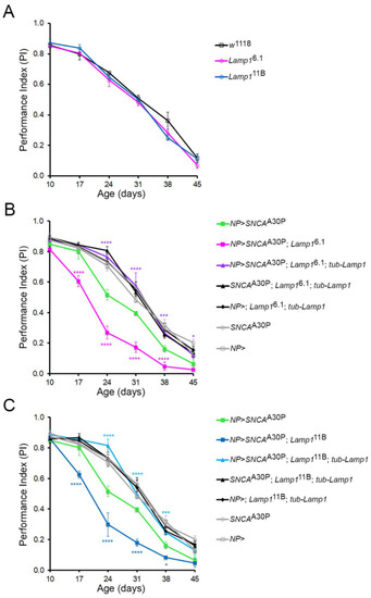
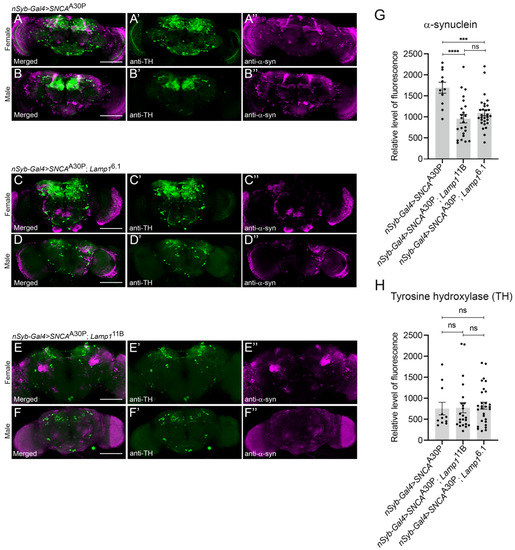
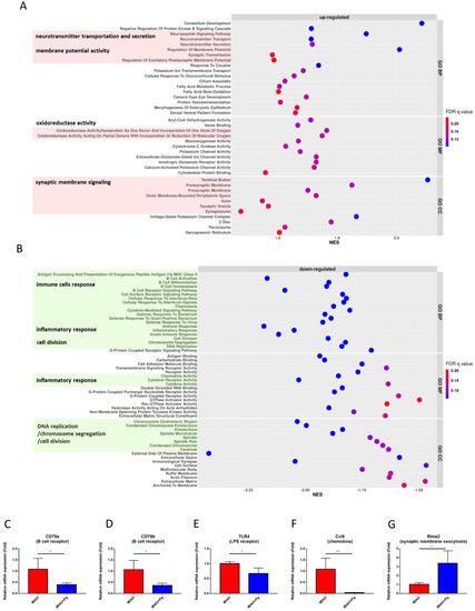
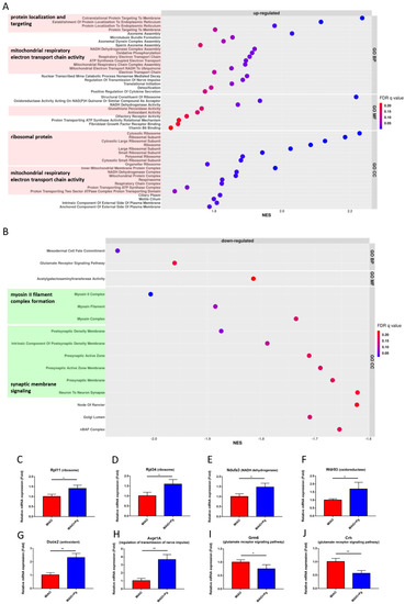
Parkinson disease (PD) is a common neurodegenerative condition affecting people predominantly at old age that is characterized by a progressive loss of midbrain dopaminergic neurons and by the accumulation of α-synuclein-containing intraneuronal inclusions known as Lewy bodies. Defects in cellular degradation processes such as the autophagy-lysosomal pathway are suspected to be involved in PD progression. The mammalian Lysosomal-associated membrane proteins LAMP1 and LAMP2 are transmembrane glycoproteins localized in lysosomes and late endosomes that are involved in autophagosome/lysosome maturation and function. Here, we show that the lack of Drosophila Lamp1, the homolog of LAMP1 and LAMP2, severely increased fly susceptibility to paraquat, a pro-oxidant compound known as a potential PD inducer in humans. Moreover, the loss of Lamp1 also exacerbated the progressive locomotor defects induced by the expression of PD-associated mutant α-synuclein A30P (α-synA30P) in dopaminergic neurons. Remarkably, the ubiquitous re-expression of Lamp1 in a mutant context fully suppressed all these defects and conferred significant resistance towards both PD factors above that of wild-type flies. Immunostaining analysis showed that the brain levels of α-synA30P were unexpectedly decreased in young adult Lamp1-deficient flies expressing this protein in comparison to non-mutant controls. This suggests that Lamp1 could neutralize α-synuclein toxicity by promoting the formation of non-pathogenic aggregates in neurons. Overall, our findings reveal a novel role for Drosophila Lamp1 in protecting against oxidative stress and α-synuclein neurotoxicity in PD models, thus furthering our understanding of the function of its mammalian homologs.
Full article
(This article belongs to the Special Issue Drosophila Models for Neurodegenerative Diseases: Achievements and Prospects)
►
Show Figures
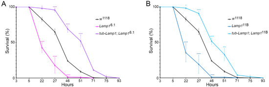
Figure 1


