by
Sumonto Mitra 1,* , Ruchi Gera 1, Julia Sundheimer 1, Marine Lemee 1, Lars U. Wahlberg 2, Bengt Linderoth 3, Maria Eriksdotter 1,4,† and Homira Behbahani 1,*,†
, Ruchi Gera 1, Julia Sundheimer 1, Marine Lemee 1, Lars U. Wahlberg 2, Bengt Linderoth 3, Maria Eriksdotter 1,4,† and Homira Behbahani 1,*,†
1
Division of Clinical Geriatrics, Center for Alzheimer Research, Department of NVS, Karolinska Institutet, 141 52 Huddinge, Sweden
2
Gloriana Therapeutics, Inc., Providence, RI 02885, USA
3
Department of Clinical Neuroscience, Karolinska Institutet, 171 77 Stockholm, Sweden
4
Theme Inflammation and Aging, Karolinska University Hospital, 141 86 Huddinge, Sweden
†
These authors supervised equally to this work.
Int. J. Mol. Sci. 2022, 23(16), 9011; https://doi.org/10.3390/ijms23169011 - 12 Aug 2022
Cited by 5 | Viewed by 3294
Abstract
There is no cure yet available for Alzheimer’s disease (AD). We recently optimized encapsulated cell biodelivery (ECB) devices releasing human mature nerve growth factor (hmNGF), termed ECB-NGF, to the basal forebrain of AD patients. The ECB-NGF delivery resulted in increased CSF cholinergic markers,
[...] Read more.
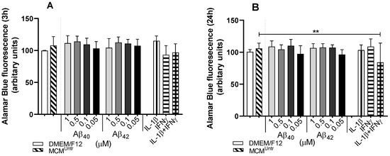
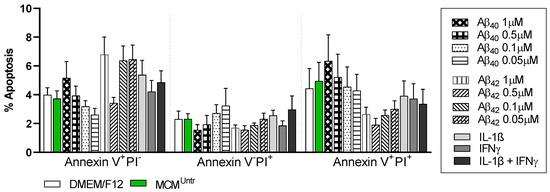
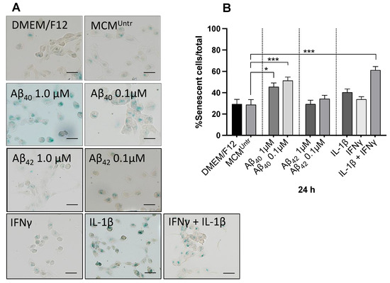
There is no cure yet available for Alzheimer’s disease (AD). We recently optimized encapsulated cell biodelivery (ECB) devices releasing human mature nerve growth factor (hmNGF), termed ECB-NGF, to the basal forebrain of AD patients. The ECB-NGF delivery resulted in increased CSF cholinergic markers, improved glucose metabolism, and positive effects on cognition in AD patients. However, some ECB-NGF implants showed altered hmNGF release post-explantation. To optimize the ECB-NGF platform for future therapeutic purposes, we initiated in-vitro optimization studies by exposing ECB-NGF devices to physiological factors present within the AD brain. We report here that microglia cells can impair hmNGF release from ECB-NGF devices in-vitro, which can be reversed by transferring the devices to fresh culture medium. Further, we exposed the hmNGF secreting human ARPE-19 cell line (NGC0211) to microglia (HMC3) conditioned medium (MCM; untreated or treated with IL-1β/IFNγ/Aβ40/Aβ42), and evaluated biochemical stress markers (ROS, GSH, ΔΨm, and Alamar Blue assay), cell death indicators (Annexin-V/PI), cell proliferation (CFSE retention and Ki67) and senescence markers (SA-β-gal) in NGC0211 cells. MCMs from activated microglia reduced cell proliferation and induced cell senescence in NGC0211 cells, which otherwise resist biochemical alterations and cell death. These data indicate a critical but reversible impact of activated microglia on NGC0211 cells.
Full article
(This article belongs to the Special Issue Advances in Neurodegenerative Diseases Research and Therapy)
▼
Show Figures

Figure 1


