by
Bożena Futoma-Kołoch 1,* , Bartłomiej Dudek 1, Katarzyna Kapczyńska 2, Eva Krzyżewska 2, Martyna Wańczyk 1, Kamila Korzekwa 1, Jacek Rybka 2, Elżbieta Klausa 3 and Gabriela Bugla-Płoskońska 1,*
, Bartłomiej Dudek 1, Katarzyna Kapczyńska 2, Eva Krzyżewska 2, Martyna Wańczyk 1, Kamila Korzekwa 1, Jacek Rybka 2, Elżbieta Klausa 3 and Gabriela Bugla-Płoskońska 1,*
1
Department of Microbiology, Institute of Genetics and Microbiology, University of Wrocław, 51-148 Wrocław, Poland
2
Department of Immunology of Infectious Diseases, Hirszfeld Institute of Immunology and Experimental Therapy, Polish Academy of Sciences, 53-114 Wrocław, Poland
3
Regional Centre of Transfusion Medicine and Blood Bank, 50-345 Wrocław, Poland
Int. J. Mol. Sci. 2017, 18(7), 1459; https://doi.org/10.3390/ijms18071459 - 11 Jul 2017
Cited by 8 | Viewed by 4013
Abstract
A new emerging phenomenon is the association between the incorrect use of biocides in the process of disinfection in farms and the emergence of cross-resistance in Salmonella populations. Adaptation of the microorganisms to the sub-inhibitory concentrations of the disinfectants is not clear, but
[...] Read more.
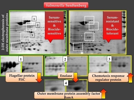
A new emerging phenomenon is the association between the incorrect use of biocides in the process of disinfection in farms and the emergence of cross-resistance in Salmonella populations. Adaptation of the microorganisms to the sub-inhibitory concentrations of the disinfectants is not clear, but may result in an increase of sensitivity or resistance to antibiotics, depending on the biocide used and the challenged Salmonella serovar. Exposure of five Salmonella enterica subsp. enterica serovar Senftenberg (S. Senftenberg) strains to triamine-containing disinfectant did not result in variants with resistance to antibiotics, but has changed their susceptibility to normal human serum (NHS). Three biocide variants developed reduced sensitivity to NHS in comparison to the sensitive parental strains, while two isolates lost their resistance to serum. For S. Senftenberg, which exhibited the highest triamine tolerance (6 × MIC) and intrinsic sensitivity to 22.5% and 45% NHS, a downregulation of flagellin and enolase has been demonstrated, which might suggest a lower adhesion and virulence of the bacteria. This is the first report demonstrating the influence of biocide tolerance on NHS resistance. In conclusion, there was a potential in S. Senftenberg to adjust to the conditions, where the biocide containing triamine was present. However, the adaptation did not result in the increase of antibiotic resistance, but manifested in changes within outer membrane proteins’ patterns. The strategy of bacterial membrane proteins’ analysis provides an opportunity to adjust the ways of infection treatments, especially when it is connected to the life-threating bacteremia caused by Salmonella species.
Full article
(This article belongs to the Special Issue Special Protein Molecules Computational Identification)
▼
Show Figures
Graphical abstract