by
Christine Delporte * , Angélic Bryla and Jason Perret
, Angélic Bryla and Jason Perret
Laboratory of Pathophysiological and Nutritional Biochemistry, Faculty of Medicine, Université Libre de Bruxelles, 808 Route de Lennik, Blg G/E CP 611, Brussels B-1070, Belgium
Int. J. Mol. Sci. 2016, 17(2), 166; https://doi.org/10.3390/ijms17020166 - 27 Jan 2016
Cited by 71 | Viewed by 9308
Abstract
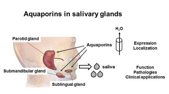
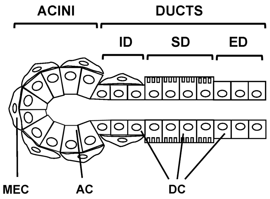
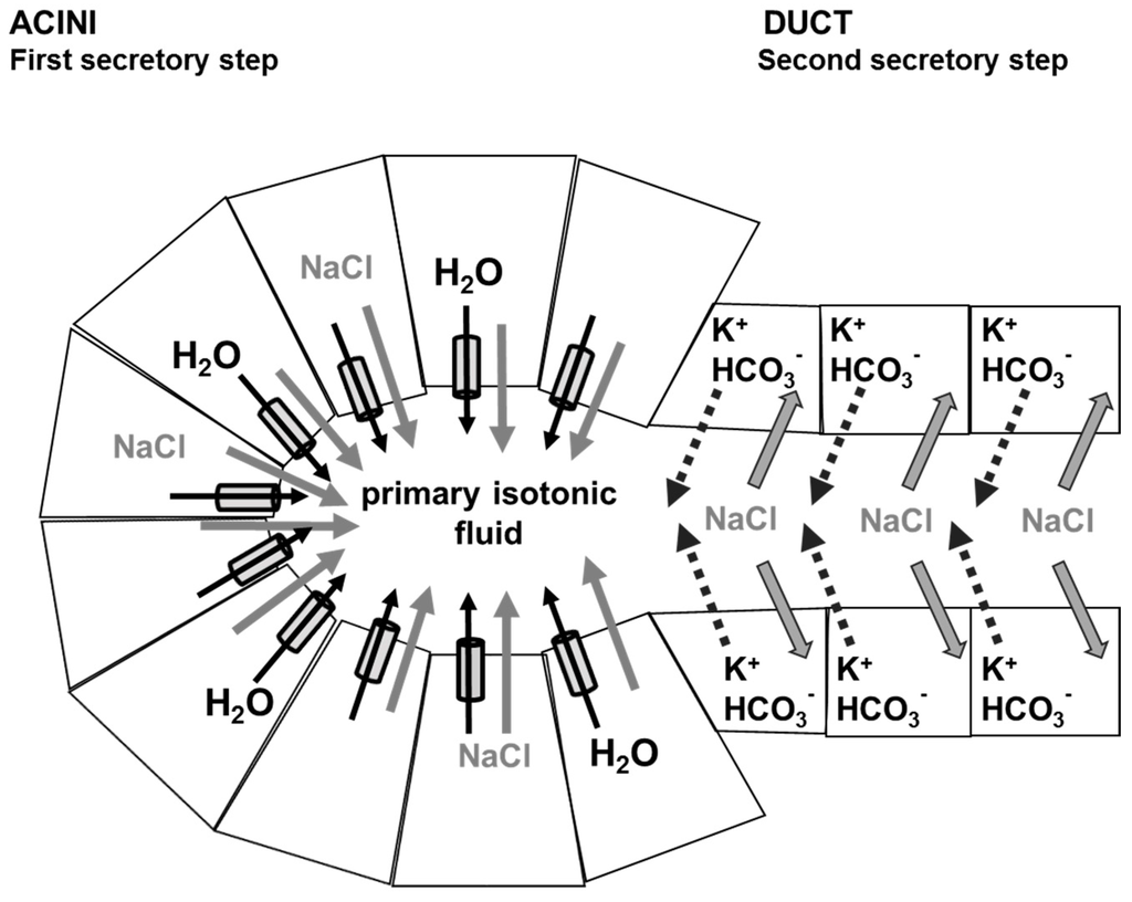
Salivary glands are involved in saliva secretion that ensures proper oral health. Aquaporins are expressed in salivary glands and play a major role in saliva secretion. This review will provide an overview of the salivary gland morphology and physiology of saliva secretion, and
[...] Read more.
Salivary glands are involved in saliva secretion that ensures proper oral health. Aquaporins are expressed in salivary glands and play a major role in saliva secretion. This review will provide an overview of the salivary gland morphology and physiology of saliva secretion, and focus on the expression, subcellular localization and role of aquaporins under physiological and pathophysiological conditions, as well as clinical applications involving aquaporins. This review is highlighting expression and localization of aquaporins in human, rat and mouse, the most studied species and is pointing out possible difference between major salivary glands, i.e., parotid, submandibular and sublingual glands.
Full article
(This article belongs to the Special Issue Aquaporin)
▼
Show Figures
Graphical abstract


