1
Institute of Chemistry and Biotechnology, Federal University of Alagoas (IQB/UFAL), Maceio, Alagoas 57072-900, Brazil
2
Federal Institute for Education, Science and Technology of Alagoas, Maceio, Alagoas 57020-600, Brazil
3
Faculty of Nutrition, Federal University of Alagoas (FANUT/UFAL), Maceio, Alagoas 57072-970, Brazil
4
Northeast Biotechnology Network (RENORBIO), Federal University of Alagoas (UFAL), Maceio, Alagoas 57072-900, Brazil
†
These authors contributed equally to this work.
Int. J. Mol. Sci. 2013, 14(10), 19846-19866; https://doi.org/10.3390/ijms141019846 - 1 Oct 2013
Cited by 62 | Viewed by 12346
Abstract
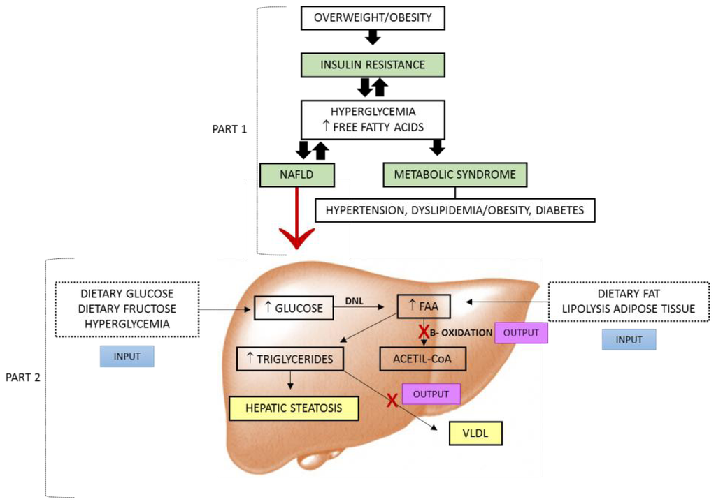
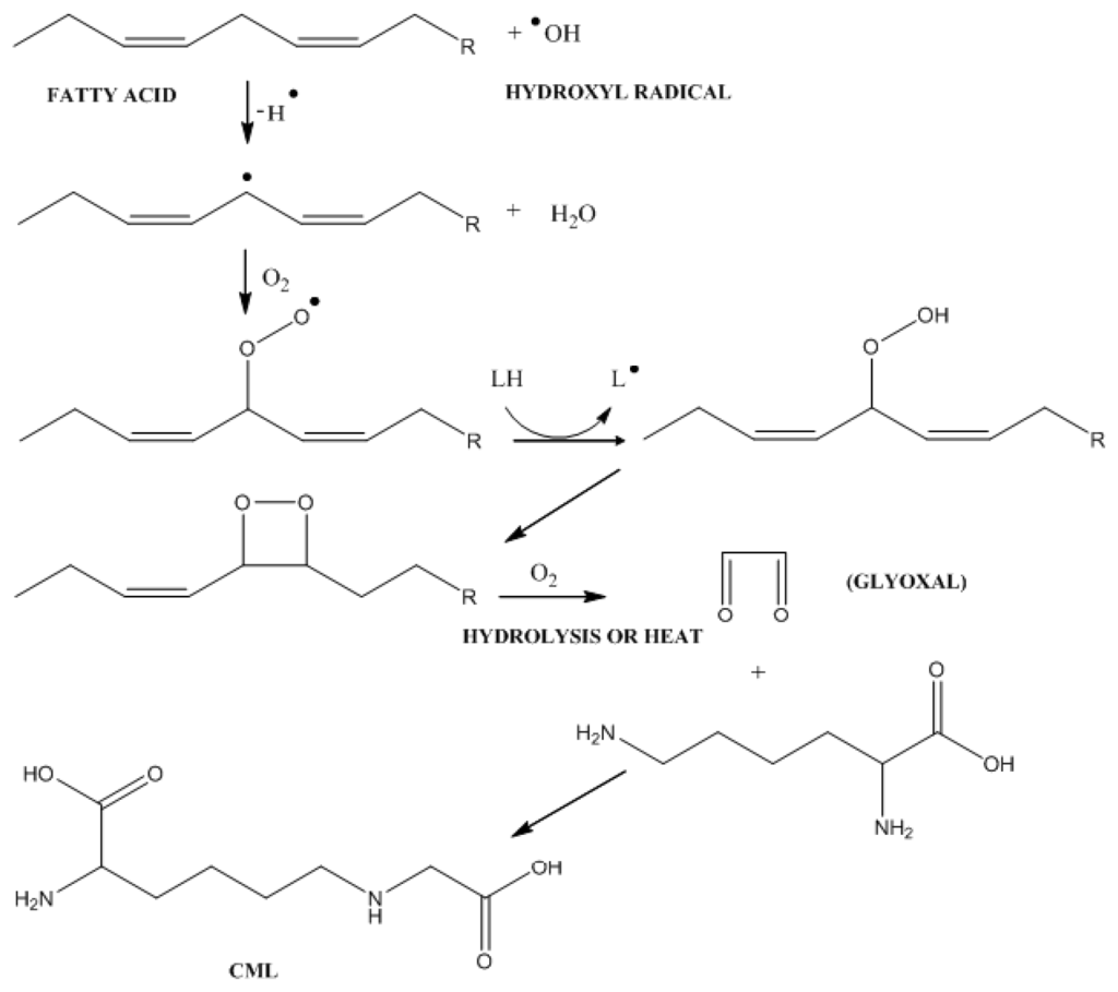
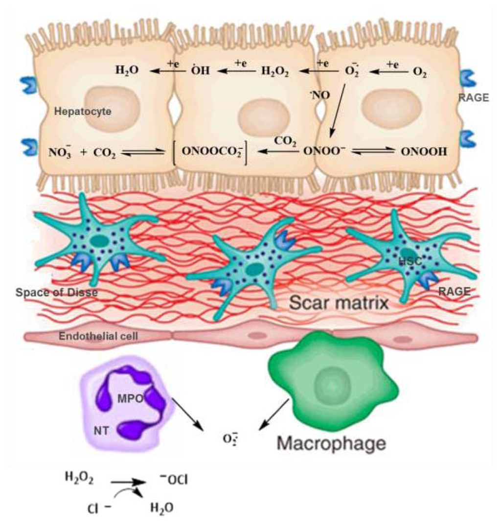
Advanced glycation end products (AGEs) are generated spontaneously in cells; however, under conditions of hyperglycemia and lipid peroxidation, their levels are higher than usual, which contribute to the development of diseases such as the nonalcoholic fatty liver disease (NAFLD). NAFLD is associated with
[...] Read more.
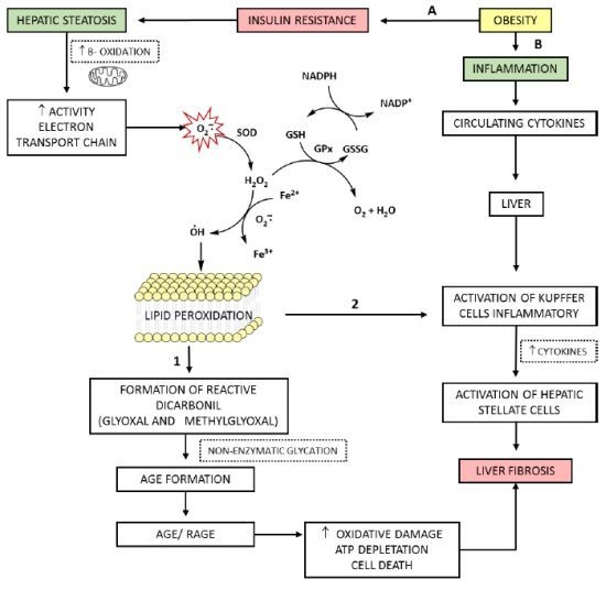
Advanced glycation end products (AGEs) are generated spontaneously in cells; however, under conditions of hyperglycemia and lipid peroxidation, their levels are higher than usual, which contribute to the development of diseases such as the nonalcoholic fatty liver disease (NAFLD). NAFLD is associated with oxidative stress (OS), which is linked to the transition of steatosis to steatohepatitis due to lipid peroxidation. The AGE-receptor interaction in hepatic stellate cells leads to an increase in reactive oxygen species and enhances the proliferation and activation of these cells, worsening liver fibrosis and disease progression. In this vicious cycle, there is production of (carboxymethyl)lysine, a biomarker for products of advanced glycation and lipid peroxidation, being a shared component between the two pathways. In this review, we aim to compile evidence to support the basic molecular mechanisms of AGEs and OS generation and their influence, independently or combined, on the evolution of NAFLD. The deeper understanding of the interrelations of AGEs + OS may help to elucidate the pathogenic pathways of NAFLD and to devise rational therapeutic interventions for this disease, with an expected positive impact on quality of life of patients.
Full article
(This article belongs to the Special Issue Non-Alcoholic Fatty Liver Disease Research)
▼
Show Figures
Graphical abstract


