by
Guido Carpino 1,2,*, Anastasia Renzi 1, Paolo Onori 1 and Eugenio Gaudio 1
and Eugenio Gaudio 1
1
Department of Anatomical, Histological, Forensic Medicine and Orthopedics Sciences, Sapienza University of Rome, Rome 00161, Italy
2
Department of Movement, Human and Health Sciences, University of Rome "Foro Italico", Piazza Lauro De Bosis 6, Rome 00135, Italy
Int. J. Mol. Sci. 2013, 14(10), 20112-20130; https://doi.org/10.3390/ijms141020112 - 9 Oct 2013
Cited by 49 | Viewed by 13076
Abstract
Nonalcoholic fatty liver disease (NAFLD) includes a spectrum of diseases ranging from simple fatty liver to nonalcoholic steatohepatitis, (NASH) which may progress to cirrhosis and hepatocellular carcinoma. NASH has been independently correlated with atherosclerosis progression and cardiovascular risk. NASH development is characterized by
[...] Read more.
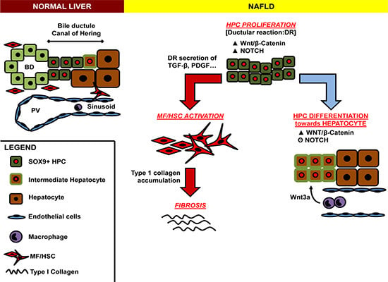
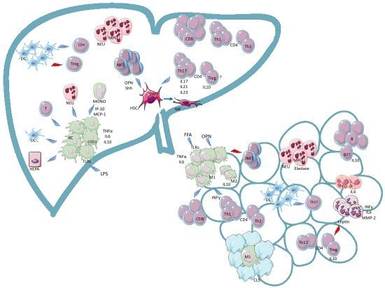
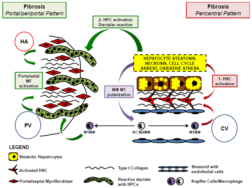
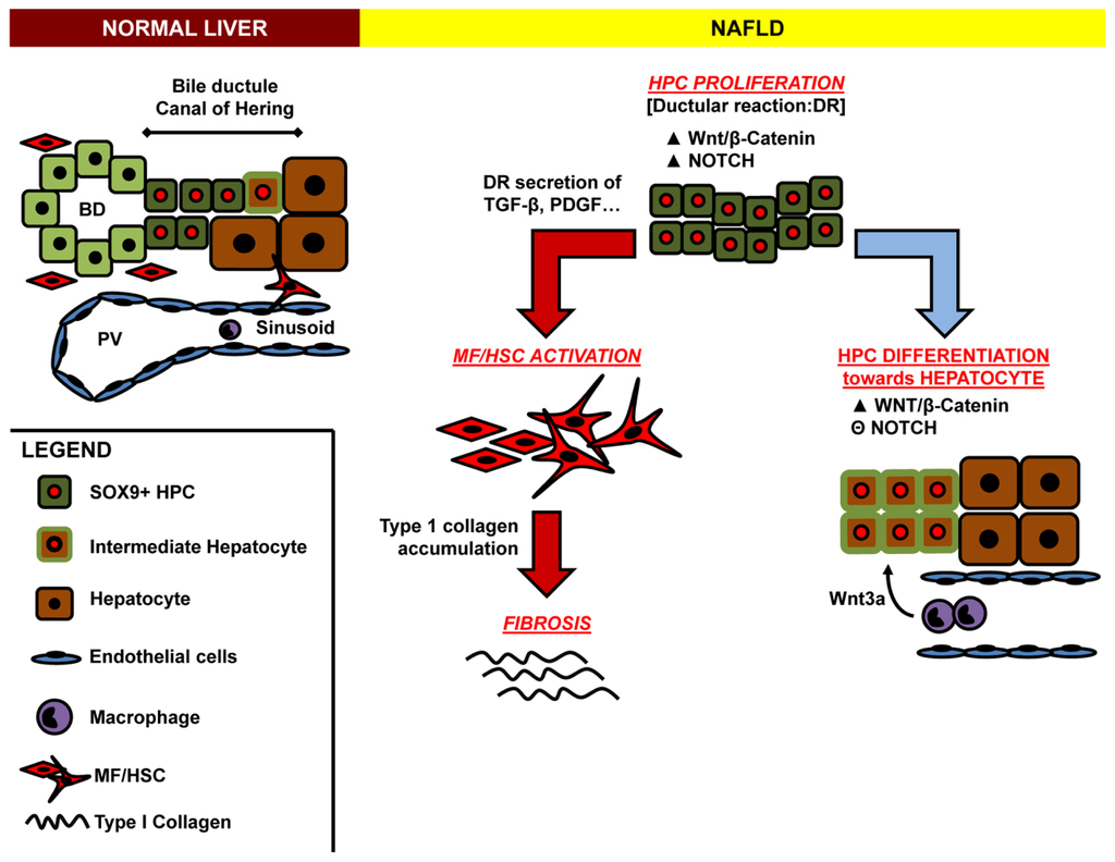
Nonalcoholic fatty liver disease (NAFLD) includes a spectrum of diseases ranging from simple fatty liver to nonalcoholic steatohepatitis, (NASH) which may progress to cirrhosis and hepatocellular carcinoma. NASH has been independently correlated with atherosclerosis progression and cardiovascular risk. NASH development is characterized by intricate interactions between resident and recruited cells that enable liver damage progression. The increasing general agreement is that the cross-talk between hepatocytes, hepatic stellate cells (HSCs) and macrophages in NAFLD has a main role in the derangement of lipid homeostasis, insulin resistance, danger recognition, immune tolerance response and fibrogenesis. Moreover, several evidences have suggested that hepatic stem/progenitor cell (HPCs) activation is a component of the adaptive response of the liver to oxidative stress in NAFLD. HPC activation determines the appearance of a ductular reaction. In NASH, ductular reaction is independently correlated with progressive portal fibrosis raising the possibility of a periportal fibrogenetic pathway for fibrogenesis that is parallel to the deposition of subsinusoidal collagen in zone 3 by HSCs. Recent evidences indicated that adipokines, a class of circulating factors, have a key role in the cross-talk among HSCs, HPCs and liver macrophages. This review will be focused on cellular cross-talk and the relative molecular networks which are at the base of NASH progression and fibrosis.
Full article
(This article belongs to the Special Issue Non-Alcoholic Fatty Liver Disease Research)
▼
Show Figures
Graphical abstract


