sensors-logo

Journal Browser

Journal Browser

Dedication to Professor Eiichi Tamiya: Over 30 Years of Outstanding Contributions to the Field of Sensors and Biosensors

A special issue of Sensors (ISSN 1424-8220).

Deadline for manuscript submissions: closed (30 September 2017) | Viewed by 127949

Special Issue Editors


E-Mail Website
Guest Editor
Department of Physical & Environmental Sciences, University of Toronto Scarborough, Room SW533, 1265 Military Trail, Toronto, ON M1C 1A4, Canada
Interests: electrochemical sensors; biosensors; immunosensors; DNA; nanomaterials
Special Issues, Collections and Topics in MDPI journals

E-Mail Website
Guest Editor
Department of Food Science and Biotechnology, Kagoshima University, 1-21-24 Korimoto, Kagoshima, Kagoshima Prefecture 890-8580, Japan
Interests: electrochemical and optical biosensors; bio-efficacy of natural products; biofunctional materials; point-of-need application; biomimetic membranes
Special Issues, Collections and Topics in MDPI journals

Special Issue Information

Dear Colleagues,

Eiichi Tamiya

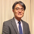
Professor Tamiya is a leading scholar in biochips, biosensors, and bioanalyses. He has a wide variety of fundamental, as well as topical research interests including nanotechnology-based bioscience and bioengineering, screening of new bacteria, enzymes and other bioactive molecules, design and creation of molecular recognition materials and environmental nanobiotechnology. After obtaining his Ph.D. degree in 1985 from the Tokyo Institute of Technology, Prof. Tamiya began his academic career as an Assistant Professor at the same university and became an Associate Professor at the Research Centre for Advanced Science and Technology in University of Tokyo, RCAST in 1988. Prof. Tamiya became a full Professor and one of the first faculty members to oversee the establishment of the School of Materials Science at Japan Advanced Institute of Science and Technology (JAIST) in 1993. Upon his leaving JAIST in 2007, he was honoured with a Prof. Emeritus position for his valuable contributions to the School and JAIST, at barely 55 years of age. He then joined Osaka University and established his Nano-Bioengineering Laboratory within the Graduate School of Engineering, where he continues to contribute to advancement of education, science and technology.

Professor Tamiya has made remarkable contributions to the field of biochips and biosensors, including the commercialization of numerous products. His work in electrochemical and optical biosensors brought upon noticeable impacts to the development of integrated microfluidic detection platforms. He has played an active leadership role in promoting research on various fields of biotechnology not only in Japan, but also on numerous international organizations and high-impact journals. He has co-authored over 323 original papers, 211 reviews and books, and 130 patents. Among the many awards received, the Osaka University Presidential Awards for Achievement (2014), and Invention Encouragement Award by the Japanese Ministry of Education, Culture, Sports and Science (2010), are the most recent honours. His passion and devotion to the field of sensors has inspired many scientists, especially young scholars globally.

We dedicate this Special Issue to celebrate Professor Eiichi Tamiya. In the last two decades, many of us, including the two Guest Editors of this Special Issue, were fortunate to study, work and collaborate with Professor Tamiya. While all of us witnessed his diligence and high originality in research as well as passion and devotion to education and professional service, we also enjoyed very much with his pleasant personality and friendship.

We appreciate the authors and reviewers for their valuable contributions to this Special Issue to celebrate Professor Eiichi Tamiya. For contributors who wish to add a personal message to Prof. Tamiya, please include the message (less than half A4, in blue font) at the beginning the submitted manuscript. These messages will be highlighted in some form, within this Special Issue. We believe that this Special Issue constitutes a timely celebration to the contributions of Professor Eiichi Tamiya, a highly respected scholar and researcher. Furthermore, we hope that the issue will also inspire scholars, especially young researchers, to continue the advancement in novel sensors.

Dr. Kagan Kerman,
Dr. Mun'delanji C. Vestergaard
Guest Editors

Published Papers (14 papers)

Order results
Result details
Select all
Export citation of selected articles as:

Research

Jump to: Review

11 pages, 3352 KiB  
Article
Electrochemical Immunoassay Using Open Circuit Potential Detection Labeled by Platinum Nanoparticles
by Kanokwan Charoenkitamorn, Phan Trong Tue, Keiko Kawai, Orawon Chailapakul and Yuzuru Takamura
Sensors 2018, 18(2), 444; https://doi.org/10.3390/s18020444 - 03 Feb 2018
Cited by 19 | Viewed by 6446
Abstract
In this work, a simple electrochemical immunoassay based on platinum nanoparticles (PtNPs) using open circuit potential (OCP) detection was developed. The detection of human chorionic gonadotropin hormone (hCG) as a model analyte, was demonstrated by direct electrical detection of PtNPs in hydrazine solution [...] Read more.
In this work, a simple electrochemical immunoassay based on platinum nanoparticles (PtNPs) using open circuit potential (OCP) detection was developed. The detection of human chorionic gonadotropin hormone (hCG) as a model analyte, was demonstrated by direct electrical detection of PtNPs in hydrazine solution using OCP measurement without any application of either potential or current to the system. Disposable screen-printed carbon electrodes (SPCEs) were utilized for the development of our immunosensor, which required a sample volume as small as 2 μL. After preparation of a sandwich-type immunosystem, hydrazine solution was dropped on the electrode’s surface, which was followed immediately by electrical detection using OCP. The change of the OCP signal originated from electrocatalytic oxidation of the hydrazine on PtNPs. Under the optimal conditions of a pH of 6.0 and a hydrazine concentration of 1 mM, a detection limit of 0.28 ng mL−1 and a linearity of 0–10 ng mL−1 were obtained. The PtNP-based OCP method is a simpler electrochemical detection procedure than those obtained from other electrochemical methods and has an acceptable sensitivity and reproducibility. The simplicity of the detection procedure and the cost-effectiveness of the disposable SPCE illustrate the attractive benefits of this sensor. Moreover, it could be applied to a simplified and miniaturized diagnostic system with minimal user manipulation. Full article
Show Figures

Figure 1

2844 KiB  
Article
Epigallocatechin Gallate-Modified Graphite Paste Electrode for Simultaneous Detection of Redox-Active Biomolecules
by Hashwin V. S. Ganesh, Meissam Noroozifar and Kagan Kerman
Sensors 2018, 18(1), 23; https://doi.org/10.3390/s18010023 - 22 Dec 2017
Cited by 22 | Viewed by 4434
Abstract
In this study, simultaneous electrochemical detection of ascorbic acid (AA), dopamine (DA), and uric acid (UA) was performed using a modified graphite paste electrode (MGPE) with epigallocatechin gallate (EGCG) and green tea (GT) powder. It was shown that the anodic peak current increased [...] Read more.
In this study, simultaneous electrochemical detection of ascorbic acid (AA), dopamine (DA), and uric acid (UA) was performed using a modified graphite paste electrode (MGPE) with epigallocatechin gallate (EGCG) and green tea (GT) powder. It was shown that the anodic peak current increased in comparison with that of the graphite paste electrode (GPE) in the cyclic voltammograms. The optimal pH for simultaneous determination of a quaternary mixture of AA–DA–UA was determined to be pH 2. The anodic peak potentials for a mixture containing AA–DA–UA were well separated from each other. The catalytic peak currents obtained at the surface of the MGPE/EGCG were linearly dependent on the AA, DA, and UA concentrations up to 23, 14, and 14 µM, respectively. The detection limits for AA, DA, and UA were 190, 90, and 70 nM, respectively. The analytical performance of this sensor has been evaluated for simultaneous detection of AA, DA, and UA in real samples. Finally, a modified electrode was prepared using GT and used for simultaneous determination of AA, DA, and UA. Based on the results, MPGE/GT showed two oxidation peaks at 0.43 and 0.6 V for DA and UA, respectively, without any oxidation peak for AA. The calibration curves at the surface of MGPE/GT were linear up to 14 µM with a detection limit of 0.18 and 0.33 µM for DA and UA, respectively. MGPEs provide a promising platform for the future development of sensors for multiplexed electrochemical detection of clinically important analytes. Full article
Show Figures

Figure 1

3137 KiB  
Article
Dual-Color Fluorescence Imaging of EpCAM and EGFR in Breast Cancer Cells with a Bull’s Eye-Type Plasmonic Chip
by Shota Izumi, Shohei Yamamura, Naoko Hayashi, Mana Toma and Keiko Tawa
Sensors 2017, 17(12), 2942; https://doi.org/10.3390/s17122942 - 19 Dec 2017
Cited by 10 | Viewed by 5270
Abstract
Surface plasmon field-enhanced fluorescence microscopic observation of a live breast cancer cell was performed with a plasmonic chip. Two cell lines, MDA-MB-231 and Michigan Cancer Foundation-7 (MCF-7), were selected as breast cancer cells, with two kinds of membrane protein, epithelial cell adhesion molecule [...] Read more.
Surface plasmon field-enhanced fluorescence microscopic observation of a live breast cancer cell was performed with a plasmonic chip. Two cell lines, MDA-MB-231 and Michigan Cancer Foundation-7 (MCF-7), were selected as breast cancer cells, with two kinds of membrane protein, epithelial cell adhesion molecule (EpCAM) and epidermal growth factor receptor (EGFR), observed in both cells. The membrane proteins are surface markers used to differentiate and classify breast cancer cells. EGFR and EpCAM were detected with Alexa Fluor® 488-labeled anti-EGFR antibody (488-EGFR) and allophycocyanin (APC)-labeled anti-EpCAM antibody (APC-EpCAM), respectively. In MDA-MB231 cells, three-fold plus or minus one and seven-fold plus or minus two brighter fluorescence of 488-EGFR were observed on the 480-nm pitch and the 400-nm pitch compared with that on a glass slide. Results show the 400-nm pitch is useful. Dual-color fluorescence of 488-EGFR and APC-EpCAM in MDA-MB231 was clearly observed with seven-fold plus or minus two and nine-fold plus or minus three, respectively, on the 400-nm pitch pattern of a plasmonic chip. Therefore, the 400-nm pitch contributed to the dual-color fluorescence enhancement for these wavelengths. An optimal grating pitch of a plasmonic chip improved a fluorescence image of membrane proteins with the help of the surface plasmon-enhanced field. Full article
Show Figures

Figure 1

6395 KiB  
Article
A Quartz Crystal Microbalance Immunosensor for Stem Cell Selection and Extraction
by Ornella Maglio, Salvatore Costanzo, Rosaria Cercola, Gerardo Zambrano, Marco Mauro, Raffaele Battaglia, Gianluca Ferrini, Flavia Nastri, Vincenzo Pavone and Angela Lombardi
Sensors 2017, 17(12), 2747; https://doi.org/10.3390/s17122747 - 28 Nov 2017
Cited by 22 | Viewed by 13463
Abstract
A cost-effective immunosensor for the detection and isolation of dental pulp stem cells (DPSCs) based on a quartz crystal microbalance (QCM) has been developed. The recognition mechanism relies on anti-CD34 antibodies, DPSC-specific monoclonal antibodies that are anchored on the surface of the quartz [...] Read more.
A cost-effective immunosensor for the detection and isolation of dental pulp stem cells (DPSCs) based on a quartz crystal microbalance (QCM) has been developed. The recognition mechanism relies on anti-CD34 antibodies, DPSC-specific monoclonal antibodies that are anchored on the surface of the quartz crystals. Due to its high specificity, real time detection, and low cost, the proposed technology has a promising potential in the field of cell biology, for the simultaneous detection and sorting of stem cells from heterogeneous cell samples. The QCM surface was properly tailored through a biotinylated self-assembled monolayer (SAM). The biotin–avidin interaction was used to immobilize the biotinylated anti-CD34 antibody on the gold-coated quartz crystal. After antibody immobilization, a cellular pellet, with a mixed cell population, was analyzed; the results indicated that the developed QCM immunosensor is highly specific, being able to detect and sort only CD34+ cells. Our study suggests that the proposed technology can detect and efficiently sort any kind of cell from samples with high complexity, being simple, selective, and providing for more convenient and time-saving operations. Full article
Show Figures

Figure 1

2626 KiB  
Article
Mediator Preference of Two Different FAD-Dependent Glucose Dehydrogenases Employed in Disposable Enzyme Glucose Sensors
by Noya Loew, Wakako Tsugawa, Daichi Nagae, Katsuhiro Kojima and Koji Sode
Sensors 2017, 17(11), 2636; https://doi.org/10.3390/s17112636 - 16 Nov 2017
Cited by 32 | Viewed by 6947
Abstract
Most commercially available electrochemical enzyme sensor strips for the measurement of blood glucose use an artificial electron mediator to transfer electrons from the active side of the enzyme to the electrode. One mediator recently gaining attention for commercial sensor strips is hexaammineruthenium(III) chloride. [...] Read more.
Most commercially available electrochemical enzyme sensor strips for the measurement of blood glucose use an artificial electron mediator to transfer electrons from the active side of the enzyme to the electrode. One mediator recently gaining attention for commercial sensor strips is hexaammineruthenium(III) chloride. In this study, we investigate and compare the preference of enzyme electrodes with two different FAD-dependent glucose dehydrogenases (FADGDHs) for the mediators hexaammineruthenium(III) chloride, potassium ferricyanide (the most common mediator in commercial sensor strips), and methoxy phenazine methosulfate (mPMS). One FADGDH is a monomeric fungal enzyme, and the other a hetero-trimeric bacterial enzyme. With the latter, which contains a heme-subunit facilitating the electron transfer, similar response currents are obtained with hexaammineruthenium(III), ferricyanide, and mPMS (6.8 µA, 7.5 µA, and 6.4 µA, respectively, for 10 mM glucose). With the fungal FADGDH, similar response currents are obtained with the negatively charged ferricyanide and the uncharged mPMS (5.9 µA and 6.7 µA, respectively, for 10 mM glucose), however, no response current is obtained with hexaammineruthenium(III), which has a strong positive charge. These results show that access of even very small mediators with strong charges to a buried active center can be almost completely blocked by the protein. Full article
Show Figures

Figure 1

1878 KiB  
Article
Separation and Analysis of Adherent and Non-Adherent Cancer Cells Using a Single-Cell Microarray Chip
by Shohei Yamamura, Eriko Yamada, Fukiko Kimura, Kumiko Miyajima and Hajime Shigeto
Sensors 2017, 17(10), 2410; https://doi.org/10.3390/s17102410 - 21 Oct 2017
Cited by 8 | Viewed by 6240
Abstract
A new single-cell microarray chip was designed and developed to separate and analyze single adherent and non-adherent cancer cells. The single-cell microarray chip is made of polystyrene with over 60,000 microchambers of 10 different size patterns (31–40 µm upper diameter, 11–20 µm lower [...] Read more.
A new single-cell microarray chip was designed and developed to separate and analyze single adherent and non-adherent cancer cells. The single-cell microarray chip is made of polystyrene with over 60,000 microchambers of 10 different size patterns (31–40 µm upper diameter, 11–20 µm lower diameter). A drop of suspension of adherent carcinoma (NCI-H1650) and non-adherent leukocyte (CCRF-CEM) cells was placed onto the chip, and single-cell occupancy of NCI-H1650 and CCRF-CEM was determined to be 79% and 84%, respectively. This was achieved by controlling the chip design and surface treatment. Analysis of protein expression in single NCI-H1650 and CCRF-CEM cells was performed on the single-cell microarray chip by multi-antibody staining. Additionally, with this system, we retrieved positive single cells from the microchambers by a micromanipulator. Thus, this system demonstrates the potential for easy and accurate separation and analysis of various types of single cells. Full article
Show Figures

Figure 1

2539 KiB  
Communication
A Multi-Fluorescent DNA/Graphene Oxide Conjugate Sensor for Signature-Based Protein Discrimination
by Shunsuke Tomita, Sayaka Ishihara and Ryoji Kurita
Sensors 2017, 17(10), 2194; https://doi.org/10.3390/s17102194 - 23 Sep 2017
Cited by 21 | Viewed by 7638
Abstract
Signature-based protein sensing has recently emerged as a promising prospective alternative to conventional lock-and-key methods. However, most of the current examples require the measurement of optical signals from spatially-separated materials for the generation of signatures. Herein, we present a new approach for the [...] Read more.
Signature-based protein sensing has recently emerged as a promising prospective alternative to conventional lock-and-key methods. However, most of the current examples require the measurement of optical signals from spatially-separated materials for the generation of signatures. Herein, we present a new approach for the construction of multi-fluorescent sensing systems with high accessibility and tunability, which allows generating protein fluorescent signatures from a single microplate well. This approach is based on conjugates between nano-graphene oxide (nGO) and three single-stranded DNAs (ssDNAs) that exhibit different sequences and fluorophores. Initially, the three fluorophore-modified ssDNAs were quenched simultaneously by binding to nGO. Subsequent addition of analyte proteins caused a partial recovery in fluorescent intensity of the individual ssDNAs. Based on this scheme, we have succeeded in acquiring fluorescence signatures unique to (i) ten proteins that differ with respect to pI and molecular weight and (ii) biochemical marker proteins in the presence of interferent human serum. Pattern-recognition methods demonstrated high levels of discrimination for this system. The high discriminatory power and simple format of this sensor system should enable an easy and fast evaluation of proteins and protein mixtures. Full article
Show Figures

Figure 1

3669 KiB  
Article
Real-Time Amperometric Recording of Extracellular H2O2 in the Brain of Immunocompromised Mice: An In Vitro, Ex Vivo and In Vivo Characterisation Study
by Caroline H. Reid and Niall J. Finnerty
Sensors 2017, 17(7), 1596; https://doi.org/10.3390/s17071596 - 08 Jul 2017
Cited by 6 | Viewed by 4364
Abstract
We detail an extensive characterisation study on a previously described dual amperometric H2O2 biosensor consisting of H2O2 detection (blank) and degradation (catalase) electrodes. In vitro investigations demonstrated excellent H2O2 sensitivity and selectivity against the [...] Read more.
We detail an extensive characterisation study on a previously described dual amperometric H2O2 biosensor consisting of H2O2 detection (blank) and degradation (catalase) electrodes. In vitro investigations demonstrated excellent H2O2 sensitivity and selectivity against the interferent, ascorbic acid. Ex vivo studies were performed to mimic physiological conditions prior to in vivo deployment. Exposure to brain tissue homogenate identified reliable sensitivity and selectivity recordings up to seven days for both blank and catalase electrodes. Furthermore, there was no compromise in pre- and post-implanted catalase electrode sensitivity in ex vivo mouse brain. In vivo investigations performed in anaesthetised mice confirmed the ability of the H2O2 biosensor to detect increases in amperometric current following locally perfused/infused H2O2 and antioxidant inhibitors mercaptosuccinic acid and sodium azide. Subsequent recordings in freely moving mice identified negligible effects of control saline and sodium ascorbate interference injections on amperometric H2O2 current. Furthermore, the stability of the amperometric current was confirmed over a five-day period and analysis of 24-h signal recordings identified the absence of diurnal variations in amperometric current. Collectively, these findings confirm the biosensor current responds in vivo to increasing exogenous and endogenous H2O2 and tentatively supports measurement of H2O2 dynamics in freely moving NOD SCID mice. Full article
Show Figures

Figure 1

Review

Jump to: Research

28 pages, 5460 KiB  
Review
Trends and Advances in Electrochemiluminescence Nanobiosensors
by Mohammad Rizwan, Noor Faizah Mohd-Naim and Minhaz Uddin Ahmed
Sensors 2018, 18(1), 166; https://doi.org/10.3390/s18010166 - 09 Jan 2018
Cited by 87 | Viewed by 9737
Abstract
The rapid and increasing use of the nanomaterials (NMs), nanostructured materials (NSMs), metal nanoclusters (MNCs) or nanocomposites (NCs) in the development of electrochemiluminescence (ECL) nanobiosensors is a significant area of study for its massive potential in the practical application of nanobiosensor fabrication. Recently, [...] Read more.
The rapid and increasing use of the nanomaterials (NMs), nanostructured materials (NSMs), metal nanoclusters (MNCs) or nanocomposites (NCs) in the development of electrochemiluminescence (ECL) nanobiosensors is a significant area of study for its massive potential in the practical application of nanobiosensor fabrication. Recently, NMs or NSMs (such as AuNPs, AgNPs, Fe3O4, CdS QDs, OMCs, graphene, CNTs and fullerenes) or MNCs (such as Au, Ag, and Pt) or NCs of both metallic and non-metallic origin are being employed for various purposes in the construction of biosensors. In this review, we have selected recently published articles (from 2014–2017) on the current development and prospects of label-free or direct ECL nanobiosensors that incorporate NCs, NMs, NSMs or MNCs. Full article
Show Figures

Graphical abstract

3271 KiB  
Review
Carbon-Based Nanomaterials in Biomass-Based Fuel-Fed Fuel Cells
by Le Quynh Hoa, Mun’delanji C. Vestergaard and Eiichi Tamiya
Sensors 2017, 17(11), 2587; https://doi.org/10.3390/s17112587 - 10 Nov 2017
Cited by 22 | Viewed by 8876
Abstract
Environmental and sustainable economical concerns are generating a growing interest in biofuels predominantly produced from biomass. It would be ideal if an energy conversion device could directly extract energy from a sustainable energy resource such as biomass. Unfortunately, up to now, such a [...] Read more.
Environmental and sustainable economical concerns are generating a growing interest in biofuels predominantly produced from biomass. It would be ideal if an energy conversion device could directly extract energy from a sustainable energy resource such as biomass. Unfortunately, up to now, such a direct conversion device produces insufficient power to meet the demand of practical applications. To realize the future of biofuel-fed fuel cells as a green energy conversion device, efforts have been devoted to the development of carbon-based nanomaterials with tunable electronic and surface characteristics to act as efficient metal-free electrocatalysts and/or as supporting matrix for metal-based electrocatalysts. We present here a mini review on the recent advances in carbon-based catalysts for each type of biofuel-fed/biofuel cells that directly/indirectly extract energy from biomass resources, and discuss the challenges and perspectives in this developing field. Full article
Show Figures

Figure 1

5633 KiB  
Review
Disease-Related Detection with Electrochemical Biosensors: A Review
by Ying Huang, Jin Xu, Junjie Liu, Xiangyang Wang and Bin Chen
Sensors 2017, 17(10), 2375; https://doi.org/10.3390/s17102375 - 17 Oct 2017
Cited by 109 | Viewed by 11188
Abstract
Rapid diagnosis of diseases at their initial stage is critical for effective clinical outcomes and promotes general public health. Classical in vitro diagnostics require centralized laboratories, tedious work and large, expensive devices. In recent years, numerous electrochemical biosensors have been developed and proposed [...] Read more.
Rapid diagnosis of diseases at their initial stage is critical for effective clinical outcomes and promotes general public health. Classical in vitro diagnostics require centralized laboratories, tedious work and large, expensive devices. In recent years, numerous electrochemical biosensors have been developed and proposed for detection of various diseases based on specific biomarkers taking advantage of their features, including sensitivity, selectivity, low cost and rapid response. This article reviews research trends in disease-related detection with electrochemical biosensors. Focus has been placed on the immobilization mechanism of electrochemical biosensors, and the techniques and materials used for the fabrication of biosensors are introduced in details. Various biomolecules used for different diseases have been listed. Besides, the advances and challenges of using electrochemical biosensors for disease-related applications are discussed. Full article
Show Figures

Figure 1

3435 KiB  
Review
Plasmonic Nanomaterial-Based Optical Biosensing Platforms for Virus Detection
by Jaewook Lee, Kenshin Takemura and Enoch Y. Park
Sensors 2017, 17(10), 2332; https://doi.org/10.3390/s17102332 - 13 Oct 2017
Cited by 38 | Viewed by 7580
Abstract
Plasmonic nanomaterials (P-NM) are receiving attention due to their excellent properties, which include surface-enhanced Raman scattering (SERS), localized surface plasmon resonance (LSPR) effects, plasmonic resonance energy transfer (PRET), and magneto optical (MO) effects. To obtain such plasmonic properties, many nanomaterials have been developed, [...] Read more.
Plasmonic nanomaterials (P-NM) are receiving attention due to their excellent properties, which include surface-enhanced Raman scattering (SERS), localized surface plasmon resonance (LSPR) effects, plasmonic resonance energy transfer (PRET), and magneto optical (MO) effects. To obtain such plasmonic properties, many nanomaterials have been developed, including metal nanoparticles (MNP), bimetallic nanoparticles (bMNP), MNP-decorated carbon nanotubes, (MNP-CNT), and MNP-modified graphene (MNP-GRP). These P-NMs may eventually be applied to optical biosensing systems due to their unique properties. Here, probe biomolecules, such as antibodies (Ab), probe DNA, and probe aptamers, were modified on the surface of plasmonic materials by chemical conjugation and thiol chemistry. The optical property change in the plasmonic nanomaterials was monitored based on the interaction between the probe biomolecules and target virus. After bioconjugation, several optical properties, including fluorescence, plasmonic absorbance, and diffraction angle, were changed to detect the target biomolecules. This review describes several P-NMs as potential candidates of optical sensing platforms and introduces various applications in the optical biosensing field. Full article
Show Figures

Figure 1

5048 KiB  
Review
Graphene-Based Materials for Biosensors: A Review
by Phitsini Suvarnaphaet and Suejit Pechprasarn
Sensors 2017, 17(10), 2161; https://doi.org/10.3390/s17102161 - 21 Sep 2017
Cited by 307 | Viewed by 23152
Abstract
The advantages conferred by the physical, optical and electrochemical properties of graphene-based nanomaterials have contributed to the current variety of ultrasensitive and selective biosensor devices. In this review, we present the points of view on the intrinsic properties of graphene and its surface [...] Read more.
The advantages conferred by the physical, optical and electrochemical properties of graphene-based nanomaterials have contributed to the current variety of ultrasensitive and selective biosensor devices. In this review, we present the points of view on the intrinsic properties of graphene and its surface engineering concerned with the transduction mechanisms in biosensing applications. We explain practical synthesis techniques along with prospective properties of the graphene-based materials, which include the pristine graphene and functionalized graphene (i.e., graphene oxide (GO), reduced graphene oxide (RGO) and graphene quantum dot (GQD). The biosensing mechanisms based on the utilization of the charge interactions with biomolecules and/or nanoparticle interactions and sensing platforms are also discussed, and the importance of surface functionalization in recent up-to-date biosensors for biological and medical applications. Full article
Show Figures

Figure 1

5052 KiB  
Review
Whispering-Gallery Mode Resonators for Detecting Cancer
by Weeratouch Pongruengkiat and Suejit Pechprasarn
Sensors 2017, 17(9), 2095; https://doi.org/10.3390/s17092095 - 13 Sep 2017
Cited by 22 | Viewed by 10719
Abstract
Optical resonators are sensors well known for their high sensitivity and fast response time. These sensors have a wide range of applications, including in the biomedical fields, and cancer detection is one such promising application. Sensor diagnosis currently has many limitations, such as [...] Read more.
Optical resonators are sensors well known for their high sensitivity and fast response time. These sensors have a wide range of applications, including in the biomedical fields, and cancer detection is one such promising application. Sensor diagnosis currently has many limitations, such as being expensive, highly invasive, and time-consuming. New developments are welcomed to overcome these limitations. Optical resonators have high sensitivity, which enable medical testing to detect disease in the early stage. Herein, we describe the principle of whispering-gallery mode and ring optical resonators. We also add to the knowledge of cancer biomarker diagnosis, where we discuss the application of optical resonators for specific biomarkers. Lastly, we discuss advancements in optical resonators for detecting cancer in terms of their ability to detect small amounts of cancer biomarkers. Full article
Show Figures

Figure 1

Back to TopTop