Journal Description
Radiation
Radiation
is an international, peer-reviewed, open access journal on scientific advances and applications of radiotherapy, immunotherapy, radiology and radiation technologies across multiple fields, published quarterly online by MDPI.
- Open Access— free for readers, with article processing charges (APC) paid by authors or their institutions.
- High Visibility: indexed within Scopus and other databases.
- Journal Rank: CiteScore - Q2 (Radiation)
- Rapid Publication: manuscripts are peer-reviewed and a first decision is provided to authors approximately 21 days after submission; acceptance to publication is undertaken in 4.6 days (median values for papers published in this journal in the first half of 2026).
- Recognition of Reviewers: APC discount vouchers, optional signed peer review, and reviewer names published annually in the journal.
- Radiation is a companion journal of Cancers.
Latest Articles
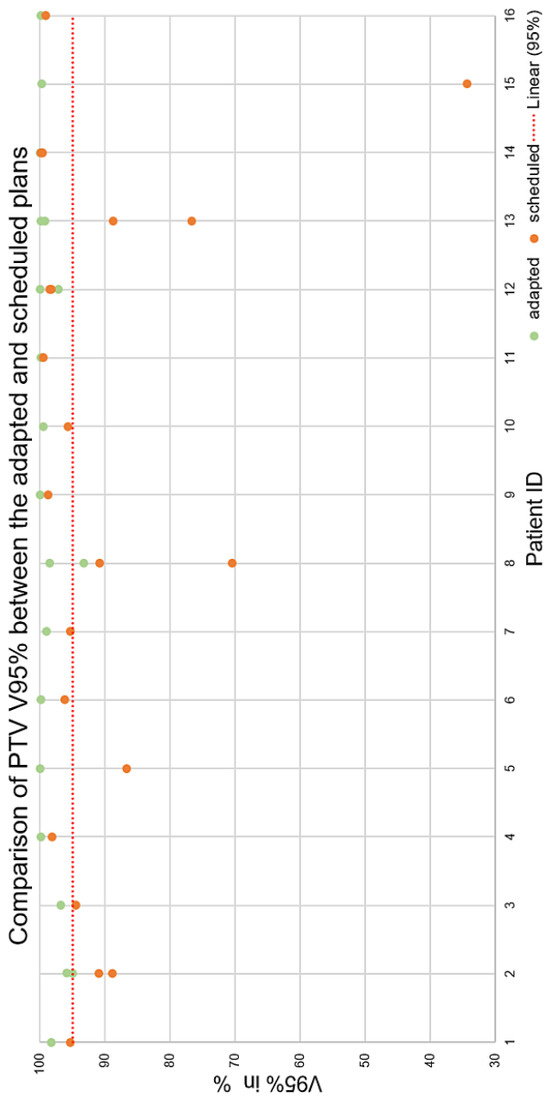
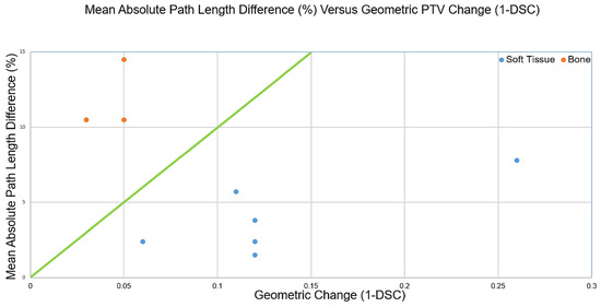
Adaptive Emergency Radiotherapy Using Diagnostic Computed Tomography Imaging: Initial Results from a Retrospective Geometric Evaluation and Clinical Considerations
Radiation 2026, 6(3), 34; https://doi.org/10.3390/radiation6030034 - 1 Sep 2026
Abstract
Background: Radiotherapy commonly requires a computed tomography (CT) dataset for treatment planning. Artificial intelligence-supported radiotherapy systems now enable online adaptive workflows that reduce uncertainties resulting from the use of diagnostic instead of planning CT images. The aim was to identify patient groups that
[...] Read more.
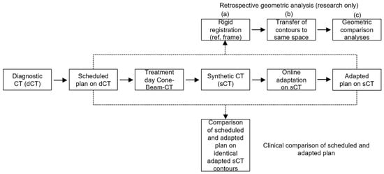
Background: Radiotherapy commonly requires a computed tomography (CT) dataset for treatment planning. Artificial intelligence-supported radiotherapy systems now enable online adaptive workflows that reduce uncertainties resulting from the use of diagnostic instead of planning CT images. The aim was to identify patient groups that may particularly benefit from adaptive emergency treatments. Methods: In this retrospective exploratory study, 16 patients comprising 21 planning target volumes (PTVs) were evaluated. PTVs were classified as predominantly soft-tissue or bony. To verify the geometric agreement between scheduled and adapted treatment plans, these subgroups were analyzed using absolute planning target volume change, the Dice similarity coefficient (DSC), and the 95th percentile Hausdorff distance, and their association with V95% PTV coverage was evaluated. Results: Bony targets showed higher geometric reproducibility than soft-tissue-dominant lesions, with higher median DSC values (0.96 vs. 0.88) and lower median HD95 values (2.6 mm vs. 5.5 mm). Online adaptation improved coverage in 20 of 21 PTVs, while predominantly soft-tissue PTVs showed potentially greater benefit from online adaptation. Conclusions: Soft-tissue-dominant PTVs showed greater geometric variability and potentially greater benefit from adaptation, whereas selected bone-dominant cases may be suitable for faster conventional workflows. Given the small exploratory cohort, these results require further confirmation.
Full article
(This article belongs to the Section Radiation and Its Application in Oncology and Radiation Protection)
►
Show Figures
Open AccessArticle
Economic Evaluation of Lutetium-177 (177Lu)–PSMA-617 in Patients with Metastatic Castration-Resistant Prostate Cancer in Italy
by
Jacopo Giuliani, Emilia Durante, Giuseppe Napoli, Marina Tommasi, Giuseppe Aprile and Francesco Fiorica
Radiation 2026, 6(3), 33; https://doi.org/10.3390/radiation6030033 - 1 Sep 2026
Abstract
Introduction: The increasing costs of cancer care have raised the need for economic evaluations that integrate treatment efficacy and resource utilization. This study aimed to assess the economic value of Lutetium-177 (177Lu)–prostate-specific membrane antigen (PSMA)-617 in patients with PSMA-positive metastatic castration-resistant
[...] Read more.
Introduction: The increasing costs of cancer care have raised the need for economic evaluations that integrate treatment efficacy and resource utilization. This study aimed to assess the economic value of Lutetium-177 (177Lu)–prostate-specific membrane antigen (PSMA)-617 in patients with PSMA-positive metastatic castration-resistant prostate cancer (mCRPC) from an Italian healthcare perspective. Patients and Methods: A cost-effectiveness analysis was performed using clinical efficacy data from randomized controlled trials (RCTs) evaluating 177Lu-PSMA-617. Two scenarios were considered. Scenario 1 evaluated 177Lu-PSMA-617 plus standard care (SC) versus SC alone using pivotal phase III trial data. Scenario 2 compared 177Lu-PSMA-617 with chemotherapy and androgen receptor pathway inhibitors (ARPIs) using phase II and III RCTs. Drug costs were based on Italian ex-factory prices. In addition, an exploratory three-state Markov cost–utility model including progression-free survival (PFS), progressive disease, and death was developed using data from the VISION trial. Results: In Scenario 1, 730 patients were analyzed. The ESMO-MCBS grade was 3, and the incremental cost per month of PFS gained with 177Lu-PSMA-617 versus SC was €20,906. In Scenario 2, 651 patients were included. The monthly cost per PFS gained with 177Lu-PSMA-617 was €19,017 versus enzalutamide and €20,565 versus abiraterone. In the exploratory cost–utility model, estimated QALYs were 1.02 with 177Lu-PSMA-617 and 0.64 with SC, corresponding to an incremental gain of 0.38 QALYs. At an incremental cost of approximately €32,000 per patient, the resulting model-based ICER was approximately €85,000/QALY. Conclusions:177Lu-PSMA-617 provides meaningful clinical benefit in appropriately selected patients with PSMA-positive mCRPC, but this benefit is associated with substantial incremental treatment costs. The exploratory model-based ICER was sensitive to treatment duration and other model assumptions. These findings support careful assessment of the clinical value and economic sustainability of 177Lu-PSMA-617 in the Italian healthcare setting. Further studies incorporating real-world resource utilization, treatment-related costs, post-progression therapies, and prospectively collected quality-of-life data are warranted.
Full article
Open AccessArticle
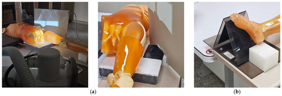
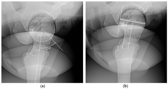
Fabrication of a 3D-Printed Fixation Device and Quantitative Analysis of Reproducibility in Hip Joint Axiolateral Radiography
by
Donghee Hong, Yeongcheol Joo and Eunhye Kim
Radiation 2026, 6(3), 32; https://doi.org/10.3390/radiation6030032 - 18 Aug 2026
Abstract
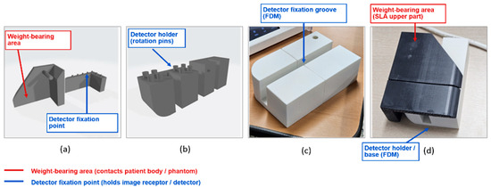
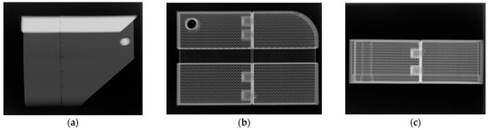
This study aimed to evaluate the utility of a 3D-printed, weight-bearing auxiliary device designed to improve reproducibility and facilitate detector fixation during axiolateral radiography of hip joints known to be affected by inter-operator variability. The experiment was conducted using a radiolucent human phantom.
[...] Read more.
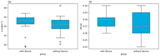
This study aimed to evaluate the utility of a 3D-printed, weight-bearing auxiliary device designed to improve reproducibility and facilitate detector fixation during axiolateral radiography of hip joints known to be affected by inter-operator variability. The experiment was conducted using a radiolucent human phantom. Multiple radiographs were taken under identical conditions. Reproducibility and utility were assessed by measuring the α angle and off-set ratio from acquired images. Results showed that the mean α angle was 51.58° (±5.14°) with the auxiliary device and 50.45° (±4.90°) without it, with no statistically significant difference between conditions (Wilcoxon signed-rank test, p = 0.2024), indicating no measurable improvement in reproducibility. The off-set ratio likewise did not differ significantly between conditions (p = 0.1704). However, using the device did not significantly affect the off-set ratio. In conclusion, the auxiliary device developed in this study allows for consistent detector angle maintenance and fixation during hip joint axiolateral projection, thereby improving reproducibility and ease of examination. This suggests its potential usefulness for achieving more accurate imaging in clinical settings.
Full article
(This article belongs to the Section Radiation in Medical Imaging)
►▼
Show Figures

Figure 1

Open AccessArticle
Urgent Carotid Revascularization for Symptomatic High-Grade Carotid Stenosis Associated with Ipsilateral Hemispheric Hypoperfusion: A Single-Centre Retrospective Observational Study
by
Antonio Marzano, Martina Pacillo, Giovanni Gagliardo di Carpinello, Marta Ascione, Claudia Panzano, Alessia Di Girolamo, Rocco Cangiano, Francesca Miceli, Luca di Marzo and Wassim Mansour
Radiation 2026, 6(3), 31; https://doi.org/10.3390/radiation6030031 - 6 Aug 2026
Abstract
►▼
Show Figures
Symptomatic high-grade carotid stenosis may present with extensive ipsilateral hemispheric hypoperfusion without a large infarct core, a grey-zone phenotype between standard early and truly urgent carotid revascularization. This single-centre retrospective observational study described the procedural and clinical outcomes of urgent carotid revascularization in
[...] Read more.
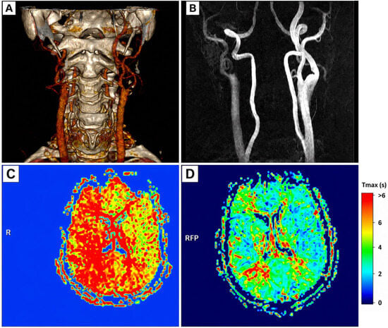
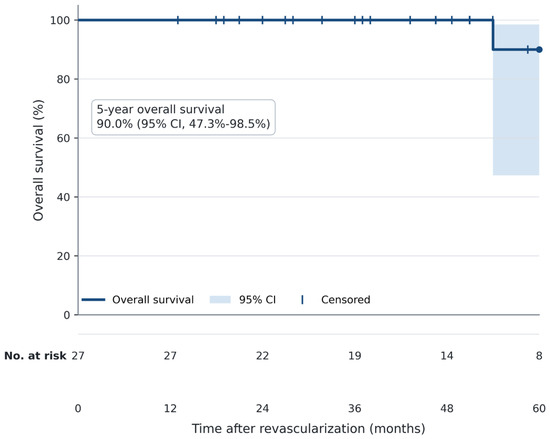
Symptomatic high-grade carotid stenosis may present with extensive ipsilateral hemispheric hypoperfusion without a large infarct core, a grey-zone phenotype between standard early and truly urgent carotid revascularization. This single-centre retrospective observational study described the procedural and clinical outcomes of urgent carotid revascularization in this imaging-defined subgroup. Consecutive patients treated between June 2015 and August 2025 were included when they had recent ipsilateral anterior-circulation symptoms, ipsilateral internal carotid stenosis > 70%, objective hemispheric hypoperfusion involving at least one third of the anterior circulation territory on CT or MR perfusion, and no intracranial haemorrhage or large established infarction. Twenty-seven patients met the criteria. Median NASCET-style stenosis was 92%, and 14 patients (51.9%) had at least one collateral-limiting feature. Revascularization was performed within 48 h in all cases, by carotid endarterectomy in 25 patients and carotid artery stenting in two. Technical success was 100%. No 30-day death, stroke, intracranial haemorrhage, major adverse event, or target-lesion reintervention occurred. Complete neurological recovery at discharge was observed in 25 patients (92.6%). During median follow-up of 48.6 months, no late ipsilateral stroke, carotid-related neurological event, neurological death, or target-lesion reintervention was recorded. In this small, selected, uncontrolled cohort, urgent carotid revascularization was technically achievable, and no prespecified 30-day safety events were observed. These descriptive findings do not establish comparative safety or clinical benefit and require prospective multicentre validation.
Full article

Figure 1
Open AccessReview
Contrast-Enhanced Ultrasound and CT Fusion Imaging in EVAR Follow-Up: Diagnostic Workflow, Endoleak Detection, and Imaging Pitfalls
by
Manuela Montatore, Gianmichele Muscatella, Ruggiero Tupputi, Eluisa Muscogiuri and Giuseppe Guglielmi
Radiation 2026, 6(3), 30; https://doi.org/10.3390/radiation6030030 - 3 Aug 2026
Abstract
►▼
Show Figures
Endovascular aneurysm repair (EVAR) has become a widely adopted treatment for abdominal aortic aneurysms, offering lower perioperative morbidity and mortality than open surgical repair. Nevertheless, long-term imaging surveillance remains essential because procedure-related complications may occur years after treatment. Among these, endoleaks represent the
[...] Read more.
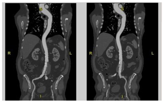
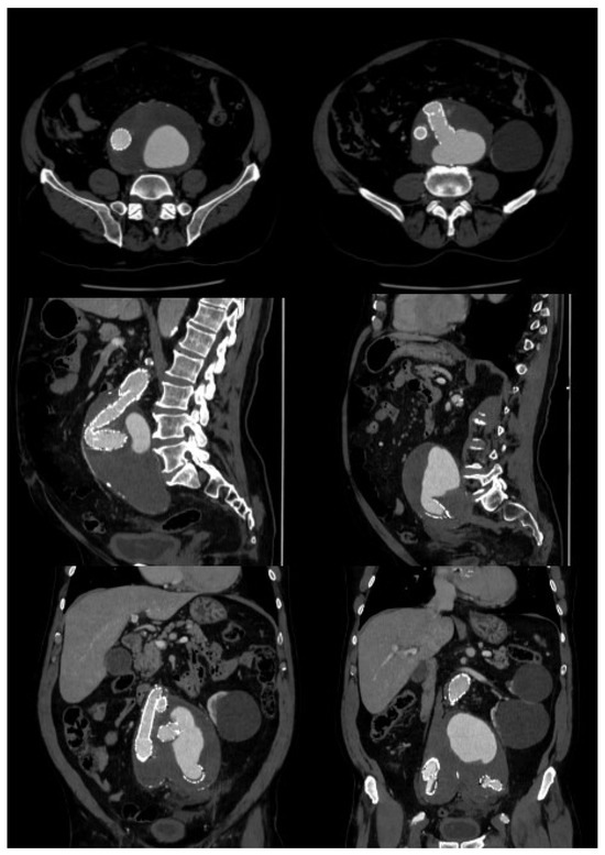
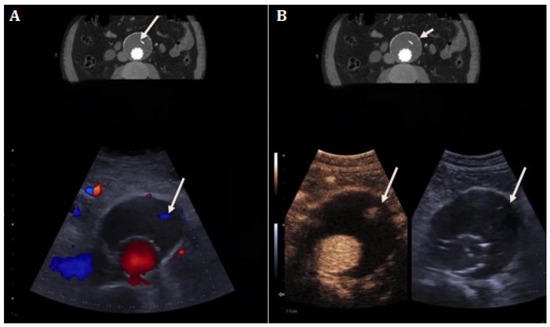
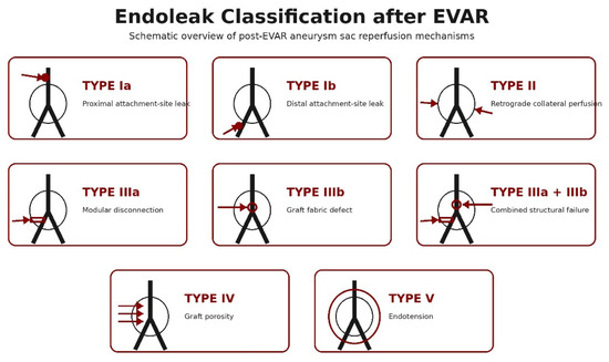
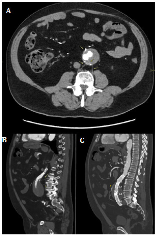
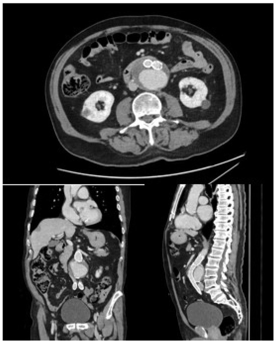
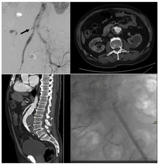
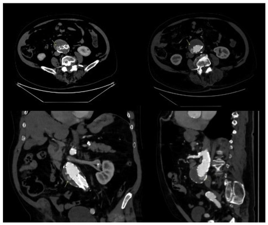
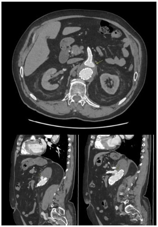
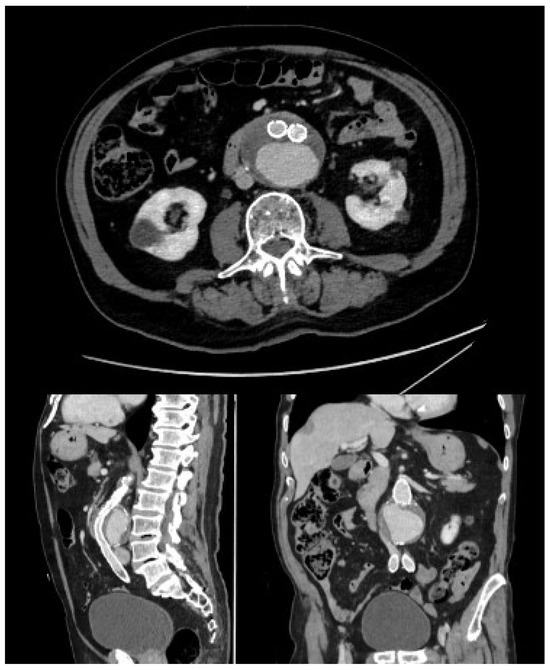
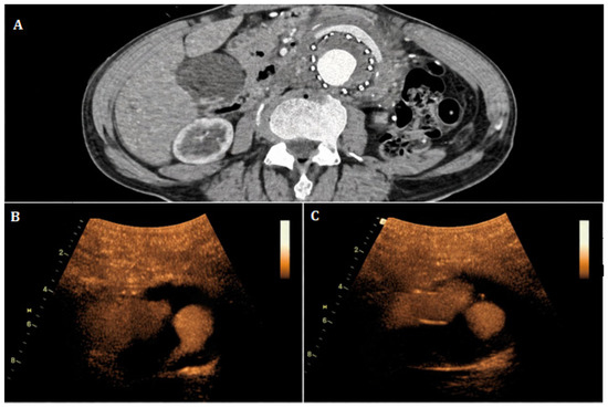
Endovascular aneurysm repair (EVAR) has become a widely adopted treatment for abdominal aortic aneurysms, offering lower perioperative morbidity and mortality than open surgical repair. Nevertheless, long-term imaging surveillance remains essential because procedure-related complications may occur years after treatment. Among these, endoleaks represent the most frequent and clinically relevant finding, particularly Type II endoleaks, which are sustained by retrograde collateral perfusion and may be associated with persistent aneurysm sac filling and sac enlargement. Computed tomography angiography (CTA) remains the reference standard for anatomical assessment after EVAR, providing detailed evaluation of endograft integrity, aneurysm sac morphology, and vascular anatomy. However, repeated CTA examinations involve cumulative radiation exposure and iodinated contrast administration and may be limited in the detection of low-flow or intermittent endoleaks. Contrast-enhanced ultrasound (CEUS) provides real-time hemodynamic assessment, improves detection of slow-flow endoleaks, and avoids both ionizing radiation and nephrotoxic contrast agents. Nevertheless, CEUS may be affected by operator dependency and limited anatomical overview. CT–ultrasound fusion imaging combines pre-acquired CTA datasets with real-time ultrasound through spatial co-registration and probe tracking, enabling simultaneous evaluation of vascular anatomy and flow dynamics. This narrative review critically synthesizes current evidence regarding CTA, CEUS, and CT–ultrasound fusion imaging in post-EVAR surveillance, with particular emphasis on the incremental clinical value of fusion imaging beyond standalone CTA and CEUS. Particular attention is devoted to diagnostic performance, technical implementation, clinical applicability, current limitations, and future perspectives of CT–CEUS fusion imaging.
Full article

Figure 1
Open AccessReview
Are Water-Based Algorithms Still Adequate for Dose Calculation in Modern HDR Interventional Radiotherapy? A Review with Special Focus on 3D-Printed Applicators and Heterogeneous Materials
by
Enrico Rosa, Bruno Fionda, Maria Vaccaro, Valentina Lancellotta, Elisa Placidi, Maria Concetta La Milia, Gabriele Ciasca, Pierpaolo Dragonetti, Andre Karius, Frank-André Siebert, Luca Tagliaferri and Marco De Spirito
Radiation 2026, 6(3), 29; https://doi.org/10.3390/radiation6030029 - 2 Aug 2026
Abstract
►▼
Show Figures
Background: High-dose-rate interventional radiotherapy (HDR IRT) relies on accurate dose calculations to ensure safe and effective treatment. The AAPM TG-43 formalism, based on homogeneous water assumptions, has long been the clinical standard, but the growing use of patient-specific applicators and heterogeneous materials challenges
[...] Read more.
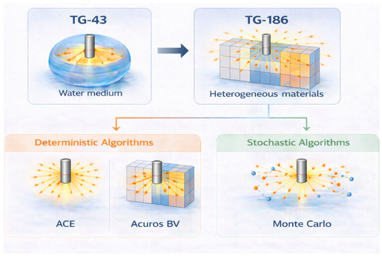
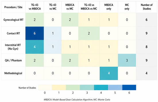
Background: High-dose-rate interventional radiotherapy (HDR IRT) relies on accurate dose calculations to ensure safe and effective treatment. The AAPM TG-43 formalism, based on homogeneous water assumptions, has long been the clinical standard, but the growing use of patient-specific applicators and heterogeneous materials challenges its accuracy. Materials and Methods: A literature narrative review was performed using PubMed and Scopus to identify studies comparing TG-43 and model-based dose calculation algorithms (MBDCAs), including deterministic methods (ACE, Acuros BV) and Monte Carlo simulations. Thirty-five studies were selected and qualitatively analyzed. Results: TG-43 yielded higher dose compared with MBDCAs, particularly in the presence of tissue heterogeneities, air gaps, shielding materials, and limited scatter conditions. Differences ranged from 2 to 5% in relatively homogeneous settings to more than 10–20% in complex geometries such as superficial mould treatments and head-and-neck IRT. Model-based approaches showed better agreement with Monte Carlo simulations and experimental measurements, especially in contact HDR IRT and applications involving 3D-printed applicators. Conclusion: While TG-43 remains clinically established and widely used, its limitations are increasingly evident in modern personalized HDR IRT. Model-based dose calculation algorithms provide greater dosimetric accuracy and should be progressively integrated into clinical practice, particularly in treatments involving significant heterogeneities.
Full article

Figure 1
Open AccessArticle
Influence of Sub-200 µm CBCT Voxel Size on Assessment of Root Canal Anatomy: A Cadaver Study
by
Francis Chan, Jay Black and Peter Parashos
Radiation 2026, 6(3), 28; https://doi.org/10.3390/radiation6030028 - 29 Jul 2026
Abstract
►▼
Show Figures
The aim of this study was to compare the accuracy of different sub-200 µm voxel reconstruction of CBCT scans in the identification of root canal anatomy. Cadaveric dentate maxillae and mandibles with soft tissues attached (69 teeth comprising 91 roots) were assessed with
[...] Read more.
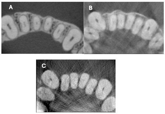
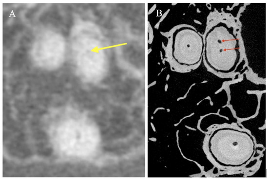
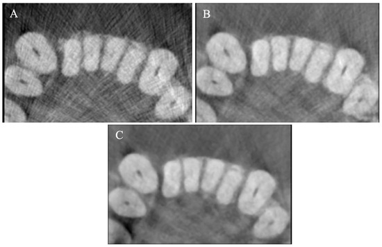
The aim of this study was to compare the accuracy of different sub-200 µm voxel reconstruction of CBCT scans in the identification of root canal anatomy. Cadaveric dentate maxillae and mandibles with soft tissues attached (69 teeth comprising 91 roots) were assessed with scans reconstructed at voxel sizes of 80 µm, 125 µm, and 160 µm. Three examiners recorded the canal configuration, presence of lateral canals, PAI score, and image quality seen on the scans. Micro-computed tomography (micro-CT) scans of the specimens reconstructed at 10 µm served as the reference standard. There was no significant difference between 80 µm and 125 µm reconstructions, but 80 µm scans were more accurate than 160 µm scans (p = 0.04). The findings suggest that canal anatomical complexity may have a greater influence on diagnostic accuracy than differences among the sub-200 µm voxel reconstructions evaluated. Lateral canals were only noted in 2% of roots in CBCT scans, but were present in 55% of roots assessed with micro-CT. There was significantly more variation in the assessment of image quality for 80 µm compared with 125 µm (p = 0.01) and 160 µm (p = 0.003) scans. Generally, 80 µm reconstructions provided marginally improved diagnostic yield compared with 160 µm scans; however, CBCT scans showed limited accuracy for more complex canal configurations. The poorer image quality of 80 µm scans may be due to the limited field of view, which causes partial volume effects rather than being reconstructed at 80 µm per se.
Full article

Figure 1
Open AccessArticle
Formulation and Physiochemical Characterization of PLGA–Chitosan–Folic Acid Nanoparticles Loaded with [225Ac]Ac-PSMA617-TFA for Targeted Alpha Therapy of Prostate Cancer
by
Yonwaba Mzizi, Bwalya Angel Witika, Honest Ndlovu, Mbongeni Shungube, Pedzisai Makoni, Sandile Sibiya, Amanda Mdlophane, Keamogetswe Ramonaheng, Mike Sathekge and Sipho Mdanda
Radiation 2026, 6(3), 27; https://doi.org/10.3390/radiation6030027 - 8 Jul 2026
Abstract
►▼
Show Figures
Background: Actinium-225 (225Ac) is receiving major attention as the radionuclide of choice for targeted alpha therapy (TAT) due to its outstanding physical properties such as a long physical half-life of 9.9 days and a short range of alpha (α)-particles which are
[...] Read more.
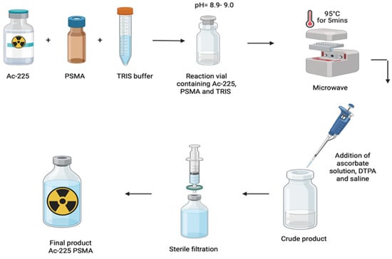
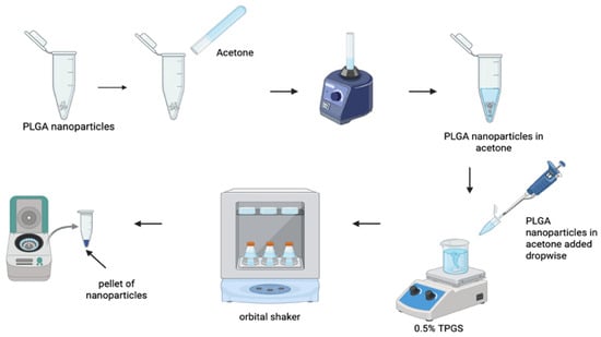
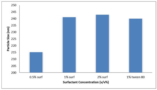
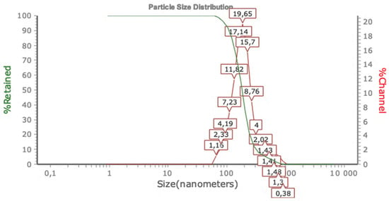
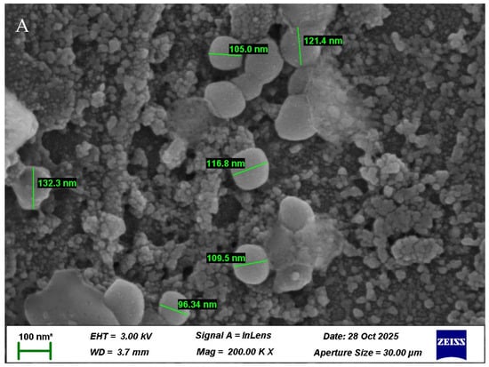
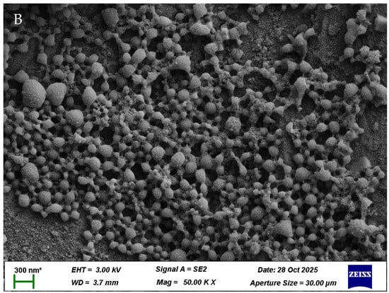
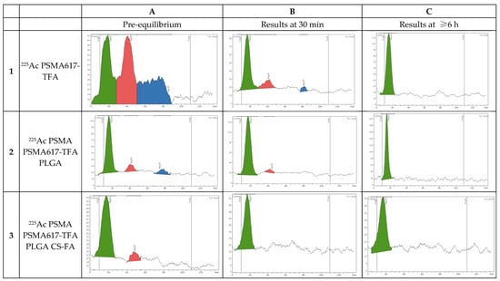
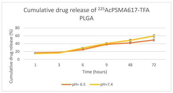
Background: Actinium-225 (225Ac) is receiving major attention as the radionuclide of choice for targeted alpha therapy (TAT) due to its outstanding physical properties such as a long physical half-life of 9.9 days and a short range of alpha (α)-particles which are responsible for the destruction of malignant tumors, whilst sparing normal surrounding tissues. Although the physical properties of 225Ac make it a desirable radionuclide for TAT, its application is challenging due to the lack of chelators available to stabilize its daughter radionuclides, resulting in the recoil effect. This occurs when there is a breakdown between the radionuclide and the chelator, therefore minimizing the therapeutic effects of the radiopharmaceutical. Nanodrug delivery systems (NDDSs) may minimize the challenge of 225Ac’s recoiling daughters and increase tumor penetration. Aim: This study aimed at using poly(lactic-co-glycolic)acid (PLGA) and chitosan (CS) nanoparticles as a delivery vehicle for targeted alpha therapy of prostate cancer in order to increase the therapeutic effect of 225Ac PSMA617-TFA. Methods and Results: PLGA nanoparticles were prepared using a nanoprecipitation method, after which they were functionalized with chitosan and folic acid. Following synthesis of 225Ac PSMA617-TFA, the radiopharmaceutical was loaded onto the nanoparticles. SEM analysis and FTIR were performed for characterization of the nanoparticles, and in-vitro drug release of 225Ac PSMA617-TFA at pH = 6.5 and pH = 7.4, respectively, was measured. The nanoparticles prepared had an average size of 200 nm and had a positive charge. This was further confirmed using a zetasizer and with scanning electron microscope (SEM) analysis. The PLGA-CS nanoparticles indicated a high encapsulation efficiency after 24 h. The results also showed a controlled release of 225Ac PSMA617-TFA over 72 h. The results of this study indicate that PLGA-CS nanoparticles are suitable for retaining 225Ac and its recoiling daughters (221Fr and 213Bi) at the tumor site, potentially providing a platform for future therapeutic evaluation. Conclusions: The results of this study indicate that PLGA-CS nanoparticles demonstrate feasibility as a drug delivery vehicle for 225Ac PSMA617-TFA, with effective retention of 225Ac and its decay daughters. However, biological validation through in vitro cellular studies and in vivo preclinical models is required before therapeutic effectiveness can be established.
Full article

Figure 1
Open AccessReview
Tissue Resilience in Radiation-Induced Injury: A Hypothesis-Generating Review of Heat Shock Protein 27 in Osteoradionecrosis of the Jaw
by
Erkan Topkan, Doga Topkan, Efsun Somay, Duriye Ozturk, Sibel Bascil and Ugur Selek
Radiation 2026, 6(3), 26; https://doi.org/10.3390/radiation6030026 - 6 Jul 2026
Abstract
►▼
Show Figures
Osteoradionecrosis of the jaw (ORNJ) remains one of the most severe late complications of head and neck radiotherapy. Current evidence suggests that ORNJ is a progressive and biologically heterogeneous disorder driven by microvascular injury, chronic hypoxia, oxidative stress, fibro-atrophic remodeling, impaired bone turnover,
[...] Read more.
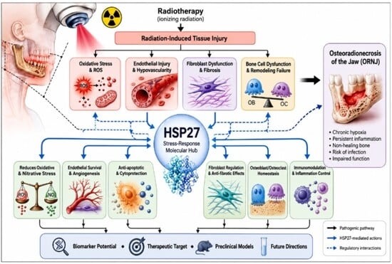
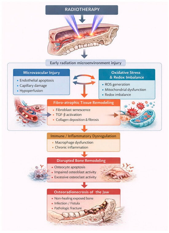
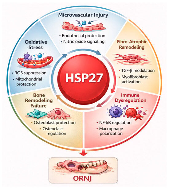
Osteoradionecrosis of the jaw (ORNJ) remains one of the most severe late complications of head and neck radiotherapy. Current evidence suggests that ORNJ is a progressive and biologically heterogeneous disorder driven by microvascular injury, chronic hypoxia, oxidative stress, fibro-atrophic remodeling, impaired bone turnover, immune dysregulation, and systemic susceptibility factors. Within this complex pathogenic network, heat shock protein 27 (HSP27) emerges as a biologically plausible but unexplored mediator. HSP27 regulates multiple stress-response pathways, including redox homeostasis, cytoskeletal stabilization, endothelial protection, apoptosis control, fibroblast activation, and osteoblast–osteoclast function, all of which overlap with key mechanisms implicated in ORNJ. However, no studies have directly investigated HSP27 expression, activation, or functional significance in irradiated mandibular tissues or ORNJ-specific cohorts. This review summarizes current knowledge of ORNJ pathobiology, examines potential mechanistic links with HSP27, and outlines future research priorities involving biomarker development, tissue-level characterization, preclinical modeling, and therapeutic targeting. Integrating HSP27 into ORNJ research may improve understanding of pathogenesis, risk stratification, and the development of novel preventive and therapeutic strategies.
Full article

Graphical abstract
Open AccessReview
Biologically Informed Radiotherapy in Glioblastoma: A Structured Framework for Imaging-Guided Clinical Decision-Making
by
Flavio Donnini, Giovanni Rubino, Giuseppe Battaglia, Pierpaolo Pastina, Tommaso Carfagno, Marta Vannini, Alfonso Cerase, Giulio Bagnacci, Armando Perrella, Salvatore Chibbaro, Maria Antonietta Mazzei and Paolo Tini
Radiation 2026, 6(3), 25; https://doi.org/10.3390/radiation6030025 - 4 Jul 2026
Abstract
Radiotherapy for glioblastoma remains anchored to postoperative structural MRI and anatomy-based target definitions, despite marked spatial heterogeneity and dynamic biological change during chemoradiotherapy. Advanced MRI, amino-acid PET, radiomics, and habitat imaging can characterize tumor biology beyond conventional anatomy, yet their relevance to radiotherapy
[...] Read more.
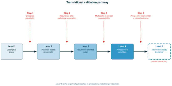
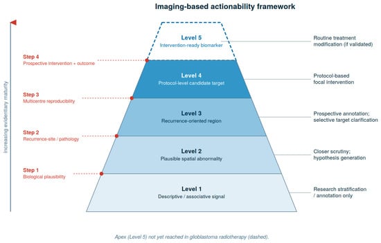
Radiotherapy for glioblastoma remains anchored to postoperative structural MRI and anatomy-based target definitions, despite marked spatial heterogeneity and dynamic biological change during chemoradiotherapy. Advanced MRI, amino-acid PET, radiomics, and habitat imaging can characterize tumor biology beyond conventional anatomy, yet their relevance to radiotherapy planning and their evidentiary maturity differ substantially. This narrative review examines the current evidence for biologically informed radiotherapy in glioblastoma and proposes an imaging-based actionability framework that organizes imaging-derived findings into five levels of evidentiary maturity, from descriptive or associative signals to intervention-ready biomarkers, to guide literature interpretation, multidisciplinary discussion, and prospective protocol design. Standard MRI-based planning remains the clinical backbone, supported by contemporary guidelines and the absence of randomized evidence demonstrating benefit from routine biologically guided target modification. Advanced MRI is discussed as the most practical serial platform for treatment-course reassessment, amino-acid PET as a selective complementary tool for metabolic clarification and recurrence assessment, and radiomics and habitat imaging as promising but not yet intervention-ready research layers. Across modalities, prospective evidence supports feasibility more consistently than clinical benefit. The proposed framework is intended to separate biologically informative findings from those sufficiently validated to justify a defined radiotherapy consequence, and to structure the translational path toward intervention-grade evidence in glioblastoma radiotherapy.
Full article
(This article belongs to the Section Radiation and Its Application in Oncology and Radiation Protection)
►▼
Show Figures

Figure 1
Open AccessArticle
Artificial-Intelligence-Driven Dose Rate Prediction Across Various 60Co Irradiator Source Configurations
by
Imen Hammami, Omer A. Magzoub, Asma Ayadi, Faouzi Hosni and Salam Labidi
Radiation 2026, 6(3), 24; https://doi.org/10.3390/radiation6030024 - 1 Jul 2026
Abstract
►▼
Show Figures
Accurate calculation of gamma dose rates in medical and industrial facilities is a critical component of comprehensive dosimetry assessment. Usually, two complementary approaches are employed to this end: experimental measurements and Monte Carlo (MC) simulations, both of which have established themselves as powerful
[...] Read more.
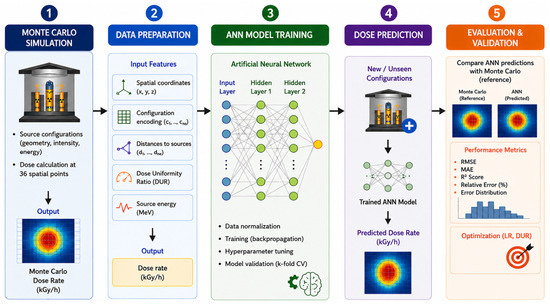
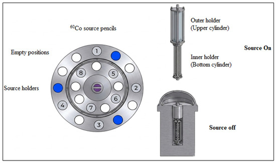
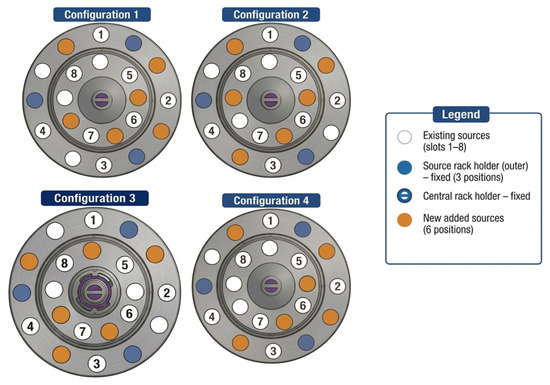
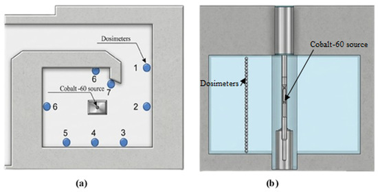
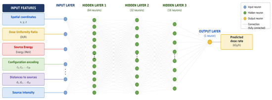
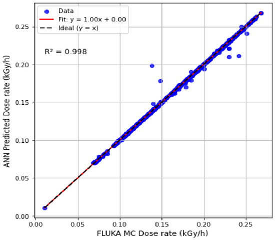
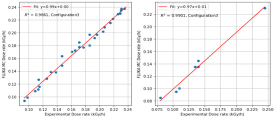
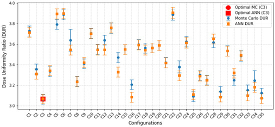
Accurate calculation of gamma dose rates in medical and industrial facilities is a critical component of comprehensive dosimetry assessment. Usually, two complementary approaches are employed to this end: experimental measurements and Monte Carlo (MC) simulations, both of which have established themselves as powerful and reliable tools in radiation protection and dosimetry practice. Given the high computational cost of Monte Carlo simulations, artificial intelligence can offer a compelling and efficient alternative for predicting dose rate distributions. This study evaluates the capability of machine learning models to predict MC-calculated dose rates and to identify the optimal 60Co source arrangement for the upcoming replenishment. The replenishment scenario involves inserting six new 60Co pencil sources. Dose rate prediction was performed using FLUKA MC simulations, complemented by an Artificial Neural Network (ANN)-based predictive model. The ANN model demonstrated strong concordance with FLUKA MC results, with deviations consistently below 1%, and exhibited reliable predictive performance on previously unseen configurations. Based on the dose uniformity ratio and the coefficient of determination, configuration 3 was identified as the optimal arrangement (R2 = 0.986). The integration of machine learning with MC simulation proves highly effective, enabling rapid and accurate dose rate prediction around the 60Co source while substantially reducing computational time and CPU resource demands.
Full article

Figure 1
Open AccessReview
Beyond Photons: Emerging Advances and Clinical Potential of Proton Beam Therapy in Gynecological Malignancies
by
Lifeng Chen, Li Wang and Hamid A. Bakshi
Radiation 2026, 6(3), 23; https://doi.org/10.3390/radiation6030023 - 30 Jun 2026
Abstract
►▼
Show Figures
Radiation therapy is central to the management of gynecological cancers, including endometrial, cervical, ovarian, and vaginal malignancies. Despite advances in photon-based techniques, treatment-related toxicity remains significant owing to the anatomical proximity of pelvic targets to critical organs at risk (OARs), including the bowel,
[...] Read more.
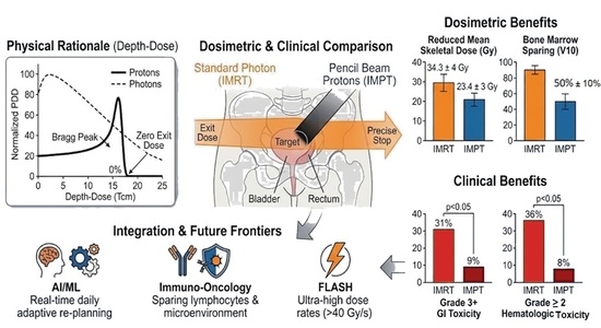
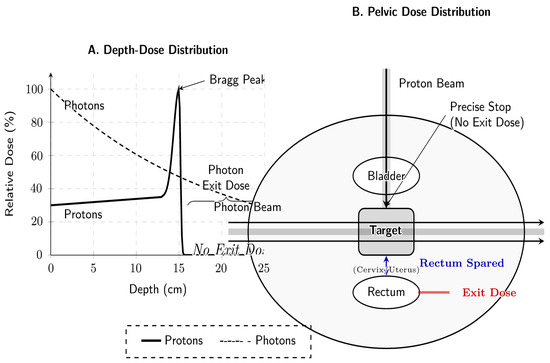
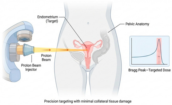
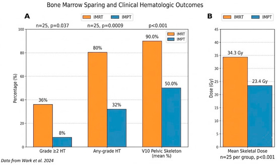
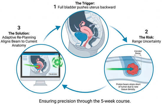
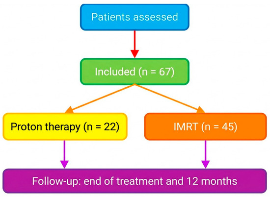
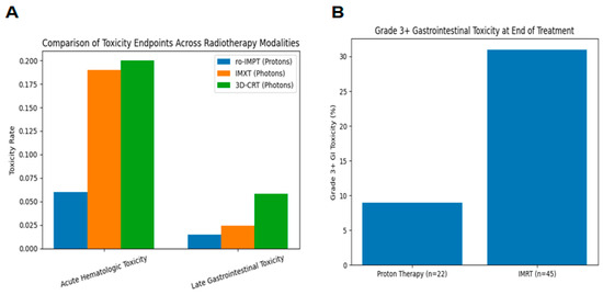
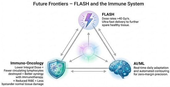
Radiation therapy is central to the management of gynecological cancers, including endometrial, cervical, ovarian, and vaginal malignancies. Despite advances in photon-based techniques, treatment-related toxicity remains significant owing to the anatomical proximity of pelvic targets to critical organs at risk (OARs), including the bowel, bladder, and bone marrow. Proton beam therapy exploits the Bragg peak to deliver a precise dose at depth with minimal exit dose, potentially reducing OAR exposure. This review develops the physical principles, dosimetric evidence, and early clinical data for proton therapy in gynecological malignancies, including cervical, endometrial, ovarian, vaginal, and vulvar cancers. Major focus is given to clinical conditions where conventional brachytherapy is not practical, and proton therapy may offer the greatest advantages, such as reirradiation for recurrent disease, post-operative pelvic irradiation, and extended field nodal treatment. This review also emphasizes current constraints that have slowed down wide clinical implementation, such as the lack of mature prospective data, cost, and accessibility. Finally, we emphasize future directions, including well-designed comparative trials, integration with systemic and immunotherapies, and adaptive treatment strategies. As the body of accumulated evidence evolves, the proton beam therapy potential for the treatment of gynecological malignancies has tremendously increased due to its role in safety and personalization of radiation treatment.
Full article

Graphical abstract
Open AccessReview
Beyond the Knife: A Review of the Burden of Localized Recurrent Pancreatic Adenocarcinoma and the Potential Role of Intraoperative Radiation Therapy
by
Jared Mount, Brandon Mount, Michael Rutenberg and John A. Stauffer
Radiation 2026, 6(3), 22; https://doi.org/10.3390/radiation6030022 - 26 Jun 2026
Abstract
►▼
Show Figures
Background: Pancreatic resection (Pancreatoduodenectomy, PD, distal pancreatectomy, DP, or total pancreatectomy, TP) is the standard of care for resectable pancreatic adenocarcinoma (PDAC). Despite advances in multimodal therapy, recurrence rates remain high, approaching 80%, and continue to drive poor overall survival. Objective: This review
[...] Read more.
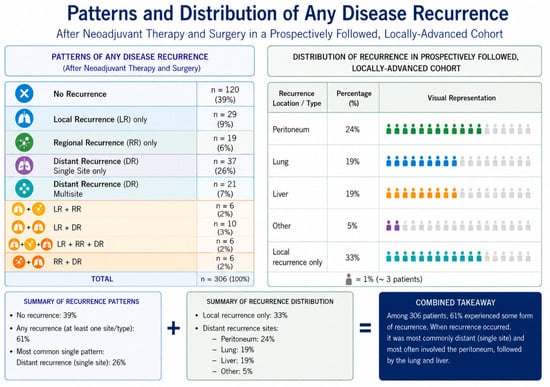
Background: Pancreatic resection (Pancreatoduodenectomy, PD, distal pancreatectomy, DP, or total pancreatectomy, TP) is the standard of care for resectable pancreatic adenocarcinoma (PDAC). Despite advances in multimodal therapy, recurrence rates remain high, approaching 80%, and continue to drive poor overall survival. Objective: This review evaluates the burden and clinical significance of localized recurrence in PDAC and critically examines the potential role of intraoperative radiation therapy (IORT) in improving locoregional disease control. Results: Distant recurrence remains the predominant pattern of failure, occurring in 55–75% of patients, most commonly involving the liver, lungs, and peritoneum. However, isolated local recurrence, observed in approximately 17–32% of patients, represents a clinically meaningful subset associated with significant morbidity and potential for subsequent metastatic progression. IORT, delivered as a single high-dose radiation treatment to the tumor bed at the time of surgery, enables precise targeting of areas at highest risk for residual microscopic disease while minimizing radiation exposure to adjacent radiosensitive structures. Retrospective and meta-analytic data, while limited, suggest that IORT is associated with improved local control and modest survival benefit without a significant increase in perioperative morbidity, though interpretation is limited by study heterogeneity and lack of randomized control trials. Conclusion: IORT represents a rational adjunct in the multimodal management of PDAC, particularly for patients at high risk of locoregional failure, including those with borderline resectable or locally advanced disease, nodal involvement, perineural invasion, or concern for margin positivity. Prospective studies are needed to better define optimal patient selection and to clarify the role of IORT in the modern treatment paradigm.
Full article

Figure 1
Open AccessArticle
Mepilex Dressings in Managing Radiation-Induced Moist Desquamation in Head and Neck Cancer
by
Shely Kagan, Yulya Kagan, Tharshini Yoganathan, Madette Galapin, Christina Yang, Britney Zhang, Shivani Verma, Henry C. Y. Wong, Amir H. Safavi, Michael C. Tjong, Shirley S. W. Tse, Shing Fung Lee, Sarah Bayrakdarian, Edward Chow and Irene Karam
Radiation 2026, 6(2), 21; https://doi.org/10.3390/radiation6020021 - 22 Jun 2026
Abstract
►▼
Show Figures
Background: Radiation dermatitis (ARD), particularly its most severe form, moist desquamation (MD), is a frequent and distressing complication of external beam radiotherapy (RT) in head and neck (H&N) cancer patients. Standard management often provides limited benefit for healing and symptom control. Silicone-based foam
[...] Read more.
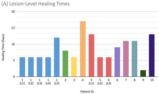
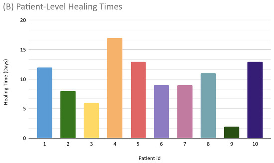
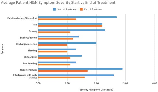
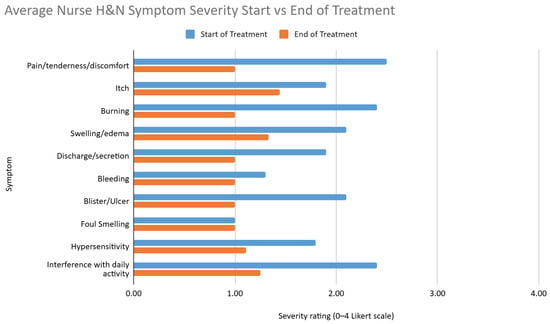
Background: Radiation dermatitis (ARD), particularly its most severe form, moist desquamation (MD), is a frequent and distressing complication of external beam radiotherapy (RT) in head and neck (H&N) cancer patients. Standard management often provides limited benefit for healing and symptom control. Silicone-based foam dressings, including Mepilex Lite and Mepilex Ag, may offer atraumatic adherence, moisture balance, and pain reduction. This study evaluated their real-world effectiveness for MD after conventional RT. Methods: Ten H&N cancer patients with clinically confirmed MD post-radiotherapy were prospectively followed until healing. Patients received Mepilex Lite or Mepilex Ag based on exudate level and infection risk, with dressings changed every three days. Patient- and healthcare provider-reported measures were collected throughout follow-up. The primary endpoint was time to MD resolution, defined as healing to grade 1 skin status. Secondary endpoints included changes in symptom burden, dressing tolerability and satisfaction, and adverse events. Results: Median age was 69 years (range 44–78). All wounds healed to grade 1, with a mean time of 8.6 days (SD 3.9). No infections or adverse events occurred. Pain, burning, and interference with daily activity decreased, and most patients reported improved comfort. Conclusions: In this small prospective cohort study, use of Mepilex dressings was associated with rapid healing, good tolerability, and improvement in patient-reported symptoms of acute radiation dermatitis. These findings suggest that Mepilex dressings may be a promising management option and warrant evaluation in larger comparative studies.
Full article

Figure 1
Open AccessSystematic Review
Systematic Review of Quantitative Metrics to Standardize Contours on Radiotherapy Clinical Trials
by
Leila T. Tchelebi, Janice Lester, Joshua J. Kim, Huaizhi Geng and Ying Xiao
Radiation 2026, 6(2), 20; https://doi.org/10.3390/radiation6020020 - 5 Jun 2026
Abstract
►▼
Show Figures
Radiation therapy quality assurance (RTQA) is critical to the success of RT and impacts trial outcomes. We aimed to evaluate the existence of standardized quantitative metrics to grade contours as part of RTQA and to identify barriers to standardization. A systematic review was
[...] Read more.
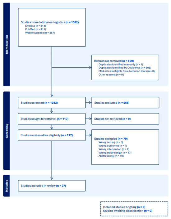
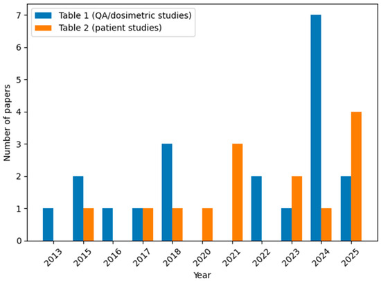
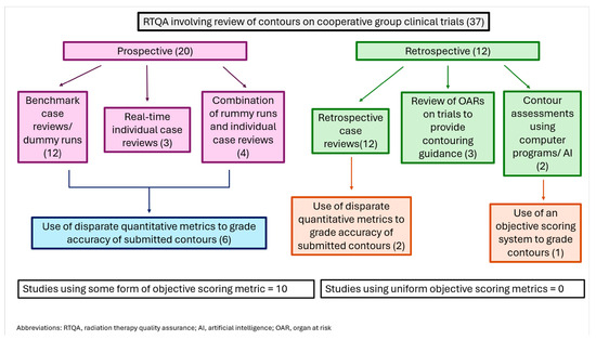
Radiation therapy quality assurance (RTQA) is critical to the success of RT and impacts trial outcomes. We aimed to evaluate the existence of standardized quantitative metrics to grade contours as part of RTQA and to identify barriers to standardization. A systematic review was conducted according to PRISMA guidelines. PubMed, Web of Science, and Embase were used to search relevant studies published from 2014–2024. Our endpoints were quantitative metrics used to guide contour scoring. We initially planned quantitative synthesis (meta-analysis) of geometric metric distributions across studies. However, the heterogeneity in evaluation methods, predominance of qualitative assessment, and lack of standardized reporting precluded meaningful quantitative pooling. We therefore performed structured qualitative synthesis, thematic analysis, and gap assessment. A total of 37 studies were included, 20 of which were prospective and 17 were retrospective. Of the prospective, 12 reported the results of benchmark case/dummy run reviews, 4 were a combination of benchmark case/dummy runs and individual QA analyses done pre- or post-treatment, 1 used knowledge-based planning to evaluate plans in real-time, and 3 involved real-time prospective QA. Only one reported the use of an objective QA scoring system. There is a concerning lack of standardized quantitative metrics to evaluate RT contours on clinical trials and a lack of methodological consistency required for systematic quantitative synthesis, reinforcing the need for standardization. Effort is needed to create guidelines for quantitatively evaluating RT contours to use across clinical trials to improve the quality and validity of RTQA.
Full article

Figure 1
Open AccessStudy Protocol
Itraconazole (ITRA) with Standard Radiotherapy (RT) and Temozolomide (TMZ) in Patients with Newly Diagnosed Glioblastoma Multiforme (GBM) ITRA-RAD: Phase I Clinical Study
by
Dusan Milanovic, Tatiana Lushchaeva, Hai Minh Ha, Raul-Ciprian Covrig, Christian Scheller, Klaus-Peter Stein, I. Erol Sandalcioglu, Antje Wiede, André Glowka and Daniel Medenwald
Radiation 2026, 6(2), 19; https://doi.org/10.3390/radiation6020019 - 2 Jun 2026
Abstract
Preclinical studies and data from other cancers suggest that inhibition of the Hedgehog (Hh) pathway also has antiproliferative effects on glioma cells. A key component of this pathway is the Smoothened (SMO) protein, which is expressed in high-grade gliomas. Itraconazole (ITRA), a widely
[...] Read more.
Preclinical studies and data from other cancers suggest that inhibition of the Hedgehog (Hh) pathway also has antiproliferative effects on glioma cells. A key component of this pathway is the Smoothened (SMO) protein, which is expressed in high-grade gliomas. Itraconazole (ITRA), a widely used antifungal agent, inhibits SMO, the PI3K/AKT/mTOR pathway, and the VEGF/VEGFR-2 axis—both of which are critical for GBM progression and angiogenesis. This protocol describes a prospective, single-center, dose-escalation phase I study with the classical 3 + 3 design in order to determine the MTD of ITRA. The study enrolls patients with a newly histologically confirmed diagnosis of GBM, without previous treatment except surgery. They will be treated with standard RT schedule (60 Gy in 30 fractions) with concurrent TMZ 75 mg/m2 and ITRA 2 × 100 mg, 200 mg, or 300 mg daily. The primary endpoint is to determine the MTD of ITRA given concurrently with RT + TMZ. Secondary endpoints include safety and tolerability of ITRA, overall survival (OS), progression-free survival (PFS), overall objective response rate, use of corticosteroids, treatment compliance, and health-related quality of life (EORTC QLQ-C30 and BN20). Participants will be monitored for one week post-treatment. All relevant statistics will be primarily descriptive.
Full article
(This article belongs to the Section Radiation and Its Application in Oncology and Radiation Protection)
Open AccessArticle
Impact of PET Imaging on Detection of Extra-Pelvic Metastases in Cervical Cancer at Diagnosis: A SEER-Based Analysis
by
Ugur Yilmaz, Ellen Jones and Lawrence B. Marks
Radiation 2026, 6(2), 18; https://doi.org/10.3390/radiation6020018 - 1 Jun 2026
Abstract
►▼
Show Figures
This study aims to evaluate the quantitative impact of FDG-PET on detecting extra-pelvic metastases in women with cervical cancer, using population-based data, following its approval by US Medicare/Medicaid in 2005 for staging patients without detectable extra-pelvic metastases on CT or MRI scans. Surveillance,
[...] Read more.
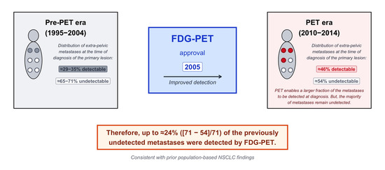
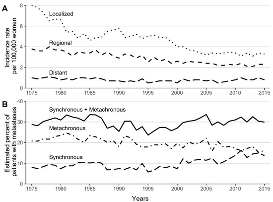
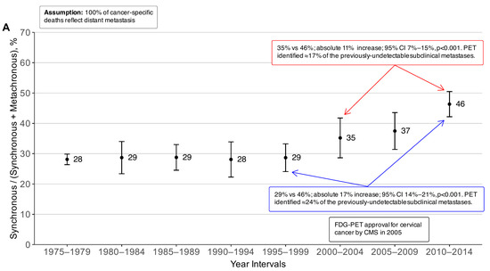
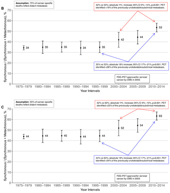
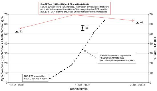
This study aims to evaluate the quantitative impact of FDG-PET on detecting extra-pelvic metastases in women with cervical cancer, using population-based data, following its approval by US Medicare/Medicaid in 2005 for staging patients without detectable extra-pelvic metastases on CT or MRI scans. Surveillance, Epidemiology, and End Results (SEER) data from eight US registries from 1975 to 2021 were analyzed. The proportions of synchronous metastases “Synchronous/(Synchronous + Metachronous)” were compared between the pre-PET era (1995–1999 and 2000–2004) and the PET era (2010–2014). The estimated fraction of extra-pelvic metastases at initial diagnosis was 35% in the pre-PET era (2000–2004) compared to 46% in the PET era (2010–2014) (absolute difference 11%; p < 0.001). The proportion of undetected metastases decreased from 65% to 54%, indicating that PET identified up to 17% (i.e., [65 − 54]/65) of previously undetectable subclinical metastases. Likewise, the comparison of data from 1995–1999 vs. 2010–2014 showed 29% vs. 46% (absolute difference 17%; p <0.001). The proportion of undetected metastases decreased from 71% to 54%, suggesting that PET identified up to 24% (i.e., [71 − 54]/71) of the previously undetectable subclinical extra-pelvic metastases. In conclusion, up to ≈17–24% of subclinical extra-pelvic metastases, previously undetectable at initial diagnosis, appear to be identified through PET, findings that are consistent with and externally validate our prior population-based observations in non-small cell lung cancer.
Full article

Graphical abstract
Open AccessArticle
Enhanced Efficacy of Multiport Synchrotron Microbeam Radiation Therapy for Brain Tumors in Rats
by
Laura Eling, Liam Day, Micah Barnes, Matthew Cameron, Michael De Veer, My-Linh Tempo, Helen Forrester, Mitzi Klein, Daniel Hausermann, Jean A. Laissue and Raphaël Serduc
Radiation 2026, 6(2), 17; https://doi.org/10.3390/radiation6020017 - 27 May 2026
Abstract
►▼
Show Figures
Microbeam Radiation Therapy (MRT) is a drastically novel approach of radiosurgery, challenging the understanding of how normal tissues respond to radiation therapy, where X-rays generated by a synchrotron light source are collimated into wafers of high-dose (hundreds of Gray), thin (50 µm) parallel
[...] Read more.
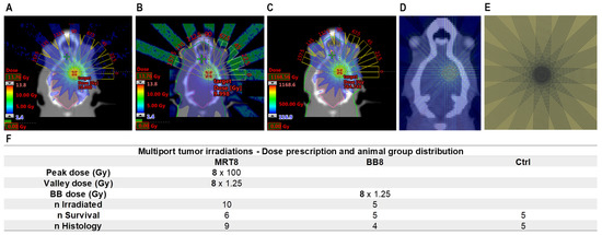
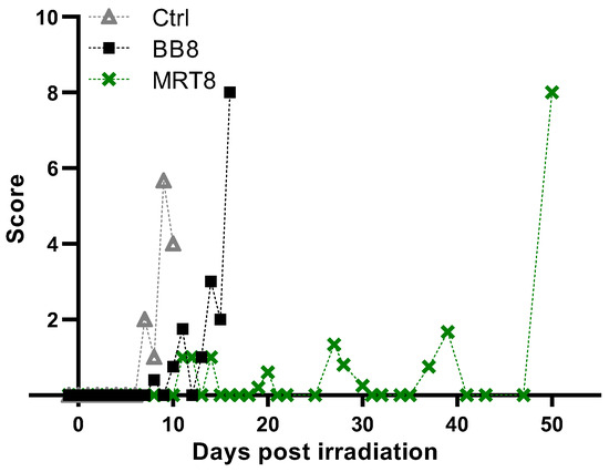
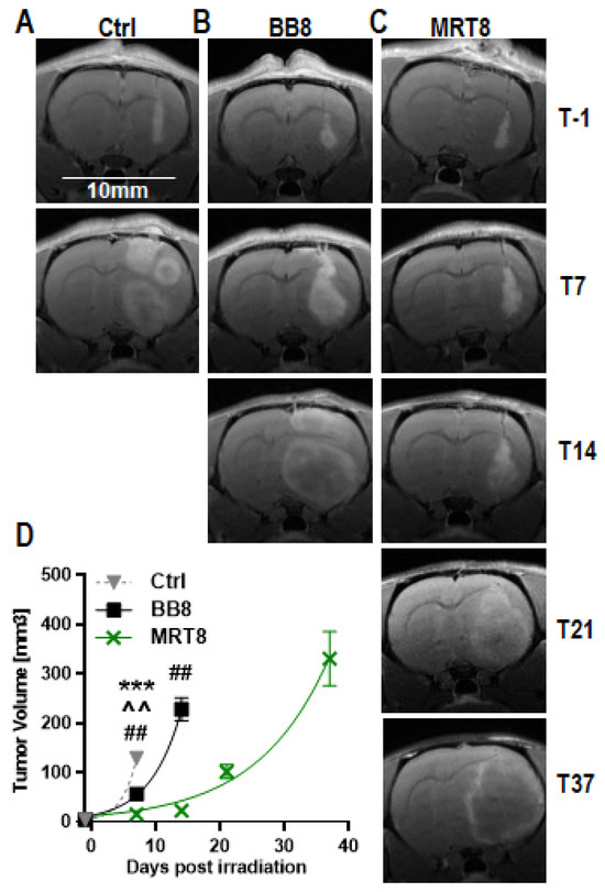
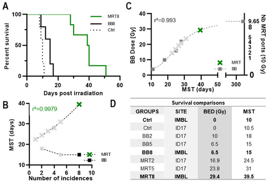
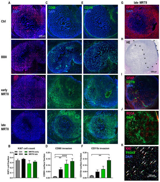
Microbeam Radiation Therapy (MRT) is a drastically novel approach of radiosurgery, challenging the understanding of how normal tissues respond to radiation therapy, where X-rays generated by a synchrotron light source are collimated into wafers of high-dose (hundreds of Gray), thin (50 µm) parallel microbeams. Recent work showed that MRT dose prescription must consider a dual approach: separating normal tissue dose constraints tied to the valley dose prescription and anti-tumor effects, depending on the prescribed number of ports. Here, we consolidate this assumption by increasing the number of MRT incidences to irradiate 9L-bearing rats with a cumulated valley dose of 10 Gy. 9L-bearing rats were irradiated by eight ports of conventional Broad Beam (BB) or MRT. Animals were followed up clinically by MR imaging, histopathology and survival analysis. MRI follow-up revealed that MRT significantly amplified antitumor effect and tumor tissue damage while improving the clinical score of animals. Ten Gy, delivered by either BB or MRT, increased median survival time to 15 and 39.5 days after irradiation, respectively. Neither complete ablation of brain tumors nor long-term survival was achieved, but multiport MRT (eight ports) showed an outstanding dose equivalence factor (×2.9), which needs to be tested in a clinical environment.
Full article

Figure 1
Open AccessSystematic Review
Advancing Nasopharyngeal Carcinoma Diagnosis: A Systematic Review of AI-Driven Machine Learning Techniques for CT, MRI, and WSI Imaging in Bioengineering
by
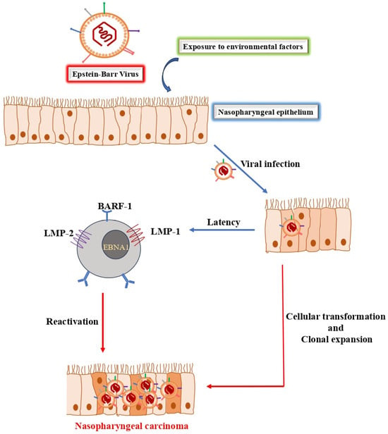
Muhammad Kabir Abdullahi, Arbab Sufyan Wadood, Md Serajun Nabi, Sarina Binti Mansor and Mohammad Faizal Ahmad Fauzi
Radiation 2026, 6(2), 16; https://doi.org/10.3390/radiation6020016 - 25 May 2026
Abstract
►▼
Show Figures
Background: Nasopharyngeal carcinoma (NPC) presents significant diagnostic and therapeutic challenges, often due to late-stage detection and its complex anatomical location. The increasing integration of artificial intelligence (AI) into oncology offers potential opportunities to enhance the precision of NPC management. This systematic review aims
[...] Read more.
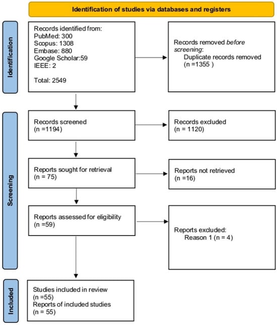
Background: Nasopharyngeal carcinoma (NPC) presents significant diagnostic and therapeutic challenges, often due to late-stage detection and its complex anatomical location. The increasing integration of artificial intelligence (AI) into oncology offers potential opportunities to enhance the precision of NPC management. This systematic review aims to synthesise the current evidence of AI applications in NPC diagnosis, prognostication, and treatment planning. Methods: A systematic literature search was conducted following PRISMA guidelines across multiple databases (PubMed, Scopus, Embase, Google Scholar, IEEE Xplore) for studies published up to June 2025. From an initial pool of 2549 articles, 55 studies meeting the inclusion criteria were selected for qualitative analysis. The review focuses on AI models applied to key diagnostic modalities: computed tomography (CT), magnetic resonance imaging (MRI), and histopathological whole-slide images (WSI). Results: AI, particularly deep learning (DL), shows promising performance in automating critical tasks across all modalities. For CT and MRI, models have been reported to achieve accurate tumor and organ-at-risk segmentation, potentially supporting radiotherapy planning, and show strong performance in predicting survival outcomes and treatment toxicity. In digital pathology, AI enables automated diagnosis and facilitates the extraction of prognostic “pathomic” features from WSIs, with some studies suggesting performance comparable to or exceeding traditional radiomics. The most significant advances are seen in multimodal AI systems that integrate radiological, pathological, and clinical data, which, in some studies, show modest improvements in prognostic performance compared to single-modality approaches. However, these findings are preliminary, as none of the reviewed multimodal models underwent rigorous external validation in large, multi-center cohorts. Reported performance varies considerably across studies, and claims of superiority should be interpreted with caution.
Full article

Figure 1
Open AccessReview
Applications of Artificial Intelligence in Prostate Cancer Radiotherapy: A Narrative Review
by
Antonio Piras, Albert Comelli, Andrea D’Aviero, Nino Dispensa, Nicola Pavan, Fabrizio Di Maida, Davide Cusumano, Calogero Casà, Antonino Daidone, Viviana Benfante, Domenico Genovesi, Tommaso Ingrassia, Domenico Di Raimondo, Antonino Tuttolomondo and Luca Boldrini
Radiation 2026, 6(2), 15; https://doi.org/10.3390/radiation6020015 - 7 May 2026
Cited by 1
Abstract
Introduction: Radiotherapy (RT) plays a crucial role in the management of prostate cancer (PC). Artificial intelligence (AI) is reshaping cancer care by providing innovative tools for diagnosis, treatment optimization, and outcome prediction. This review provides an end-to-end synthesis of AI applications across the
[...] Read more.
Introduction: Radiotherapy (RT) plays a crucial role in the management of prostate cancer (PC). Artificial intelligence (AI) is reshaping cancer care by providing innovative tools for diagnosis, treatment optimization, and outcome prediction. This review provides an end-to-end synthesis of AI applications across the prostate RT workflow, critically evaluating their clinical maturity, level of evidence, and current barriers to real-world implementation. Methods: A literature review of PubMed/MEDLINE and Embase was conducted to investigate the impact of AI on prostate RT. Only original articles published up to 1 August 2025 were included. The 27 selected studies were categorized into the following clusters: adaptive radiotherapy, autocontouring, autoplanning, prediction, synthetic computed tomography (CT), quality assurance (QA), and tracking. Results: Autocontouring was the most represented cluster, followed by prediction, autoplanning, and adaptive RT. Fewer studies addressed tracking, QA, and synthetic CT. Conclusions: AI shows significant potential across multiple phases of the prostate RT workflow; however, most evidence is based on retrospective or technical validation studies. Further research is required to establish clinical benefit and support integration into personalized treatment strategies.
Full article
(This article belongs to the Section Radiation and Its Application in Oncology and Radiation Protection)
Highly Accessed Articles
Latest Books
E-Mail Alert
News
27 July 2026
Meet Us at the 29th International Conference on Medical Image Computing and Computer-Assisted Intervention, 27 September–1 October 2026, Strasbourg, France
1 September 2026
MDPI INSIGHTS: The CEO’s Letter #38 – 2 Million Published Articles, Outstanding Reviewers, Michele Parrinello Award, AIS 2026 & WSF-12
Topics
Topic in
Applied Sciences, Toxics, IJERPH, Biology, Cancers, Radiation
Disease Risks from Environmental Radiological Exposure
Topic Editors: Valentina Venuti, Francesco CaridiDeadline: 31 December 2026
Topic in
Cancers, Radiation, Tomography, Physics, QuBS
Innovations in Physics and Radiobiology Studies of Particle Therapy
Topic Editors: Yidong Yang, Francesco Giuseppe Cordoni, Fada GuanDeadline: 31 October 2027
Special Issues
Special Issue in
Radiation
Advances in Palliative Radiotherapy
Guest Editor: Alysa FairchildDeadline: 30 October 2026